selenium, selenoproteins and neurodegenerative diseases
TRANSCRIPT
This journal is©The Royal Society of Chemistry 2015 Metallomics, 2015, 7, 1213--1228 | 1213
Cite this:Metallomics, 2015,
7, 1213
Selenium, selenoproteins andneurodegenerative diseases
Barbara Rita Cardoso,*ab Blaine R. Roberts,a Ashley I. Bush†a andDominic J. Hare†*acd
It is unsurprising that our understanding of the role of selenium in neurological function is somewhat
immature, considering its relatively recent discovery as an essential element to human health.
Selenocysteine, the 21st amino acid, is the defining feature of the 25 selenoprotein-encoding genes so
far discovered within the human genome. The low abundance of these proteins in the brain belies the
integral role they play in normal neurological function, from well-characterised antioxidant activity in the
periphery to poorly understood mechanisms that modulate mitochondrial function and response to
brain pathology. Selenium has been identified as playing a role in several neurodegenerative disorders,
including Alzheimer’s and Parkinson’s disease, though its function as a ‘cause or effect’ of disease
process remains unclear. This review discusses selenium metabolism in detail, specifically with regard to
the role it plays within the central nervous system, and examines the most current literature investigating
how selenium may be involved in chronic diseases of the central nervous system.
Introduction
The observation that selenium (Se) prevented necrotic liverdamage in mice by Schwarz and Foltz in 1957 was the firstdirect experimental evidence that this element was essential tonormal health.1 Almost twenty years later, Flohe et al.2 con-firmed the importance of Se by identifying it as an essentialcofactor of glutathione peroxidase (GPx). Twenty-five mamma-lian selenoprotein-encoding genes have since been identified,3
all containing selenocysteine (Sec) residues, which are analo-gous to cysteine (Cys) with a selenol moiety replacing the thiolgroup.4 From these genes, it is estimated that more than 30 uniqueselenoproteins are synthesised in mammals,5 and it is likely more
a The Florey Institute of Neuroscience and Mental Health, The University of
Melbourne, 30 Royal Parade, Parkville, Victoria, 3052, Australia.
E-mail: [email protected]; Tel: +61 450 633 537b Faculty of Pharmaceutical Sciences, Department of Food and Experimental
Nutrition, University of Sao Paulo, Sao Paulo, Brazilc Elemental Bio-imaging Facility, University of Technology Sydney, Thomas Street,
Broadway, New South Wales, 2007, Australia. E-mail: [email protected];
Tel: +61 3 9035 9549d Exposure Biology, Frank R. Lautenberg Environmental Health Sciences Laboratory,
Department of Preventive Medicine, Icahn School of Medicine at Mount Sinai,
New York, USA
Barbara Rita Cardoso
Barbara Rita Cardoso is anutritionist, and holds a Mastersdegree in Human Applied Nutri-tion. She obtained her PhD inFood Science from the Universityof Sao Paulo in 2014. She joinedthe Florey Institute of Neuro-science and Mental Health in2014 to investigate the associa-tion between selenium status andcognition as part of The AustralianImaging, Biomarker & LifestyleFlagship Study of Ageing (AIBL). Blaine R. Roberts
Blaine Roberts is the head of theNeuroproteomics Laboratory atthe Florey Institute of Neuro-science and Mental Health. Hereceived his PhD in 2007 fromOregon State University beforemoving to Australia in 2008to pursue the evolving fieldof metalloproteomics and itsapplication to neurodegenerativediseases.
† Co-senior authors.
Received 16th March 2015,Accepted 13th May 2015
DOI: 10.1039/c5mt00075k
www.rsc.org/metallomics
Metallomics
CRITICAL REVIEW
Ope
n A
cces
s A
rtic
le. P
ublis
hed
on 1
3 M
ay 2
015.
Dow
nloa
ded
on 1
0/4/
2021
6:5
4:47
AM
. T
his
artic
le is
lice
nsed
und
er a
Cre
ativ
e C
omm
ons
Attr
ibut
ion
3.0
Unp
orte
d L
icen
ce.
View Article OnlineView Journal | View Issue

1214 | Metallomics, 2015, 7, 1213--1228 This journal is©The Royal Society of Chemistry 2015
will be characterised in the future. Selenoproteins have diverseroles throughout the body, acting as antioxidants, modulatorsof immune function, detoxification agents for heavy metals andxenobiotics, and participants in thyroid hormone metabolism.6
Selenium is essential to human health, and the antioxidantactivity (or lack thereof) of specific selenoproteins is of parti-cular interest in diseases of the central nervous system. Thebrain is the most metabolically active of all organs in the body,and although it represents only 2% of the total mass, cerebralmetabolism accounts for about 20% of the oxygen and 25%of the glucose consumed by the human body.7 In concert withthis high metabolic activity, the high concentration of poly-unsaturated fatty acids in the brain makes it more vulnerable toperoxidation. A selective vulnerability within certain neuronsto oxidative stress may arise from deficiencies in antioxidantenzyme activity,8 which is a likely upstream event in thepathogenesis of several neurodegenerative diseases,9 includingAlzheimer’s disease (AD),10 Parkinson’s disease (PD)11 andother neurodegenerative disorders. Selenoproteins, with theirinherent antioxidant activity, thus play essential roles as part ofthe free radical defence system, though the function of theseproteins is not necessarily restricted to this singular classifica-tion. This review summarises the current data and importantconcepts concerning selenoproteins on brain function and theassociation between Se and diseases of the brain.
Selenoprotein synthesis
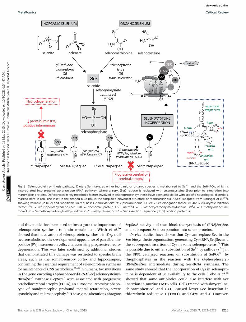
Dietary Se first must be converted to selenide (Se2�), whichserves as the donor for the incorporation of Se into seleno-proteins. Inorganic Se forms, such as selenite (SeO3
2�), areconverted to Se2� via the glutathione–glutaredoxin and thio-redoxin pathways, while organic forms are metabolised to Se2�
by selenocysteine lyase (Scly) or trans-selenation (analogous totrans-sulfuration).12
The incorporation of Se into proteins was described in detailby Turanov et al.,13 recently reviewed by Labunskyy et al.,14 and
is summarised in Fig. 1. After conversion of dietary Se to Se2�,selenophosphate (SePO3
3�) is produced in a reaction catalysedby selenophosphate synthase 2.4 Selenocysteine (Sec) is incor-porated into proteins via its own unique tRNA, (tRNA[Ser]Sec).tRNA[Ser]Sec decodes the UGA codon as Sec instead of a stopcodon, as is typical of other amino acids, and thus preventspremature termination. This is possible due to the presence ofa Sec insertion sequence (SECIS) element, a specific cis-actingstem-loop structure, in the 30-untranslated region (UTR) ofselenoprotein mRNA that works as a platform for RNA-bindingproteins. SECIS forms complexes with two trans-acting elements:the specific elongation factor (EFsec) and the SECIS bindingprotein 2 (SBP2).15 EFsec interacts with SBP2 to recruit tRNA[Ser]Secand mediates insertion of Sec into nascent protein chains inresponse to the UGA codon.15 Among the three distinctdomains of SBP2, the Sec incorporation domain and a COOH-terminal RNA-binding domain are specifically involved inSECIS binding. Additional SECIS binding proteins have beenidentified, including ribosomal L30, which is part of thebasal Sec insertion machinery and has a similar RNA-bindingdomain to SBP2;16 a eukaryotic initiation factor (eIF4a3) thatlinks Se status and differential selenoprotein expression,17 andnucleolin, which demonstrates a high affinity for SECIS, thoughits complete function is not currently known.18 Recently,Brocker et al.19 used Escherichia coli formate dehydrogenasemutants to reassign 64 codons encoding a critical Sec residue,finding that 58 of these were able to site-specifically incorporateSec. At 15 sense codons selenoprotein synthesis could bepromoted compared to the 3 stop codons. This study providescritical new information about how selenoprotein productioncan be manipulated through genetic recoding, and providesexciting new opportunities for additional studies to furtherelucidate the mechanisms involved in selenoprotein synthesis,which remains a relatively new frontier in molecular biology.
It is known that selenoprotein synthesis is critical forviability of life, as shown by tRNA[Ser]Sec (Trsp) knockout mice,which present an embryonically lethal phenotype. However,Trsp null mice allow tissue-specific manipulation of tRNA[Ser]Sec,
Ashley I. Bush
Ashley Bush heads the OxidationBiology Unit at the Florey Insti-tute of Neuroscience and MentalHealth, is Professor of Neuro-science at The University ofMelbourne, NHMRC AustraliaFellow, co-director of biomarkerdevelopment for the AustralianImaging Biomarker Lifestyle Studyof Ageing (AIBL), Chief ScientificOfficer of the CooperativeResearch Center for MentalHealth, and has staff appoint-ments at the MassachusettsGeneral Hospital.
Dominic J. Hare
Dominic Hare is a ChancellorsPostdoctoral Fellow at the Univer-sity of Technology, Sydney, Head ofthe Analytical NeurochemistryDevelopment Group at theFlorey Institute of Neuroscienceand Mental Health, and anAdjunct Assistant Professor atthe Icahn School of Medicine atMount Sinai in New York. Hereceived his PhD at UTS in2009, and works primarily instudying the roles of metals inneurodegeneration.
Critical Review Metallomics
Ope
n A
cces
s A
rtic
le. P
ublis
hed
on 1
3 M
ay 2
015.
Dow
nloa
ded
on 1
0/4/
2021
6:5
4:47
AM
. T
his
artic
le is
lice
nsed
und
er a
Cre
ativ
e C
omm
ons
Attr
ibut
ion
3.0
Unp
orte
d L
icen
ce.
View Article Online
This journal is©The Royal Society of Chemistry 2015 Metallomics, 2015, 7, 1213--1228 | 1215
and this model has been used to investigate the importance ofselenoprotein synthesis to brain metabolism. Wirth et al.20
showed that inactivation of selenoprotein synthesis in Trsp nullneurons abolished the developmental appearance of parvalbumin-positive (PV) interneuron cells, characterising progressive neuro-degeneration. This was later confirmed by additional studiesthat demonstrated this damage was restricted to specific brainareas, such as the somatosensory cortex and hippocampus,confirming the essential requirement of selenoprotein synthesisfor maintenance of CNS metabolism.21,22 In humans, two mutationsin the gene encoding O-phosphoseryl-tRNA[Sec]:selenocysteinyl-tRNA[Sec] synthase (SepSecS) were associated with progressivecerebellocerebral atrophy (PCCA), an autosomal-recessive pheno-type of nondysmorphic profound mental retardation, severespasticity and microencephaly.23 These gene alterations abrogate
SepSecS activity and thus block the synthesis of tRNA[Ser]Sec,and subsequent Se incorporation into selenoproteins.
In vivo studies have shown that Cys can replace Sec in theSec biosynthetic organisation, generating Cys-tRNA[Ser]Sec andthe subsequent insertion of Cys in some selenoproteins.24 Thisis possible due to either substitution of Se2� by sulfide (S2�) inthe SPS2 catalysed reaction; or substitution of SePO3
3� bythiophosphates in the reaction with the O-phosphoseryl-tRNA[Ser]Sec intermediate during Sec-tRNA synthesis. Thesame study showed that the incorporation of Cys in selenopro-teins is dependent of Se availability to the cells. Tobe et al.25
showed that some antibiotics could also interfere with Secinsertion in murine EMT6 cells. Cells treated with doxycycline,chloramphenicol and G418 caused lower Sec insertion inthioredoxin reductase 1 (Trxr1), and GPx1 and 4. However,
Fig. 1 Selenoprotein synthesis pathway. Dietary Se intake, as either inorganic or organic species is metabolised to Se2�, and the SeH3PO3, which isincorporated into proteins via a unique tRNA pathway, where a seryl (Ser) residue is replaced with selenocysteine (Sec) prior to integration intomammalian proteins. Deficiencies in key metabolic factors involved in selenoprotein synthesis have been associated with specific neurological disorders,marked here in red. The inset in the dashed blue box is the simplified cloverleaf structure of mammalian tRNA[Sec] (adapted from Birringer et al.196),showing variable (in blue) and modifiable (in red) bases. Abbreviations: C = pseudouridine; EFSec = Sec elongation factor; eIF4a3 = eukaryotic initiationfactor; i6A = N6-isopentenyladenosine; L30 = ribosomal protein L30; mcm5U = 5-methoxycarbonylmethyluridine; m1A = 1-methyladenosine;mcm5Um = 5-methoxycarbonylmethyluridine-20-O-methylribose; SBP2 = Sec insertion sequence (SCIS) binding protein-2.
Metallomics Critical Review
Ope
n A
cces
s A
rtic
le. P
ublis
hed
on 1
3 M
ay 2
015.
Dow
nloa
ded
on 1
0/4/
2021
6:5
4:47
AM
. T
his
artic
le is
lice
nsed
und
er a
Cre
ativ
e C
omm
ons
Attr
ibut
ion
3.0
Unp
orte
d L
icen
ce.
View Article Online

1216 | Metallomics, 2015, 7, 1213--1228 This journal is©The Royal Society of Chemistry 2015
Sec was also exchanged by arginine and tryptophan in addi-tion to Cys. The specific mechanisms that are associated withmis-insertion of amino acids in place of Sec and how they areassociated with changes in selenoproteins activity need to beclarified, but these reports show another way selenoproteinhierarchy is potentially regulated, which may also includeas-yet undetermined environmental factors.
Selenoprotein P and brain seleniumhierarchy
Selenoprotein expression is modulated by Se availability, and ahierarchy is observed in response to dietary Se intake. In casesof Se deficiency the synthesis of some proteins is maintained ata higher level than that of others, suggesting that insufficientavailable Se results in competition between transcripts ofdifferent selenoproteins.26 Iodothyronine deiodinase 1 is atopthis hierarchy and is comparatively the least affected byrestricted dietary Se; glutathione peroxidases GPx2 and GPx4are less vulnerable to Se deficiency than GPx1 and GPx3; andselenoprotein P (SelP) is in an intermediary position on thisselenoprotein transcription ladder.27
Some mechanisms underlying Se regulation of the seleno-protein transcriptome have been identified. During Se defi-ciency, non-essential selenoproteins are down regulated vianonsense-mediated mRNA decay, which decodes the UGA-Seccodon as a termination codon causing RNA degradation.28,29
Also, Se levels directly correlate with the degree of Um34 methyla-tion (the single methyl group differentiating the two tRNA[Ser]Secisoforms30), one step of maturation of tRNA[Ser]Sec which can bealtered as a result of some mutations. The Um34-containingisoform of tRNA[Ser]Se; 5-methoxycarbonylmethyl-20-O-methyl-uridine (mcm5Um), is recruited for efficient expression of thestress-related selenoproteins; and the non-Um34-containing iso-form, 5-methoxycarbonylmethyluridine (mcm5U), is not requiredspecifically. The distinct use of these tRNA[Ser]Sec isoforms showsthat this mechanism contributes to Se regulation for the purposeof selenoprotein synthesis.28
The hypothesis that explains the hierarchy between seleno-protein syntheses suggests that they are classified according totheir importance to the body. However, it is important to notethat Se concentration is highly variable within the periphery(i.e. liver, kidneys, etc.), though in the brain Se levels remainrelatively stable by comparison during periods of deficiency.31
Additionally, the ability of organs outside the CNS to retain Seis also variable. The CNS, together with reproductive andendocrine organs, has the highest priority for Se both uptakeand retention during Se deficiency, although the Se concen-tration in the CNS is low in comparison with other organs.32
Though interrelated, supply and retention are not exclusivelylinked, and it is thus a testament to the importance of Se inbrain function that rigorous mechanisms to ensure adequate Selevels are available in times of dietary shortage are in place. Thebrain’s top position on a tissue-specific grading further indicatesthat Se is essential for its normal metabolism.33 This is primarily
due to the high expression of apolipoprotein E receptor-2(apoER2) at the blood–brain barrier (see below) to facilitate Seuptake and strong SelP-apoER2 interaction within the CNSproviding an adequate pool of available Se. Selenium deficientrats presented approximately 29% lower Se levels in the braincompared to animals with a sufficient Se supply, which is notnearly as dramatic as the 99% and 92% decrease in Se concen-tration in the liver and kidneys of deficient animals.34 Sedistribution is heterogeneous among different brain regions,and grey matter appears to have preference for greater uptake.Imaging using a 75Se radiotracer combined with autoradio-graphic localisation and proteomic analysis to profile Se levelsin different brain regions of rats fed a Se-replete diet revealedthat Se was enriched in choroid plexus, putamen, parietalinferior lobule and occipital cortex.35 This observation has beenreflected in human brains, where the highest Se concentrationoccurs in grey matter, with the putamen, parietal inferior lobuleand occipital cortex in particular, while the lowest levels arefound in the cerebellum and medulla.36
The mechanism that maintains Se levels in the brain at theexpense of other organs, even during times of deprivation, ismainly orchestrated by SelP, which is recognised as the mostimportant Se supply to different tissue types. The role of SelP todeliver Se to cells is facilitated by the presence of ten SECISforms: one is situated in the larger N-terminal region, whichacts as an enzyme; and nine are located in a Sec-rich C-terminaldomain, providing a mechanism for transporting the highlyreactive Se atom safely, incorporated into the protein as Sec.37
Deletion of the conserved, non-SECIS containing 30UTR of SelPdoes not affect the efficiency of incorporation of multiple Secresidues into SelP in vitro whilst still remaining sensitive toambient Se levels, indicating that the 30UTR are not involved inSe incorporation per se.38 The structure of plasma SelP remainsunresolved, and there is some contention as to the number ofcirculating isoforms of the protein. Ballihaut et al.39 recentlyreported three isoforms in a human plasma standard referencematerial, two of which incorporated Se, suggesting the thirdwas in fact a truncated variant.
Burk et al.40 recently proposed the mechanism by which Seis delivered to the brain (represented in Fig. 2a). SelP fromplasma is endocytosed following docking with apoER2 at theblood–brain barrier (BBB) in brain capillary endothelial cells(BCECs) and choroid plexus epithelial cells, which facilitates Setransport, in an as-yet unknown chemical form, into the brain.Selenium is then delivered to brain cells via SelP synthesised byastrocytes on the abluminal side of BCECs, composing boththe main transport mechanism and distinct pool of Se. Directtransport of SelP to neurons from BCECs has not beenexcluded. Neurons regulate Se levels via apoER2-mediateduptake of SelP,41 which has obvious implications for AD dueits role in neurodevelopment and synaptic function. In theperiphery, apoER2 is widely expressed in the body and is alsoinvolved in Se uptake in myoblasts42 and testis cells.43 In theCNS, apoER2 is also expressed in choroid plexus and on PVinterneurons in the hippocampus, inferior colliculus, medialseptum, red nucleus, reticular thalamus and cerebellum.40,44
Critical Review Metallomics
Ope
n A
cces
s A
rtic
le. P
ublis
hed
on 1
3 M
ay 2
015.
Dow
nloa
ded
on 1
0/4/
2021
6:5
4:47
AM
. T
his
artic
le is
lice
nsed
und
er a
Cre
ativ
e C
omm
ons
Attr
ibut
ion
3.0
Unp
orte
d L
icen
ce.
View Article Online

This journal is©The Royal Society of Chemistry 2015 Metallomics, 2015, 7, 1213--1228 | 1217
ApoER2 expression is regulated by physiological factors relatedto developmental stage; fetal brain mRNA ApoER2 are morethan 9 times greater than that observed in adult brain, andapparently not influenced by Se status.40 Although deletion ofapoER2 gene does not affect the whole-body Se levels, apoER2�/�
and SelP�/� mice have lower Se levels in brain,45,46 and thismay be associated with uptake of Se during specific develop-mental windows.40 In this study, fetal apoER2�/� mice brainscontain 75% less Se levels compared to age-matched apoER2+/+
mice, decreasing to 39% when reaching adulthood. Moreover,apoER2�/�mice had no SelP in BCECs or choroid plexus cells at
the BBB, suggesting a key role of ApoER2 in brain Se uptake,and also the presence of an unknown redundancy mechanismfor Se transport. It remains unclear if other members of thelipoprotein receptor family may participate of SelP uptake,since Burk et al.47 observed an alternative mechanism thatdoes not appear to depend directly on selenoproteins partici-pating on the transfer of Se from pregnant dam to the fetus,and it is unclear if this mechanism is conserved in the post-natal period.
Though primarily expressed in astrocytes, neurons have alsobeen identified as a source of endogenous SelP48,49 in all the
Fig. 2 (a) SelP, the master protein driving Se bioavailability is synthesised in the liver from both inorganic and organoselenium compounds accessiblethrough dietary sources. At the blood–brain barrier, SelP releases Se into the CNS via the ApoER2 receptor, which is in turn incorporated into newly –formed SelP in astrocytes (figure adapted from Burk et al.35), or is transported directly to neurons (dashed arrow). SelP is made available to neurons via thesame membrane-bound ApoER2, where additional selenoproteins essential to neurological function (blue box) are biosynthesised. (b) In cases ofdeficient Se intake Um34 methylation is downregulated, causing premature termination of protein synthesis via failure of tRNA[Ser]Sec to decode theUGA codon as Sec instead of a stop codon, and misincorpoartion of selenocysteine (Sec) as cysteine (Cys; in red). These three pathological features mayhave downstream effects on a range of neurological disorders (red boxes).
Metallomics Critical Review
Ope
n A
cces
s A
rtic
le. P
ublis
hed
on 1
3 M
ay 2
015.
Dow
nloa
ded
on 1
0/4/
2021
6:5
4:47
AM
. T
his
artic
le is
lice
nsed
und
er a
Cre
ativ
e C
omm
ons
Attr
ibut
ion
3.0
Unp
orte
d L
icen
ce.
View Article Online

1218 | Metallomics, 2015, 7, 1213--1228 This journal is©The Royal Society of Chemistry 2015
areas of the brain, though particularly high expression wasidentified in the putamen and substantia nigra.50 In contrast toapoER2�/� animals, SelP�/� mice present generalised Se defi-ciency, and in brain this gene deletion causes severe neuro-logical dysfunction.47,51 SelP�/�mice showed decreased densityof PV interneurons in different brain areas, most marked in theinferior colliculus. This was associated with higher oxidativedamage and a behavioural phenotype, characterised by impair-ments in contextual fear extinction, latent inhibition andsensorimotor gating. These data suggest that besides the effecton PV-expressing neurons causing increased vulnerability tooxidative damage, the behavioural deficits resulting from SelPdeletion are likely associated with impaired GABAergic functionof the inferior colliculus.44 Both SelP�/� and apoER2�/� mice,when maintained on a Se-deficient diet, are more vulnerable toneuronal damage and eventual neurodegeneration. In this way,Burk et al.40 reported that extremely Se-deficient apoER2�/� andSelP�/� mice displayed neurological dysfunction, while thelittermate wild type mice did not show equivalent disruption,despite a decrease in brain Se levels. In another importantstudy,46 progressive neurodegeneration was evaluated in brainsof SelP�/� postnatal mice maintained on a Se-deficient dietafter weaning. Higher lipid peroxidation and decreased dendritelength and spine density was observed, and the areas moresusceptible to neurodegeneration during this post-weaning devel-opmental window were the forebrain bundle, somatosensorycortex and lateral striatum. In the hippocampus decreased dendritelength and spine density was also present, which might beassociated with the disrupted hippocampal-dependent learningand alteration to synaptic transmission observed in these mice.
As Scly is essential for breakdown of Sec into L-analine andSe2�, it has been proposed that this enzyme plays importantrole in Se metabolism in different tissue, and interacts withSelP to maintain adequate selenoprotein function in the brain.Although Scly�/� mice do not demonstrate neurological dys-function,51 deletion of both Scly and SelP exacerbates theneurological phenotype of SelP�/� mice, which displayimpaired motor coordination and locomotion compared withSelP null mice alone. Moreover, Scly�/�SelP�/� mice have lowerexpression of Trxr1, GPx1 and selenoprotein M (SelM) thanSelP�/� mice, showing that interplay of Scly and SelP helpsmaintain selenoprotein levels within the brain.52
Selenium and other selenoproteins:role in brain function
Selenium appears to have a multifaceted role in the nervoussystem, with a number of studies showing its importance tonormal brain physiological function. The antioxidant activity ofselenoproteins in the CNS is well established, and lower levelsof Se has been associated with brain injury.53 Selenium treat-ment as organic selenomethionine and inorganic SeO3
2�
resulted in a similar increased activity of antioxidant enzymes,scavenging of reactive oxygen species (ROS) and reduced pro-tein carbonyl content of patulin-induced brain damaged rats,54
and reduced the pro-oxidant effects of glyceryl trinitrate in arat model of migraine headaches.55 Administration of SeO3
2�
to suckling pups whose dams had methimazole-inducedhypothyroidism decreased total oxidant status and reduceddegenerative changes in the granular cell layer of the dentalgyrus,56 and increased levels of brain-derived neurothophicfactor (BDNF) in the hippocampus and cerebellum of bothhypothyroid and control groups.57 In a rat model of postnatalprotein malnutrition oral Se and zinc (Zn) supplementationincreased not only antioxidant enzyme activity, but also correctedthe neurobehavioral deficits caused by a protein-deficient diet.58
Besides the role of Se as essential component of the anti-oxidant system in the brain, studies have also shown that Secan act in response to oxidative stress through regulationof Ca2+ channels and mitochondrial biogenesis. Mice dorsalganglion root cells treated with Se showed reduced Ca2+ releaseinduced by H2O2 when compared with non-treated cells, withcorresponding decreases in apoptosis and lipid peroxidationlevels, and increased levels of GPx and glutathione (GSH)activity.59 In a traumatic brain injury animal model exhibitingoxidative stress, increased apoptosis and activation of transientreceptor potential vanilloid 1 (TRVP1) cation channels, Setreatment modulated Ca2+ entry through TRVP1 in neurons,reduced levels of ROS, prevented apoptosis, and reduced activityof caspases 3 and 9. Since brain injury induces Ca2+ influx andmitochondrial dysfunction in neurons, these data show that Secan increase cell viability not only as an antioxidant, but also bymodulating apoptotoic pathways and mitochondrial function.60
Studies with murine hippocampal HT22 cells treated withSeO3
2� clearly showed improved mitochondrial function, evenafter glutamate exposure and hypoxia. This was associated withactivation of complexes I, II + III and IV, and increasedmitochondrial respiratory rate. Selenium administrationincreased levels of peroxisome proliferator-activated receptorg-coactivator-1a (PGC-1a) and nuclear respiratory factor 1(NRF1), two important nuclear transcriptional factors asso-ciated with mitochondrial biogenesis. This role of Se appearsto be mediated through activation of the cyclic adenosinemonophosphate response element-binding protein (CREB)and activated protein kinase (AKT) cycles.61–63
Synthetic organoselenium compounds have been studied aspotential antioxidant therapies due their lower toxicity com-pared to inorganic forms of Se. Diphenyl diselenide [(PhSe)2]mimics GPx activity, producing both beneficial or harmfuleffects, depending on dose and route of administration.64 Acutetreatment of rat hippocampal slices with (PhSe)2 modifiedvarious proteins involved in glutamate signaling damage toastrocytes.65 Conversely, (PhSe)2 demonstrated a neuroprotectiveactivity in animal models of autoimmune encephalomyelitis andhypothyroidism due its antioxidant and anti-inflammatoryrole.66,67 A derivate of (PhSe)2, m-trifluoromethyl (m-CF3-PhSe)2
and the Se-containing salicylic acid derivative 5–50-diselanediylbis-(2-hydroxybenzoic acid) both exhibit antinociceptive action byinteraction with serotoninergic system, as well as an anti-inflammatory role in mice.68,69 Selenium supplementation asa promoter of antioxidant activity does appear to be somewhat
Critical Review Metallomics
Ope
n A
cces
s A
rtic
le. P
ublis
hed
on 1
3 M
ay 2
015.
Dow
nloa
ded
on 1
0/4/
2021
6:5
4:47
AM
. T
his
artic
le is
lice
nsed
und
er a
Cre
ativ
e C
omm
ons
Attr
ibut
ion
3.0
Unp
orte
d L
icen
ce.
View Article Online

This journal is©The Royal Society of Chemistry 2015 Metallomics, 2015, 7, 1213--1228 | 1219
dependent on the chemical form; 3-methyl-1-pheny-2-(phenyl-seleno)oct-2-en-1-one was less effective as an antioxidant com-pared to SeO4
2�, though it did reduce adenosine deaminaseactivity, suggesting a potential immunomodulatory property.54
Musik et al.70 examined two other Se-containing molecules;(4-(o-tolyl-)-selenosemicarbazide of 2-chlorobenzoic acid; 3-(2-chlorobenzoylamino-)-2-(o-tolylimino-)-4-methyl-4-selenazoline)reporting differential impacts on brain antioxidant mechanisms,including GPx and superoxide dismutase-1 (SOD-1). These con-tradictory results regarding Se supplementation on brain functionare likely due to the variety of chemical structures, dose, route ofadministration and the animal models used. More targetedstudies regarding the role and the safety of these compoundsare required before they can be considered as potential anti-oxidant therapeutics in humans.
The apparent neuroprotective role of Se supplementation inthe brain is not only directly associated with antioxidant effectsof organic and inorganic Se species, but also suggests a role inde novo selenoprotein synthesis, as demonstrated by increasedactivity of certain selenoproteins following Se treatment (Fig. 2).Methamphetamine treatment decreased levels of GPx1 andGPx4 in SH-SY5Y cells; an effect that was arrested with additionof SeO3
2� 24 hours after methamphetamine exposure.71 Intheir patulin-induced brain damaged rats, Song et al.72 showedthat, although both SeO3
2� and selenomethionine were able tooffer some degree of increased antioxidant activity, the organicform of Se was more efficient in increasing the expression andactivity of TrxR, GPx1 and GPx4.
The GPx family of enzymes are expressed in both neuronsand glia,73,74 and are recognised as one of the most importantmechanisms protecting against ROS-induced damage.75 Amongthese isoenzymes, GPx4 protects cells against lipid hydroperoxidedamage76 and is the most expressed GPx in brain, found inneurons of cerebellum, hippocampus and hypothalamus.77
Following brain injury, GPx4 is also observed in reactive astro-cytes, suggesting that this GPx may have an important role inthe protective cellular response to stress-induced oxidativedamage and apoptosis.78 Autooxidation of dopamine to thereactive dopamine quinone causes a decrease in GPx4 activityby covalently modifying this mitochondrial enzyme,79 suggest-ing that antioxidant properties of GPx4 are restricted by dopa-mine breakdown in neurons. Wirth et al.20 showed that micewith a neuron-specific deletion of GPx4 had mild neurologicaldysfunction, and that GPx4 deficiency may contribute to dys-function in other selenoproteins expressed in the brain, includingTrxR, SelM and selenoprotein W (SelW).
TrxR is abundant throughout the CNS. In the mouse brainTrxR1 (the cytosolic isoform) is abundantly expressed in glialcells of the cerebellar white matter and TrxR2 (the mitochon-drial isoform) is observed mainly in the cell bodies of neuronslocalised in the Purkinje and molecular cell layers in thecerebellum.80 This selenoenzyme plays an important role inmaintaining the redox balance inside cells. It forms, togetherwith thioredoxin (Trx), a disulfide reductase system.81 Mitozoet al.75 showed that TrxR inhibition resulted in increased DNAdamage and poor cell viability following a H2O2 challenge in
neuronal C6 cells, though it did not impede the ability of N2acells to dispose of peroxides. However, specific inhibitors usedto estimates the relative role of the Trx2, glutathione systemand catalase for hydrogen peroxide detoxification in differentpreparations from rat brain showed that Trx2-system contri-butes to over 60% of the intra-mitochondrial H2O2 turnover,whereas the glutathione system contributes only about 20%to H2O2 detoxification reactions in the rat hippocampus.82
Recently, Cohen-Kutner et al.,83 using two thioredoxin mimeticpeptides (NAc-Cys-Pro Cys-amide; CB3 and Ac-Cys-Gly-Pro-Cys-amide; CB4) as a treatment for oxidative stress in SH-SY5Yhuman neuroblastoma cells, observed anti-inflammatory andantioxidant effects of both peptides, as well as reduced apop-tosis markers. The potentially protective role of TrxR is alsoimportant for dopaminergic cells, which exhibit high levels ofoxidative stress when undergoing parkinsonian degeneration.In cells exposed to 6-hydroxydopamine (6-OHDA) and paraquat,inhibition of TrxR potentiated mitochondrial dysfunction,decreased maximal and reserve respiratory capacity, andincreased H2O2 levels and cell death through oxidative stress.Also, TrxR2 deficient cells showed decreased basal mitochon-drial oxygen consumption rates.84 Immunoreactivity of TrxR2was increased in the spinal neurons and hippocampus of ageddogs, suggesting that this selenoenzyme is important for main-taining neuronal homeostasis following the increase in oxidativestress experienced during normal ageing.85 Human astrocytomas,which are the most common form of gliomas, were found toexpress TrxR in a manner that correlated with both tumourgrading and Ki-67 labelling index (a marker of cell proliferation),with both quantitative reverse transcription polymerase chainreaction and immunostaining confirming higher TrxR levelscorrespond to tumour severity.86 Though no clear causative linkbetween TrxR activity and astrocytoma proliferation has beenidentified, this association with cancer progression and TrxRexpression is a tantalising research direction considering that,besides its antioxidant role, the TrxR class of enzymes contributesto several physiological and biochemical functions, including cellgrowth, maintenance and programmed death.
Most selenoproteins have the conserved Cys-X-X-Sec (or CXXU)redox motif, including selenoproteins H, W, T and O. Otherselenoproteins include Cys-X-X-Cys (CXXC) in TxrRs, Cys-X-Sec(CXU) in selenoprotein-15,87 Sec-X-X-Cys (UXXC) in SelP,88 as wellas Sec-X-X-Ser (UXXS) and Sec-X-X-Thr (UXXT), identified in arange of prokaryotic selenoproteins with mammalian homologs.89
Their role it is not well elucidated, though it is thought they act asthiol–disulfide oxidoreductases that participate in the formationof disulfide bonds.37,90–94 SelM has an endoplasmatic reticulum(ER)/Golgi-directing signaling peptide with a short N-terminalextension, the CXXU motif, and a thioredoxin-like domain inthe middle.95 It is characterised by a two-layer a/b/a sandwichwith a babbba secondary structure pattern, and an ER retentionsignal tetrapeptide in the C-terminal.96,97 In the mouse brain,SelM expression is highest in the olfactory bulb and cerebellum,with intermediate levels found in the hypothalamus and brainstem and low expression in the hippocampus and cerebralcortex.98 The precise role of SelM is not well understood; some
Metallomics Critical Review
Ope
n A
cces
s A
rtic
le. P
ublis
hed
on 1
3 M
ay 2
015.
Dow
nloa
ded
on 1
0/4/
2021
6:5
4:47
AM
. T
his
artic
le is
lice
nsed
und
er a
Cre
ativ
e C
omm
ons
Attr
ibut
ion
3.0
Unp
orte
d L
icen
ce.
View Article Online

1220 | Metallomics, 2015, 7, 1213--1228 This journal is©The Royal Society of Chemistry 2015
studies using mice overexpressing this protein have shown that itpromotes antioxidant activity in brain, in this case increasing theactivity of other antioxidant enzymes including SOD-1 and GPx,which could be enhanced with further dietary supplementation ofSe.99–102 SelM knockdown primary cortical neurons show dis-turbed Ca2+ homeostasis compared to controls.99 Pitts et al.98
observed that the deletion of SelM gene in mice did not result inmorphological changes to the brain, nor did it result in motor orcognitive deficits and anxiety-like behavior. However, these micepresented an obesity phenotype accompanied by higher leptinlevels and elevated hypothalamic leptin resistance when com-pared with wild type controls. As SelM is expressed in highquantities in the hypothalamus, the authors hypothesised thatdeletion of this gene adversely affected hypothalamic thyrodoxinbalance and cause metabolic dysfunction, indicating that SelMplays an important role in maintaining redox balance in brainareas associated with high metabolic output, though furtherinvestigation is obviously required to confirm this supposition.
Yim et al.100 showed that the association between SelMoverexpression and Se treatment via intraperitoneal injectioncontributed to the activation of ERK MAPK signaling pathway,an important mechanism involved in the modulation of proteinphosphorylation. Additionally, hippocampal mouse cells andC8-D1A cerebellar cells overexpressing SelM exhibited decreasedcytosolic Ca2+ in response to oxidative stress, without affectingbasal Ca2+ levels, suggesting that SelM can modulate the apoptoticpathway by mediating Ca2+ release from internal cell stores.99
Another proposed mechanism for the role of SelM in providingneuroprotection was the observation that SelM interacts withgalectin-1,103 a b-galactoside-binding lectin associated with modu-lation of microglia activation in neuroinflammation, neuronalmyelination and neuronal stem cell proliferation.104,105 Addition-ally, it was recently shown that overexpression of SelM couldchange the expression of other proteins; Kim et al.102 showed that,when compared with wild type controls, mice overexpressing SelMupregulated expression of several different genes, includingcreatine-kinase B-type and LDH-B, essential for energy homeo-stasis; the E3 ubiquitin-protein ligase RING1 that participates incellular defense against environmental insult; eIF-4H, whichfacilitates the binding of initiator rRNA to ribosomes, regulatingprotein synthesis; and SytXV, that regulates Ca2+-dependentmembrane trafficking, and thus is crucial for maintenance ofadequate neurotransmitter release. On the other hand, these micedemonstrated downregulation of CENP-N, which mediates mito-tic progression, assembly of kinetochore proteins and chromo-some segregation; dihydropyrimidinase-related protein 2, thatplays important role in neurite guidance; and proteasome subunitK. This study strongly suggests that the protective role of SelMmay in fact be carried out indirectly through the modulation ofdifferent genes that regulate cell viability.
SelW is abundant in brain of chickens,106,107 and is highlystable in rat brain even during Se-deficiency. In vivo, where Sesupply is entirely dictated by the media in which cells aregrown, SelW expression is highly dependent on ambient Seconcentrations. Low levels (10�9 mol L�1) of Se yielded thehighest levels of SelW mRNA expression, which progressively
increased over time, along with better general embryonic chickenneuronal health. High levels of Se (up to 10�5 mol L�1) wereobviously neurotoxic, with SelW mRNA expression suppressed.108
This clearly indicates that neurons depend on an external Se poolto which it can draw from, and has a place lower on the Se brainhierarchy. It is more likely that delivery of Se from astrocytesas SelP to endogenous neuronal apoE2 receptors is the morecontrolled mechanism of neuronal Se uptake, as opposed to directtranslocation of SelP immediately following BBB transit. Thisstudy, though clearly useful in investigating what, if any, bufferingcapacity neurons have for excessive Se, does however highlight avery pertinent point when considering in vitro studies of Se (or anyother low-level essential element) metabolism: what conclusionscan appropriately be drawn when the multicellular interplay thattypically regulates Se hierarchy are removed and the system isviewed in isolation? As such, these studies should be alsoconsidered somewhat in vacuo; in this case purely as anexample of how excessive Se levels impact upon SelW expres-sion and neurotoxicity.
In murine brain tissue, SelW is expressed in axons anddendrites of neurons in several nuclei, mainly the cortex, hippo-campus and cerebellum. It is highly abundant in apical dendritesof most pyramidal neurons in the hippocampus, cingulate cortexand piriform cortex, and is also expressed in synapses.51,109,110
However, as observed for other selenoproteins, SelW expression isdependent on the master regulator SelP, as SelP deleted micepresent a reduced level of SelW mRNA.110 SelW is present insurvival motor neuron protein (SMN) complexes, the majorcomponents of the spliceosome (the machinery that carries outpre-mRNA splicing).111 This particularly interesting study showedthat SelW is in part dependent of SMN-complexes due to itsneuronal transport along neurites, since SelW was decreased by17% in neurites of SMN-depleted NSC-34 cells. The role of SelW inthe brain remains unclear: its redox motif suggests that thisselenoprotein acts as antioxidant in different cells, though inmuscle cell cultures depletion of SelW was compensated for byalternate antioxidant proteins, suggesting its major function isnot antioxidant activity alone.112 Also, because SelW is highlyexpressed in brain areas associated with synaptic plasticity,Raman et al.110 suggested that it is essential for maintainingredox homeostasis and mediates proper synaptic adaptation anddevelopment.
Several selenoproteins are not highly expressed in brain,such as selenoproteins S (SelS) and H (SelH), though recentstudies are attempting to determine their role in CNS, suggestingthat whilst they exist in low abundance, they may be essentialparticipants in normal brain function. Speciation of seleno-proteins is analytically challenging, and in situ hybridisationoften lacks the necessary sensitivity to probe selenoprotein RNAwhere expression is very low. Selenoprotein S appears to have aneuroprotective effect, and is found mainly in neurons, sparinglyin astrocytes; and animal studies have reported that it is typicallyconfined to the hippocampus and cortex.113–115 SelS expressionis induced in reactive astrocytes as part of an activation programtriggered by astrogliosis-inducing stimuli.114 As a result, SelSoverexpression decreased ER-stress and IL-6 release in astrocytes.
Critical Review Metallomics
Ope
n A
cces
s A
rtic
le. P
ublis
hed
on 1
3 M
ay 2
015.
Dow
nloa
ded
on 1
0/4/
2021
6:5
4:47
AM
. T
his
artic
le is
lice
nsed
und
er a
Cre
ativ
e C
omm
ons
Attr
ibut
ion
3.0
Unp
orte
d L
icen
ce.
View Article Online

This journal is©The Royal Society of Chemistry 2015 Metallomics, 2015, 7, 1213--1228 | 1221
These results were confirmed by Liu et al.,115 who observedincreased SelS expression after 3–7 days of ischemic reperfusion,suggesting that SelS modulates astrocyte function by reducing thesynthesis and release of inflammatory cytokines and limitingdegradation of unfolded proteins. Considering that previous stu-dies have shown that SelS has an important anti-inflammatory rolein different tissue types,116,117 other work regarding the role andtherapeutic potential of this selenoprotein in brain are encour-aged. In the same way, although SelH is only moderately expressedin the brain,118 overexpression in neuronal cells improved mito-chondrial biogenesis and functional performance. Overexpressionof SelH in HT-22 neurons increased the respiratory function ofmitochondria and preserved UVB stress-induced mitochondrialmembrane potential depolarisation. The study of neurons eitheroverexpressing SelH or with the gene ablated revealed that thisselenoprotein improved mitochondrial biogenesis signaling viaPGC-1a, NRF1 and mitochondrial transcription factor A (Tfam),likely mediated by modulation of CREB dependent PGC-1a activa-tion.61,62 The protective role of SelH is also possibly linked to itscapacity to maintain genomic stability, protecting the cells againstreplicative senescence under chronic oxidative stress.119 Accordingto this hypothesis, besides its direct antioxidant role arising fromthe presence of the CXXU motif, which prevents ribosomal DNAoxidation in the nucleoli, SelH may induce de novo synthesis ofGSH and phase II detoxification enzymes. Although this theorywas not evaluated in brain cells, it does pose one possiblyimportant role of this selenoprotein in neurodegenerative diseasesassociated with ageing. The examples described here exemplify theways by which selenoproteins can exert a neuroprotective role,even though they are expressed at relatively low levels.
Selenium in brain diseasesAlzheimer’s disease
Alzheimer’s disease (AD) is a neurodegenerative condition charac-terised by progressive loss of memory and cognition, and com-promising of daily activities. Its key pathological features areneuron death, loss of synaptic connections, extracellular deposi-tion of beta-amyloid (Ab) protein aggregates (plaques), and intra-cellular precipitation of hyperphosphorylated tau protein, whichmanifest as neurofibrillary tangles.120–122 Oxidative stress is acentral component of AD pathogenesis, with ROS adverselyaffecting mitochondrial function, synaptic transmission, axonaltransport, and stimulating neuroinflammation.123–125
Studies in humans have reported negative correlations betweencognitive decline and Se levels.126 Selenium status in the elderlywith mild cognitive impairment (MCI) and AD compared tohealthy cognitively intact controls revealed a decrease in the Secontent of erythrocytes that correlated with a decrease in cognitivefunction. However, plasma Se was not significantly differentbetween healthy and MCI participants.127 As plasma is a markerof recent exposure and erythrocytes tend to report longer-term Sestatus, these data suggest that chronic deficiency correlates withcognition decline. Gonzalez-Domınguez et al.128 reported lower Selevels in serum from AD patients in comparison to MCI elderly,
although both groups were not considered deficient. Even in‘‘mild’’ AD patients (defined as a Mini-Mental State Examination[MMSE] score Z20) Se levels in plasma were reported to be lowercompared to healthy age-matched elderly subjects.129 These dataare in agreement with previous studies that suggest that a lack ofSe might increase the risk of dementia.127,130 Supplementationwith compounds containing Se has shown potential for stimulat-ing cognitive improvement, although this effect cannot be directlyattributed to Se alone.131,132 Cardoso et al.133 showed that the dailyintake of one Brazil nut over 6 months, which provides about288 mg of Se, was associated with cognitive performance improve-ment assessed by constructional praxis and animal naming; bothsubtests of the CERAD battery. These data are promising; thoughas Brazil nuts contain other antioxidant components, more studiesare required to elucidate the precise effects of selenium intake oncognition via this dietary source.
Animal models and in vitro studies have also provided someinsight into the role of Se on AD pathogenesis. A Se deficientdiet was associated with increased Ab plaque formation in thebrains of Tg2576 transgenic mice.134 Sodium selenate treat-ment reduced tau phosphorylation in both cell culture and tautransgenic animal models. In cells, the treatment antagonisedthe protein phosphatase 2A inhibitor OA,135 activated theprotein phosphatase 2A and dephosphorylated tau.136 Thistreatment prevented and reversed memory and motor deficits;neurofibrillary tangles formation and neurodegeneration intransgenic animals with tau dysfunction. Although SeO3
2�
appears to be more toxic to cells, the treatment with thisinorganic Se compound to streptozotocin toxin model of mentalimpairment improved tolerance to oxidative stress and pre-vented cognitive decline.137 In hippocampal and cortical cells,it was associated with lower activity of a- and b-secretases andcorresponding reduced production of Ab1–40 and Ab1–42.138
Treatment of triple-transgenic AD mice expressing mutantforms of human APPswe, PS1M146V, and tauP301L with seleno-methionine resulted in reduced total and phosphorylated tau,lower inflammatory biomarkers and improvement in cogni-tion.139 Organoselenium compounds also display interestingeffects in improving AD pathology in murine models: p, p0-methoxyl-diphenyl diselenide treatment to streptozotocin-exposed mice slowed memory decline, reduced oxidative stressand normalised acetyl-cholinesterase activity.140 Selenium-containing 8-hydroxyquinoline derivates bound to Cu2+, Fe2+
and Zn2+ inhibited aggregation and disassembled Cu2+-inducedaggregates of Ab.141
Some studies have shown an association between certainselenoproteins and pathological features of AD. In the post-mortem brain SelP was found to colocalise with senile plaquesand neurofibrillary tangles,142 and was elevated in the choroidplexus and cerebrospinal fluid of AD patients.143 Consideringthat oxidative stress is a hallmark of AD, these data couldsuggest that SelP might act by protecting neurons against thehallmark lesions damage of the disease via its prominentantioxidant role, or by transporting Se to the synthesis of otherantioxidant selenoproteins. SelP encodes two His-rich regionsthat act as high-affinity binding sites for transition metals,
Metallomics Critical Review
Ope
n A
cces
s A
rtic
le. P
ublis
hed
on 1
3 M
ay 2
015.
Dow
nloa
ded
on 1
0/4/
2021
6:5
4:47
AM
. T
his
artic
le is
lice
nsed
und
er a
Cre
ativ
e C
omm
ons
Attr
ibut
ion
3.0
Unp
orte
d L
icen
ce.
View Article Online
1222 | Metallomics, 2015, 7, 1213--1228 This journal is©The Royal Society of Chemistry 2015
potentially blocking metal-mediated Ab1–42 aggregation andsubsequent ROS generation.144,145 This protein also appearsto modulate tau aggregation, with Du et al.146 showing that theHis-rich domain of SelP inhibited aggregation and dis-assembled formed aggregates of tau induced by the presenceof Cu+/Cu2+. These two His-rich regions associate with theacidic tail of a-tubulin via a charge–charge interaction. Thissuggests that SelP possibly participates in microtubule events,which are associated with maintenance of cell polarity, intra-cellular transport and cell division and migration, although theexact role of SelP within microtubules has yet to be elucidated.147
Overexpression of SelM, as well as Se treatment contributesto activation of ERK signalling, which induces a decrease intau phosphorylation, a-secretase and g-secretase activity andan increase in b-secretase.100 Selenium treatment to SelM-overexpressing mice resulted in significant ROS inhibition,reduced mitochondrial damage, Ab aggregation;101 and increasedg-secretase activity.102
Parkinson’s disease
Parkinson’s disease (PD) is a neurodegenerative disorder charac-terised by loss of pigmented dopaminergic neurons in thesubstantia nigra (SN) pars compacta together with the presenceof intraneuronal proteinacious inclusions of a-synculein termedLewy bodies. Several mechanisms have been proposed thatinduce toxicity inside the cytoplasm of neurons and also in theextracellular space, including a-synuclein/neuromelaninpathology,148 ceruloplasmin dysfunction,149 copper and cupro-protein dyshomeostasis11 and an iron/dopamine-mediated oxi-dative stress cycle.150 Dopaminergic neurons are particularlyvulnerable to oxidative damage, potentially due to their propen-sity to accumulate iron with age,151,152 and thus oxidative stresspresents a central role on PD pathogenesis,153–155 and a logicaltarget of selenoprotein antioxidant activity.
An association between Se status and PD pathology is notclear. Shahar et al.156 measured plasma levels in 1,012 elderly(Z65 years of age) Italian subjects and were able to associateperformance-based assessments of coordination with circulatingSe concentrations. Although there was no measurable associa-tion between plasma Se and PD, older subjects demonstrateda significant association between circulating Se levels anddecreased performance in neurological coordination tests.In other smaller PD cohorts (o100 PD subjects) no differencein plasma Se levels compared to healthy controls have beenidentified.157,158
As noted by Shahar et al.,156 causal influence between Se andPD pathology is difficult to ascertain, particularly consideringmotor function is often related to malnutrition, and therebycirculating Se levels. Dietary Se may indeed be such a con-founding factor that any possible causal, or even indicativerelationship between circulating Se and PD (or any otherchronic illness, for that matter) impossible to elucidate. Sele-nium in primary food sources is heavily dependent on the Secontent of soil, which is highly variable around the world; soilSe levels in parts of Scandinavia, New Zealand and a wide beltof land extending from Siberia through north-eastern China are
notably low, and are particularly replete in the US Midwest andAmazon regions of the Americas.159 In contrast to Shahar’scomments that PD pathology may cause malnutrition and Sedeficiency, a large cohort of PD patients and controls fromeastern China, (238 PD; 302 controls) reported that plasma Se(and iron) levels were significantly increased in patients withidiopathic PD.160 Clearly, epidemiological studies with a higherstatistical power are required to confirm or deny a possibleassociation between circulating Se and PD. Additionally, it isadvisable that several different biological matrices (e.g. erythro-cytes, hair, nails etc.) should be used to evaluate Se levels. Thiswould provide a more comprehensive overview of Se levels, andcould potentially be used to identify times of exposure. Further,speciation of selenoproteins, as opposed to measuring total Selevels provides another avenue to identify specific Se function-ality and its possible role in disease process.
Several studied have suggested a Se-deficient diet may con-tribute to dopaminergic cell vulnerability,161,162 particularlyprior to exposure to parkinsonian neurotoxins.163 Conversely,Se pretreatment somewhat mitigated 1-methyl-4-phenyl-1,2,3,6-tetrahydropyridine (MPTP)-induced depletion of striatal dopa-mine in a dose dependent manner, though only a high dose of3 mg kg�1 of Se (as SeO3
2�) produced a statistically significanteffect.164 In a paraquat animal model of PD when treated withSeO3
2� at a low dose (in drinking water at 11.8 mg L�1) presentednormalisation of motor activity when co-administered with thetoxin.165
SelP is localised within cell bodies, axons and presynapticterminals of SN neurons, and in PD patients this selenoproteinco-localises within Lewy bodies.50 Like SelP, GPx4 is alsoreduced in the SN of the PD brain, but is increased relative tocell density in surviving neurons.166 Hauser et al.79 showed thatGPx4 is covalently modified by dopamine quinone, an oxidisedand reactive metabolite of dopamine, and that this deleteriousproduct of neurotransmitter breakdown may result in degrada-tion and reduced activity of this antioxidant selenoprotein. Morestudies are necessary to elucidate the precise mechanisms bywhich GPx metabolism is altered during PD progression.
Other neurodegenerative diseases
A number of other progressive neurological disorders have beenstudied with regard to a possible association with Se metabolismand selenoprotein activity. Multiple sclerosis (MS) is one suchchronic, progressive, inflammatory disease affecting the CNSwith a possible link to Se. Although the aetiology of MS remainselusive, it is believed that disease onset and development dependon a genetic and environmental interaction. The pathogenesis ofthis disease is characterised by abnormal immune responseagainst self-antigens, resulting in inflammation and massiveneurodegeneration characterised by marked demyelination.Additionally, infiltration of pro-inflammatory and autoreactiveleukocytes into the brain initiates a cascade of events includingblood–brain-barrier disruption, microglial activation and excito-toxicity; all potentiating oligodendrocyte death and axondamage.167–169 There is growing evidence that the involvementof oxidative stress in MS pathogenesis as a crucial factor in
Critical Review Metallomics
Ope
n A
cces
s A
rtic
le. P
ublis
hed
on 1
3 M
ay 2
015.
Dow
nloa
ded
on 1
0/4/
2021
6:5
4:47
AM
. T
his
artic
le is
lice
nsed
und
er a
Cre
ativ
e C
omm
ons
Attr
ibut
ion
3.0
Unp
orte
d L
icen
ce.
View Article Online

This journal is©The Royal Society of Chemistry 2015 Metallomics, 2015, 7, 1213--1228 | 1223
initiating and perpetuating mechanisms involved in neuro-degeneration. Glutathione peroxidase activity, as well otherantioxidant enzymes, is decreased in MS patients when com-pared to the control subjects.170,171 In a proteomic study,among a set of 44 proteins that discriminated experimentalautoimmune encephalomyelitis (EAE) rats from control groups,GPx3 was increased in EAE animals.172 Selenium levels tend todecrease in MS, as observed in a cohorts of Iranian173 andPolish patients.174 Complementing the suggestive data regardinga role of Se in MS pathogenesis, Chanaday et al.66 observed thattreatment with diphenyl diselenide to the EAE animal modelreduced signs of the disease phenotype, partly by preventing thereduction in body weight that accompanies the disease progres-sion and decreased the time and the severity of symptoms. Inthese animals, the treatment also improved immune systemfunction by diminishing the extent of infiltrating macrophagesand controlling mononuclear cell proliferation.
Amyotrophic lateral sclerosis (ALS) is another degenerativedisease, in this case characterised by motor neuron loss in thebrain and spinal cord, which leads to muscle atrophy, weaknessand fasciculation.175 Although the cause of ALS is unknown,several studies indicate a link between genetic and environ-mental factors, including excessive exposure to inorganic Se.This association was first suggested in 1977, when researchersobserved a higher prevalence of ALS in a known seleniferousarea.176 In a northern Italian locale, where some inhabitantsconsumed drinking water with unusually high inorganic Secontent, a higher risk of ALS was observed, with dose–responserelation.177,178 ALS patients presented higher SeO3
2� levels andlower organoselenium compounds, particularly SelP, in CSFcompared with healthy control subjects.179 This finding issomething of a curiosity, as SeO3
2� is not typically found athigh levels in biological fluids; conversion to non-redox activeforms occurs rapidly through a reaction with GSH, as SeO3
2� istoxic,180 though improved analytical technology has identifiedthe presence of SeO3
2� at low ng mL�1 concentrations in urine181
and serum.182 It should be noted that the same method used inthis study previously failed to identify SeO3
2� in CSF samples.183
Selenite is, however, toxic, though paradoxically it has been shownto offer some degree of neuroprotection against lead neurotoxicityin Caenorhabditis elegans,184 though this may result from conver-sion to other selenoproteins rather than activity of the SeO3
2�
itself. Selenium in excess has neurotoxic effects to motor neuronalcells in vitro. Human neuroblastoma SKNBE cells were morevulnerable to Se toxicity when compared with kidney and prostaticcells when treated with organic and inorganic Se, even at a lowdose (0.1 mM).185 Selenite induced degeneration of cholinergicand GABAergic neurons and impaired locomotor activity inCaenorhabditis elegans.186–188 The elevated SeO3
2� in CSF of ALSpatients may be due to excessive exposure to this compound fromenvironmental sources, alteration of the body’s detoxificationcapacity or even genetic modifications.
Huntington’s disease (HD) is an autosomal dominant neuro-degenerative disease caused by increased repetition of CAGtrinucleotide in exon 1 of the Huntington gene, resultingin polyQ-expanded sequence in N-terminal region of the
Huntington protein. This genetic alteration makes the Huntingtonprotein more prone to aggregation, which compromises protea-somal activity, impairs mitochondrial function, increases oxidativestress and contributes to neurodegeneration.189–191 Clinically, HDis characterised by chorea, psychiatric disturbance and cognitivedecline.192,193 Although Se levels in plasma were not differentbetween HD patients and healthy controls, HD patients presenteda decrease in regional brain Se content; specifically in the putamen,dorso-lateral prefrontal cortex, primary visual cortex, cingulategyrus and cerebellum.194 Treatment of N171-82Q HD mice withSeO3
2� significantly protected against decreased brain weightand reduced Huntington protein aggregation and oxidisedglutathione.194 In another HD mice model (3-nitropropionicacid exposure) treatment with ((Z)-2,3-bis(4-chlorophenyl-selanyl)prop-2-en-1-ol)(bis selenide) improved locomotor activityand motor coordination, restored succinate dehydrogenaseenzyme activity and protected against oxidative stress.195
Although there are few studies investigating Se in HD, thesefindings suggest that Se may be a potential avenue of explora-tion with regard to prevention and treatment of HD, and morestudies are therefore encouraged.
Conclusions
Selenium has an essential role in the brain; not only due to itsdirect antioxidant role, but also via its participation in main-taining redox balance, mitochondrial dynamics, regulation ofCa2+ channels and modulation of neurogenesis. The relevance ofSe in the pathogenesis of different neurological diseases hasbeen shown by several studies in humans, animal models andcell culture that all associate Se deficiency with a disturbance inbrain metabolism. It is important to note that the stronghierarchical nature of Se regulation in the brain, and its abilityto remain somewhat efficiently isolated from Se deficiency in theperiphery, and thereby in vitro studies that control ambient Selevels in a manner not reflective of in vivo Se metabolism aresuited only to investing a specific selenoprotein in isolation, asopposed to the interplay between the 30+ known selenoproteinsin toto. Future research is required to fully elucidate the roleof different selenoproteins in brain cells, and in turn this willprovide potential uses of selenoproteins as biomarkers of normalor pathogenic biological processes in CNS. As recognition of theimportance Se has in maintaining normal brain function, studiesexamining the benefits of Se supplementation are emerging, butliterature is still unclear regarding the precise effects of differentforms and doses of Se. Thus, additional experiments investigatingthe potential neurotoxicity of Se overload are also required tobetter assess the safe range of Se exposures.
Notes and references
1 K. Schwarz and C. M. Foltz, J. Am. Chem. Soc., 1957,79, 3292.
2 L. Flohe, W. Gunzler and H. Schock, FEBS Lett., 1973, 32,132–134.
Metallomics Critical Review
Ope
n A
cces
s A
rtic
le. P
ublis
hed
on 1
3 M
ay 2
015.
Dow
nloa
ded
on 1
0/4/
2021
6:5
4:47
AM
. T
his
artic
le is
lice
nsed
und
er a
Cre
ativ
e C
omm
ons
Attr
ibut
ion
3.0
Unp
orte
d L
icen
ce.
View Article Online

1224 | Metallomics, 2015, 7, 1213--1228 This journal is©The Royal Society of Chemistry 2015
3 G. V. Kryukov, S. Castellano, S. V. Novoselov, A. V. Lobanov,O. Zehtab, R. Guigo and V. N. Gladyshev, Science, 2003,300, 1439–1443.
4 M. Roman, P. Jitaru and C. Barbante, Metallomics, 2014, 6,25–54.
5 J. Gromadzinska, E. Reszka, K. Bruzelius, W. Wsowicz andB. Åkesson, Eur. J. Nutr., 2008, 47, 29–50.
6 D. H. Holben and A. M. Smith, J. Am. Diet. Assoc., 1999, 99,836–843.
7 M. Belanger, I. Allaman and P. J. Magistretti, Cell Metab.,2011, 14, 724–738.
8 X. Wang and E. K. Michaelis, Front. Aging Neurosci., 2010,2, 12.
9 A. Federico, E. Cardaioli, P. Da Pozzo, P. Formichi,G. N. Gallus and E. Radi, J. Neurol. Sci., 2012, 322, 254–262.
10 W. R. Markesbery, Free Radical Biol. Med., 1997, 23, 134–147.11 K. M. Davies, S. Bohic, A. Carmona, R. Ortega, V. Cottam,
D. J. Hare, J. P. M. Finberg, S. Reyes, G. M. Halliday,J. F. B. Mercer and K. L. Double, Neurobiol. Aging, 2014,35, 858–866.
12 M. Navarro-Alarcon and C. Cabrera-Vique, Sci. TotalEnviron., 2008, 400, 115–141.
13 A. A. Turanov, A. V. Lobanov, D. L. Hatfield and V. N.Gladyshev, Nucleic Acids Res., 2013, 41, 6952–6959.
14 V. M. Labunskyy, D. L. Hatfield and V. N. Gladyshev,Physiol. Rev., 2014, 94, 739–777.
15 J. N. Gonzalez-Flores, N. Gupta, L. W. DeMong and P. R.Copeland, J. Biol. Chem., 2012, 287, 38936–38945.
16 A. L. Bifano, T. Atassi, T. Ferrara and D. M. Driscoll, BMCMol. Biol., 2013, 14, 12.
17 M. E. Budiman, J. L. Bubenik, A. C. Miniard, L. M.Middleton, C. A. Gerber, A. Cash and D. M. Driscoll, Mol.Cell, 2009, 35, 479–489.
18 A. C. Miniard, L. M. Middleton, M. E. Budiman, C. A.Gerber and D. M. Driscoll, Nucleic Acids Res., 2010, 38,4807–4820.
19 M. J. Brocker, J. M. L. Ho, G. M. Church, D. Soll andP. O’Donoghue, Angew. Chem., Int. Ed., 2014, 53, 319–323.
20 E. K. Wirth, M. Conrad, J. Winterer, C. Wozny, B. A.Carlson, S. Roth, D. Schmitz, G. W. Bornkamm,V. Coppola, L. Tessarollo, L. Schomburg, J. Kohrle, D. L.Hatfield and U. Schweizer, FASEB J., 2010, 24, 844–852.
21 E. K. Wirth, B. S. Bharathi, D. Hatfield, M. Conrad,M. Brielmeier and U. Schweizer, Biol. Trace Elem. Res.,2014, 158, 203–210.
22 S. Seeher, B. A. Carlson, A. C. Miniard, E. K. Wirth,Y. Mahdi, D. L. Hatfield, D. M. Driscoll and U. Schweizer,Biochem. J., 2014, 462, 67–75.
23 O. Agamy, B. Ben Zeev, D. Lev, B. Marcus, D. Fine, D. Su,G. Narkis, R. Ofir, C. Hoffmann, E. Leshinsky-Silver,H. Flusser, S. Sivan, D. Soll, T. Lerman-Sagie and O. S. Birk,Am. J. Hum. Genet., 2010, 87, 538–544.
24 X.-M. Xu, A. A. Turanov, B. A. Carlson, M.-H. Yoo, R. A.Everley, R. Nandakumar, I. Sorokina, S. P. Gygi, V. N.Gladyshev and D. L. Hatfield, Proc. Natl. Acad. Sci. U. S. A.,2010, 107, 21430–21434.
25 R. Tobe, S. Naranjo-Suarez, R. A. Everley, B. A. Carlson, A. A.Turanov, P. A. Tsuji, M.-H. Yoo, S. P. Gygi, V. N. Gladyshev andD. L. Hatfield, J. Biol. Chem., 2013, 288, 14709–14715.
26 L. Schomburg and U. Schweizer, Biochim. Biophys. Acta,Gen. Subj., 2009, 1790, 1453–1462.
27 R. Sunde, in Selenium – Its molecular biology and role inhuman health, ed. D. L. Hatfield, M. Berry and V. N.Gladyshev, Springer, 2012, pp. 137–152.
28 M. T. Howard, B. A. Carlson, C. B. Anderson and D. L.Hatfield, J. Biol. Chem., 2013, 288, 19401–19413.
29 A. Seyedali and M. J. Berry, RNA, 2014, 20, 1248–1256.30 J. Y. Kim, B. A. Carlson, X.-M. Xu, Y. Zeng, S. Chen, V. N.
Gladyshev, B. J. Lee and D. L. Hatfield, Biochem. Biophys.Res. Commun., 2011, 409, 814–819.
31 B. A. Zachara, H. Pawluk, E. Bloch-Boguslawska, K. M.Sliwka, J. Korenkiewicz, Z. Skok and K. Ryc, Arch. Environ.Health, 2001, 56, 461–466.
32 D. Behne and W. Wolters, J. Nutr., 1983, 113, 456–461.33 M. Haratake, K. Koga, M. Inoue, T. Fuchigami and
M. Nakayama, Metallomics, 2011, 3, 1019–1026.34 M. Kuhbacher, J. Bartel, B. Hoppe, D. Alber, G. Bukalis,
A. U. Brauer, D. Behne and A. Kyriakopoulos, J. Neurochem.,2009, 110, 133–142.
35 M. Kuhbacher, J. Bartel, D. Alber, G. Bukalis and H.Kuhn, 2014, http://www.laborjournal.de/editorials/ed424/Ms421_v425_web.pdf, accessed 11 May 2015.
36 P. Ramos, A. Santos, N. R. Pinto, R. Mendes, T. Magalhaesand A. Almeida, Biol. Trace Elem. Res., 2014, 163, 89–96.
37 L. R. Ferguson, N. Karunasinghe, S. Zhu and A. H. Wang,Mutat. Res., 2012, 733, 100–110.
38 S. P. Shetty, R. Shah and P. R. Copeland, J. Biol. Chem.,2014, 289, 25317–25326.
39 G. Ballihaut, L. E. Kilpatrick, E. L. Kilpatrick and W. C.Davis, Metallomics, 2012, 4, 533–538.
40 R. F. Burk, K. E. Hill, A. K. Motley, V. P. Winfrey, S. Kurokawa,S. L. Mitchell and W. Zhang, FASEB J., 2014, 28, 3579–3588.
41 A. E. Clatworthy, W. Stockinger, R. H. Christie, W. J.Schneider, J. Nimpf, B. T. Hyman and G. W. Rebeck,Neuroscience, 1999, 90, 903–911.
42 S. Kurokawa, K. E. Hill, W. H. McDonald and R. F. Burk,J. Biol. Chem., 2012, 287, 28717–28726.
43 G. E. Olson, V. P. Winfrey, S. K. NagDas, K. E. Hill andR. F. Burk, J. Biol. Chem., 2007, 282, 12290–12297.
44 M. W. Pitts, A. V. Raman, A. C. Hashimoto, C. Todorovic,R. A. Nichols and M. J. Berry, Neuroscience, 2012, 208, 58–68.
45 I. Masiulis, T. A. Quill, R. F. Burk and J. Herz, Biol. Chem.,2009, 390, 67–73.
46 S. W. Caito, D. Milatovic, K. E. Hill, M. Aschner, R. F. Burkand W. M. Valentine, Brain Res., 2011, 1398, 1–12.
47 R. F. Burk, G. E. Olson, K. E. Hill, V. P. Winfrey, A. K.Motley and S. Kurokawa, FASEB J., 2013, 27, 3249–3256.
48 H. Steinbrenner, L. Alili, E. Bilgic, H. Sies and P. Brenneisen,Free Radical Biol. Med., 2006, 40, 1513–1523.
49 M. Scharpf, U. Schweizer, T. Arzberger, W. Roggendorf,L. Schomburg and J. Kohrle, J. Neural Transm., 2007, 114,877–884.
Critical Review Metallomics
Ope
n A
cces
s A
rtic
le. P
ublis
hed
on 1
3 M
ay 2
015.
Dow
nloa
ded
on 1
0/4/
2021
6:5
4:47
AM
. T
his
artic
le is
lice
nsed
und
er a
Cre
ativ
e C
omm
ons
Attr
ibut
ion
3.0
Unp
orte
d L
icen
ce.
View Article Online
This journal is©The Royal Society of Chemistry 2015 Metallomics, 2015, 7, 1213--1228 | 1225
50 F. P. Bellinger, A. V. Raman, R. H. Rueli, M. T. Bellinger,A. S. Dewing, L. A. Seale, M. A. Andres, J. H. Uyehara-Lock,L. R. White, G. W. Ross and M. J. Berry, J. Parkinson’s Dis.,2012, 2, 115–126.
51 A. V. Raman, M. W. Pitts, A. Seyedali, A. C. Hashimoto,L. A. Seale, F. P. Bellinger and M. J. Berry, Genes, BrainBehav., 2012, 11, 601–613.
52 C. N. Byrns, M. W. Pitts, C. A. Gilman, A. C. Hashimoto andM. J. Berry, J. Biol. Chem., 2014, 289, 9662–9674.
53 K.-M. Fang, F.-C. Cheng, Y.-L. Huang, S.-Y. Chung,Z.-Y. Jian and M.-C. Lin, Biol. Trace Elem. Res., 2013, 152,66–74.
54 P. Bitencourt, L. Belle, G. Bonfanti, L. Cargnelutti,K. D. Bona, P. Silva, F. Abdalla, R. Zanette, R. Guerra,C. Funchal and M. Moretto, Hum. Exp. Toxicol., 2013, 32,942–949.
55 M. Nazıroglu, O. Çelik, A. C. Uguz and A. Butun, Biol. TraceElem. Res., 2015, 164, 72–79.
56 I. Ben Amara, H. Fetoui, F. Guermazi and N. Zeghal, Int.J. Dev. Neurosci., 2009, 27, 719–726.
57 A. S. Abedelhaffez and A. Hassan, Acta Physiol. Hung., 2013,100, 197–210.
58 O. L. Adebayo, G. A. Adenuga and R. Sandhir, Nutr. Neurosci.,2014, 17, 268–278.
59 A. C. Uguz and M. Nazıroglu, Neurochem. Res., 2012, 37,1631–1638.
60 M. Nazıroglu, N. S- enol, V. Ghazizadeh and V. Yuruker, Cell.Mol. Neurobiol., 2014, 34, 895–903.
61 N. Mendelev, S. L. Mehta, S. Witherspoon, Q. He, J. Z.Sexton and P. A. Li, Mitochondrion, 2011, 11, 76–82.
62 S. L. Mehta, N. Mendelev, S. Kumari and P. Andy Li, Int.J. Biochem. Cell Biol., 2013, 45, 604–611.
63 S. Kumari, S. L. Mehta and P. A. Li, PLoS One, 2012,7, e39382.
64 A. S. de Freitas and J. B. T. Rocha, Neurosci. Lett., 2011, 503,1–5.
65 A. P. Ardais, G. G. Viola, M. S. Costa, F. Nunes, G. A. Behr,F. Klamt, J. C. F. Moreira, D. O. Souza, J. B. T. Rocha andL. O. Porciuncula, Toxicol. Sci., 2010, 113, 434–443.
66 N. L. Chanaday, A. F. de Bem and G. A. Roth, Neurochem.Int., 2011, 59, 1155–1162.
67 G. Roseni Mundstock Dias, R. Medeiros Golombieski,R. de Lima Portella, G. Pires do Amaral, F. Antunes Soares,J. B. Teixeira da Rocha, C. Wayne Nogueira and N. VargasBarbosa, Neuroendocrinology, 2014, 100, 45–59.
68 C. A. Bruning, B. M. Gai, S. M. Soares, F. Martini and C. W.Nogueira, Pharmacol., Biochem. Behav., 2014, 125, 15–20.
69 P. M. Chagas, S. G. Rosa, M. H. M. Sari, C. E. S. Oliveira,R. F. S. Canto, S. C. A. da Luz, A. L. Braga and C. W.Nogueira, Pharmacol., Biochem. Behav., 2014, 118, 87–95.
70 I. Musik, M. Kiełczykowska and J. Kocot, BioMetals, 2013,26, 763–771.
71 S. M. Barayuga, X. Pang, M. A. Andres, J. Panee andF. P. Bellinger, Neurotoxicology, 2013, 37, 240–246.
72 E. Song, C. Su, J. Fu, X. Xia, S. Yang, C. Xiao, B. Lu, H. Chen,Z. Sun, S. Wu and Y. Song, Life Sci., 2014, 109, 37–43.
73 T. Garcia, J. L. Esparza, M. R. Nogues, M. Romeu,J. L. Domingo and M. Gomez, Hippocampus, 2010, 20,218–225.
74 S. Zhang, C. Rocourt and W.-H. Cheng, Mech. Ageing Dev.,2010, 131, 253–260.
75 P. A. Mitozo, L. F. de Souza, G. Loch-Neckel, S. Flesch,A. F. Maris, C. P. Figueiredo, A. R. S. dos Santos, M. Farinaand A. L. Dafre, Free Radical Biol. Med., 2011, 51, 69–77.
76 M.-H. Yoo, X. Gu, X.-M. Xu, J. Y. Kim, B. A. Carlson,A. D. Patterson, H. Cai, V. N. Gladyshev and D. L.Hatfield, Antioxid. Redox Signaling, 2010, 12, 819–827.
77 M. W. Pitts, C. N. Byrns, A. N. Ogawa-Wong, P. Kremer andM. J. Berry, Biol. Trace Elem. Res., 2014, 161, 231–245.
78 N. E. Savaskan, A. Borchert, A. U. Brauer and H. Kuhn, FreeRadical Biol. Med., 2007, 43, 191–201.
79 D. N. Hauser, A. A. Dukes, A. D. Mortimer and T. G.Hastings, Free Radical Biol. Med., 2013, 65, 419–427.
80 J. R. Godoy, M. Funke, W. Ackermann, P. Haunhorst,S. Oesteritz, F. Capani, H. P. Elsasser and C. H. Lillig,Biochim. Biophys. Acta, 2011, 1810, 2–92.
81 D. Silva-Adaya, M. E. Gonsebatt and J. Guevara, Oxid. Med.Cell. Longevity, 2014, 2014, 1–13.
82 A. P. Kudin, B. Augustynek, A. K. Lehmann, R. Kovacs andW. S. Kunz, Biochim. Biophys. Acta, Bioenerg., 2012, 1817,1901–1906.
83 M. Cohen-Kutner, L. Khomsky, M. Trus, H. Ben-Yehuda,J. M. Lenhard, Y. Liang, T. Martin and D. Atlas, Redox Biol.,2014, 2, 447–456.
84 P. Lopert, B. J. Day and M. Patel, PLoS One, 2012, 7, e50683.85 J. H. Ahn, J. H. Choi, J. M. Song, C. H. Lee, K.-Y. Yoo,
I. K. Hwang, J. S. Kim, H.-C. Shin and M. H. Won, Exp.Gerontol., 2011, 46, 946–952.
86 H. Esen, F. Erdi, B. Kaya, B. Feyzioglu, F. Keskin andL. S. Demir, J. Neuro-Oncol., 2014, 121, 451–458.
87 L. V. Papp, J. Lu, A. Holmgren and K. K. Khanna, Antioxid.Redox Signaling, 2007, 9, 775–806.
88 R. F. Burk and K. E. Hill, Annu. Rev. Nutr., 2005, 25, 215–235.89 Y. Zhang and V. N. Gladyshev, PLoS Genet., 2008,
4, e1000095.90 A. Dikiy, S. V. Novoselov, D. E. Fomenko, A. Sengupta,
B. A. Carlson, R. L. Cerny, K. Ginalski, N. V. Grishin, D. L.Hatfield and V. N. Gladyshev, Biochemistry, 2007, 46,6871–6882.
91 F. Musiani, S. Ciurli and A. Dikiy, J. Proteome Res., 2011, 10,968–976.
92 M. E. Moustafa and H. A. Antar, Biochem. Genet., 2012, 50,736–747.
93 F. Li, P. B. Lutz, Y. Pepelyayeva, E. S. J. Arner, C. A. Bayseand S. Rozovsky, Proc. Natl. Acad. Sci. U. S. A., 2014, 111,6976–6981.
94 S.-J. Han, B. C. Lee, S. H. Yim, V. N. Gladyshev andS.-R. Lee, PLoS One, 2014, 9, e95518.
95 S. Guariniello, G. Colonna, R. Raucci, M. Costantini, G. DiBernardo, F. Bergantino, G. Castello and S. Costantini,Biochim. Biophys. Acta, Proteins Proteomics, 2014, 1844,447–456.
Metallomics Critical Review
Ope
n A
cces
s A
rtic
le. P
ublis
hed
on 1
3 M
ay 2
015.
Dow
nloa
ded
on 1
0/4/
2021
6:5
4:47
AM
. T
his
artic
le is
lice
nsed
und
er a
Cre
ativ
e C
omm
ons
Attr
ibut
ion
3.0
Unp
orte
d L
icen
ce.
View Article Online
1226 | Metallomics, 2015, 7, 1213--1228 This journal is©The Royal Society of Chemistry 2015
96 A. D. Ferguson, V. M. Labunskyy, D. E. Fomenko, D. Araç,Y. Chelliah, C. A. Amezcua, J. Rizo, V. N. Gladyshev andJ. Deisenhofer, J. Biol. Chem., 2006, 281, 3536–3543.
97 J. L. Martin, Structure, 1995, 3, 245–250.98 M. W. Pitts, M. A. Reeves, A. C. Hashimoto, A. Ogawa,
P. Kremer, L. A. Seale and M. J. Berry, J. Biol. Chem., 2013,288, 26121–26134.
99 M. A. Reeves, F. P. Bellinger and M. J. Berry, Antioxid. RedoxSignaling, 2010, 12, 809–818.
100 S. Y. Yim, K. R. Chae, S. B. Shim, J. T. Hong, J. Y. Park,C. Y. Lee, H. J. Son, Y. Y. Sheen and D. Y. Hwang, Int. J. Mol.Med., 2009, 24, 91–96.
101 P. Chen, R.-R. Wang, X.-J. Ma, Q. Liu and J.-Z. Ni,Int. J. Mol. Sci., 2013, 14, 4385–4399.
102 Y. Kim, J. S. Goo, I. Y. Kim, J. E. Kim, M. H. Kwak, J. Go,S. Shim, J. T. Hong, D. Y. Hwang and J. K. Seong, Int. J. Mol.Med., 2014, 34, 1688–1698.
103 X. Qiao, J. Tian, P. Chen, C. Wang, J. Ni and Q. Liu, Int.J. Mol. Sci., 2013, 14, 22233–22245.
104 S. C. Starossom, I. D. Mascanfroni, J. Imitola, L. Cao,K. Raddassi, S. F. Hernandez, R. Bassil, D. O. Croci,J. P. Cerliani, D. Delacour, Y. Wang, W. Elyaman, S. J.Khoury and G. A. Rabinovich, Immunity, 2012, 37, 249–263.
105 K. Kajitani, Y. Kobayakawa, H. Nomaru, T. Kadoya, H. Horieand Y. Nakabeppu, NeuroReport, 2014, 25, 171–176.
106 J.-L. Li, H.-F. Ruan, H.-X. Li, S. Li, S.-W. Xu and Z.-X. Tang,Mol. Biol. Rep., 2010, 38, 4015–4022.
107 B.-R. Ou, M.-J. Jiang, C.-H. Lin, Y.-C. Liang, K.-J. Lee andJ.-Y. Yeh, BioMetals, 2011, 24, 323–333.
108 J.-L. Li, H.-X. Li, S. Li, X.-J. Gao, S.-W. Xu and Z.-X. Tang,BioMetals, 2012, 25, 459–468.
109 I. J. Cajigas, G. Tushev, T. J. Will, S. T. Dieck, N. Fuerst andE. M. Schuman, Neuron, 2012, 74, 453–466.
110 A. V. Raman, M. W. Pitts, A. Seyedali, A. C. Hashimoto,F. P. Bellinger and M. J. Berry, Brain Behav., 2013, 3, 562–574.
111 F. Rage, N. Boulisfane, K. Rihan, H. Neel, T. Gostan,E. Bertrand, R. Bordonne and J. Soret, RNA, 2013, 19,1755–1766.
112 W. Xiao-Long, Y. Chuan-Ping, X. Kai and Q. Ou-Jv, Biochemistry,2010, 75, 201–207.
113 Y. Zhang, Y. Zhou, U. Schweizer, N. E. Savaskan, D. Hua,J. Kipnis, D. L. Hatfield and V. N. Gladyshev, J. Biol. Chem.,2008, 283, 2427–2438.
114 N. Fradejas, M. Del Carmen Serrano Perez, P. Tranque andS. Calvo, Glia, 2011, 59, 959–972.
115 L. X. Liu, X. Y. Zhou, C. S. Li, L. Q. Liu, S. Y. Huang andS. N. Zhou, J. Neurol. Sci., 2013, 34, 1671–1678.
116 A. A. Turanov, V. A. Shchedrina, R. A. Everley, A. V.Lobanov, S. H. Yim, S. M. Marino, S. P. Gygi, D. L.Hatfield and V. N. Gladyshev, Biochem. J., 2014, 462, 555–565.
117 X. A. Du, H. M. Wang, X. X. Dai, Y. Kou, R. P. Wu, Q. Chen,J. L. Cao, X. Y. Mo and Y. M. Xiong, Osteoarthritis andCartilage, 2015, 23, 210–216.
118 S. V. Novoselov, G. V. Kryukov, X.-M. Xu, B. A. Carlson,D. L. Hatfield and V. N. Gladyshev, J. Biol. Chem., 2007,282, 11960–11968.
119 R. T. Y. Wu, L. Cao, B. P. C. Chen and W.-H. Cheng, J. Biol.Chem., 2014, 289, 34378–34388.
120 P. H. Reddy, Brain Res., 2011, 1415, 136–148.121 P. Mao and P. H. Reddy, Biochim. Biophys. Acta, Mol. Basis
Dis., 2011, 1812, 1359–1370.122 Y. Yoshiyama, V. M. Y. Lee and J. Q. Trojanowski,
J. Neurol., Neurosurg. Psychiatry, 2013, 84, 784–795.123 R. H. Swerdlow, J. M. Burns and S. M. Khan, Biochim.
Biophys. Acta, Mol. Basis Dis., 2014, 1842, 1219–1231.124 A. Picco, M. C. Polidori, M. Ferrara, R. Cecchetti, D. Arnaldi,
M. Baglioni, S. Morbelli, P. Bastiani, I. Bossert, G. Fiorucci,A. Brugnolo, M. E. Dottorini, F. Nobili and P. Mecocci, Eur.J. Nucl. Med. Mol. Imaging, 2013, 41, 764–775.
125 S. Haider, S. Saleem, T. Perveen, S. Tabassum, Z. Batool,S. Sadir, L. Liaquat and S. Madiha, Age, 2014, 36, 1291–1302.
126 B. R. Cardoso, V. Silva Bandeira, W. Jacob-Filho and S. M.Franciscato Cozzolino, J. Trace Elem. Med. Biol., 2014, 28,422–426.
127 B. R. Cardoso, T. P. Ong, W. Jacob-Filho, O. Jaluul,M. I. D. A. A. Freitas and S. M. F. Cozzolino, Br. J. Nutr.,2010, 103, 803–806.
128 R. Gonzalez-Domınguez, T. Garcıa-Barrera and J. L. Gomez-Ariza, BioMetals, 2014, 27, 539–549.
129 M. G. M. Olde Rikkert, F. R. Verhey, J. W. C. Sijben,F. H. Bouwman, P. L. J. Dautzenberg, M. Lansink,W. M. W. Sipers, D. Z. B. van Asselt, A. M. J. van Hees,M. Stevens, B. Vellas and P. Scheltens, J. Alzheimer’s Dis.,2014, 41, 261–271.
130 H. Vural, H. Demirin, Y. Kara, I. Eren and N. Delibas,J. Trace Elem. Med. Biol., 2010, 24, 169–173.
131 P. Scheltens, P. J. G. H. Kamphuis, F. R. J. Verhey, M. G. M.Olde Rikkert, R. J. Wurtman, D. Wilkinson, J. W. R. Twiskand A. Kurz, Alzheimer’s Dementia, 2010, 6, 1–10.e11.
132 E. Kesse-Guyot, L. Fezeu, C. Jeandel, M. Ferry, V. Andreeva,H. Amieva, S. Hercberg and P. Galan, Am. J. Clin. Nutr.,2011, 94, 892–899.
133 B. R. Cardoso, D. Apolinario, V. da Silva Bandeira, A. L.Busse, R. M. Magaldi, W. Jacob-Filho and S. M. F.Cozzolino, Eur. J. Nutr., 2015, 1–10, DOI: 10.1007/s00394-014-0829-2.
134 M. Haratake, S. Yoshida, M. Mandai, T. Fuchigami andM. Nakayama, Metallomics, 2013, 5, 479–483.
135 J. van Eersel, Y. D. Ke, X. Liu, F. Delerue, J. J. Kril, J. Gotzand L. M. Ittner, Proc. Natl. Acad. Sci. U. S. A., 2010, 107,13888–13893.
136 N. M. Corcoran, D. Martin, B. Hutter-Paier, M. Windisch,T. Nguyen, L. Nheu, L. E. Sundstrom, A. J. Costello andC. M. Hovens, J. Clin. Neurosci., 2010, 17, 1025–1033.
137 T. Ishrat, K. Parveen, M. M. Khan, G. Khuwaja, M. B. Khan,S. Yousuf, A. Ahmad, P. Shrivastav and F. Islam, Brain Res.,2009, 1281, 117–127.
138 A.-R. Gwon, J.-S. Park, J.-H. Park, S.-H. Baik, H.-Y. Jeong,D.-H. Hyun, K. W. Park and D.-G. Jo, Neurosci. Lett., 2010,469, 391–395.
139 G. Song, Z. Zhang, L. Wen, C. Chen, Q. Shi, Y. Zhang, J. Niand Q. Liu, J. Alzheimer’s Dis., 2014, 41, 85–99.
Critical Review Metallomics
Ope
n A
cces
s A
rtic
le. P
ublis
hed
on 1
3 M
ay 2
015.
Dow
nloa
ded
on 1
0/4/
2021
6:5
4:47
AM
. T
his
artic
le is
lice
nsed
und
er a
Cre
ativ
e C
omm
ons
Attr
ibut
ion
3.0
Unp
orte
d L
icen
ce.
View Article Online
This journal is©The Royal Society of Chemistry 2015 Metallomics, 2015, 7, 1213--1228 | 1227
140 S. Pinton, C. A. Bruning, C. E. Sartori Oliveira, M. Prigoland C. W. Nogueira, J. Nutr. Biochem., 2013, 24, 311–317.
141 Z. Wang, Y. Wang, W. Li, F. Mao, Y. Sun, L. Huang andX. Li, ACS Chem. Neurosci., 2014, 5, 952–962.
142 F. P. Bellinger, Q. P. He, M. T. Bellinger and Y. Lin,J. Alzheimer’s Dis., 2008, 15, 465–472.
143 R. H. L. H. Rueli, A. C. Parubrub, A. S. T. Dewing,A. C. Hashimoto, M. T. Bellinger, E. J. Weeber, J. H.Uyehara-Lock, L. R. White, M. J. Berry and F. P.Bellinger, J. Alzheimer’s Dis., 2015, 44, 379–383.
144 X. Du, H. Li, Z. Wang, S. Qiu, Q. Liu and J. Ni, Metallomics,2013, 5, 861–870.
145 X. Du, Z. Wang, Y. Zheng, H. Li, J. Ni and Q. Liu, Inorg.Chem., 2014, 53, 1672–1678.
146 X. Du, Y. Zheng, Z. Wang, Y. Chen, R. Zhou, G. Song, J. Niand Q. Liu, Inorg. Chem., 2014, 53, 11221–11230.
147 X. Du, S. Qiu, Z. Wang, R. Wang, C. Wang, J. Tian andQ. Liu, Int. J. Mol. Sci., 2014, 15, 10199–10214.
148 G. M. Halliday, A. Ophof, M. Broe, P. H. Jensen, E. Kettle,H. Fedorow, M. I. Cartwright, F. M. Griffiths, C. E.Shepherd and K. L. Double, Brain, 2005, 128, 2654–2664.
149 S. Ayton, P. Lei, J. A. Duce, B. X. W. Wong, A. Sedjahtera,P. A. Adlard, A. I. Bush and D. I. Finkelstein, Ann. Neurol.,2013, 73, 554–559.
150 D. J. Hare, P. Lei, S. Ayton, B. R. Roberts, R. Grimm, J. L.George, D. P. Bishop, A. D. Beavis, S. J. Donovan, G. McColl,I. Volitakis, C. L. Masters, P. A. Adlard, R. A. Cherny, A. I. Bush,D. I. Finkelstein and P. A. Doble, Chem. Sci., 2014, 5, 2160.
151 L. Zecca, A. Stroppolo, A. Gatti, D. Tampellini, M. Toscani,M. Gallorini, G. Giaveri, P. Arosio, P. Santambrogio andR. G. Fariello, Proc. Natl. Acad. Sci. U. S. A., 2004, 101, 9843.
152 D. Hare, S. Ayton, A. Bush and P. Lei, Front. Aging Neurosci.,2013, 5, 34.
153 C. Venkateshappa, G. Harish, R. B. Mythri, A. Mahadevan,M. M. S. Bharath and S. K. Shankar, Neurochem. Res., 2011,37, 358–369.
154 G. S. Gaki and A. G. Papavassiliou, NeuroMol. Med., 2014,16, 217–230.
155 A. Recasens and B. Dehay, Front. Neuroanat., 2014, 8, a159.156 A. Shahar, K. V. Patel, R. D. Semba, S. Bandinelli,
D. R. Shahar, L. Ferrucci and J. M. Guralnik, Mov. Disord.,2010, 25, 1909–1915.
157 S. Younes-Mhenni, M. Aissi, N. Mokni, A. Boughammoura-Bouatay, S. Chebel, M. Frih-Ayed, A. Kerkeni, M. Bost,G. Chazot, M. T. Sfar and M. H. Sfar, Tunis. Med., 2013,91, 402–405.
158 L. K. Mischley, J. Allen and R. Bradley, J. Neurol. Sci., 2012,318, 72–75.
159 G. F. Combs, Br. J. Nutr., 2001, 85, 517–547.160 H.-W. Zhao, J. Lin, X.-B. Wang, X. Cheng, J.-Y. Wang, B.-L.
Hu, Y. Zhang, X. Zhang and J.-H. Zhu, PLoS One, 2013,8, e83060.
161 M. L. Vizuete, V. Steffen, A. Machado and J. Cano, Eur.J. Pharmacol., 1994, 270, 183–187.
162 H.-C. Kim, W.-K. Jhoo, E.-J. Shin and G. Bing, Brain Res.,2000, 862, 247–252.
163 D. J. Hare, P. A. Adlard, P. A. Doble and D. I. Finkelstein,Metallomics, 2013, 5, 91–109.
164 H. A. Khan, Neurochem. Int., 2010, 57, 489–491.165 J. H. Ellwanger, P. Molz, D. R. Dallemole, A. Pereira Dos
Santos, T. E. Muller, L. Cappelletti, M. Goncalves da Silva,S. I. Rech Franke, D. Pra and J. A. Pegas Henriques,Nutrition, 2015, 31, 359–365.
166 F. P. Bellinger, M. T. Bellinger, L. A. Seale, A. S. Takemoto,A. V. Raman, T. Miki, A. B. Manning-Bog, M. J. Berry,L. R. White and G. W. Ross, Mol. Neurodegener., 2011, 6, 8.
167 R. Gandhi, A. Laroni and H. L. Weiner, J. Neuroimmunol.,2010, 221, 7–14.
168 B. Broux, P. Stinissen and N. Hellings, Crit. Rev. Immunol.,2013, 33, 283–306.
169 G. G. Ortiz, F. P. Pacheco-Moises, M. A. Macıas-Islas,L. J. Flores-Alvarado, M. A. Mireles-Ramırez, E. D. Gonzalez-Renovato, V. E. Hernandez-Navarro, A. L. Sanchez-Lopez andM. A. Alatorre-Jimenez, Arch. Med. Res., 2014, 45, 687–697.
170 S. Ljubisavljevic, I. Stojanovic, T. Cvetkovic, S. Vojinovic,D. Stojanov, D. Stojanovic, V. Bojanic, D. Stokanovic andD. Pavlovic, NeuroImmunoModulation, 2014, 21, 13–20.
171 I. Tasset, E. Aguera, F. Sanchez-Lopez, M. Feijoo, A. I.Giraldo, A. H. Cruz, F. Gascon and I. Tunez, Clin. Biochem.,2012, 45, 440–444.
172 T. Rosenling, M. P. Stoop, A. Attali, H. van Aken, E. Suidgeest,C. Christin, C. Stingl, F. Suits, P. Horvatovich, R. Q. Hintzen,T. Tuinstra, R. Bischoff and T. M. Luider, J. Proteome Res.,2012, 11, 2048–2060.
173 M. Mehrpour, A. Kyani, M. Tafazzoli, F. Fathi and M. T.Joghataie, Magn. Reson. Chem., 2013, 51, 102–109.
174 K. Socha, J. Kochanowicz, E. Karpinska, J. Soroczynska,M. Jakoniuk, Z. Mariak and M. H. Borawska, Nutr. J., 2014,13, 62.
175 L. P. Rowland and N. A. Shneider, N. Engl. J. Med., 2001,344, 1688–1700.
176 A. W. Kilness and F. H. Hichberg, JAMA, J. Am. Med. Assoc.,1977, 237, 2843–2844.
177 M. Vinceti, F. Bonvicini, K. J. Rothman, L. Vescovi andF. Wang, Environ. Health, 2010, 9, 77.
178 M. Vinceti, D. Guidetti, M. Pinotti, S. Rovesti, M. Merlin,L. Vescovi, M. Bergomi and G. Vivoli, Epidemiology, 1996, 7,529–532.
179 M. Vinceti, N. Solovyev, J. Mandrioli, C. M. Crespi, F. Bonvicini,E. Arcolin, E. Georgoulopoulou and B. Michalke, Neuro-toxicology, 2013, 38, 25–32.
180 C. M. Weekley and H. H. Harris, Chem. Soc. Rev., 2013, 42,8870–8894.
181 S. Kokarnig, A. Tsirigotaki, T. Wiesenhofer, V. Lackner,K. A. Francesconi, S. A. Pergantis and D. Kuehnelt, J. TraceElem. Med. Biol., 2015, 29, 83–90.
182 O. Palacios, J. R. Encinar, D. Schaumloffel and R. Lobinski,Anal. Bioanal. Chem., 2006, 384, 1276–1283.
183 B. Michalke and A. Berthele, J. Anal. At. Spectrom., 2011, 26,165–170.
184 W.-H. Li, Y.-C. Shi, I.-L. Tseng and V. H.-C. Liao, PLoS One,2013, 8, e62387.
Metallomics Critical Review
Ope
n A
cces
s A
rtic
le. P
ublis
hed
on 1
3 M
ay 2
015.
Dow
nloa
ded
on 1
0/4/
2021
6:5
4:47
AM
. T
his
artic
le is
lice
nsed
und
er a
Cre
ativ
e C
omm
ons
Attr
ibut
ion
3.0
Unp
orte
d L
icen
ce.
View Article Online
1228 | Metallomics, 2015, 7, 1213--1228 This journal is©The Royal Society of Chemistry 2015
185 T. Maraldi, M. Riccio, L. Zambonin, M. Vinceti, A. De Poland G. Hakim, Neurotoxicology, 2011, 32, 180–187.
186 K. L. Morgan, A. O. Estevez, C. L. Mueller, B. Cacho-Valadez, A. Miranda-Vizuete, N. J. Szewczyk andM. Estevez, Toxicol. Sci., 2010, 118, 530–543.
187 A. O. Estevez, K. L. Morgan, N. J. Szewczyk, D. Gems andM. Estevez, Neurotoxicology, 2014, 41, 28–43.
188 A. O. Estevez, C. L. Mueller, K. L. Morgan, N. J. Szewczyk,L. Teece, A. Miranda-Vizuete and M. Estevez, Neurotoxicol-ogy, 2012, 33, 1021–1032.
189 J. P. Vonsattel, R. H. Myers, T. J. Stevens, R. J. Ferrante,E. D. Bird and E. P. Richardson Jr., J. Neuropathol. Exp.Neurol., 1985, 44, 559–577.
190 A. Bush, W. Pettingell, G. Multhaup, M. D. Paradis,J. Vonsattel, J. Gusella, K. Beyreuther, C. Masters andR. Tanzi, Science, 1994, 265, 1464–1467.
191 T.-C. Ju, H.-M. Chen, Y.-C. Chen, C.-P. Chang, C. Changand Y. Chern, Biochim. Biophys. Acta, Mol. Basis Dis., 2014,1842, 1668–1680.
192 A. D. Ha and V. S. C. Fung, Curr. Opin. Neurol., 2012, 25,491–498.
193 J. Paulsen, R. Ready, J. Hamilton, M. Mega andJ. Cummings, J. Neurol., Neurosurg. Psychiatry, 2001,71, 310–314.
194 Z. Lu, E. Marks, J. Chen, J. Moline, L. Barrows,M. Raisbeck, I. Volitakis, R. A. Cherny, V. Chopra,A. I. Bush, S. Hersch and J. H. Fox, Neurobiol. Dis., 2014,71C, 34–42.
195 C. F. Bortolatto, C. R. Jesse, E. A. Wilhelm, P. M. Chagasand C. W. Nogueira, Neurotoxic. Res., 2012, 23, 214–224.
196 M. Birringer, S. Pilawa and L. Flohe, Nat. Prod. Rep., 2002,19, 693–718.
Critical Review Metallomics
Ope
n A
cces
s A
rtic
le. P
ublis
hed
on 1
3 M
ay 2
015.
Dow
nloa
ded
on 1
0/4/
2021
6:5
4:47
AM
. T
his
artic
le is
lice
nsed
und
er a
Cre
ativ
e C
omm
ons
Attr
ibut
ion
3.0
Unp
orte
d L
icen
ce.
View Article Online