self study paralleling rx technique
DESCRIPTION
Intraoral radiograph techniqueTRANSCRIPT
The following slides describe the Paralleling Technique.
Paralleling Technique
In navigating through the slides, you should click on the left mouse button when you see the mouse holding an x-ray tubehead or you are done reading a slide. Hitting “Enter” or “Page Down” will also work. To go back to the previous slide, hit “backspace” or “page up”.
Patient Preparation
Prior to starting to take films, the patient must be positioned properly. Seat the patient and ask them to remove their glasses and any removable appliances. Adjust the headrest to support the head while taking films. Raise or lower the chair to a comfortable height for the operator. Place the lead apron and thyroid collar on the patient. You are now ready to begin taking films.
It is a good idea to inform the patient about the number of films you will be taking so they know what to expect.
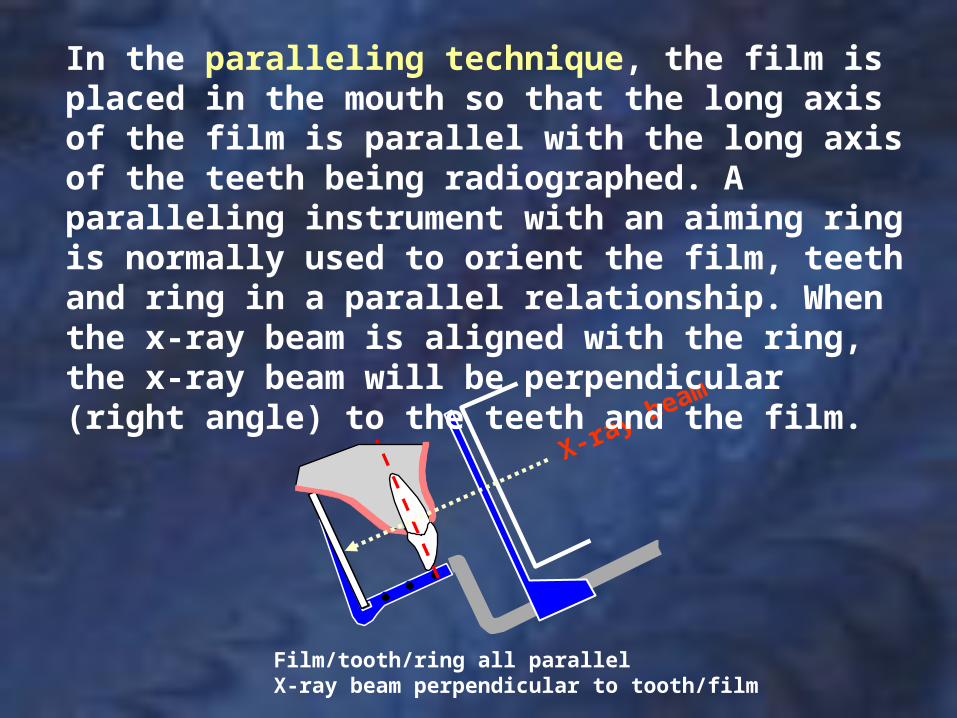
Film/tooth/ring all parallelX-ray beam perpendicular to tooth/film
X-ray beam
In the paralleling technique, the film is placed in the mouth so that the long axis of the film is parallel with the long axis of the teeth being radiographed. A paralleling instrument with an aiming ring is normally used to orient the film, teeth and ring in a parallel relationship. When the x-ray beam is aligned with the ring, the x-ray beam will be perpendicular (right angle) to the teeth and the film.

Paralleling Technique (Advantages)
There are two techniques for taking periapical films, the paralleling and the bisecting angle techniques. When comparing the two techniques, the advantages of the paralleling technique are:
1. Better dimensional accuracy: the paralleling technique results in less distortion of the image of the teeth. (The shape of the teeth and the relationship of the teeth to surrounding structures is more accurate).
2. When using the paralleling instrument with the aiming ring, the alignment of the x-ray beam is simplified.
(continued next slide)
Paralleling Technique (Advantages)
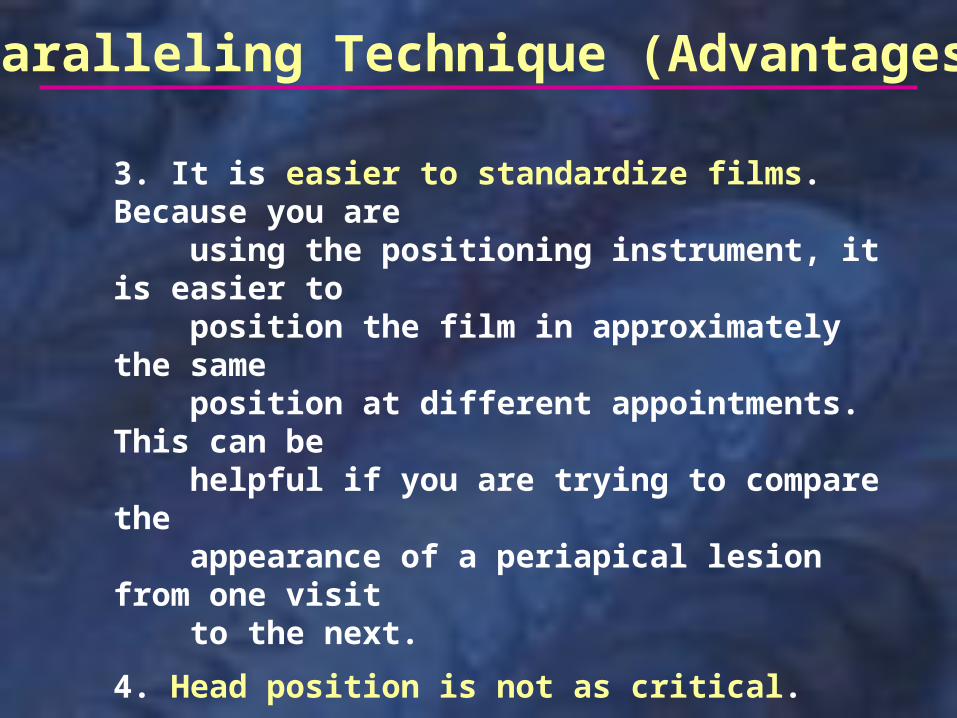
3. It is easier to standardize films. Because you are using the positioning instrument, it is easier to position the film in approximately the same position at different appointments. This can be helpful if you are trying to compare the appearance of a periapical lesion from one visit to the next.
4. Head position is not as critical. Because of the paralleling instrument, with its aiming ring, it is easy to properly align the x-ray beam no matter how the head is positioned.
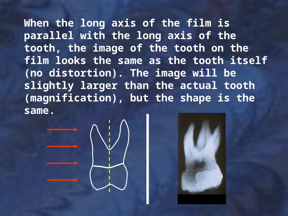
When the long axis of the film is parallel with the long axis of the tooth, the image of the tooth on the film looks the same as the tooth itself (no distortion). The image will be slightly larger than the actual tooth (magnification), but the shape is the same.
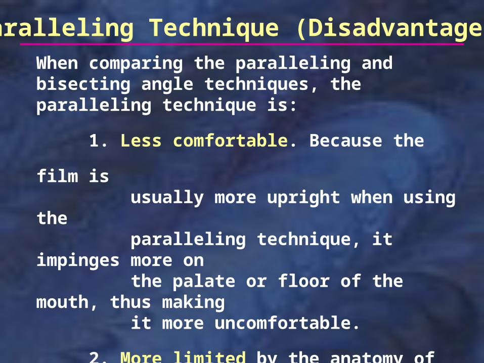
Paralleling Technique (Disadvantages)When comparing the paralleling and bisecting angle techniques, the paralleling technique is:
1. Less comfortable. Because the film is usually more upright when using the paralleling technique, it impinges more on the palate or floor of the mouth, thus making it more uncomfortable.
2. More limited by the anatomy of the patient’s mouth. A shallow palate or floor of the mouth makes it harder to position the film using the paralleling technique.
correct incorrect
Paralleling Film Placement
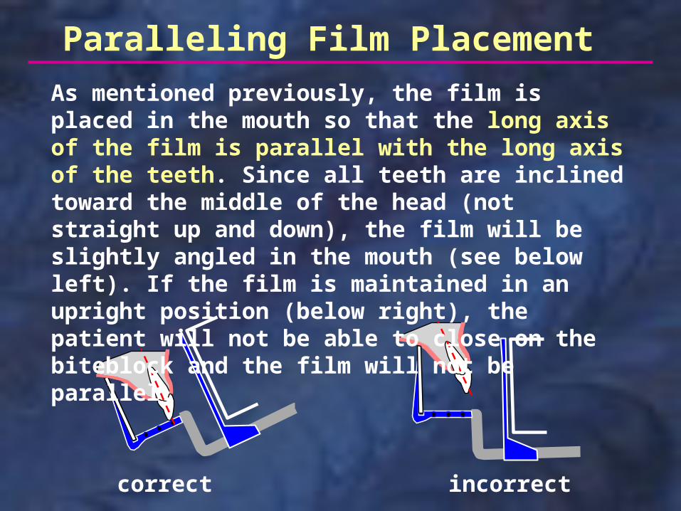
As mentioned previously, the film is placed in the mouth so that the long axis of the film is parallel with the long axis of the teeth. Since all teeth are inclined toward the middle of the head (not straight up and down), the film will be slightly angled in the mouth (see below left). If the film is maintained in an upright position (below right), the patient will not be able to close on the biteblock and the film will not be parallel.
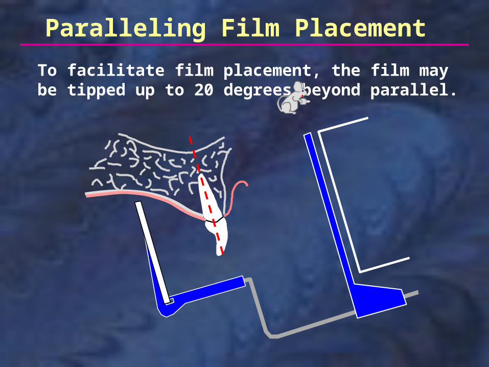
To facilitate film placement, the film may be tipped up to 20 degrees beyond parallel.
Paralleling Film Placement
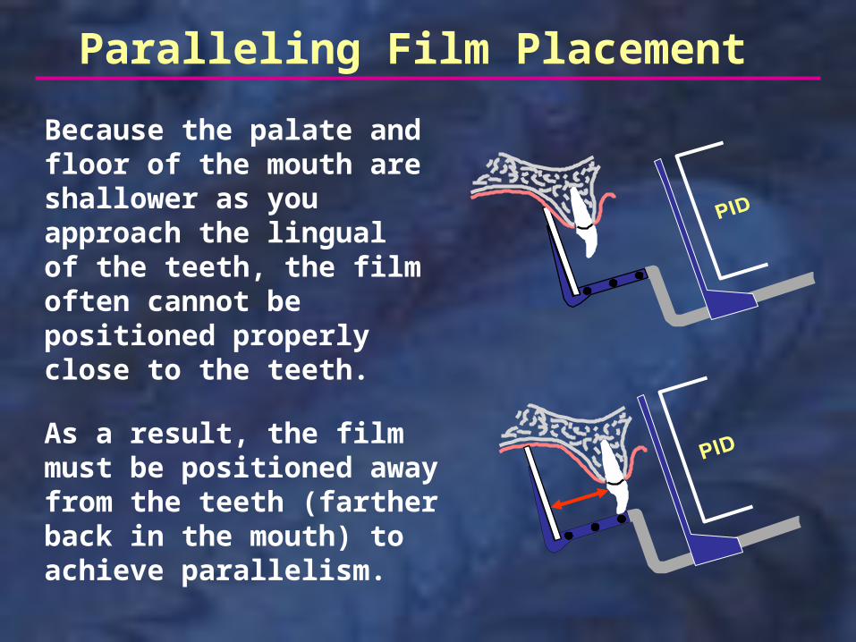
As a result, the film must be positioned away from the teeth (farther back in the mouth) to achieve parallelism.
Because the palate and floor of the mouth are shallower as you approach the lingual of the teeth, the film often cannot be positioned properly close to the teeth.
Paralleling Film Placement
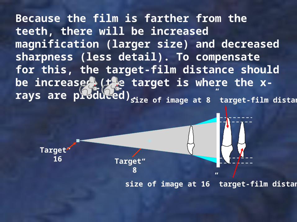
Because the film is farther from the teeth, there will be increased magnification (larger size) and decreased sharpness (less detail). To compensate for this, the target-film distance should be increased (the target is where the x-rays are produced).
Target 16” Target
8”
size of image at 8” target-film distance
size of image at 16” target-film distance
Long PID Short PIDRecessed target
Medium PIDRecessed target
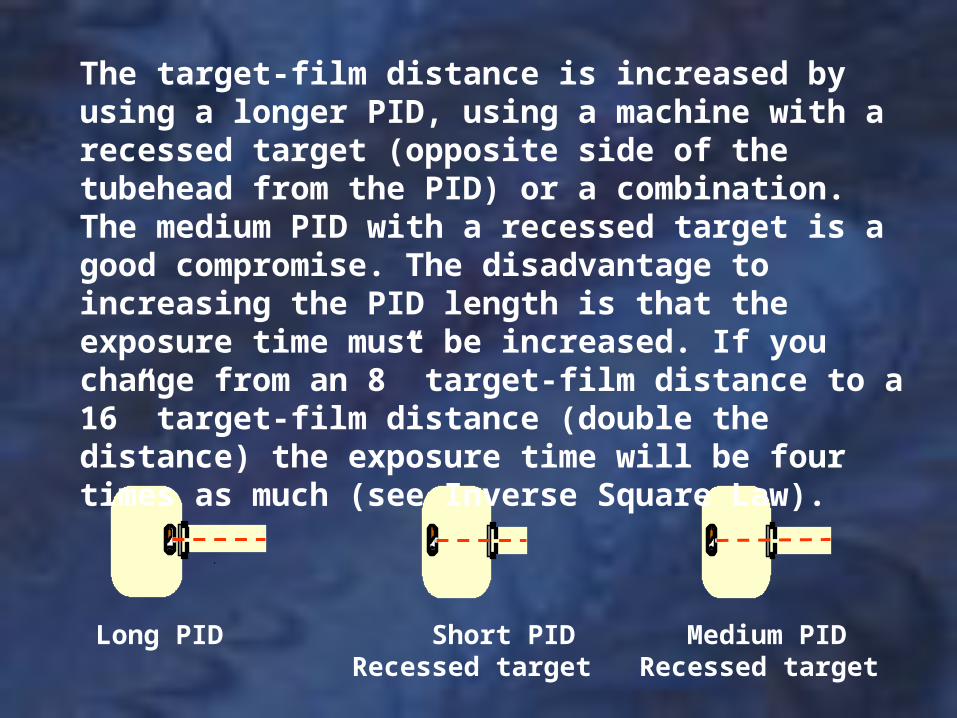
The target-film distance is increased by using a longer PID, using a machine with a recessed target (opposite side of the tubehead from the PID) or a combination. The medium PID with a recessed target is a good compromise. The disadvantage to increasing the PID length is that the exposure time must be increased. If you change from an 8” target-film distance to a 16” target-film distance (double the distance) the exposure time will be four times as much (see Inverse Square Law).
Best OKOK
Paralleling TechniqueHead Position
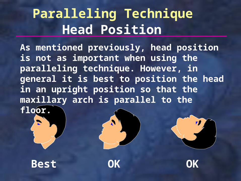
As mentioned previously, head position is not as important when using the paralleling technique. However, in general it is best to position the head in an upright position so that the maxillary arch is parallel to the floor.
#2#1
anterior posterior
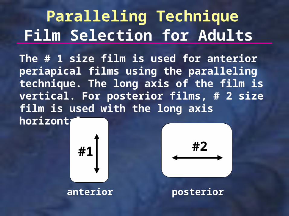
Paralleling TechniqueFilm Selection for Adults
The # 1 size film is used for anterior periapical films using the paralleling technique. The long axis of the film is vertical. For posterior films, # 2 size film is used with the long axis horizontal.
#0 #0
anterior posterior
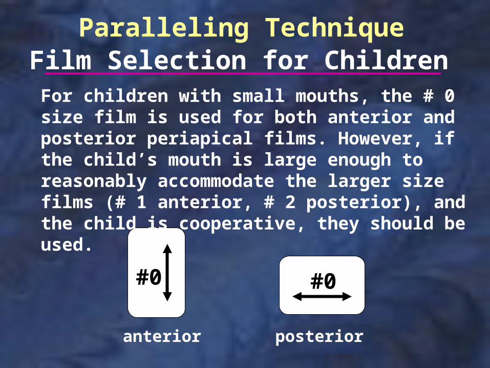
Paralleling TechniqueFilm Selection for Children
For children with small mouths, the # 0 size film is used for both anterior and posterior periapical films. However, if the child’s mouth is large enough to reasonably accommodate the larger size films (# 1 anterior, # 2 posterior), and the child is cooperative, they should be used.
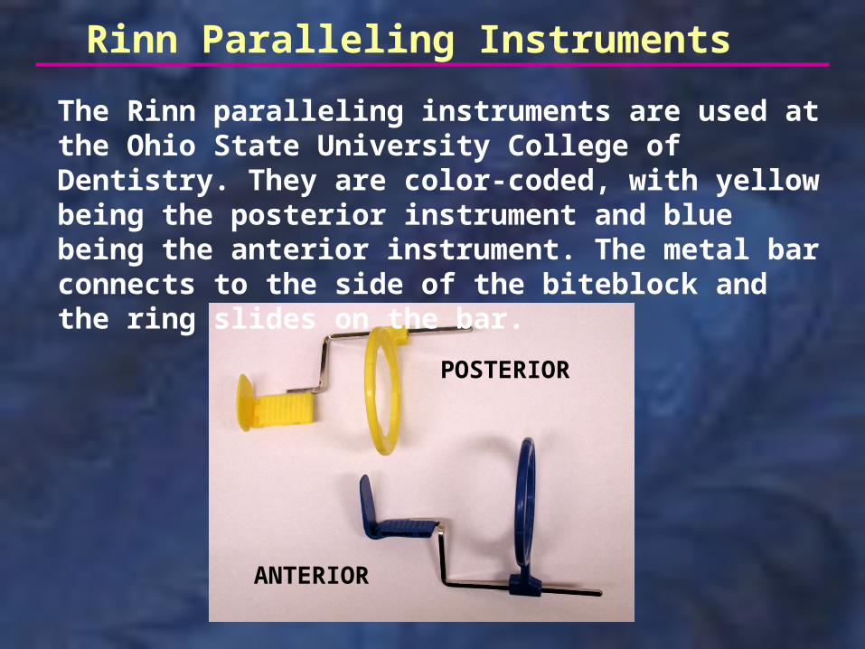
Rinn Paralleling Instruments
ANTERIOR
POSTERIOR
The Rinn paralleling instruments are used at the Ohio State University College of Dentistry. They are color-coded, with yellow being the posterior instrument and blue being the anterior instrument. The metal bar connects to the side of the biteblock and the ring slides on the bar.
front back
oppositeside
toward tube
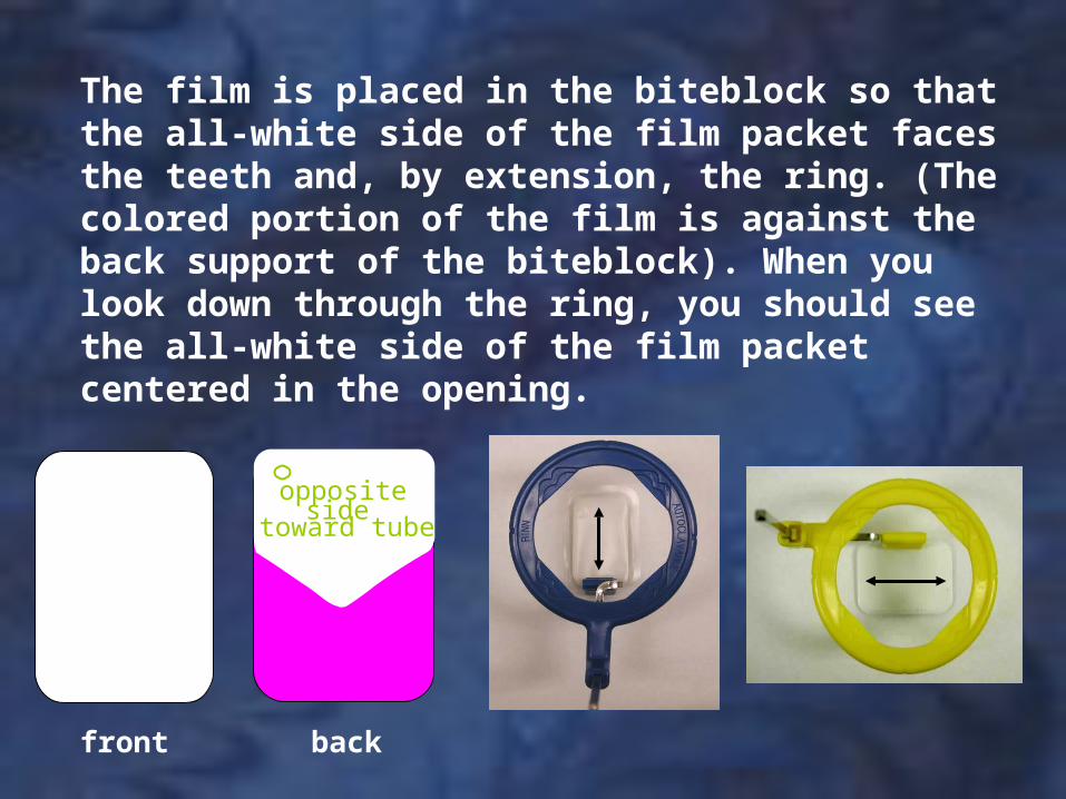
The film is placed in the biteblock so that the all-white side of the film packet faces the teeth and, by extension, the ring. (The colored portion of the film is against the back support of the biteblock). When you look down through the ring, you should see the all-white side of the film packet centered in the opening.
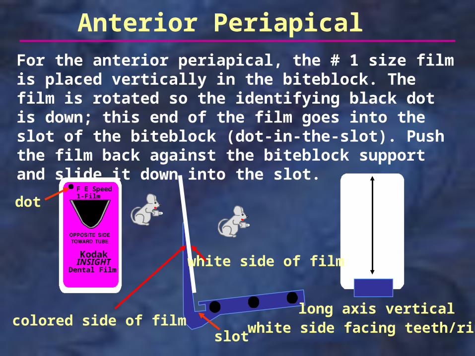
Anterior Periapical
long axis vertical
F E Speed1-Film
INSIGHTDental Film
Kodak
dot
slot
For the anterior periapical, the # 1 size film is placed vertically in the biteblock. The film is rotated so the identifying black dot is down; this end of the film goes into the slot of the biteblock (dot-in-the-slot). Push the film back against the biteblock support and slide it down into the slot.
colored side of film
white side of film
white side facing teeth/ring
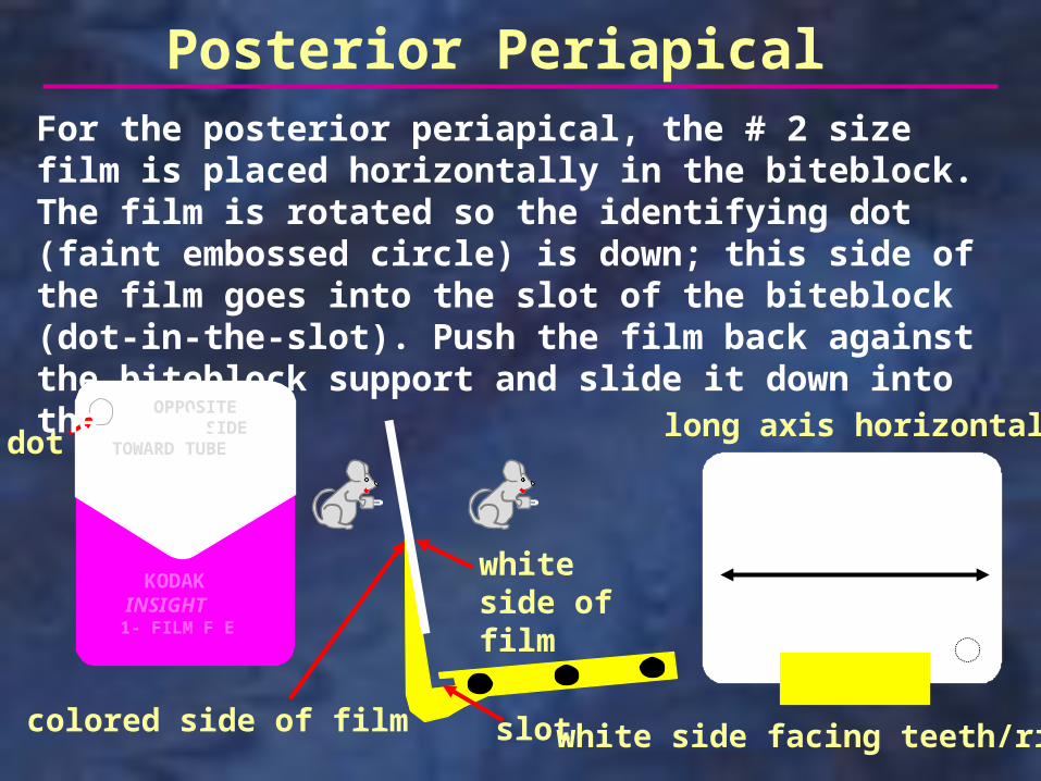
Posterior Periapical
long axis horizontal OPPOSITE SIDETOWARD TUBE
KODAK INSIGHT1- FILM F E
slot
dot
For the posterior periapical, the # 2 size film is placed horizontally in the biteblock. The film is rotated so the identifying dot (faint embossed circle) is down; this side of the film goes into the slot of the biteblock (dot-in-the-slot). Push the film back against the biteblock support and slide it down into the slot.
colored side of film
white side of film
white side facing teeth/ring
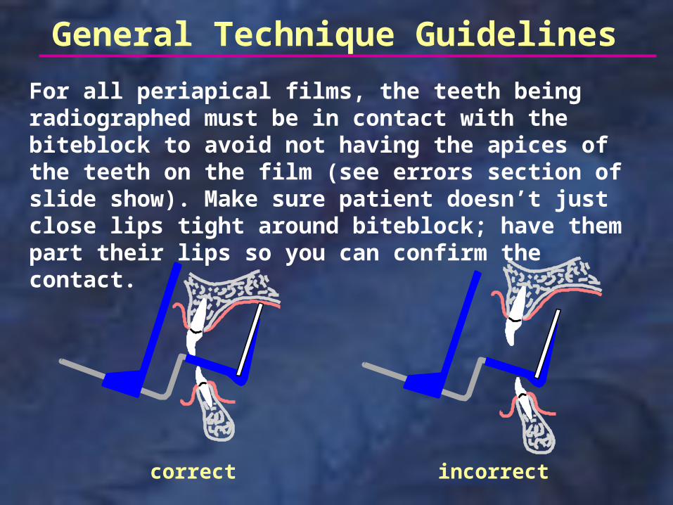
For all periapical films, the teeth being radiographed must be in contact with the biteblock to avoid not having the apices of the teeth on the film (see errors section of slide show). Make sure patient doesn’t just close lips tight around biteblock; have them part their lips so you can confirm the contact.
correct incorrect
General Technique Guidelines

As shown above, cotton rolls may be used in any area of the mouth to help support the biteblock, especially if an edentulous region or uneven teeth oppose the teeth being radiographed. Using a cotton roll also makes it more comfortable for the patient to bite in some situations. The cotton roll should be placed against the arch opposite the one being radiographed.
General Technique Guidelines
General Technique Guidelines
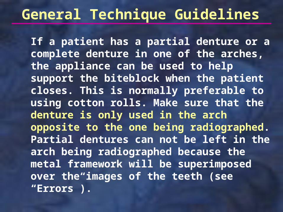
If a patient has a partial denture or a complete denture in one of the arches, the appliance can be used to help support the biteblock when the patient closes. This is normally preferable to using cotton rolls. Make sure that the denture is only used in the arch opposite to the one being radiographed. Partial dentures can not be left in the arch being radiographed because the metal framework will be superimposed over the images of the teeth (see “Errors”).
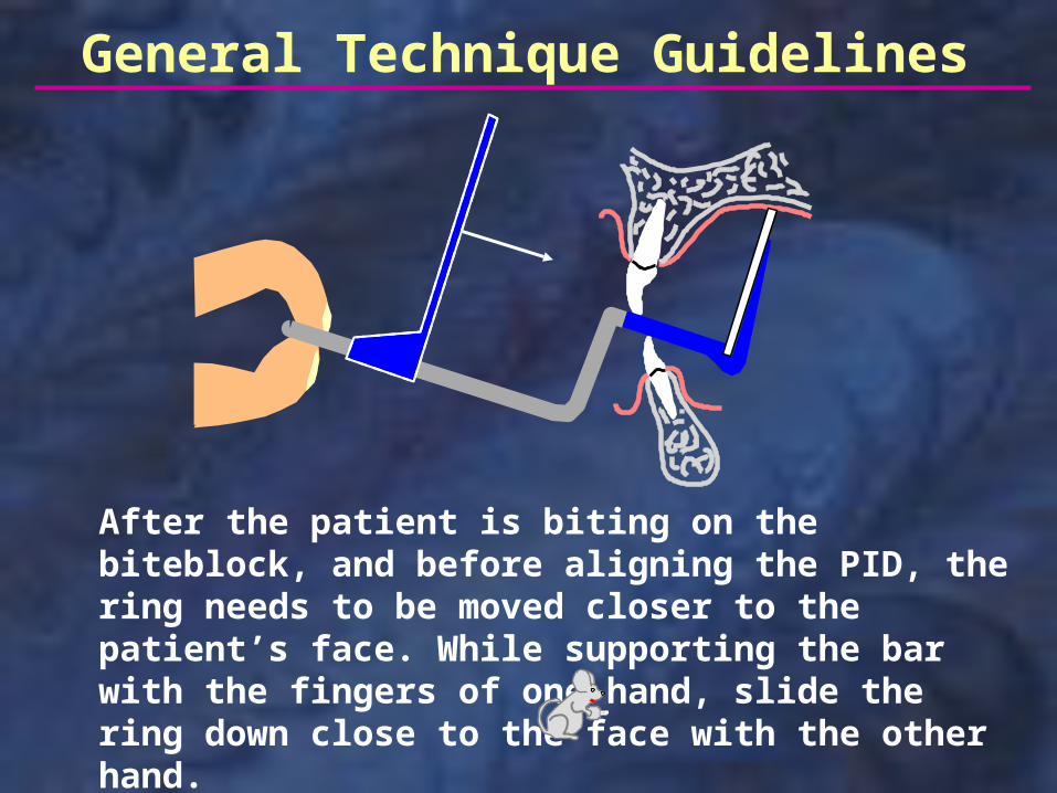
After the patient is biting on the biteblock, and before aligning the PID, the ring needs to be moved closer to the patient’s face. While supporting the bar with the fingers of one hand, slide the ring down close to the face with the other hand.
General Technique Guidelines
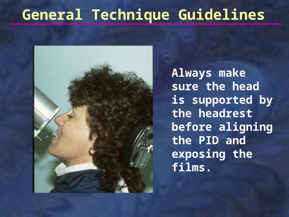
Always make sure the head is supported by the headrest before aligning the PID and exposing the films.
General Technique Guidelines
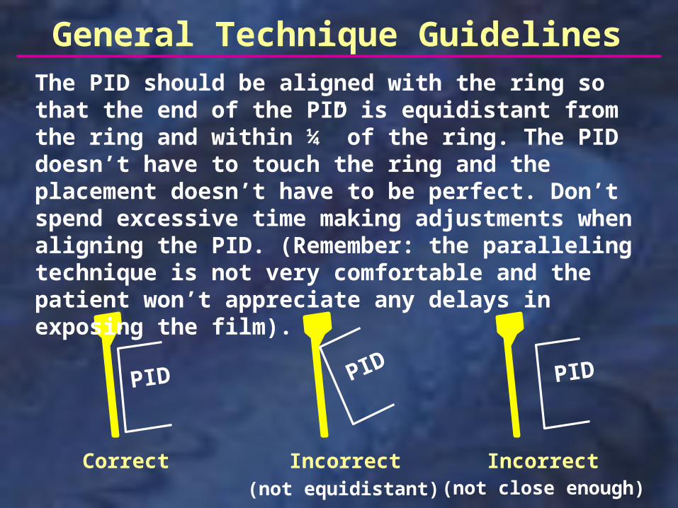
The PID should be aligned with the ring so that the end of the PID is equidistant from the ring and within ¼” of the ring. The PID doesn’t have to touch the ring and the placement doesn’t have to be perfect. Don’t spend excessive time making adjustments when aligning the PID. (Remember: the paralleling technique is not very comfortable and the patient won’t appreciate any delays in exposing the film).
Incorrect IncorrectCorrect
PID PID PID
(not equidistant) (not close enough)
General Technique Guidelines
Maxillary Central-lateral
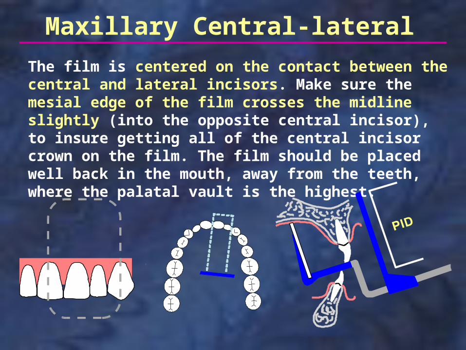
The film is centered on the contact between the central and lateral incisors. Make sure the mesial edge of the film crosses the midline slightly (into the opposite central incisor), to insure getting all of the central incisor crown on the film. The film should be placed well back in the mouth, away from the teeth, where the palatal vault is the highest.
Maxillary Central-lateral
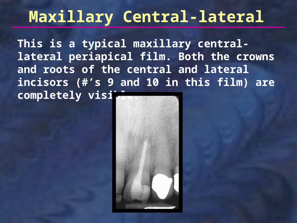
This is a typical maxillary central-lateral periapical film. Both the crowns and roots of the central and lateral incisors (#’s 9 and 10 in this film) are completely visible.
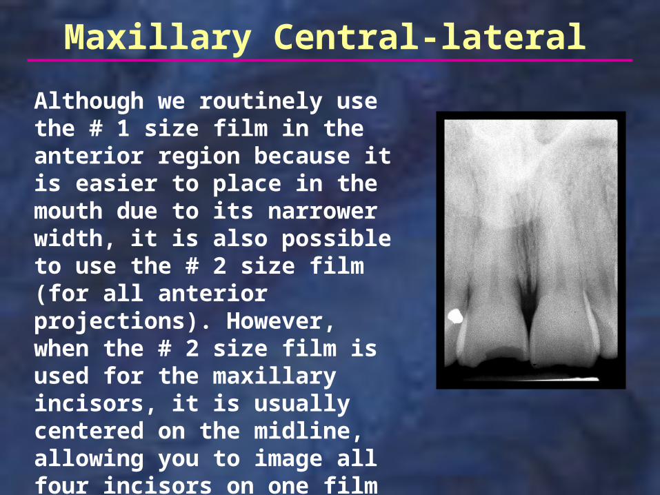
Although we routinely use the # 1 size film in the anterior region because it is easier to place in the mouth due to its narrower width, it is also possible to use the # 2 size film (for all anterior projections). However, when the # 2 size film is used for the maxillary incisors, it is usually centered on the midline, allowing you to image all four incisors on one film (the film at right is slightly cropped, cutting off the distal of the laterals).
Maxillary Central-lateral
Maxillary Canine
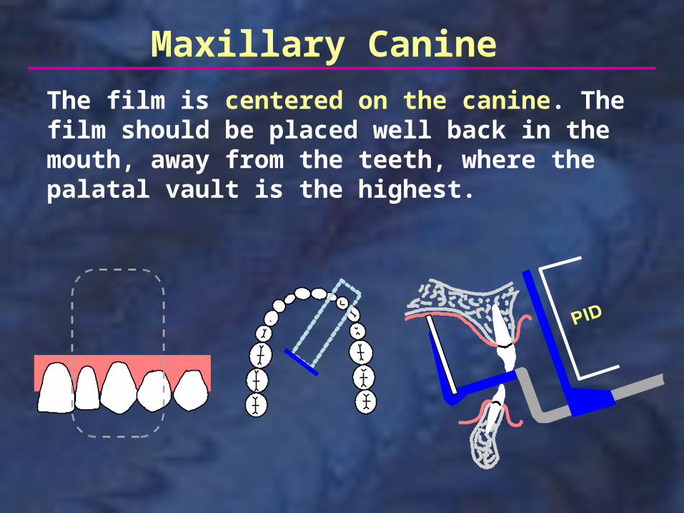
The film is centered on the canine. The film should be placed well back in the mouth, away from the teeth, where the palatal vault is the highest.
Maxillary Canine
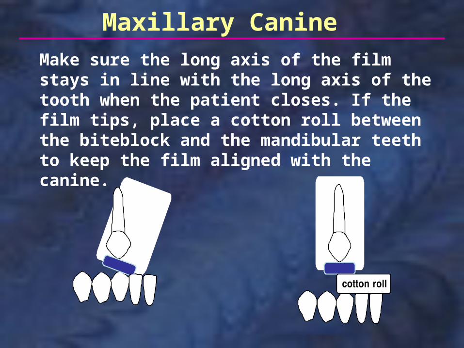
Make sure the long axis of the film stays in line with the long axis of the tooth when the patient closes. If the film tips, place a cotton roll between the biteblock and the mandibular teeth to keep the film aligned with the canine.
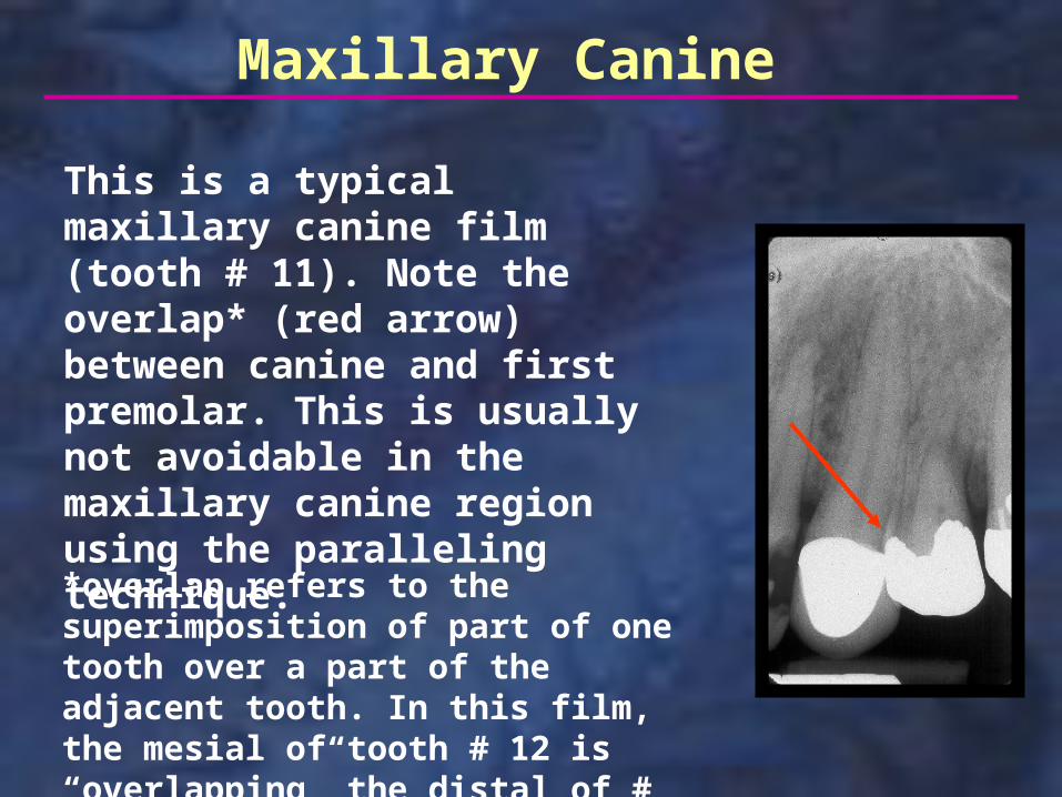
This is a typical maxillary canine film (tooth # 11). Note the overlap* (red arrow) between canine and first premolar. This is usually not avoidable in the maxillary canine region using the paralleling technique.
Maxillary Canine
*overlap refers to the superimposition of part of one tooth over a part of the adjacent tooth. In this film, the mesial of tooth # 12 is “overlapping” the distal of # 11.
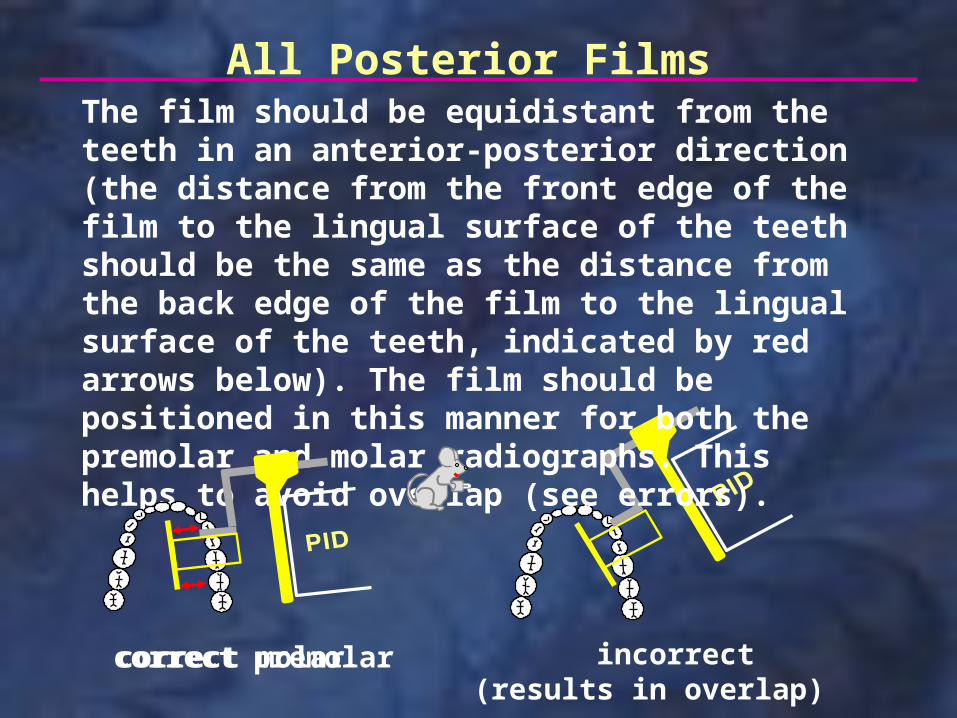
The film should be equidistant from the teeth in an anterior-posterior direction (the distance from the front edge of the film to the lingual surface of the teeth should be the same as the distance from the back edge of the film to the lingual surface of the teeth, indicated by red arrows below). The film should be positioned in this manner for both the premolar and molar radiographs. This helps to avoid overlap (see errors).
correct premolar incorrect(results in overlap)
All Posterior Films
correct molar
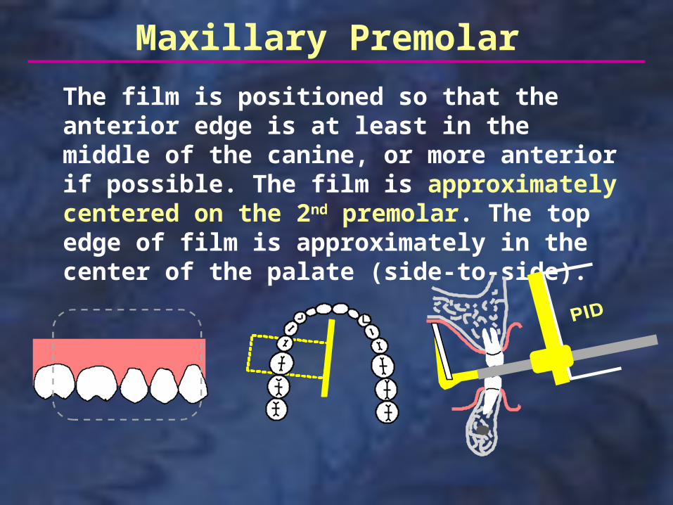
The film is positioned so that the anterior edge is at least in the middle of the canine, or more anterior if possible. The film is approximately centered on the 2nd premolar. The top edge of film is approximately in the center of the palate (side-to-side).
Maxillary Premolar
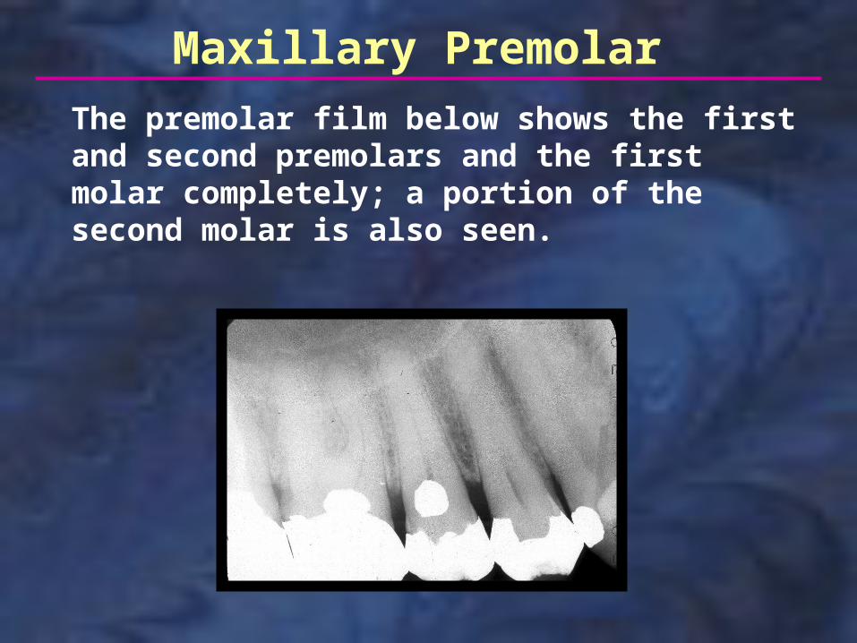
Maxillary Premolar
The premolar film below shows the first and second premolars and the first molar completely; a portion of the second molar is also seen.
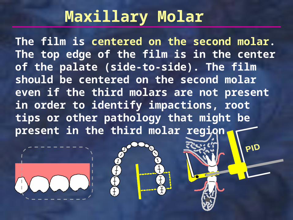
The film is centered on the second molar. The top edge of the film is in the center of the palate (side-to-side). The film should be centered on the second molar even if the third molars are not present in order to identify impactions, root tips or other pathology that might be present in the third molar region.
Maxillary Molar
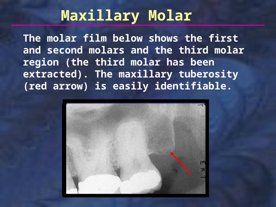
Maxillary Molar
The molar film below shows the first and second molars and the third molar region (the third molar has been extracted). The maxillary tuberosity (red arrow) is easily identifiable.
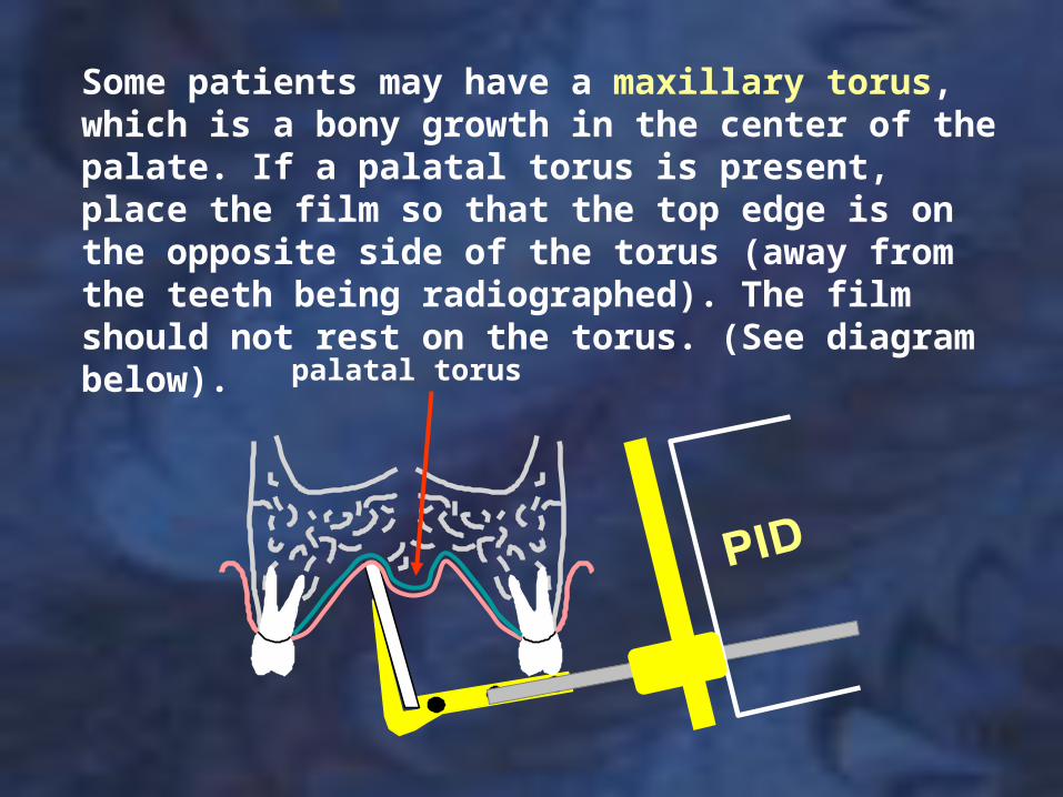
palatal torus
Some patients may have a maxillary torus, which is a bony growth in the center of the palate. If a palatal torus is present, place the film so that the top edge is on the opposite side of the torus (away from the teeth being radiographed). The film should not rest on the torus. (See diagram below).
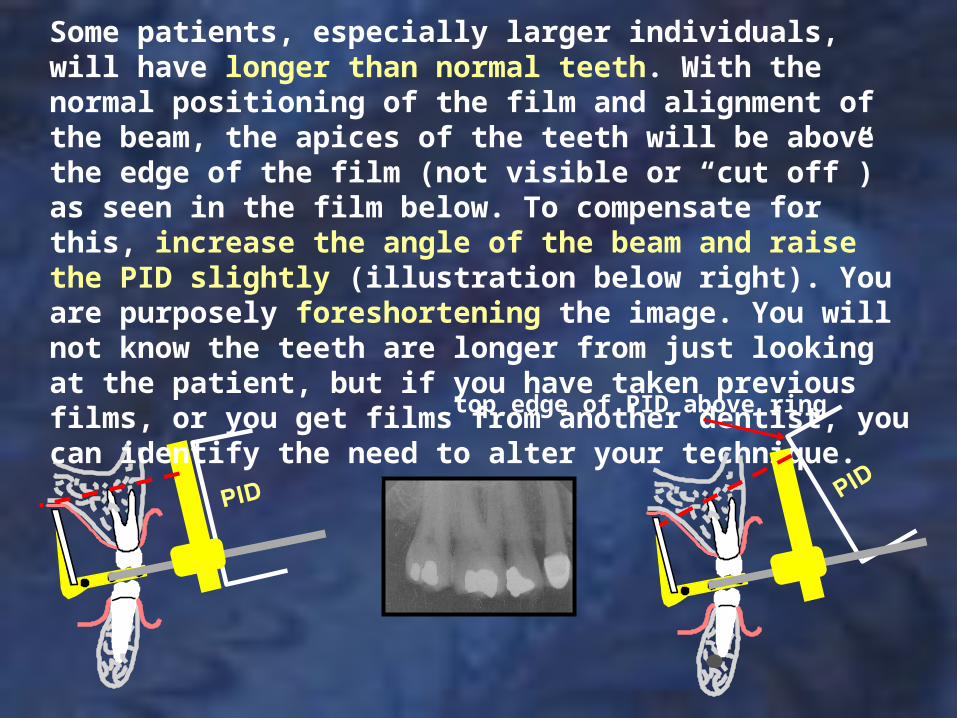
Some patients, especially larger individuals, will have longer than normal teeth. With the normal positioning of the film and alignment of the beam, the apices of the teeth will be above the edge of the film (not visible or “cut off”) as seen in the film below. To compensate for this, increase the angle of the beam and raise the PID slightly (illustration below right). You are purposely foreshortening the image. You will not know the teeth are longer from just looking at the patient, but if you have taken previous films, or you get films from another dentist, you can identify the need to alter your technique.
top edge of PID above ring
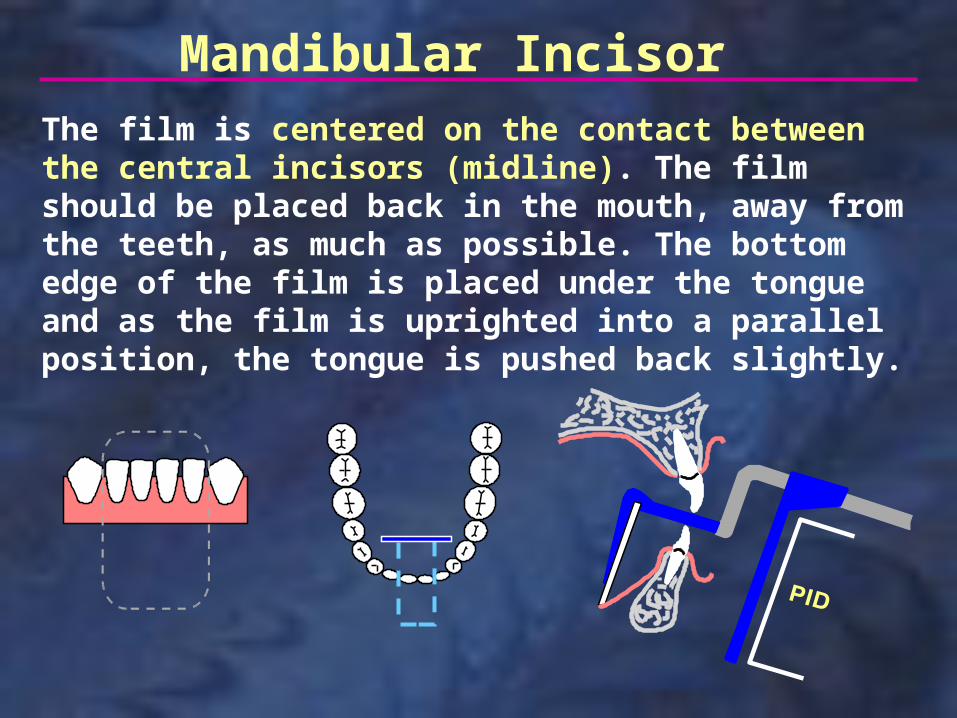
Mandibular IncisorThe film is centered on the contact between the central incisors (midline). The film should be placed back in the mouth, away from the teeth, as much as possible. The bottom edge of the film is placed under the tongue and as the film is uprighted into a parallel position, the tongue is pushed back slightly.
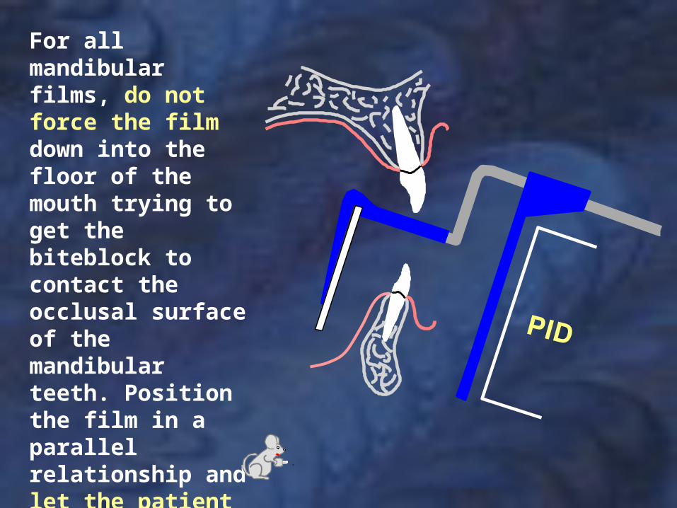
For all mandibular films, do not force the film down into the floor of the mouth trying to get the biteblock to contact the occlusal surface of the mandibular teeth. Position the film in a parallel relationship and let the patient guide the film into place as they close their mouth. Have the patient bite slowly and gently.
Mandibular Incisor
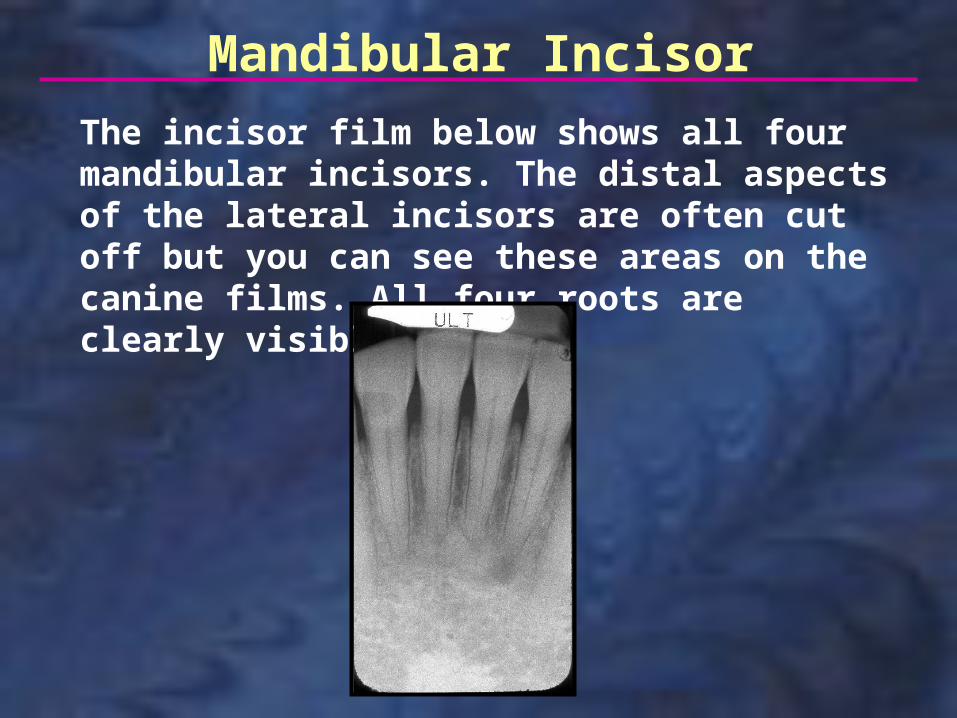
The incisor film below shows all four mandibular incisors. The distal aspects of the lateral incisors are often cut off but you can see these areas on the canine films. All four roots are clearly visible.
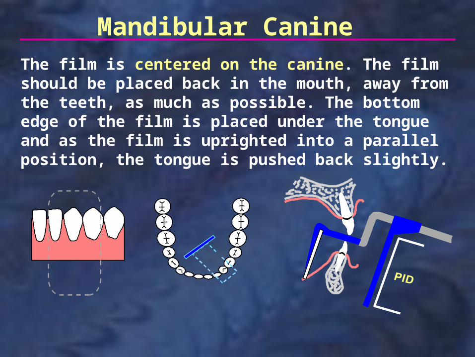
Mandibular CanineThe film is centered on the canine. The film should be placed back in the mouth, away from the teeth, as much as possible. The bottom edge of the film is placed under the tongue and as the film is uprighted into a parallel position, the tongue is pushed back slightly.
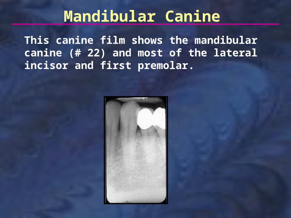
Mandibular Canine
This canine film shows the mandibular canine (# 22) and most of the lateral incisor and first premolar.
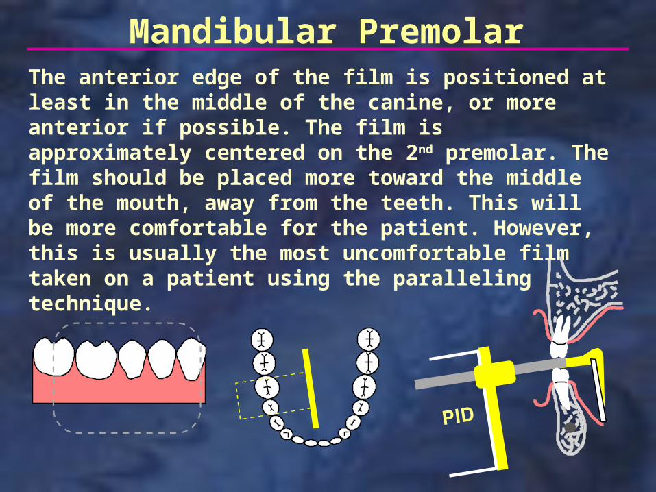
The anterior edge of the film is positioned at least in the middle of the canine, or more anterior if possible. The film is approximately centered on the 2nd premolar. The film should be placed more toward the middle of the mouth, away from the teeth. This will be more comfortable for the patient. However, this is usually the most uncomfortable film taken on a patient using the paralleling technique.
Mandibular Premolar
Mandibular Premolar
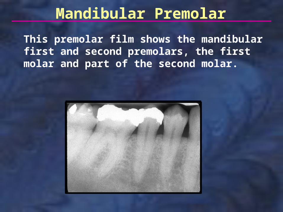
This premolar film shows the mandibular first and second premolars, the first molar and part of the second molar.
Mandibular Molar
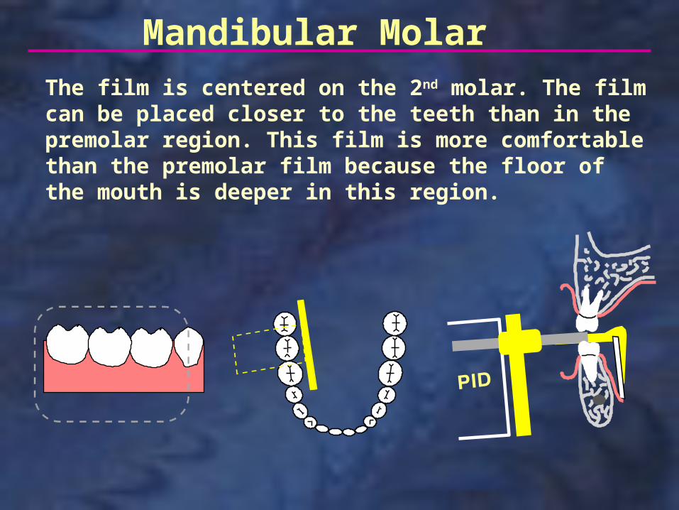
The film is centered on the 2nd molar. The film can be placed closer to the teeth than in the premolar region. This film is more comfortable than the premolar film because the floor of the mouth is deeper in this region.
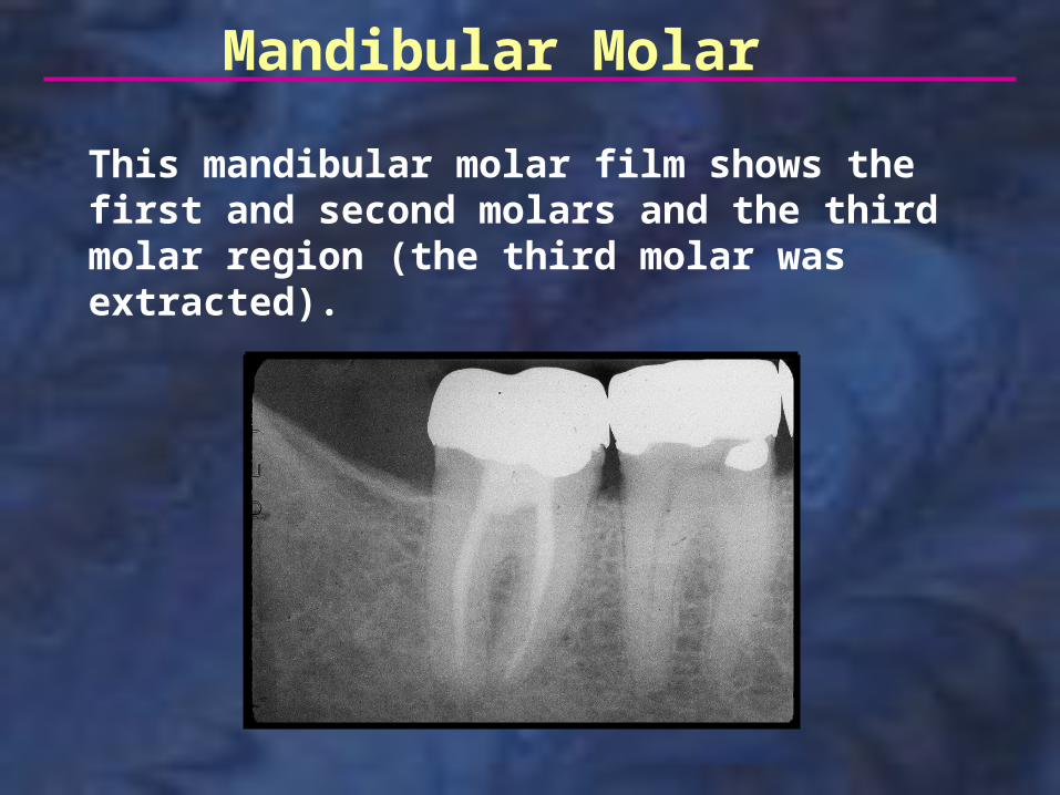
Mandibular Molar
This mandibular molar film shows the first and second molars and the third molar region (the third molar was extracted).
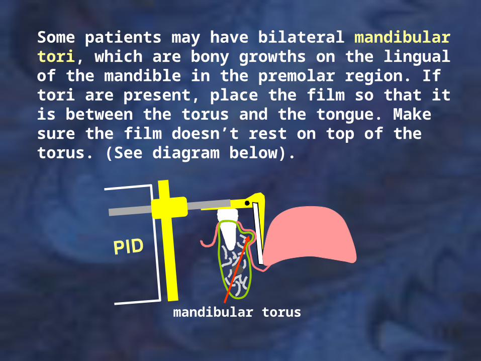
mandibular torus
Some patients may have bilateral mandibular tori, which are bony growths on the lingual of the mandible in the premolar region. If tori are present, place the film so that it is between the torus and the tongue. Make sure the film doesn’t rest on top of the torus. (See diagram below).
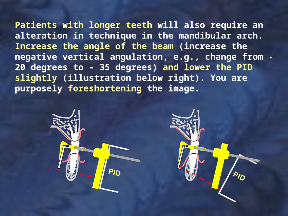
Patients with longer teeth will also require an alteration in technique in the mandibular arch. Increase the angle of the beam (increase the negative vertical angulation, e.g., change from - 20 degrees to - 35 degrees) and lower the PID slightly (illustration below right). You are purposely foreshortening the image.
Adult full-mouth series, Paralleling Technique
# 1# 2 # 2
R L
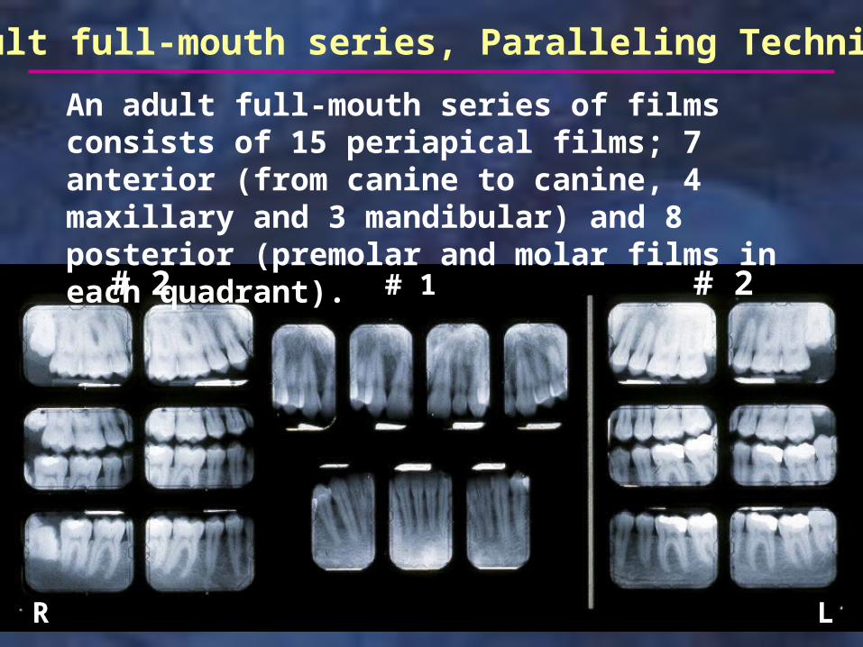
An adult full-mouth series of films consists of 15 periapical films; 7 anterior (from canine to canine, 4 maxillary and 3 mandibular) and 8 posterior (premolar and molar films in each quadrant).
Anterior First
When taking films on a patient, you should always start with the anterior films. If you are doing a full series, start with the maxillary canine film and then finish all the anterior films, both maxillary and mandibular. Then complete the posterior films, starting with the premolar, then molar, in each quadrant. When doing only a few films on a patient, start with the most anterior film and work your way back in the mouth. This sequence of taking films allows the patient to get used to the procedure with a minimum of discomfort and helps to avoid stimulation of the gag reflex.
Paralleling Technique Errors
The following slides identify some of the most common errors seen when using the paralleling technique.
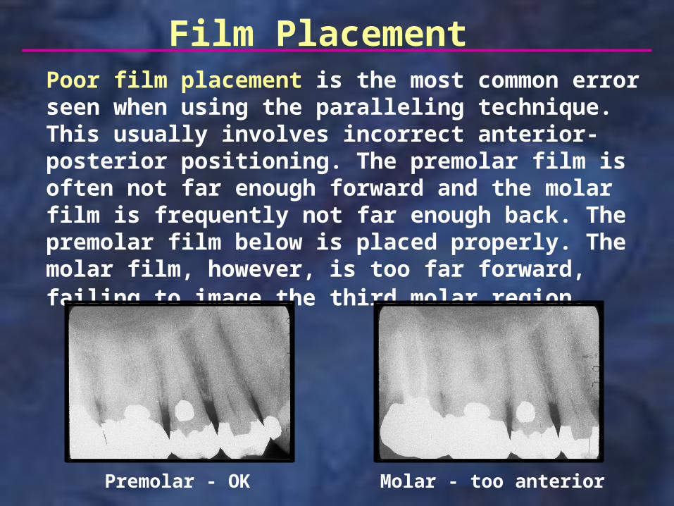
Poor film placement is the most common error seen when using the paralleling technique. This usually involves incorrect anterior-posterior positioning. The premolar film is often not far enough forward and the molar film is frequently not far enough back. The premolar film below is placed properly. The molar film, however, is too far forward, failing to image the third molar region.
Premolar - OK Molar - too anterior
Film Placement
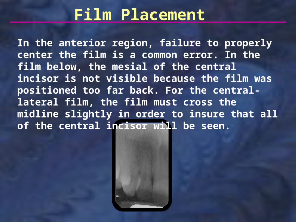
In the anterior region, failure to properly center the film is a common error. In the film below, the mesial of the central incisor is not visible because the film was positioned too far back. For the central-lateral film, the film must cross the midline slightly in order to insure that all of the central incisor will be seen.
Film Placement
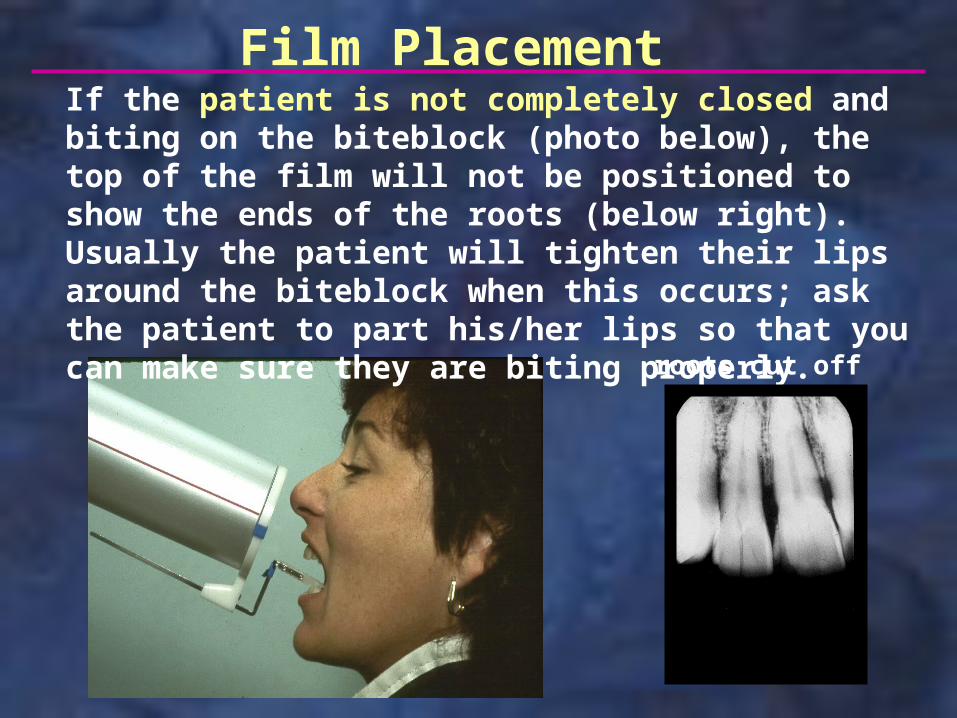
If the patient is not completely closed and biting on the biteblock (photo below), the top of the film will not be positioned to show the ends of the roots (below right). Usually the patient will tighten their lips around the biteblock when this occurs; ask the patient to part his/her lips so that you can make sure they are biting properly.
roots cut off
Film Placement
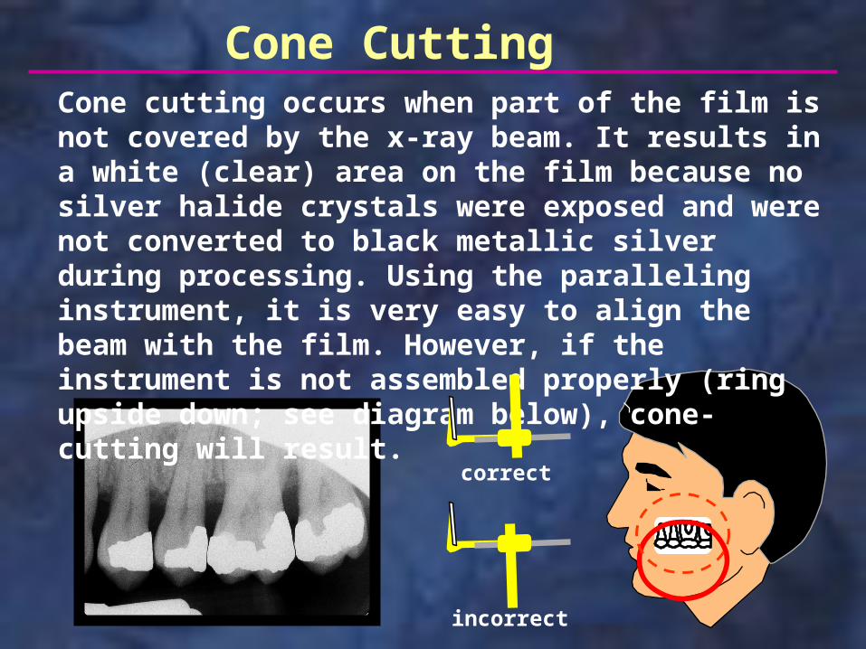
Cone CuttingCone cutting occurs when part of the film is not covered by the x-ray beam. It results in a white (clear) area on the film because no silver halide crystals were exposed and were not converted to black metallic silver during processing. Using the paralleling instrument, it is very easy to align the beam with the film. However, if the instrument is not assembled properly (ring upside down; see diagram below), cone-cutting will result.
correct
incorrect
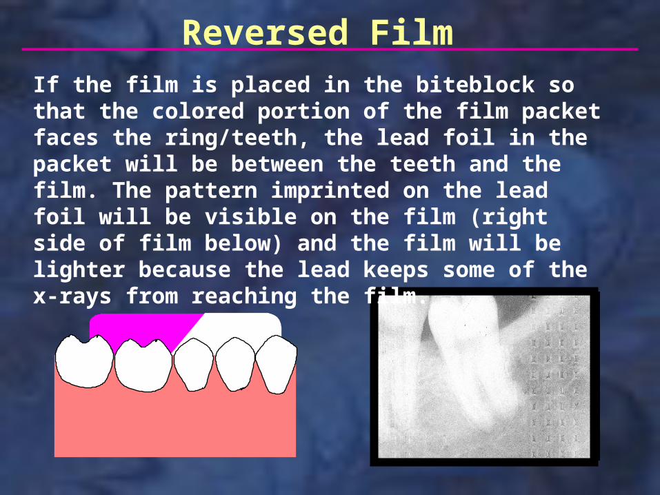
Reversed FilmIf the film is placed in the biteblock so that the colored portion of the film packet faces the ring/teeth, the lead foil in the packet will be between the teeth and the film. The pattern imprinted on the lead foil will be visible on the film (right side of film below) and the film will be lighter because the lead keeps some of the x-rays from reaching the film.
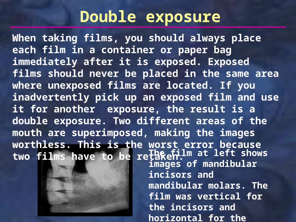
Double exposureWhen taking films, you should always place each film in a container or paper bag immediately after it is exposed. Exposed films should never be placed in the same area where unexposed films are located. If you inadvertently pick up an exposed film and use it for another exposure, the result is a double exposure. Two different areas of the mouth are superimposed, making the images worthless. This is the worst error because two films have to be retaken.
The film at left shows images of mandibular incisors and mandibular molars. The film was vertical for the incisors and horizontal for the molars.
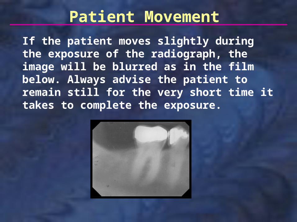
Patient Movement
If the patient moves slightly during the exposure of the radiograph, the image will be blurred as in the film below. Always advise the patient to remain still for the very short time it takes to complete the exposure.
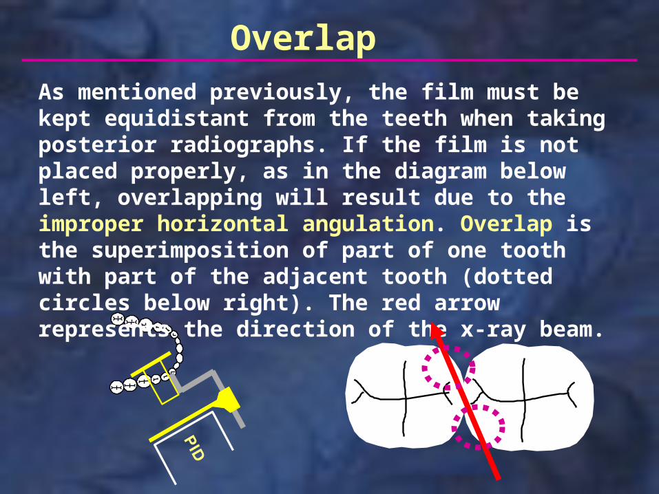
Overlap
As mentioned previously, the film must be kept equidistant from the teeth when taking posterior radiographs. If the film is not placed properly, as in the diagram below left, overlapping will result due to the improper horizontal angulation. Overlap is the superimposition of part of one tooth with part of the adjacent tooth (dotted circles below right). The red arrow represents the direction of the x-ray beam.
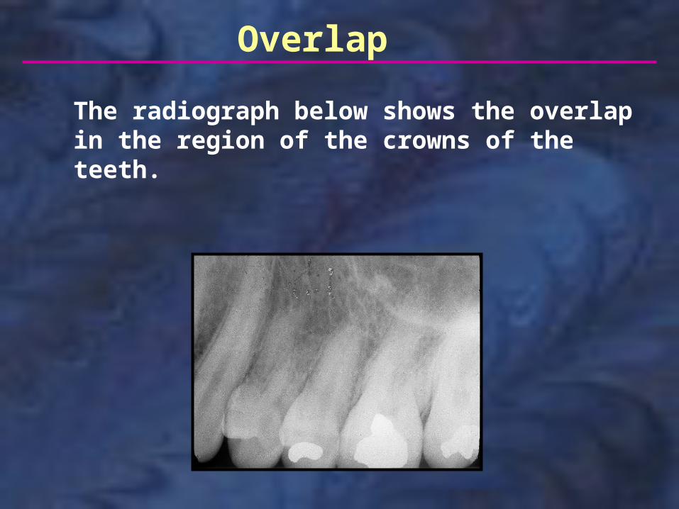
Overlap
The radiograph below shows the overlap in the region of the crowns of the teeth.
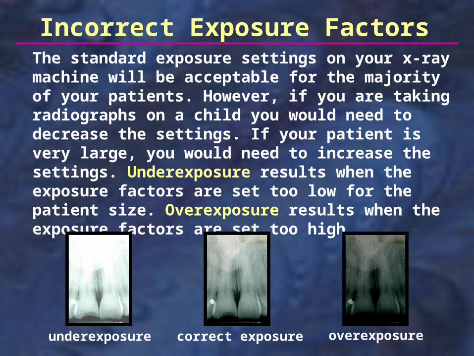
overexposureunderexposure
Incorrect Exposure Factors
correct exposure
The standard exposure settings on your x-ray machine will be acceptable for the majority of your patients. However, if you are taking radiographs on a child you would need to decrease the settings. If your patient is very large, you would need to increase the settings. Underexposure results when the exposure factors are set too low for the patient size. Overexposure results when the exposure factors are set too high.
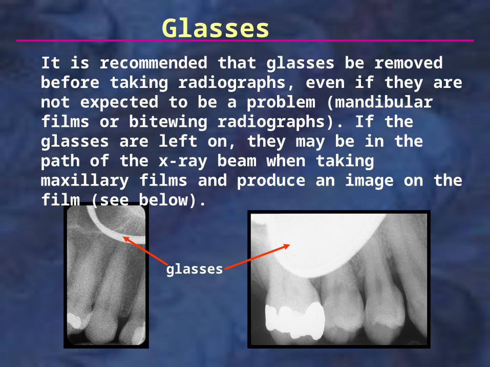
Glasses It is recommended that glasses be removed before taking radiographs, even if they are not expected to be a problem (mandibular films or bitewing radiographs). If the glasses are left on, they may be in the path of the x-ray beam when taking maxillary films and produce an image on the film (see below).
glasses
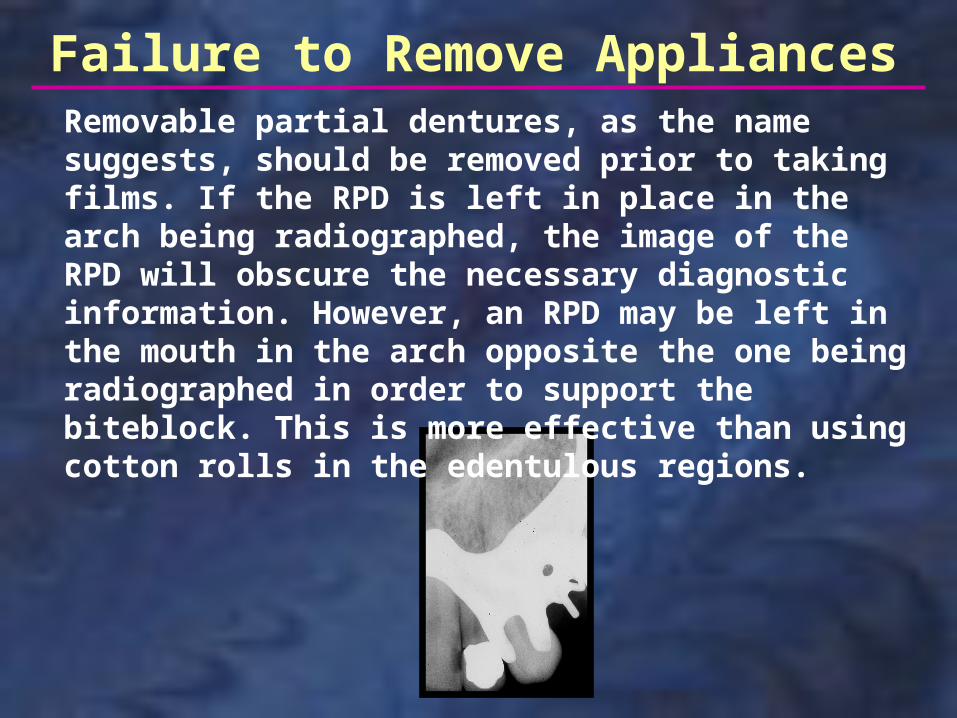
Failure to Remove AppliancesRemovable partial dentures, as the name suggests, should be removed prior to taking films. If the RPD is left in place in the arch being radiographed, the image of the RPD will obscure the necessary diagnostic information. However, an RPD may be left in the mouth in the arch opposite the one being radiographed in order to support the biteblock. This is more effective than using cotton rolls in the edentulous regions.
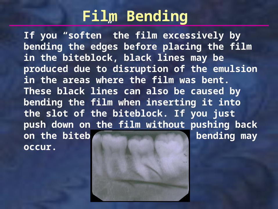
If you “soften” the film excessively by bending the edges before placing the film in the biteblock, black lines may be produced due to disruption of the emulsion in the areas where the film was bent. These black lines can also be caused by bending the film when inserting it into the slot of the biteblock. If you just push down on the film without pushing back on the biteblock support, this bending may occur.
Film Bending
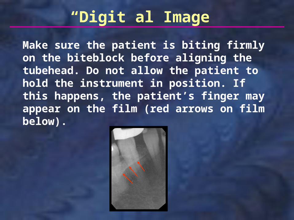
“Digit”al Image
Make sure the patient is biting firmly on the biteblock before aligning the tubehead. Do not allow the patient to hold the instrument in position. If this happens, the patient’s finger may appear on the film (red arrows on film below).
This concludes the section on Paralleling Technique. Additional self-study modules are available at: http://dent.osu.edu/radiology/resources.htm
If you have any questions, you may e-mail me at: [email protected]
Robert M. Jaynes, DDS, MSDirector, Radiology GroupCollege of DentistryOhio State University