semecarpus anacardium (bhallataka) alters the glucose
TRANSCRIPT
Hindawi Publishing CorporationEvidence-Based Complementary and Alternative MedicineVolume 2011, Article ID 142978, 9 pagesdoi:10.1155/2011/142978
Research Article
Semecarpus anacardium (Bhallataka) Alters the GlucoseMetabolism and Energy Production in Diabetic Rats
Jaya Aseervatham,1 Shanthi Palanivelu,2 and Sachdanandam Panchanadham1
1 Department of Medical Biochemistry, Dr. ALM Post Graduate Institute of Basic Medical Sciences, University of Madras,Taramani Campus, Chennai 600 113, India
2 Department of Pathology, Dr. ALM Post Graduate Institute of Basic Medical Sciences, University of Madras,Taramani Campus, Chennai 600 113, India
Correspondence should be addressed to Sachdanandam Panchanadham, [email protected]
Received 12 March 2010; Revised 11 May 2010; Accepted 27 June 2010
Copyright © 2011 Jaya Aseervatham et al. This is an open access article distributed under the Creative Commons AttributionLicense, which permits unrestricted use, distribution, and reproduction in any medium, provided the original work is properlycited.
Glucose produced by gluconeogenesis and glycogenolysis plays an important role in aggravating hyperglycemia in diabetes,and altered mitochondrial function is associated with impaired energy production. The present study focuses on the effectof Semecarpus anacardium on carbohydrate metabolism and energy production in diabetic rats. Diabetes was induced by theadministration of Streptozotocin at a dose of 50 mg/kg.b.wt. Three days after the induction, Semecarpus anacardium at a dose of300 mg/kg.b.wt was administered for 21 days. After the experimental duration, the activities of the enzymes involved in Glycolysis,TCA cycle, gluconeogenesis, and glycogen were assayed in the liver and kidney of the experimental animals. In addition, to thecomplexes the protein expression of AKT and PI3K were assayed. The levels of the enzymes involved in Glycolysis and TCA cycleincreased, while that of gluconeogensis decreased. The activities of the mitochondrial complexes were also favorably modulated.The expressions of PI3K and AKT also increased in the skeletal muscle. These effects may be attributed to the hypoglycemic andthe antioxidative activity of Semecarpus anacardium. The results of the study revealed that Semecarpus anacardium was able torestore the altered activities of the enzymes involved in carbohydrate metabolism and energy production.
1. Introduction
Diabetes mellitus is a metabolic disorder characterized byhyperglycemia due to defect in insulin secretion, action, orboth. Liver plays an important role in the glucose home-ostasis through glycolysis, glycogenesis, and gluconeogenesis.The net glucose uptake by the liver depends on the activitiesof glucokinase and glucose 6 phosphatase. The activity ofhepatic glucokinase is markedly decreased and activity ofglucose-6-phosphatase is almost doubled [1]. Glucose, takenup by secondary active transporter proteins, is degradedto pyruvate, which is then introduced into the citric acidcycle after its decarboxylation to acetyl coenzyme A. TheKrebs cycle provides NADH for oxidative phosphorylationto generate the electron gradient for ATP formation.
Medicinal plants have played a significant role in variousancient traditional systems of medicine. They are rich sou-
rces of bioactive compounds and thus serve as impor-tant raw materials for drug production and have becomea target for the search of new drugs [2]. Semecarpusanacardium L. (Anacardiaceae) (SA) commonly known asBhallataka or marking nut is used in indigenous systems ofmedicine for the treatment of various diseases [3]. Manycompounds mainly biflavonoids, phenolics, bhilawanols,sterols, Anacardic acid, and glycosides have been identifiedas constituents of S. anacardium nut extract. On the basisof chemical and spectral data, several biflavonoids, suchas Jeediflavanone, Galluflavanone, Nalluflavanone, Semecar-petin, and Anacardiflavanone have been characterized [4].Several monophenolic compounds known as Semecarpol(C17H28O) and Bhilawanol were also isolated [5]. Thedrug is used as milk extract to treat many diseases asdetailed by many texts specially Caraka Samhita [6]. Earlierstudies from our laboratory have proved the presence of
2 Evidence-Based Complementary and Alternative Medicine
Table 1
Group I Control animals—Normal healthy controls received olive oil (0.5 mL) orally by gastric intubation for 21 days daily
Group II Diabetes induced—(50 mg/kg b.wt.) Streptozotocin dissolved in 0.5 mL of 0.1 M citrate buffer PH 4.5.
Group III SA treated—Three days after the induction of diabetes, SA (300 mg/kg body weight dissolved in 0.5 mL olive oil)was administered by gastric intubation for 21 days daily.
Group IV Metformin treated—Three days after the induction of diabetes Metformin (500 mg/kg body weight dissolved in0.5 mL physiological saline) was administered by gastric intubation for 21 days daily.
Group V Drug control—Animals received SA at a dose of 300 mg/kg b. wt. in olive oil (0.5 mL) orally by gastric intubationfor 21 days daily.
poly phenols in the nut milk extract [7]. TLC, HPLC, andHPTLC analysis of the nut and milk extract confirmed thepresence of the above compounds [8–11]. The kernel oilcontains oleic acid, 60.6; linoleic acid, 17.1; palmitic acid,16; stearic acid, 3.8; arachidic acid, 1.4%. Studies have alsoreported that the drug has anti-inflammatory, antiarthritic,anthelmentic, antioxidative and anticancer activity [12,13]. The nut milk extract has anticancer, hepatoprotectiveactivity, anti-inflammatory, antioxidant property [14], andhypoglycemic activity [15]. Since Insulin suppresses hepaticglucose output by stimulating glycogen synthesis and inhibit-ing glycogenolysis and gluconeogenesis, a drug that wouldstimulate the insulin production or sensitize the peripheraltissues to insulin would be of beneficial effect to treatDiabetes Mellitus. The holistic medical approach provided bytraditional medicine prompted us to undertake the presentstudy to prove the effect of SA on the altered activities ofcarbohydrate-metabolizing enzymes and energy productionin diabetic rats. The effect of SA on the expression of PI3Kand AKT was also investigated.
2. Material and Methods
Male Albino rats of Wister strain weighing 260 ± 10 gwere used in this study. The animals were housed inpolypropylene cages under a control environment with 12 hlight/dark cycles and a temperature between 27 and 37◦Cand were given a commercial diet with water ad libitum. Allexperiments involving animals were conducted according toNIH guidelines, after obtaining approval from the MadrasInstitute’s Ethical Committee IEAC no 02/075/06. The milkextract of SA was prepared according to the Formulary ofSiddha Medicine, by boiling the nuts (200 g) with 500 mLmilk. Decanting the decoction, 500 mL of milk was addedto the boiling nuts and again boiled for some time. Thedecoction was recovered and the process was repeated againwith the milk (500). All the three portions of milk nutdecoction were mixed with ghee (1.5 kg) and boiled tilldehydration. Then it was filtered and stored. Olive oil wasused as a vehicle for the suspending of the extract andadministering to rats [16].
3. Experimental Design
Male albino Wister rats weighing 250–270 g were dividedinto five groups of six animals each (see Table 1).
3.1. Biochemical Analysis. After the experimental period,the animals were killed by cervical decapitation. The liver,kidney, and skeletal muscle were excised immediately andimmersed in ice-cold physiological saline. Ten per centhomogenate was prepared with fresh tissue in 0.01 M Tris-HCl buffer (pH 7.4) and was used for the following assays.Hexokinase was assayed by the method of Brandstrupet al. [17]. Phosphogluco-isomerase was assayed by themethod of Horrocks et al. [18]. Phosphofructokinase wasassayed kinetically by the method of Reinhart and Lardy[19]. Aldolase was estimated by the method of King [20].LDH was assayed by the method of King [21]. Glucose-6-Phosphate Dehydrogenase was assayed by the method ofBeutler and West [22]. Glucose-6-phosphatase and Fructose1, 6-bisphosphatase were assayed according to the method ofJ. M. Gancedo and C. Gancedo [23]. Glycogen in liver andkidney was estimated by the method of Morales et al. [24].
Mitochondria were isolated by the method of Johnsonet al. [25]. Isocitrate Dehydrogenase was assayed accordingto the method of [20]. The activity of SDH was assayedaccording to the method of Slater and Borner Jr. [26]. MalateDehydrogenase was assayed by the method of Mehler etal. [27]. α-ketoglutarate dehydrogenase was assayed by themethod Reed and Mukherjee [28]. Complex I activity wasmeasured by following the decrease in absorbance due to oxi-dation of NADH to NAD at 340 with 425 nm as the referencewavelength by the method of Hatefi [29]. Complex II activitywas measured by following the decrease in absorbancedue to coupled reduction of 2,6-dichlorophenolindophenol(DCPIP) at 60 mM with 750 nm as the reference wavelength[30]. Complex III activity was measured by following theincrease in absorbance due to reduction of ferricytochromec at 550 with 580 nm as the reference wavelength (ε =19 mM−1 cm−1) [31]. Complex IV activity was measured byfollowing the oxidation of cytochrome c Fe2+ [32].
3.2. Western Blot Analysis of PI3K and AKT in Skeletal Muscleof Control and Experimental Animals. The protein concen-tration of the skeletal muscle for PI3K, AKT was estimatedand the samples (equal amount of protein; 50 μg) wereboiled with sample solubilizing buffer (SSB) for 5 min andseparated on 10% sodium dodecyl sulfate-polyacrylamidegel electrophoresis (SDS–PAGE). The gel was transferredonto a nitrocellulose membrane (Hybond C+, Amersham lifesciences) at 30 V for 5 h. Membrane was then washed thricewith PBS and blocking was done with TBST buffer (20 mM
Evidence-Based Complementary and Alternative Medicine 3
Tris, 500 mM NaCl, and 0.1% Tween 20, pH 7.5) containing5% nonfat dry milk. Then, the membrane was incubatedwith primary antibody (rabbit polyclonal anti-PI3K) andAKT (mouse) in TBST buffer containing 1% nonfat drymilk and agitated gently at room temperature for 3 h.After incubation with the primary antibody, the blots werewashed thrice for 5 min with TBST buffer and incubatedfor 75 min at room temperature with horseradish peroxidase(HRP) conjugated secondary antibody (1 : 500 dilutions) inphosphate-free TBST buffer containing 5% nonfat driedmilk. The bands were detected using DAB/hydrogen peroxidechromogen system.
3.3. Statistical Analysis. The values are expressed as mean± SD for six rats in each group. Statistically significantdifferences between the groups were calculated using one-way analysis of variance (ANOVA), followed by student-Newman-Keuls for multiple comparisons using statisticalpackage for social sciences (SPSS) computer package. Valuesof P < .05 were considered to be significant.
4. Results
4.1. Activities of Glycolysis and Gluconeogenic Enzymes. Theactivities of the carbohydrate-metabolizing enzymes in liverand kidney are given in Figures 1(a) and 1(b). In Group IIanimals, there was a significant decrease (P < .05) in theactivities of Hexokinase, phosphoglucoisomerase, and phos-phofructokinase in the liver and kidney. The values decreasedby 38% for hexokinase, 33.6% for phosphoglucoisomerase,and 39.5% for phosphofructokinase in liver, and in the kid-ney the activities decreased by 44.4% for hexokinase, 27% forphosphoglucoisomerase, and 39% for phosphofructokinase.These were significantly increased (P < .05) upon drugadministration (Group III and Group IV).
The activities of aldolase increased (P < .05) significantlyin untreated diabetic animals (Group II) which were revertedback to near normal levels upon SA (Group III) andMetformin (Group IV) treatment. A marked decrease inthe activity of G6PDH was seen in the liver and kidney forGroup II animals. In liver, the activity decreased by 57.5%,and in kidney, the activity decreased by 29.1%. These weresignificantly increased (P < .05) in the SA and Metformin-treated groups (Group III and Group IV). Nonsignificantvalues were obtained when Group I and Group V animalswere compared.
The activities of Fructose 1,6 bis phosphatase (FBPase)and glucose-6-phosphatase (G-6pase) increased in liver andkidney of diabetic untreated animals (Group II). There was1.92-fold increase in FBPase in liver and G-6Pase increased by1.3-fold. In the kidney, the fold increase was 1.7 and 1.3 forboth the enzymes, respectively. These decreased significantly(P < .05) upon SA and Metformin treatment. Nonsignificantvalues were obtained when the the Metformin treated(Group IV) and and SA treated (Group III) were compared.No significant changes were obtained when Group I andGroup V animals were compared.
The glycogen content was found to be decreased in theliver (P < .05) and increased (P < .05) in the kidney
of Group II diabetic animals. These were reverted back tonear normal levels on administration of SA. When GroupIII and Group IV animals were compared, SA was found tobe more effective than Metformin. No significant changeswere observed when Group I and Group V animals werecompared.
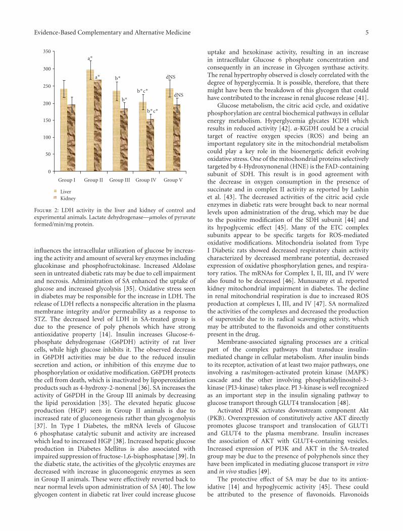
The activities of LDH increased (P < .05) significantly inuntreated diabetic animals (Group II) which were revertedback to near normal levels upon SA (Group III) andMetformin (Group IV) treatment (Figure 2).
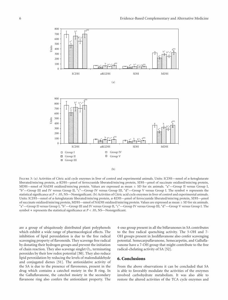
4.2. Modification of Energy Production by SA. The effects ofSA and Metformin on the activities of TCA cycle enzymes inthe kidney and liver are given in Figures 3(a) and 3(b). InGroup II animals, the activities of ICDH, αKGDH, SDH, andMDH were found to be decreased in the liver and kidney.The decrease for ICDH was 19.5% in liver and 16.2% inkidney, αKGDH; 35.4% and 34.6% in liver and kidney, SDH;29.9% and 34.6% for liver and kidney, and for MDH it was30% and 26% for liver and kidney, respectively. These valueswere significantly increased (P < .05) in Group III andGroup IV animals. When Group I and Group V animals werecompared, nonsignificant values were obtained.
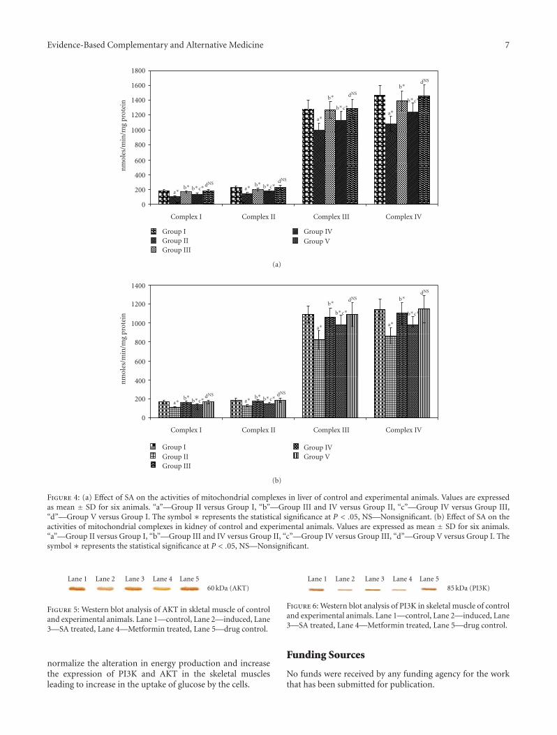
In untreated diabetic animals (Group II), marked inhi-bition (P < .05) of the activities of mitochondrial complexeswas found as shown in Figures 4(a) and 4(b) when comparedto normal animals. These were restored to near normal levels(P < .05) upon drug administration (Group III and GroupIV). No significance was found when control (Group I) anddrug control group (Group V) animals were compared.
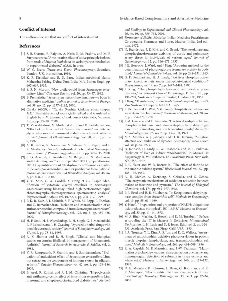
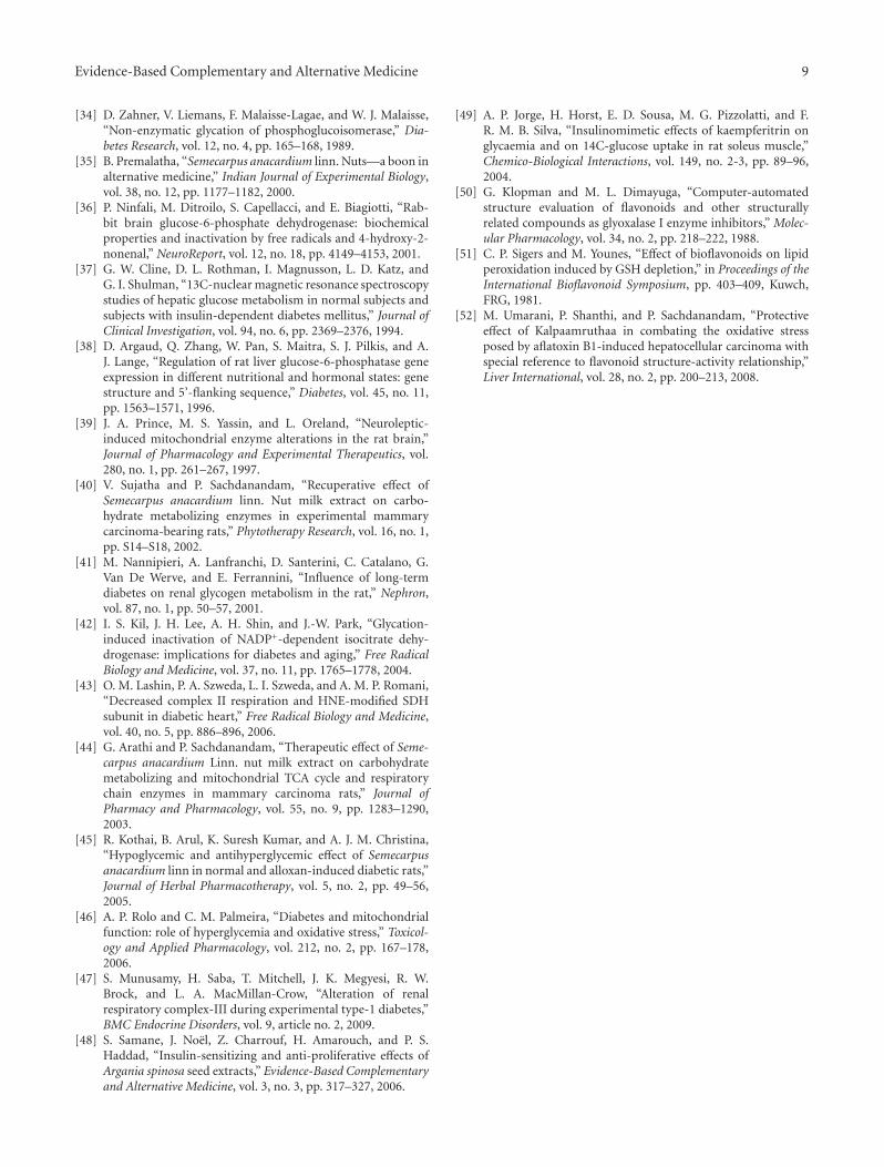
4.3. SA Increases the Protein Expression on PI3K and AKT inthe Skeletal Muscle of Control and Experimental Animals. Theprotein expressions of AKT and PI3K are shown in Figures5 and 6. In Group II animals, there was decrease in proteinexpression of PI3K and AKT when compared to GroupIII, indicating the insufficiency of insulin to maintain thenormal signaling and the uptake of glucose through GLUT4in the muscles of the animals which results in hyperglycemia.After treatment of the animals with SA, increase in proteinexpression was seen.
5. Discussion
Liver plays a key role in the maintenance of glucosehomeostasis. Following a carbohydrate rich meal, it removesa major part of the excess glucose that is absorbed andreleases it in between meals during starvation and exercise[33]. The glucostat function of the liver is based on thereversible shift between glycogen synthesis and degradationas well as between glycolysis and gluconeogenesis.
The reaction catalyzed by hexokinase serves as the entrypoint for glucose into glycolysis, glycogen synthesis, andthe hexose monophosphate. Inhibition of hexokinase leadsto impaired oxidation of glucose via glycolysis, resulting inhyperglycemia and decreased ATP production [34]. Highglucose concentration nonenzymatically glycates phospho-glucoisomerase and inhibits the proportion of glucose 6-phosphate metabolized via the glycolytic pathway. Insulin

4 Evidence-Based Complementary and Alternative Medicine
0
20
40
60
80
120
100
Group I Group II Group III Group IV Group V
a∗
a∗
a∗
a∗
a∗
a∗
a∗
a∗
b∗
b∗
b∗
b∗
b∗
b∗b∗b∗
b∗c∗
b∗c∗
b∗c∗b∗c∗
b∗c∗
b∗c∗b∗c∗
b∗c∗
dNS
dNS
dNSdNS
dNS
dNS
dNS
dNS
(a)
0
10
20
30
40
50
60
70
80
90
Group I Group II Group III Group IV Group V
a∗a∗
a∗a∗ a∗
a∗
a∗
a∗
b∗b∗
b∗
b∗
b∗
b∗
b∗ b∗ b∗
b∗c∗b∗c∗
b∗c∗
b∗c∗
b∗c∗
b∗c∗
b∗c∗
b∗c∗
dNS
dNS
dNS
dNS
dNS
dNS dNS
dNS
HexPGIPFKAldolase
G6PDG6ptaseFrul.6bi ptaseGlycogen
(b)
Figure 1: (a) Glycolytic and gluconeogenic enzymes in liver of control and experimental animals. Units: Hexokinase—nmoles of glucose-phosphate liberated/min/mg protein, Phosphoglucoisomerase—nmoles of fructose liberated/min/mg protein, Phosphofructokinase—nmoles of substrate formed/min/mg protein, Aldolase—nmoles of glyceraldehyde liberated/min/mg protein, Glucose 6-phosphatase—nmoles of phosphorous liberated/min/mg protein, Fructose-1,6-diphosphatase—nmoles of phosphorous liberated/min/mg protein.Glycogen—mg/g tissue. Values are expressed as mean ± SD for six animals. Comparisons are made between “a”—Group II versusGroup I, “b”—Group III and IV versus Group II, “c”—Group IV versus Group III, and “d”—Group V versus Group I. The symbol∗ represents the statistical significance at P < .05, NS—Nonsignificant. (b) Glycolytic and gluconeogenic enzymes in the kidney ofcontrol and experimental animals. Units: Hexokinase—nmoles of glucose-phosphate liberated/min/mg protein, Phosphoglucoisomerase—nmoles of fructose liberated/min/mg protein, Phosphofructokinase—nmoles of substrate formed/min/mg protein, Aldolase—nmolesof glyceraldehyde liberated/min/mg protein, Glucose 6-phosphatase—nmoles of phosphorous liberated/min/mg protein, Fructose-1,6-diphosphatase—nmoles of phosphorous liberated/min/mg protein. Glycogen—mg/g tissue.Values are expressed as mean ± SD for sixanimals. Comparisons are made between “a”—Group II versus Group I, “b”—Group III and IV versus Group II, “c”—Group IV versusGroup III, and “d”—Group V versus Group I. The symbol ∗ represents the statistical significance at P < .05, NS—Nonsignificant.
Evidence-Based Complementary and Alternative Medicine 5
0
50
100
150
200
250
300
350
Group I Group II Group III Group IV Group V
a∗
a∗
b∗
b∗b∗c∗
b∗c∗
dNS
dNS
LiverKidney
Figure 2: LDH activity in the liver and kidney of control andexperimental animals. Lactate dehydrogenase—μmoles of pyruvateformed/min/mg protein.
influences the intracellular utilization of glucose by increas-ing the activity and amount of several key enzymes includingglucokinase and phosphofructokinase. Increased Aldolaseseen in untreated diabetic rats may be due to cell impairmentand necrosis. Administration of SA enhanced the uptake ofglucose and increased glycolysis [35]. Oxidative stress seenin diabetes may be responsible for the increase in LDH. Therelease of LDH reflects a nonspecific alteration in the plasmamembrane integrity and/or permeability as a response toSTZ. The decreased level of LDH in SA-treated group isdue to the presence of poly phenols which have strongantioxidative property [14]. Insulin increases Glucose-6-phosphate dehydrogenase (G6PDH) activity of rat livercells, while high glucose inhibits it. The observed decreasein G6PDH activities may be due to the reduced insulinsecretion and action, or inhibition of this enzyme due tophosphorylation or oxidative modification. G6PDH protectsthe cell from death, which is inactivated by lipoperoxidationproducts such as 4-hydroxy-2-nonenal [36]. SA increases theactivity of G6PDH in the Group III animals by decreasingthe lipid peroxidation [35]. The elevated hepatic glucoseproduction (HGP) seen in Group II animals is due toincreased rate of gluconeogenesis rather than glycogenolysis[37]. In Type I Diabetes, the mRNA levels of Glucose6 phosphatase catalytic subunit and activity are increasedwhich lead to increased HGP [38]. Increased hepatic glucoseproduction in Diabetes Mellitus is also associated withimpaired suppression of fructose-1,6-bisphosphatase [39]. Inthe diabetic state, the activities of the glycolytic enzymes aredecreased with increase in gluconeogenic enzymes as seenin Group II animals. These were effectively reverted back tonear normal levels upon administration of SA [40]. The lowglycogen content in diabetic rat liver could increase glucose
uptake and hexokinase activity, resulting in an increasein intracellular Glucose 6 phosphate concentration andconsequently in an increase in Glycogen synthase activity.The renal hypertrophy observed is closely correlated with thedegree of hyperglycemia. It is possible, therefore, that theremight have been the breakdown of this glycogen that couldhave contributed to the increase in renal glucose release [41].
Glucose metabolism, the citric acid cycle, and oxidativephosphorylation are central biochemical pathways in cellularenergy metabolism. Hyperglycemia glycates ICDH whichresults in reduced activity [42]. α-KGDH could be a crucialtarget of reactive oxygen species (ROS) and being animportant regulatory site in the mitochondrial metabolismcould play a key role in the bioenergetic deficit evolvingoxidative stress. One of the mitochondrial proteins selectivelytargeted by 4-Hydroxynonenal (HNE) is the FAD-containingsubunit of SDH. This result is in good agreement withthe decrease in oxygen consumption in the presence ofsuccinate and in complex II activity as reported by Lashinet al. [43]. The decreased activities of the citric acid cycleenzymes in diabetic rats were brought back to near normallevels upon administration of the drug, which may be dueto the positive modification of the SDH subunit [44] andits hypoglycemic effect [45]. Many of the ETC complexsubunits appear to be specific targets for ROS-mediatedoxidative modifications. Mitochondria isolated from TypeI Diabetic rats showed decreased respiratory chain activitycharacterized by decreased membrane potential, decreasedexpression of oxidative phosphorylation genes, and respira-tory ratios. The mRNAs for Complex I, II, III, and IV werealso found to be decreased [46]. Munusamy et al. reportedkidney mitochondrial impairment in diabetes. The declinein renal mitochondrial respiration is due to increased ROSproduction at complexes I, III, and IV [47]. SA normalizedthe activities of the complexes and decreased the productionof superoxide due to its radical scavenging activity, whichmay be attributed to the flavonoids and other constituentspresent in the drug.
Membrane-associated signaling processes are a criticalpart of the complex pathways that transduce insulin-mediated change in cellular metabolism. After insulin bindsto its receptor, activation of at least two major pathways, oneinvolving a ras/mitogen-activated protein kinase (MAPK)cascade and the other involving phosphatidylinositol-3-kinase (PI3-kinase) takes place. PI 3-kinase is well recognizedas an important step in the insulin signaling pathway toglucose transport through GLUT4 translocation [48].
Activated PI3K activates downstream component Akt(PKB). Overexpression of constitutively active AKT directlypromotes glucose transport and translocation of GLUT1and GLUT4 to the plasma membrane. Insulin increasesthe association of AKT with GLUT4-containing vesicles.Increased expression of PI3K and AKT in the SA-treatedgroup may be due to the presence of polyphenols since theyhave been implicated in mediating glucose transport in vitroand in vivo studies [49].
The protective effect of SA may be due to its antiox-idative [14] and hypoglycemic activity [45]. These couldbe attributed to the presence of flavonoids. Flavonoids
6 Evidence-Based Complementary and Alternative Medicine
0
100
200
300
400U
nit
s
500
600
700
800
ICDH αKGDH SDH MDH
a∗
b∗
b∗c∗
b∗c∗
dNS
dNS
a∗ a∗b b∗
b∗
b∗c∗
b∗c∗
dNS
dNS
a∗
(a)
0
100
200
300
400
500
600
700
800
900
Un
its
ICDH αKGDH SDH MDH
Group IGroup II
Group IV
Group VGroup III
a∗
b∗ b∗c∗
b∗c∗
dNS
dNS
a∗ a∗b
b
b
b∗c
b∗c
dNS
dNS
a∗
(b)
Figure 3: (a) Activities of Citric acid cycle enzymes in liver of control and experimental animals. Units: ICDH—nmol of α-ketoglutarateliberated/min/mg protein, α-KDH—μmol of ferrocyanide liberated/min/mg protein, SDH—μmol of succinate oxidized/min/mg protein,MDH—nmol of NADH oxidized/min/mg protein. Values are expressed as mean ± SD for six animals. “a”—Group II versus Group I,“b”—Group III and IV versus Group II, “c”—Group IV versus Group III, “d”—Group V versus Group I. The symbol ∗ represents thestatistical significance at P < .05, NS—Nonsignificant. (b) Activities of Citric acid cycle enzymes in liver of control and experimental animals.Units: ICDH—nmol of α-ketoglutarate liberated/min/mg protein, α-KDH—μmol of ferrocyanide liberated/min/mg protein, SDH—μmolof succinate oxidized/min/mg protein, MDH—nmol of NADH oxidized/min/mg protein. Values are expressed as mean± SD for six animals.“a”—Group II versus Group I, “b”—Group III and IV versus Group II, “c”—Group IV versus Group III, “d”—Group V versus Group I. Thesymbol ∗ represents the statistical significance at P < .05, NS—Nonsignificant.
are a group of ubiquitously distributed plant polyphenolswhich exhibit a wide range of pharmacological effects. Theinhibition of lipid peroxidation is due to the free radicalscavenging property of flavonoids. They scavenge free radicalby donating their hydrogen groups and prevent the initiationof chain reaction. They also scavenge singlet O2, terminatingperoxides by their low redox potential [50]. They also reducelipid peroxidation by reducing the levels of malondialdehydeand conjugated dienes [51]. The antioxidative activity ofthe SA is due to the presence of flavonones, present in thedrug which contains a catechol moiety in the B ring. Inthe Galluflavanone, the catechol moiety in the secondaryflavanone ring also confers the antioxidant property. The
4-oxo group present in all the biflavanones in SA contributesto the free radical quenching activity. The 5-OH and 7-OH groups present in Jeediflavanone also confer scavengingpotential. Semecarpuflavanone, Semecarpetin, and Gallufla-vanone have a 7-OH group that might contribute to the freeradical-chelating activity of SA [52].
6. Conclusions
From the above observations it can be concluded that SAis able to favorably modulate the activities of the enzymesinvolved carbohydrate metabolism. It was also able torestore the altered activities of the TCA cycle enzymes and
Evidence-Based Complementary and Alternative Medicine 7
0
200
400
600
800
nm
oles
/min
/mg
prot
ein
1000
1200
1400
1600
1800
Complex I Complex II Complex III Complex IV
Group IGroup II
Group IVGroup V
Group III
a∗b∗ b∗c∗ b∗c∗dNS dNS
a∗
a∗
b∗
b∗
b∗
b∗c∗b∗c∗
dNS
dNS
a∗
(a)
0
200
400
600
800
nm
oles
/min
/mg
prot
ein
1000
1200
1400
Complex I Complex II Complex III Complex IV
Group IGroup II
Group IVGroup V
Group III
a∗b∗ b∗c∗ b∗c∗dNS dNS
a∗
a∗
b∗
b∗b∗
b∗c∗ b∗c∗
dNSdNS
a∗
(b)
Figure 4: (a) Effect of SA on the activities of mitochondrial complexes in liver of control and experimental animals. Values are expressedas mean ± SD for six animals. “a”—Group II versus Group I, “b”—Group III and IV versus Group II, “c”—Group IV versus Group III,“d”—Group V versus Group I. The symbol ∗ represents the statistical significance at P < .05, NS—Nonsignificant. (b) Effect of SA on theactivities of mitochondrial complexes in kidney of control and experimental animals. Values are expressed as mean ± SD for six animals.“a”—Group II versus Group I, “b”—Group III and IV versus Group II, “c”—Group IV versus Group III, “d”—Group V versus Group I. Thesymbol ∗ represents the statistical significance at P < .05, NS—Nonsignificant.
Lane 1 Lane 2 Lane 3 Lane 4 Lane 560 kDa (AKT)
Figure 5: Western blot analysis of AKT in skletal muscle of controland experimental animals. Lane 1—control, Lane 2—induced, Lane3—SA treated, Lane 4—Metformin treated, Lane 5—drug control.
normalize the alteration in energy production and increasethe expression of PI3K and AKT in the skeletal musclesleading to increase in the uptake of glucose by the cells.
Lane 1 Lane 2 Lane 3 Lane 4 Lane 585 kDa (PI3K)
Figure 6: Western blot analysis of PI3K in skeletal muscle of controland experimental animals. Lane 1—control, Lane 2—induced, Lane3—SA treated, Lane 4—Metformin treated, Lane 5—drug control.
Funding Sources
No funds were received by any funding agency for the workthat has been submitted for publication.
8 Evidence-Based Complementary and Alternative Medicine
Conflict of Interest
The authors declare that no conflict of interests exist.
References
[1] S. B. Sharma, R. Rajpoot, A. Nasir, K. M. Prabhu, and M. P.Suryanarayana, “Ameliorative effect of active principle isolatedfrom seeds of Eugenia Jambolana on carbohydrate metabolismin experimental diabetes,” eCAM. In press.
[2] W. C. Evans, Trease and Evans’ Pharmacognosy, Saunders,London, UK, 14th edition, 1996.
[3] K. R. Kirthikar and B. D. Basu, Indian medicinal plants.Mahendra Palsing, Dehra Dun, India: M/s. Bishen Singh, pp.667–669, 1933.
[4] S. S. N. Murthy, “New bioflavonoid from Semecarpus anac-ardium Linn,” Clin Acta Turcica, vol. 20, pp. 33–37, 1992.
[5] B. Premalatha, “Semecarpus anacardium linn. nuts—a boon inalternative medicine,” Indian Journal of Experimental Biology,vol. 38, no. 12, pp. 1177–1182, 2000.
[6] Caraka (600BC), “Caraka Samhita Chikitsa sthan chapter1(2),” Bhallataka ksira,ksoudra,taila, edited and translated inEnglish by P. V. Sharma, Choukhamba Orientalia, Varanasi,India, pp. 13–19 , 2000.
[7] T. Vijayalakshmi, V. Muthulakshmi, and P. Sachdanandam,“Effect of milk extract of Semecarpus anacardium nuts onglycohydrolases and lysosomal stability in adjuvant arthritisin rats,” Journal of Ethnopharmacology, vol. 58, no. 1, pp. 1–8, 1997.
[8] A. K. Sahoo, N. Narayanan, S. Sahana, S. S. Rajan, and P.K. Mukherjee, “In vitro antioxidant potential of Semecarpusanacardium L,” Pharmacologyonline, vol. 3, pp. 327–335, 2008.
[9] S. G. Aravind, R. Arimboor, M. Rangan, S. N. Madhavan,and C. Arumughan, “Semi-preparative HPLC preparation andHPTLC quantification of tetrahydroamentoflavone as markerin Semecarpus anacardium and its polyherbal formulations,”Journal of Pharmaceutical and Biomedical Analysis, vol. 48, no.3, pp. 808–813, 2008.
[10] Y. G. Shin, G. A. Cordell, Y. Dong et al., “Rapid iden-tification of cytotoxic alkenyl catechols in Semecarpusanacardium using bioassay-linked high performance liquidchromatography-electrospray/mass spectrometric analysis,”Phytochemical Analysis, vol. 10, no. 4, pp. 208–212, 1999.
[11] P. K. R. Nair, S. J. Melnick, S. F. Wnuk, M. Rapp, E. Escalon,and C. Ramachandran, “Isolation and characterization of ananticancer catechol compound from Semecarpus anacardium,”Journal of Ethnopharmacology, vol. 122, no. 3, pp. 450–456,2009.
[12] H. F. Smit, H. J. Woerdenbag, R. H. Singh, G. J. Meulenbeld,R. P. Labadie, and J. H. Zwaving, “Ayurvedic herbal drugs withpossible cytostatic activity,” Journal of Ethnopharmacology, vol.47, no. 2, pp. 75–84, 1995.
[13] A. K. Sharma and R. H. Singh, “Clinical and biologicalstudies on Amrita Bhallatak in management of RheumatoidArthritis,” Journal of Research in Ayurveda & Siddha, vol. 2,1981.
[14] V. R. Ramprasath, P. Shanthi, and P. Sachdanandam, “Eval-uation of antioxidant effect of Semecarpus anacardium Linn.nut extract on the components of immune system in adjuvantarthritis,” Vascular Pharmacology, vol. 42, no. 4, pp. 179–186,2005.
[15] B. Arul, R. Kothai, and A. J. M. Christina, “Hypoglycemicand antihyperglycemic effect of Semecarpus anacardium Linnin normal and streptozotocin-induced diabetic rats,” Methods
and Findings in Experimental and Clinical Pharmacology, vol.26, no. 10, pp. 759–762, 2004.
[16] Formulary of Siddha Medicine, Indian Medicine PractitionersCo-operative Pharmacy and Stores, Madras, India, 2nd edi-tion, 1972.
[17] N. Brandstrup, J. E. Kirk, and C. Bruni, “The hexokinase andphosphoglucoisomerase activities of aortic and pulmonaryartery tissue in individuals of various ages,” Journal ofGerontology, vol. 12, pp. 166–171, 1957.
[18] J. E. Horrocks, J. Ward, and J. King, “A routine method for thedetermination of phosphoglucose isomerase activity in bodyfluid,” Journal of Clinical Pathology, vol. 16, pp. 248–251, 1963.
[19] G. D. Reinhart and H. A. Lardy, “Rat liver phosphofructok-inase: kinetic activity under near-physiological conditions,”Biochemistry, vol. 19, no. 7, pp. 1477–1484, 1980.
[20] J. King, “The phosphohydrolases-acid and alkaline phos-phatases,” in Practical Clinical Enzymology, D. Van, Ed., pp.191–208, Nostrand Company Limited, London, UK, 1965.
[21] J. King, “Transferases,” in Practical Clinical Enzymology, p. 263,Van Nostrand Company, NJ, USA, 1965.
[22] E. Beutler and C. West, “Glucose-6-phosphate dehydrogenasevariants in the chimpanzee,” Biochemical Medicine, vol. 20, no.3, pp. 364–370, 1978.
[23] J. M. Gancedo and C. Gancedo, “Fructose-1,6-diphosphatase,phosphofructokinase and glucose-6-phosphate dehydroge-nase from fermenting and non fermenting yeasts,” Archiv furMikrobiologie, vol. 76, no. 2, pp. 132–138, 1971.
[24] M.A. Morales, A. J. Jabbagy, and H. R. Terenizi, “Mutationaffecting accumulation of glycogen neurospora,” News Letter,vol. 30, p. 24, 1973.
[25] D. Johnson, H. Lardy, R. W. Estabrook, and M. E. Pullman,“Isolation of liver or kidney mitochondria,” in Methods inEnzymology, R. W. Estabrook, Ed., Academic Press, New York,NY, USA, 1967.
[26] E. C. Slater and W. D. Borner Jr., “The effect of fluoride onthe succinic oxidase system,” Biochemical Journal, vol. 52, pp.185–196, 1952.
[27] A. H. Mehler, A. Kornberg, S. Grisolia, and S. Ochoa,“The enzymatic mechanisms of oxidation reduction betweenmalate or isocitrate and pyruvate,” The Journal of BiologicalChemistry, vol. 174, pp. 961–977, 1948.
[28] L. J. Reed and B. B. Mukherjee, “α-Ketoglutarate dehydroge-nase complex from Escherichia coli,” Methods in Enzymology,vol. 13, pp. 55–61, 1969.
[29] Y. Hatefi, “Preparation and properties of NADH: ubiquinoneoxidoreductase (complexI), EC 1.6.5.3,” Methods in Enzymol-ogy, vol. 53, pp. 11–14, 1978.
[30] M. A. Birch-Machin, N. Howell, and D. M. Turnbull, “Defectsat coupling site II,” in Methods in Toxicology: MitochondrialDysfunction, L. H. Lash and D. P. Jones, Eds., vol. 2, pp. 324–331, Academic Press, San Diego, Calif, USA, 1993.
[31] I. A. Trounce, Y. L. Kim, A. S. Jun, and D. C. Wallace, “Assess-ment of mitochondrial oxidative phosphorylation in patientmuscle biopsies, lymphoblasts, and transmitochondrial celllines,” Methods in Enzymology, vol. 264, pp. 484–509, 1996.
[32] R. A. Capaldi, M. F. Marusich, and J.-W. Taanman, “Mam-malian cytochrome-c oxidase: characterization of enzyme andimmunological detection of subunits in tissue extracts andwhole cells,” Methods in Enzymology, vol. 260, pp. 117–132,1995.
[33] D. E. Malarkey, K. Johnson, L. Ryan, G. Boorman, and R.R. Maronpot, “New insights into functional aspects of livermorphology,” Toxicologic Pathology, vol. 33, no. 1, pp. 27–34,2005.
Evidence-Based Complementary and Alternative Medicine 9
[34] D. Zahner, V. Liemans, F. Malaisse-Lagae, and W. J. Malaisse,“Non-enzymatic glycation of phosphoglucoisomerase,” Dia-betes Research, vol. 12, no. 4, pp. 165–168, 1989.
[35] B. Premalatha, “Semecarpus anacardium linn. Nuts—a boon inalternative medicine,” Indian Journal of Experimental Biology,vol. 38, no. 12, pp. 1177–1182, 2000.
[36] P. Ninfali, M. Ditroilo, S. Capellacci, and E. Biagiotti, “Rab-bit brain glucose-6-phosphate dehydrogenase: biochemicalproperties and inactivation by free radicals and 4-hydroxy-2-nonenal,” NeuroReport, vol. 12, no. 18, pp. 4149–4153, 2001.
[37] G. W. Cline, D. L. Rothman, I. Magnusson, L. D. Katz, andG. I. Shulman, “13C-nuclear magnetic resonance spectroscopystudies of hepatic glucose metabolism in normal subjects andsubjects with insulin-dependent diabetes mellitus,” Journal ofClinical Investigation, vol. 94, no. 6, pp. 2369–2376, 1994.
[38] D. Argaud, Q. Zhang, W. Pan, S. Maitra, S. J. Pilkis, and A.J. Lange, “Regulation of rat liver glucose-6-phosphatase geneexpression in different nutritional and hormonal states: genestructure and 5’-flanking sequence,” Diabetes, vol. 45, no. 11,pp. 1563–1571, 1996.
[39] J. A. Prince, M. S. Yassin, and L. Oreland, “Neuroleptic-induced mitochondrial enzyme alterations in the rat brain,”Journal of Pharmacology and Experimental Therapeutics, vol.280, no. 1, pp. 261–267, 1997.
[40] V. Sujatha and P. Sachdanandam, “Recuperative effect ofSemecarpus anacardium linn. Nut milk extract on carbo-hydrate metabolizing enzymes in experimental mammarycarcinoma-bearing rats,” Phytotherapy Research, vol. 16, no. 1,pp. S14–S18, 2002.
[41] M. Nannipieri, A. Lanfranchi, D. Santerini, C. Catalano, G.Van De Werve, and E. Ferrannini, “Influence of long-termdiabetes on renal glycogen metabolism in the rat,” Nephron,vol. 87, no. 1, pp. 50–57, 2001.
[42] I. S. Kil, J. H. Lee, A. H. Shin, and J.-W. Park, “Glycation-induced inactivation of NADP+-dependent isocitrate dehy-drogenase: implications for diabetes and aging,” Free RadicalBiology and Medicine, vol. 37, no. 11, pp. 1765–1778, 2004.
[43] O. M. Lashin, P. A. Szweda, L. I. Szweda, and A. M. P. Romani,“Decreased complex II respiration and HNE-modified SDHsubunit in diabetic heart,” Free Radical Biology and Medicine,vol. 40, no. 5, pp. 886–896, 2006.
[44] G. Arathi and P. Sachdanandam, “Therapeutic effect of Seme-carpus anacardium Linn. nut milk extract on carbohydratemetabolizing and mitochondrial TCA cycle and respiratorychain enzymes in mammary carcinoma rats,” Journal ofPharmacy and Pharmacology, vol. 55, no. 9, pp. 1283–1290,2003.
[45] R. Kothai, B. Arul, K. Suresh Kumar, and A. J. M. Christina,“Hypoglycemic and antihyperglycemic effect of Semecarpusanacardium linn in normal and alloxan-induced diabetic rats,”Journal of Herbal Pharmacotherapy, vol. 5, no. 2, pp. 49–56,2005.
[46] A. P. Rolo and C. M. Palmeira, “Diabetes and mitochondrialfunction: role of hyperglycemia and oxidative stress,” Toxicol-ogy and Applied Pharmacology, vol. 212, no. 2, pp. 167–178,2006.
[47] S. Munusamy, H. Saba, T. Mitchell, J. K. Megyesi, R. W.Brock, and L. A. MacMillan-Crow, “Alteration of renalrespiratory complex-III during experimental type-1 diabetes,”BMC Endocrine Disorders, vol. 9, article no. 2, 2009.
[48] S. Samane, J. Noel, Z. Charrouf, H. Amarouch, and P. S.Haddad, “Insulin-sensitizing and anti-proliferative effects ofArgania spinosa seed extracts,” Evidence-Based Complementaryand Alternative Medicine, vol. 3, no. 3, pp. 317–327, 2006.
[49] A. P. Jorge, H. Horst, E. D. Sousa, M. G. Pizzolatti, and F.R. M. B. Silva, “Insulinomimetic effects of kaempferitrin onglycaemia and on 14C-glucose uptake in rat soleus muscle,”Chemico-Biological Interactions, vol. 149, no. 2-3, pp. 89–96,2004.
[50] G. Klopman and M. L. Dimayuga, “Computer-automatedstructure evaluation of flavonoids and other structurallyrelated compounds as glyoxalase I enzyme inhibitors,” Molec-ular Pharmacology, vol. 34, no. 2, pp. 218–222, 1988.
[51] C. P. Sigers and M. Younes, “Effect of bioflavonoids on lipidperoxidation induced by GSH depletion,” in Proceedings of theInternational Bioflavonoid Symposium, pp. 403–409, Kuwch,FRG, 1981.
[52] M. Umarani, P. Shanthi, and P. Sachdanandam, “Protectiveeffect of Kalpaamruthaa in combating the oxidative stressposed by aflatoxin B1-induced hepatocellular carcinoma withspecial reference to flavonoid structure-activity relationship,”Liver International, vol. 28, no. 2, pp. 200–213, 2008.

Submit your manuscripts athttp://www.hindawi.com
Stem CellsInternational
Hindawi Publishing Corporationhttp://www.hindawi.com Volume 2014
Hindawi Publishing Corporationhttp://www.hindawi.com Volume 2014
MEDIATORSINFLAMMATION
of
Hindawi Publishing Corporationhttp://www.hindawi.com Volume 2014
Behavioural Neurology
EndocrinologyInternational Journal of
Hindawi Publishing Corporationhttp://www.hindawi.com Volume 2014
Hindawi Publishing Corporationhttp://www.hindawi.com Volume 2014
Disease Markers
Hindawi Publishing Corporationhttp://www.hindawi.com Volume 2014
BioMed Research International
OncologyJournal of
Hindawi Publishing Corporationhttp://www.hindawi.com Volume 2014
Hindawi Publishing Corporationhttp://www.hindawi.com Volume 2014
Oxidative Medicine and Cellular Longevity
Hindawi Publishing Corporationhttp://www.hindawi.com Volume 2014
PPAR Research
The Scientific World JournalHindawi Publishing Corporation http://www.hindawi.com Volume 2014
Immunology ResearchHindawi Publishing Corporationhttp://www.hindawi.com Volume 2014
Journal of
ObesityJournal of
Hindawi Publishing Corporationhttp://www.hindawi.com Volume 2014
Hindawi Publishing Corporationhttp://www.hindawi.com Volume 2014
Computational and Mathematical Methods in Medicine
OphthalmologyJournal of
Hindawi Publishing Corporationhttp://www.hindawi.com Volume 2014
Diabetes ResearchJournal of
Hindawi Publishing Corporationhttp://www.hindawi.com Volume 2014
Hindawi Publishing Corporationhttp://www.hindawi.com Volume 2014
Research and TreatmentAIDS
Hindawi Publishing Corporationhttp://www.hindawi.com Volume 2014
Gastroenterology Research and Practice
Hindawi Publishing Corporationhttp://www.hindawi.com Volume 2014
Parkinson’s Disease
Evidence-Based Complementary and Alternative Medicine
Volume 2014Hindawi Publishing Corporationhttp://www.hindawi.com