sensory physiology, ch 10 - dr. scott croes' website · extraocular muscles • six extrinsic...
TRANSCRIPT

1
Sensory Physiology, Ch 10 Outline of class lecture After studying this chapter you should be able to:
1. Briefly describe the three layers (tunics) of the eye. 2. Describe the structures and functions of the fibrous tunic 3. Describe the structures and functions of the vascular tunic. 4. Describe the following: Accommodation, visual acuity and the way it is rated; myopia,
hyperopia, and astigmatism. 5. Explain what LASIK stands for and what the procedure entails. 6. Describe the structures and functions of the nervous tunic. 7. Describe the physiology of vision 8. Explain the difference between dry and wet age-related macular degeneration (AMD) 9. Describe the structures of the lens 10. Explain the interior structures of the eye. 11. Describe the structures of the outer, middle ear and inner ear. 12. Explain the mechanism of hearing 13. Describe the structure and functions of the vestibular apparatus and its role in maintaining
equilibrium. 14. Describe the following clinical conditions: strabismus, glaucoma, pterygium 15. Discuss the Clinical Applications from the study guide and be able to describe the disorders
from the Applications to Health located at the end of this chapter. Sensory Physiology: The Eye
• Ophthalmology: Study of structure, function, and diseases of the eye.
Accessory Structures of Eye Eyelashes and Eyebrows
• Help protect the eyeballs from foreign objects, perspiration, and direct rays of the sun.
Extraocular Muscles • Six extrinsic skeletal muscles move each eye
Disruption of Binocular Vision results in diplopia (double vision) Strabismus
• Is a misalignment of the visual axes and loss of binocular vision.
– Afflicts
• Due to: – An under acting or overacting eye muscle – Abnormal innervation, physical trauma, or a central
processing problem • Can result in permanent loss of vision in the “weaker” eye if not
corrected during early childhood • Initial therapy usually includes intermittent patching of the good
eye before more invasive procedures such as surgery.

2
Structures of the Eyeball Wall of the eye consists of three layers: 1. Fibrous tunic (outer), 2. Vascular tunic (middle), and 3. Nervous Tunic or Retina (Inner) Fibrous Tunic (outer layer)
• Fibrous tunic: Consists of • Sclera: The “white” of the eye • Cornea: Transparent, avascular structure that forms the anterior surface of the eyeball.
• Curved surface assists Pterygium (tur RIDGE ium)
• A benign thickening of the conjunctiva that grows onto the cornea. Eventually is may cause vision disruption by interfering with the normal smooth surface of the cornea.
– Most commonly caused by sun exposure. Vascular Tunic (Middle Layer)
• Consists of three Parts: • Iris
– The colored portion of the eye – Regulates the amount of light entering the eyeball by contraction/relaxation of smooth
muscle that surrounds the pupil. • Pupil: The hole at the center

3
• Choroid – Highly vascular with dark pigment produced by melanocytes.
• Blood vessels provide oxygen and nourishment to the retina • Black pigment functions to
• Ciliary Body – Consists of – Ciliary processes contain suspensory ligaments (zonular fibers) that insert into the
lens capsule, functioning to anchor it in place. • Secretes aqueous humor (plasma filtrate) that provides
• Fluid is drained into the scleral venous sinus (canal of Schlemm), which returns it to the venous blood.
• Glaucoma is a condition is which there is an over production or an inadequate drainage of aqueous humor that can lead to excessive accumulation of fluid, which results in increased intraocular pressure. This may produce damage to the retina and loss of vision
– Ciliary Muscle • Circular band of smooth muscle that alters the shape of the • Contraction: Releases tension on the suspensory ligaments and thus the lens
– the lens becomes more rounded, allowing he eye to focus on nearby objects. • Lens contains elastic fibers which causes it to become more rounded
when not being pulled. • Presbyopia (old eye or
vision): Inability of the eye to focus sharply on nearby objects due to loss of lens elasticity with age.
• • Relaxation: Increases tension
on the suspensory ligaments and thus the lens – the lens flattens, allowing the eye to focus on far away objects

4
Focusing of the Eyes
• Accommodation: Refers to changes in the thickness of the lens to keep an image focused on the retina as the distance between the eyes and object varies.
• When viewing an object, light rays are bent (refracted) to focus the object onto the retina – The bending of – Curvature of the cornea remains constant while the curvature of the lens can change
depending on the lens’s elasticity and the contraction of the ciliary muscle. Visual Acuity
• Visual acuity is a rating of the ability to see objects in sharp detail (sharpness of vision). • Person with normal (20/20) vision can read a line of letters marked “20/20” while standing 20
feet from a Snellen eye chart. • A 20/15 rating: Person at 20 feet is able to see details that would require someone with normal
vision to move up to 15 feet to see. –
• A 20/50 rating: Person has move up to 20 feet to discern details that a person with normal vision can see from 50 feet away.
– Vision is worse than average – Visual acuity below 20/200, with
Visual Problems due to Alterations in Refraction
• Refraction errors are optical imperfections that prevent the eye from properly bending (refracting) light, causing blurred vision.
– Cause of refractive errors include: 1) the overall length of the eye, 2) the curvature of the lens, and 3) the curvature of the cornea.
– The primary refractive errors are nearsightedness, farsightedness and astigmatism.
• Nearsighted (myopia): Can see close objects but – Due to the eyeball being too long or lens is too strong (too steeply curved) causing
the image to be focused in front of the retina when looking at a distant object. – Corrected with diverging (concave) lenses which spreads the light rays apart as if
the object were closer and thus the image is focused further back onto the retina.
• Farsighted (hyperopia): Can see distant objects but – Due to the eyeball being too short or the lens is to weak (too flat) causing the image
to be focused behind the retina. – Corrected with converging (convex) lenses which provide additional focusing
(converging) of light needed to bring the image forward to the retina.

5
• Astigmatism: Either the cornea or the lens has an irregular curvature resulting in blurred vision.
LASIK
• LASIK (Laser-Assisted In-Situ Keratomileusis): Surgery to correct the curvature of the cornea for conditions such as farsightedness, nearsightedness, and astigmatism.
– Procedure: • A circular flap is • The underlying cornea is reshaped with a laser • Corneal flap is repositioned over the treated area and the flap reattaches to rest
of cornea within 24 hours. Nervous Tunic or Retina (inner layer)
• Nervous tunic or retina: Contains photoreceptors (rods and cones) that function to turn visual data into nerve impulses.
– Contains two portions: Pigment Epithelium
• Pigment Epithelium – Lies between the choroid and the neural portion of the retina. – Functions:
• Synthesize • Phagocytosis of the shed outer segments of the photoreceptors (~10%/day). • Delivery of nutrients from the blood to the photoreceptors. • Contains enzymes to
• Photoreceptors: Rods and Cones – Rods (120 million/retina): Specialized for vision in
dim light, such as moonlight • No color vision – see
– Cones (6 million/retina): Specialized for color vision and sharpness of vision – Contain 3 types of cones.
• Blue cones: • Green cones: Sensitive to green light • Red cones: • Color vision results from various
combinations of these 3 types of cones.

6
Structure of Rods and Cones • Specialized neurons that consist of an outer and an inner segment.
– Outer segment contains visual pigments embeded in membrane disks – Inner segment contains major organelles and synthesizes visual pigments.
• Synapses with • Visual Pigments (photopigments):
– Consists of two components: 1) Opsin, an integral protein in the disc plasma membrane that is bound to retinal and 2) retinal, the light absorbing part, which is synthesized from vitamin A.
– Rods and cones contain the same retinal pigment but differ in their types of opsin.
• Rods contain rhodopsin and cones contain derivatives of rhodopsin called color pigments or photo opsins
– Type of opsin present determines the wavelength of light that can be absorbed by retinal.
• Their stimulation in various – New disks containing visual pigments are continuously assembled at the base of the
outer segment. – After ~10 days, the disks are shed at the outer segment tip and phagocytosed by the
pigment cells.

7
Photoreception • In darkness, Na+ channels in the outer segment membrane are kept open allowing Na+ to flow
in. – Although Na+ is continuously pumped back out, the transmembrane potential is about -
40 mV, rather than the -70 mV of a typical resting neuron. – At -40 mV the photoreceptor is continuously releasing inhibitory neurotransmitters
onto the associated bipolar cell. • The bipolar cell does not send a nerve impulse to the brain under these
conditions. – The movement of Na+ into and out of the
• When a photon of light is absorbed by the retinal portion of opsin the retinal changes shape from its inactive 11-cis form to its active 11-trans form.
– The 11-trans form detaches from the opsin which starts a chain of reaction that ultimately closes Na+ channels and prevents its entry into the cell while active transport continues to remove Na+ from the cell causing the cell become hyperpolarized.
• The arrival of a photon thus reduces the dark current. • The detachment of retinal from opsin is
• The rate of neurotransmitter release declines as • The decreased inhibitory neurotransmitter release indicates to the adjacent bipolar cell that
the associated photoreceptor has absorbed a photon and sends a neural impulse to the brain.

8
Recovery After Stimulation • Once activated by a photon the 11-trans form of retinal must be enzymatically converted to the
11-cis form and recombined with opsin. – 11-trans form is transported fro the photoreceptors to the pigment epithelial cells where
it is converted back to the 11-cis form and then transported back to the photoreceptors. – Bleaching contributes to the lingering visual impression you have after you see a
flashbulb go off and see “ghost” images – Bleaching is not noticeable under normal circumstances because the eyes are
continually making small involuntary changes in position that move the image across the retina’s surface.
Clinical Application: Night Blindness • Deficiency in vitamin A can result in a decline in retinal pigment and a corresponding decrease
in vision. • At fist daylight vision is not affected because of the increased amount of photon stimulation
activates remaining visual pigments. • Problem first becomes apparent at night, which dim light proves insufficient to activate the
rods. – This condition is known as
Dark Adaptation • In the light, bleaching reaction results in an increased amount of activated visual pigments in
the rods and cones. • activated visual pigments in the rods and cones.
– • Dark adaptation occurs due to an increased amounts of inactivated visual pigments that
become highly sensitive to even the lowest amount of light stimulation. Fovea Centralis
• Fovea centralis is a small depression within the macula lutea and contains the densest concentration of cones.
– Area of highest visual – Foveal cones have long slender bodies; the blood vessels and other retinal cells are all
displaced to the side rather than resting directly on top of the cones. This allows light to pass unimpeded to the cones.
Other Retinal Neurons
• Bipolar cell layer – Transmits signal form
• Ganglion cell layer
– Ganglion cell axons extend posteriorly to the optic disc (blind spot) and exit the eyeball as the optic nerve.
– Transmit signals of vision to the midbrain and thalamus and then to the visual cortex.

9
Optic Disk • Optic disk (blind spot): Region where the axons of ganglion neurons exit the eyeball as the
optic nerve. – Area contains no rods or cones; thus we cannot see an image that strikes this region
and hence the name.
Age-related Macular Degeneration
• Age-related Macular degeneration (AMD), is the leading cause of blindness in people aged 60 and older.
• There are two major types of AMD, "dry“ and "wet" macular degeneration.
• Dry (non-neovascular) AMD: Most common type, accounts for
– Accumulation of drusen (waste products from photoreceptors) accumulates between the retina and the choroid.
– Usually progresses slowly and rarely progresses to legal blindness.
• Wet (neovascular) AMD: Least common, accounts for
– Accumulation of drusen and abnormal blood vessel growth (neovascularization) occurs underneath the retina in the choroid layer.
• As the name implies, the appearance is wet: blood and serum leak out of the new frail blood vessels, and may lift the macula causing visual distortions and permanent tissue damage.
– Retinal detachment and scarring may occur, causing significant loss of vision and many times, legal blindness.

10
Lens • Lens: Lies posterior to the iris and pupil and helps focus images on the retina for clear vision.
– Held in position by zonular fibers (suspensory ligamments) that are attached to lens capsule.
• Lens consists of transparent elastic proteins called Interior of the Eye
• The lens divides the interior of eye into two cavities – the Anterior Cavity and Posterior Cavity (vitreous chamber)
• Anterior Cavity – anterior to the lens • Aqueous humor:
• Continually secreted by ciliary processes, flows through anterior cavity and is drained by the scleral venous sinus (Canal of Schlemm)
• Helps maintain eye shape and
• Posterior Cavity (Vitreous Chamber) – Between the lens and retina. • Vitreous body (humor)
• Jelly-like substance that looks like raw egg white. • Formed during embryonic development and is not replaced thereafter.
•
• Function: Contributes to intraocular pressure, helps to prevent the eyeball from collapsing, and holds the retina flush against the internal portions of the eyeball.
• Eye Floaters • As you age, the gel-like vitreous humor composed of millions
of fine collagen fibers shrink and tend to clump together. These bits of debris cast tiny shadows onto your retina, and you perceive these shadows as eye floaters. Floaters move as your eyes move. They appear to zoom away when you try to look directly at them, and drift slowly when your eyes stop moving.
• Floaters can happen at any age. They most often occur between ages 50 and 75.
11
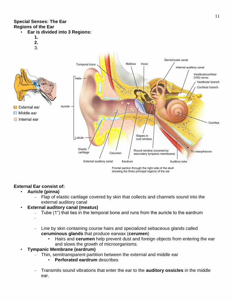
Special Senses: The Ear Regions of the Ear
• Ear is divided into 3 Regions: 1. 2. 3.
External Ear consist of:
• Auricle (pinna) – Flap of elastic cartilage covered by skin that collects and channels sound into the
external auditory canal • External auditory canal (meatus)
– Tube (1”) that lies in the temporal bone and runs from the auricle to the eardrum – – Line by skin containing course hairs and specialized sebaceous glands called
ceruminous glands that produce earwax (cerumen) • Hairs and cerumen help prevent dust and foreign objects from entering the ear
and slows the growth of microorganisms. • Tympanic Membrane (eardrum)
– Thin, semitransparent partition between the external and middle ear • Perforated eardrum describes
– Transmits sound vibrations that enter the ear to the auditory ossicles in the middle ear.

12
Middle Ear consists of: • Tympanic Cavity
– Small air filled cavity hollowed out of the temporal bone that houses the auditory ossicles
– Connected to the nasaopharynx via the auditory tube (Eustachian tube or pharyngotympanic tube)
• Functions to equalize air pressure within the tympanic cavity with that of
• Auditory Ossicles – Bones that are named for their shape:
• Malleus = hammer; attached to internal surface of eardrum • Incus = anvil; intermediate bone • Stapes = stirrup;
– Vibrations of tympanum convert arriving sound waves into mechanical movements that are transmitted via the auditory ossicles to the oval window.
• Because tympanum is 22 times larger and heavier than the oval window, a 1 µm movement of the tympanum produces a 22 µm deflection of the oval window
• Amplification allows us to
– Articulations of ossicles are by way of synovial joints an the bones are attached to the surrounding temporal bone by ligaments.
• Tensor tympani muscle (attached to malleus) and stapedium muscle (attached to stapes) are skeletal muscles that protect the inner ear against prolonged loud noise, but not brief ones such as a gunshot.
– Oval and round window.
• Oval window: Membrane-covered opening between the middle and inner ear. –
• Round window: Opening directly below the oval window
13
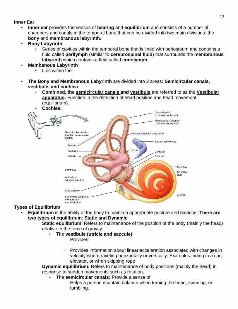
Inner Ear • Inner ear provides the senses of hearing and equilibrium and consists of a number of
chambers and canals in the temporal bone that can be divided into two main divisions: the bony and membranous labyrinth.
• Bony Labyrinth • Series of cavities within the temporal bone that is lined with periosteum and contains a
fluid called perilymph (similar to cerebrospinal fluid) that surrounds the membranous labyrinth which contains a fluid called endolymph.
• Membanous Labyrinth • Lies within the
• The Bony and Membranous Labyrinth are divided into 3 areas: Semicircular canals, vestibule, and cochlea
• Combined, the semicircular canals and vestibule are referred to as the Vestibular apparatus: Function in the detection of head position and head movement (equilibrium).
• Cochlea:
Types of Equilibrium
• Equilibrium is the ability of the body to maintain appropriate posture and balance. There are two types of equilibrium: Static and Dynamic
– Static equilibrium: Refers to maintenance of the position of the body (mainly the head) relative to the force of gravity.
• The vestibule (utricle and saccule): – Provides
– Provides information about linear acceleration associated with changes in
velocity when traveling horizontally or vertically. Examples: riding in a car, elevator, or when skipping rope
– Dynamic equilibrium: Refers to maintenance of body positions (mainly the head) in response to sudden movements such as rotation.
• The semicircular canals: Provide a sense of – Helps a person maintain balance when turning the head, spinning, or
tumbling.

14
Structures and Functions of the Vestibule: Saccule and Utricle • The vestibule consists of two saclike otolithic organs – the saccule and the utricle.
– Function: Provides information about linear acceleration and head position. – In the wall of each utricle and saccule is a sensory structure called a macula.
• Each macula consists of hair cells, an otolithic membrane, and otoliths. – Hair cells: Neuroepithelial cells innervated by the vestibular branch of the
vestibulochchlear nerve (VIII). – Have numerous (70 or more) stereocilia (microvilli) and one kinocilium (normal
cilium), that extends beyond the stereocilia. – Supporting cells: Support the hair cells
• Secret – Otolithic membrane: Gelatinous glycoprotein layer that surrounds and floats directly
over the hair cells. • Contains small protein/ calcium carbonate crystals called otoliths (ear stones)
that increase the mass of the membrane. • Results in a
How the Vestibule Works to Maintain Equilibrium
• Changes in head position due to gravity, linear acceleration, or linear deceleration pulls the otolithic membrane resulting in bending of the stereocilia of the hair cells.

15
• If you tilt your head in any direction, the otolithic membrane is pulled by gravity in the direction of the tilt and slides over the hair cells and bends the hairs, which stimulates them.
• Similarly, if you are sitting upright in a car that suddenly accelerates forward, the otolithic membrane, due to its inertia, slides backward and stimulates the hair cells by bending them
• The bending of the sterocilia of the hair cells leads to the generation of nerve impulses that are transmitted to the brain (medulla and cerebellum) via the vestibular branch of the vestibulocochlear (VIII) nerve.
• The cerebellum is the primary site for equilibrium processing. • Sensory information will then go from the medulla/cerebellum to the thalamus and up to the
cortex. Semicircular Canals
• Semicircular canals: Contain receptors that function in detection of movement (dynamic equilibrium).
– Canals project in three different planes at nearly right angles to each other. – Ampulla: Enlargement at – The crista ampularis is the receptor for movement (dynamic equilibrium) and made up
of a crista and a cupula • Crista: • Cupula: Gelatinous glycoprotein mass that covers the hair cells of the crista.
• The cupula has a higher density than that of the surrounding endolymph and essentially rests above the receptor surface.

16
How the Semicircular Canals Work to Maintain Equilibrium • When the head moves, the attached simicircular canals and hair cells move with it. The
endolymph, however, is not attached and lags behind due to its inertia. As the moving hair cells drag along the stationary fluid, the hairs bend.
• Bending of the stereocilia is transduced into nerve impulses that are transmitted to the brain via the vestibular branch of the vestibulocochlear nerve.
– When the endolymph stops moving, the elastic nature of the cupula allows it to return to its normal position.
– If rotation continues, the moving endolymph finally catches up. If the head rotation stops suddenly, the fluid has built up momentum and cannot stop immediately. The fluid continues to rotate in the direction of the head rotation, leaving the person with a turning sensation.
Vertigo
• Vertigo:
– Result of a disturbance in the vestibular system (i.e., structures of the inner ear, the vestibular nerve, brainstem, and cerebellum) causing a conflict between the signals sent to the brain.
• Example: Visual input vs. the inner ear signals – Can result from:
• Head injuries • Tumor • Infections of the inner ear • Alcohol • Migraine headaches
Nystagmus
• Nystagmus: • Causes:
– Brain's control of eye movements is poor, resulting in an inability to look steadily at an object.
– Vision problems (myopia or hyperopia) – Optic nerve damage
Structures of the Cochlea
• Is subdivided into three channels that spiral around a central bony core. • The partitions that separate the 3 channels are shaped like the letter “Y”
• Cochlear duct (Scala media): Lies between the wings of the Y and is composed of the membranous labyrinth
• Contains endolymph and organ of Corti (Spiral organ) • • Cochlear duct is separated from the scala vestibuli by the vestibular
membrane and from the scala tympani by the basilar membrane. • Scala vestibuli: Channel above the cochlear duct which begins at the oval window and
contain perilymph. • Scala tympani: Channel below the cochlear duct that ends as the round window and
contains perilymph. • The scala tympani and scala vestibuli

17
Organ of Corti (Spiral Organ)
• Organ of Corti: The organ of hearing and lies on the basilar membrane •
– Hair cells function to convert a mechanical vibration (stimulus) into an electrical signal (nerve impulse).
– At their basilar ends, hair cells synapse with the cochlear branch of the vestibulocochlear or auditory nerve (VIII).
Mechanism of Hearing 1. The auricle directs sound waves into the external auditory canal
•
2. Sound waves strike the tympanic membrane and cause it to vibrate at the same frequency as the incoming sound waves.
• Eardrum vibrates slowly in response to low-frequency (low-pitched) sounds and rapidly in response to high-frequency (high-pitched) sounds.
3. The central area of the eardrum connects to the malleus. The eardrum’s vibrations are transmitted from the malleus to the incus and then to the stapes.
4.

18
5. Movement of the oval window sets up fluid pressure waves in the perilymph of the cochlea. – As the oval window bulges inward, it pushes on the perilymph of the scala vestibuli;
these pressure waves are transmitted to the scala tympani and eventually to the round window, causing it to bulge outward into the middle ear.
6. The pressure waves cause the vestibular membrane to vibrate which in turn initiates fluid pressure waves in the endolymph of the cochlear duct.
7. The pressure waves in the endolymph cause the basilar membrane to vibrate and as a result, the hair cells or the organ of Corti move against the tectorial membrane.
– Bending of the hairs is transduced into electrical impulses that travel via the cochlear nerve to that areas of the brain involved with interpretation of sound
8. The hair cells of the organ of Corti are arranged in rows; a very soft sound may stimulate only a few hair cells in a portion of one row.
– As the volume of a sound increases, additional hair cells are stimulated as the vibrations of the basilar membrane increase.
9. Areas of the basilar membrane vibrate at different frequencies. – Sound waves of high frequency are detected closer to the oval window whereas low
frequency sounds have optimal vibration (resonance) further away from the oval window.