sensory system, ear,eye anatomy
TRANSCRIPT

ANATOMY OF SENSORY SYSTEM
Prepared by: Mr.Maulik chaudhary

Contents
Structure of eye
Structure of ear
Structure of nose
Structure of tongue

Introduction
• The special senses of hearing, sight, smell and taste all have specialised sensory receptors that collect and transmit information to specific areas of the brain.
• Incoming nerve impulses from sensory receptors in the ears, eyes, nose and mouth are integrated and coordinated within the brain allowing perception of this sensory information.

Anatomy of Ear

Introduction
• The ear is the organ of hearing and is also involved in balance.
• It is supplied by the 8th cranial nerve= vestibulocochlear nerve, which is stimulated by vibrations caused by sound waves.

Structure
• The ear is divided into three distinct parts
1) outer ear
2) middle ear (tympanic cavity)
3) inner ear. • The outer ear collects the sound waves and directs them to
the middle ear, which in turn transfers them to the inner ear, where they are converted into nerve impulses and transmitted to the hearing area in the cerebral cortex.


Outer ear
• The outer ear consists of the auricle (pinna) and the external acoustic meatus (auditory canal).
• The auricle (pinna)
• The auricle is the visible part of the ear that projects from the side of the head.
• It is composed of fibroelastic cartilage covered with skin. It is deeply grooved and ridged; the most prominent outer ridge is the helix.
• The lobule (earlobe) is the soft pliable part at the lower extremity, composed of fibrous and adipose tissue which is supplied with blood.

• External acoustic meatus (auditory canal)
• This is a slightly ‘S’-shaped tube about 2.5 cm long extending from the auricle to the tympanic membrane (eardrum).
• There are various numbers of ceruminous glands along the auditory tube, the function of this ceruminous gland is to secrete a cerumen( ear wax)
• Cerumen= a sticky material containing protective substances including the bacteriocidal enzyme lysozyme and immunoglobulins.

• Foreign materials, e.g. dust, insects and microbes, are prevented from reaching the tympanic membrane by wax, hairs and the curvature of the meatus.
• Tympanic membrane=The tympanic membrane (eardrum) completely separates the external acoustic meatus from the middle ear.
• It is oval-shaped with the slightly broader edge upwards and is formed by three types of tissue
• the outer covering of hairless skin,
• the middle layer of fibrous tissue and the inner lining of mucous membrane continuous with that of the middle ear.

Middle ear (tympanic cavity)
• This is an irregular-shaped air-filled cavity within the portion of the temporal bone
• The middle ear cavity is lined with either simple squamous or cuboidal epithelium
• Walls of the middle ear:
• The lateral wall of the middle ear is formed by the tympanic membrane.
• The roof and floor are formed by the temporal bone.

• The posterior wall is formed by the temporal bone with openings leading to the mastoid antrum through which air passes to the air cells within the mastoid process.
• The medial wall is a thin layer of temporal bone in which there are two openings:
• oval window
• round window

• The oval window is occluded by part of a small bone called the stapes and the round window, by a fine sheet of fibrous tissue.
• Pharyngotympanic (auditory or Eustachian) tube which is about 4 cm long


Auditory ossicles
• These are three very small bones only a few millimetres in size & are located in the middle ear.


• The malleus.
• This is the hammer-shaped bone. The handle is in contact with the tympanic membrane and the head forms a movable joint with the incus.
• The incus.
• This is the middle anvil-shaped bone. Its body articulates with the malleus, the long process with the stapes.
• The stapes.
• This is the medial stirrup-shaped bone. Its head articulates with the incus and its footplate fits into the oval window.

Inner ear
• The inner ear or labyrinth (meaning ‘maze’) contains the organs of hearing and balance.
• It is described in two parts, the bony labyrinth and the membranous labyrinth and is divided into three main regions:
• 1) vestibule (containing the utricle and saccule)
• 2) three semicircular canals
• 3) the cochlea.


The vestibule
• This is the expanded part nearest the middle ear. The oval and round windows are located in its lateral wall.
The semicircular canals
• These are three tubes arranged so that one is situated in each of the three planes of space.
The cochlea
• This resembles a snail’s shell.

Physiology of hearing
?
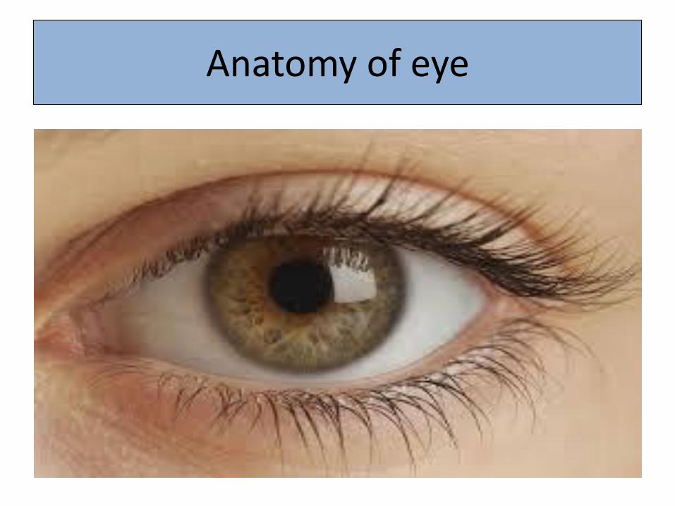
Anatomy of eye

• The eye is the organ of sight. It is situated in the orbital cavity and supplied by the optic nerve (2nd cranial nerve).
• It is almost spherical in shape and about 2.5 cm in diameter.
• The space between the eye and the orbital cavity is occupied by adipose tissue.
• Structurally the two eyes are separate but, unlike the ears, some of their activities are coordinated so that they normally function as a pair.
• It is possible to see with only one eye (monocular vision), but three-dimensional vision is impaired when only one eye is used, especially in relation to the judgement of speed and distance.

Structure
There are three layers of tissue in the walls of the eye:
• the outer fibrous layer: sclera and cornea
• the middle vascular layer or uveal tract: consisting of the choroid, ciliary body and iris
• the inner nervous tissue layer: the retina.


Inside structure of eye
• Structures inside the eyeball include the lens, aqueous fluid and vitreous body.
• Sclera and cornea
• The sclera is the white part of the eye, forms the outermost layer of the posterior and lateral aspects of the eyeball.
• and is continuous anteriorly with the cornea. It consists of a firm fibrous membrane that maintains the shape of the eye and gives attachment to the extrinsic muscles of the eye.

• Anteriorly the sclera continues as a clear transparent epithelial membrane = the cornea.
• Light rays pass through the cornea to reach the retina.
• The cornea is convex anteriorly and is involved in refracting (bending) light rays to focus them on the retina.

• Choroid
• The choroid lines the posterior five-sixths of the inner surface of the sclera. It is very rich in blood vessels and
• is deep chocolate brown in colour. Light enters the eye through the pupil, stimulates the sensory receptors in the retina and is then absorbed by the choroid.

• Ciliary body
• The ciliary body is the anterior continuation of the choroid consisting of ciliary muscle (smooth muscle fibres) and secretory epithelial cells.
• As many of the smooth muscle fibres are circular, the ciliary muscle acts like a sphincter.
• The lens is attached to the ciliary body by radiating suspensory ligaments, like the spokes of a wheel.
• Contraction and relaxation of the ciliary muscle fibres, which are attached to these ligaments, control the size and thickness of the lens.

• The epithelial cells secrete aqueous fluid into the anterior segment of the eye,
• i.e. the space between the lens and the cornea (anterior and posterior chambers)

• Iris
• The iris is the visible coloured ring at the front of the eye and extends anteriorly from the ciliary body, lying behind the cornea and in front of the lens.
• It divides the anterior segment of the eye into anterior and posterior chambers which contain aqueous fluid secreted by the ciliary body.
• It is a circular body composed of pigment cells and two layers of smooth muscle fibres, one circular and the other radiating
• In the centre is an aperture called the pupil.
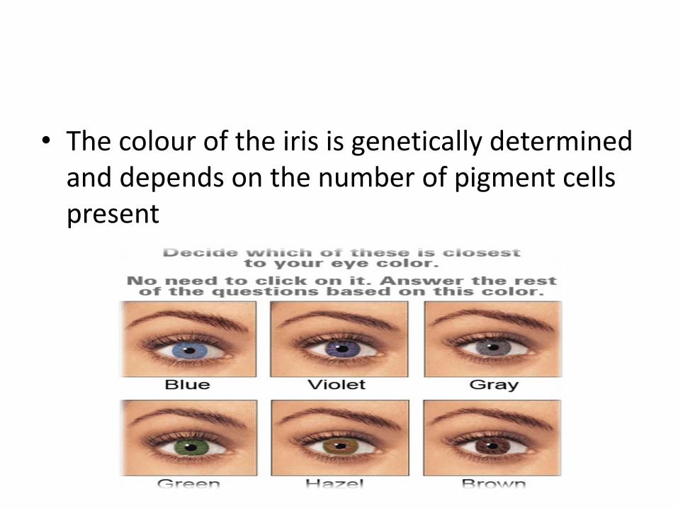
• The colour of the iris is genetically determined and depends on the number of pigment cells present

• Lens
• The lens is a highly elastic circular biconvex body, lying immediately behind the pupil.
• It consists of fibres enclosed within a capsule and is suspended from the ciliary body by the suspensory ligament.
• Its thickness is controlled by the ciliary muscle through the suspensory ligament
• Lense having refractory power.

• Inside lense there is a light sensitivity layer
• The light-sensitive layer consists of sensory receptor cells, rods and cones, which contain photosensitive pigments that convert light rays into nerve impulses.
• Near the centre of the posterior part is the macula lutea, or yellow spot.
• In the centre of the yellow spot is a little depression called the fovea centralis, consisting of only cones.
• Towards the anterior part of the retina there are fewer cones than rods.

• The small area of the retina where optic nerve leave is called as a optic disc or blind spot. It has a no light sensitivity cells.

Interior of the eye
• The anterior segment of the eye, i.e. the space between the cornea and the lens, is incompletely divided into anterior and posterior chambers by the iris
• Both chambers contain a clear aqueous fluid secreted into the posterior chamber by the ciliary glands.
• The intraocular pressure remains fairly constant between 1.3 and 2.6 kPa (10 to 21 mmHg) An increase in this pressure causes glaucoma

• Aqueous fluid supplies nutrients and removes wastes from the transparent structures in the front of the eye that have no blood supply, i.e. the cornea, lens and lens capsule.
• Behind the lens and filling the posterior segment (cavity) of the eyeball is the vitreous body.
• This is a soft, colourless, transparent, jelly-like substance composed of 99% water, some salts an mucoprotein.
• It maintains sufficient intraocular pressure to support the retina against the choroid and prevent the eyeball from collapsing.

• The eye keeps its shape because of the intraocular pressure exerted by the vitreous body and the aqueous fluid. It remains fairly constant throughout life.

Blood supply to the eye
• The eye is supplied with arterial blood by the ciliary arteries and the central retinal artery. These are branches of the ophthalmic artery, a branch of the internal carotid artery.

Venous supply
• Venous drainage is by a number of veins, including the central retinal vein.

Nerve supply
• Optic nerves (second cranial nerves)
• The fibres of the optic nerve originate in the retina and they converge to form the optic nerve about 0.5 cm to the nasal side of the macula lutea at the optic disc.
• The nerve pierces the choroid and sclera to pass backwards and medially through the orbital cavity.
• It then passes through the optic foramen of the sphenoid bone to meet the nerve from the other eye at the optic chiasma.
• Optic chiasma
• This is situated immediately in front of and above the pituitary gland, which is in the hypophyseal fossa of the sphenoid bone.
• In the optic chiasma the nerve fibres of the optic nerve from the nasal side of each retina cross over to the opposite side.


• Optic tracts
• These are the pathways of the optic nerves, posterior to the optic chiasma
• Size of the pupils
• Pupil size contributes to clear vision by controlling the amount of light entering the eye.
• In bright light the pupils are constricted. In dim light they are dilated.
• If the pupils were dilated in bright light, too much light would enter the eye and damage the sensitive retina.
• In dim light, if the pupils were constricted, insufficient lightwould enter the eye to activate the light-sensitive pigments in the rods and cones, which stimulate the nerve endings in the retina enabling vision.

• Extraocular muscles of the eye
• These include the muscles of the eyelids and those that move the eyeballs.
• The eyeball is moved by six extrinsic muscles, attached at one end to the eyeball and at the other to the walls of the orbital cavity.
• There are four straight (rectus) muscles and two oblique muscles

• Nerve supply to the muscles of the eye
• The oculomotor nerves supply the intrinsic eye muscles of the iris and ciliary body & extrinsic muscles.
Physiology of Eye
?