separation of components plasma = less dense hematocrit “packed cells” more dense platelets /...
TRANSCRIPT
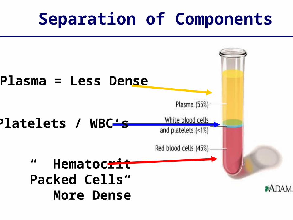
Separation of Components
Plasma = Less Dense
Hematocrit“Packed Cells”
More Dense
Platelets / WBC’s
Hematocrite value (H.V.) or packed cell volume (PCV)
•Definition-:• H.V. is the volume of RBCs in 100 ml blood or it is the
percentage of the volume of RBCs to the volume of whole blood.
• Volume of RBCs• × = 100
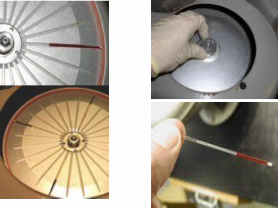
• Volume of blood• Materials:-• Microhematocrite tube (75 mm long, 1 mm pore, heparinized )• Microhematocrite centrifuge .• Microhematocrite tube reader.• Sterile lancet , 70 % ethyl alcohol.
• Procedure:-• Obtain blood drop by pricking the thumb.• Micro hematocrite tube is filled up to it's 2/3 with blood sample by
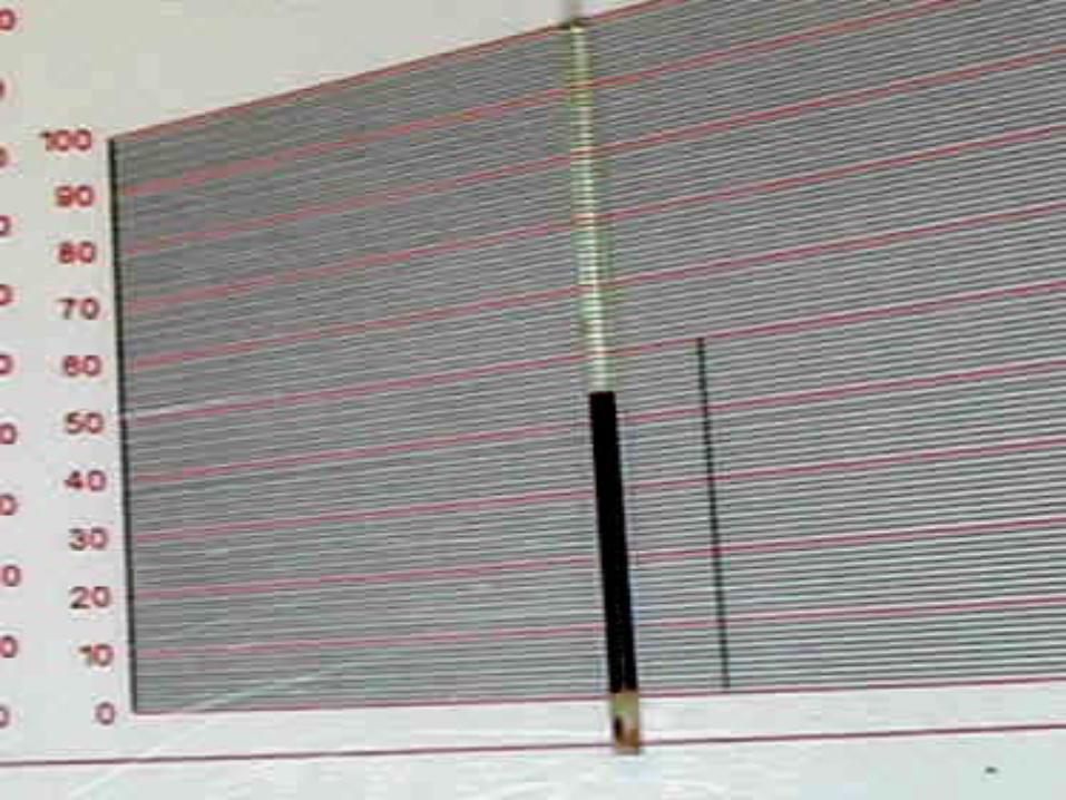
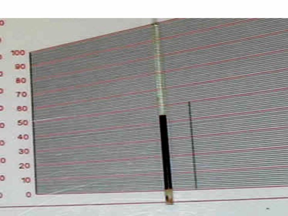
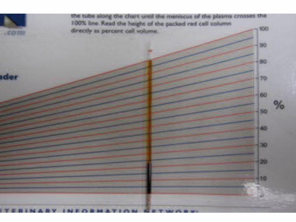
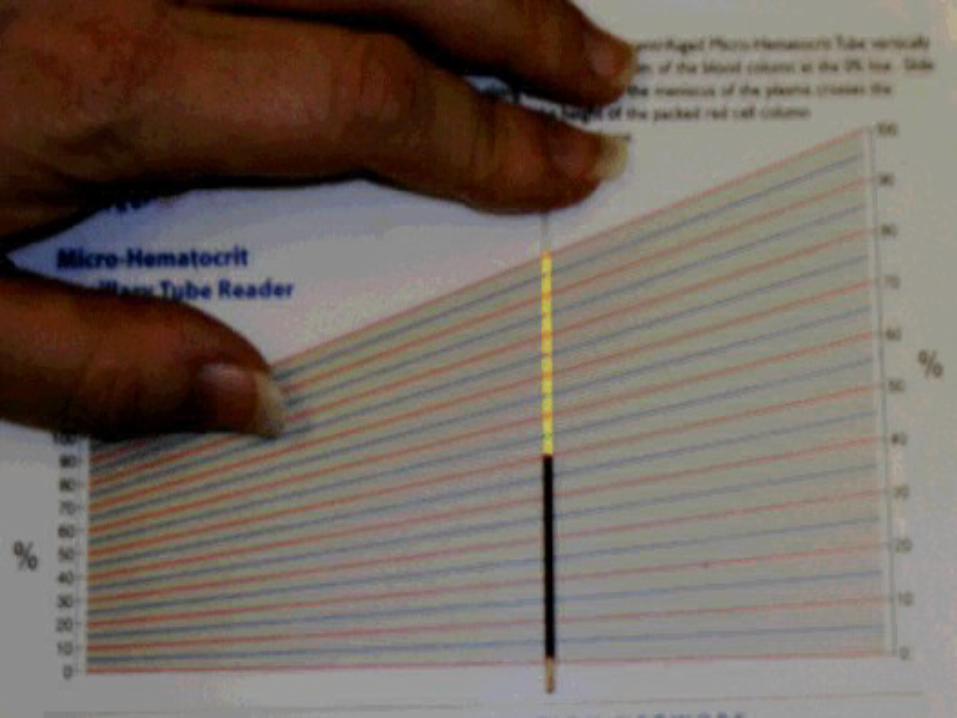
touch the drop of blood by one end of the tube.• Close the empty end of the tube by plasticine .• Centrifuge the tube at 13000 per minute for 5 minutes .• Remove the tube and read H.V. by putting the tube on special micro
hematocrite scale.
• Normal value:-• adult male. 45%- 47 % • adult female. 42 % -44%• Higher in new born. 45-49%
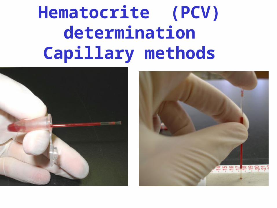
Hematocrite (PCV) determination
Capillary methods
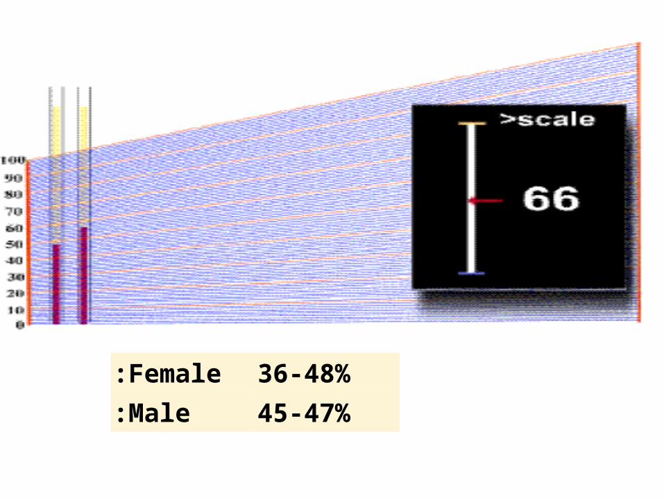
Female: 36-48%Male: 45-47%
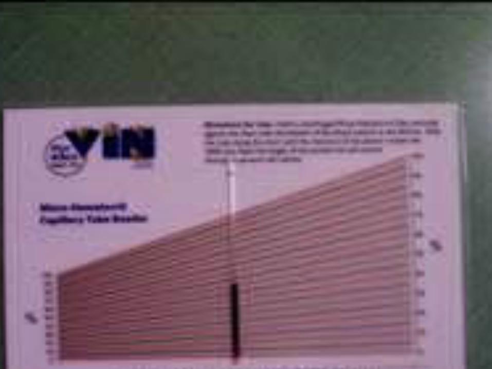
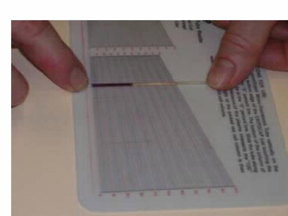
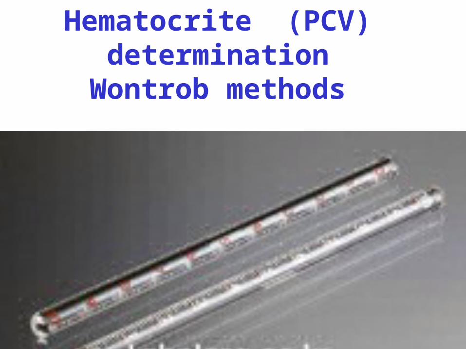
Hematocrite (PCV) determination
Wontrob methods
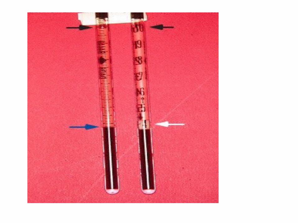
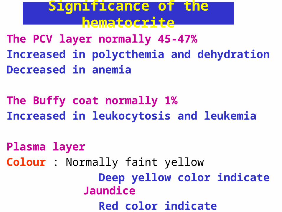
Significance of the hematocrite
The PCV layer normally 45-47%
Increased in polycthemia and dehydration
Decreased in anemia
The Buffy coat normally 1%
Increased in leukocytosis and leukemia
Plasma layer
Colour : Normally faint yellow
Deep yellow color indicate Jaundice
Red color indicate Hemolysis
Determination of Hb values• Haemoglobin can be measured manual or automatic.• Two method are commonly used:
1. Oxyhaemoglobin (Hbo2method).
2. Haemiglobincyanid. (HicN;Cyanmethaemoglobin )
• A major a advantage of the HicN method is availability of stable and reliable reference preparation.
• Other methods: Sahli’s acid haematin and alkalin-haematin method.
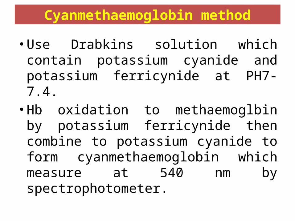
Cyanmethaemoglobin method
• Use Drabkins solution which contain potassium cyanide and potassium ferricynide at PH7-7.4.• Hb oxidation to methaemoglbin by
potassium ferricynide then combine to potassium cyanide to form cyanmethaemoglobin which measure at 540 nm by spectrophotometer.
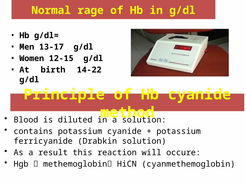
Normal rage of Hb in g/dl
• Hb g/dl= • Men 13-17 g/dl• Women 12-15 g/dl• At birth 14-22 g/dl
Principle of Hb cyanide method• Blood is diluted in a solution:• contains potassium cyanide + potassium ferricyanide (Drabkin
solution)• As a result this reaction will occure: • Hgb methemoglobin HiCN (cyanmethemoglobin)
Methods
• Prepare the working solution of diluted drabkin (one volume
drabkin+ four volumes distilled water)
• Set spectrophotometer to 0.0 reading at 540 wave length, using
blank (diluted drabkin solution)
• 10µL of blood in 2.5 mL of working solution in the cuvet, Cover
and invert the tube several times.
• Let it stand at R.T for 5-10 minutes to insure complete conversion
• Put the cuvet in the spectrophotometer.
• Continue reading patient samples and determine Hb value of the
sample by multiplying the absorbance by the factor (36.77) .