sequence similarity between the erythrocyte binding domain 1 of the plasmodium vivax duffy binding...
TRANSCRIPT

RESEARCH Open Access
Sequence similarity between the erythrocytebinding domain 1 of the Plasmodium vivax Duffybinding protein and the V3 loop of HIV-1 strainMN reveals binding residues for the DuffyAntigen Receptor for ChemokinesMichael J Bolton1, Robert F Garry2*
Abstract
Background: The surface glycoprotein (SU, gp120) of the human immunodeficiency virus (HIV) must bind to achemokine receptor, CCR5 or CXCR4, to invade CD4+ cells. Plasmodium vivax uses the Duffy Binding Protein (DBP)to bind the Duffy Antigen Receptor for Chemokines (DARC) and invade reticulocytes.
Results: Variable loop 3 (V3) of HIV-1 SU and domain 1 of the Plasmodium vivax DBP share a sequence similarity.The site of amino acid sequence similarity was necessary, but not sufficient, for DARC binding and contained aconsensus heparin binding site essential for DARC binding. Both HIV-1 and P. vivax can be blocked from binding totheir chemokine receptors by the chemokine, RANTES and its analog AOP-RANTES. Site directed mutagenesis ofthe heparin binding motif in members of the DBP family, the P. knowlesi alpha, beta and gamma proteinsabrogated their binding to erythrocytes. Positively charged residues within domain 1 are required for binding ofP. vivax and P. knowlesi erythrocyte binding proteins.
Conclusion: A heparin binding site motif in members of the DBP family may form part of a conserved erythrocytereceptor binding pocket.
IntroductionHuman immunodeficiency virus type 1 (HIV-1) and thehuman malaria parasite Plasmodium vivax both use che-mokine receptors in obligate steps of cell invasion. HIV-1 uses CCR5 and CXCR4 as the major coreceptors forinfecting CD4+ cells (macrophages, T-lymphocytes, andother cell types) in vivo, while P. vivax uses the Duffyantigen receptor for chemokines (DARC) for invadinghuman reticulocytes [1,2]. Alleles of CCR5 and DARCassociated with decreased functional protein expressionconfer resistance to HIV and P. vivax, respectively, andchemokines can inhibit in vitro infection by eitherpathogen [1,3-5]. The HIV surface glycoprotein (SU,gp120) undergoes a conformational change upon
binding to CD4 and then presents a chemokine receptorbinding surface predicted to include a hydrophobic coresurrounded by positive residues contributed by con-served and variable regions including the base of theV3 loop. The V3 loop putatively extends toward the cellsurface and contacts the chemokine receptor at a secondsite in the second extracellular loop. Individual aminoacid mutations in the V3 loop can change chemokinereceptor specificity.P. vivax and the simian malaria, P. knowlesi, use Duffy
binding proteins (PvDBP and PkDBP) to invade humanerythrocytes. These proteins belong to a family of ery-throcyte binding proteins with conserved regions. Theerythrocyte binding domains of PvDBP and PkDBP (orP. knowlesi a protein) have been shown to map to the330 amino-acid cysteine-rich region II known as theDuffy-binding-like (DBL) domains [6]. Other membersof the family include the P. knowlesi b and g proteins
* Correspondence: [email protected] of Microbiology and Immunology Tulane University1430 Tulane Avenue New Orleans, Louisiana 70112 USAFull list of author information is available at the end of the article
Bolton and Garry Virology Journal 2011, 8:45http://www.virologyj.com/content/8/1/45
© 2011 Bolton and Garry; licensee BioMed Central Ltd. This is an Open Access article distributed under the terms of the CreativeCommons Attribution License (http://creativecommons.org/licenses/by/2.0), which permits unrestricted use, distribution, andreproduction in any medium, provided the original work is properly cited.

and the P. falciparum erythrocyte-binding antigen (EBA-175), which use DBLs to bind to other receptors.Here we report the identification of an amino acid
sequence similarity between the V3 loop of HIV-1 strainMN and a site in Plasmodium erythrocyte binding pro-teins that contains a consensus heparin binding site. BothHIV-1 and P. vivax can be blocked from binding to theirchemokine receptors by the chemokine RANTES. Muta-genesis studies suggest that the heparin binding sitemotif in members of the DBP family may form part of aconserved erythrocyte receptor binding pocket.
Materials and methodsSequence comparisonsWilliam Pearson’s LALIGN program, which implementsa linear-space local similarity algorithm, was used toperform regional alignments. Sequence and structuralcomparisons were performed for the V3 loop of SU ofHIV-1 strain MN, accession: AAT67509; P. vivax DBP,ACD76813; P. knowlesi DBP, XP_002261904; P. falci-parum erythrocyte binding protein EBA-175 (F1), acces-sion AAA29600. Plasmodium proteins are members ofpfam05424 (a member of the superfamily cl05146).
ErythrocytesBlood was collected in 10% citrate phosphate dextrose(CPD) and stored at 4°C unwashed for up to 4 weeks,or washed in RPMI with malaria supplements andstored in malaria culture medium at 50% hematocrit forup to 2 weeks. The DARC+ human erythrocytes used inthe erythrocyte binding assay and the P. knowlesi ery-throcyte invasion assay had the phenotype Fy(a-b+) asdetermined by standard blood banking methods usinganti-Fya and anti-Fyb antisera (Gamma Biologicals,Houston, TX). Erythrocytes were washed three times inDMEM (Gibco BRL) and resuspended to a hematocritof 10% in complete DMEM for the erythrocyte bindingassay. Erythrocytes used in the P. knowlesi erythrocyteinvasion assay were washed three times and resuspendedto a hematocrit of 10% using malaria complete RPMI.
Cell Culture and Transfection of COS-7 CellsCOS-7 cells (ATCC CRL 1651; Rockville, MD) were cul-tured in DMEM with 10% heat inactivated FBS (GibcoBRL) in a humidified 5% CO2 incubator at 37°C. Cellswere seeded in polystyrene dishes with 3.5-cm diameterwells and grown for 24 h to 30-50% confluence beforetransfection with 1 mg of pHVDR22 plasmid DNA and10 ml of Lipofectamine (Gibco BRL).
P. knowlesi in vitro cultureWhole blood from rhesus macaques was collected in10% CPD and allowed to separate overnight at 4°C.The erythrocyte phase was washed in RPMI with
L-glutamine and supplemented with 25 mM HEPES,300 mM hypoxanthine, 10 mM thymidine, 1.0 mMsodium pyruvate, and 11 mM glucose. This RPMI withmalaria supplements was then used to prepare malariaculture medium by adding to a final concentration of0.24% sodium bicarbonate and 0.2% Albumax-I (LifeTech, Gibco BRL). Cultures were maintained at a hema-tocrit of 10% in malaria culture medium under an atmo-sphere of 5% O2, 5% CO2, balanced N2 (Air Liquide,Houston, TX) at 38°C.
Percoll Purification of Schizont-infected ErythrocytesCultures of P. knowlesi at 5-10% infected erythrocyteswere washed three times in RPMI with malaria supple-ments and 10% FBS and brought up to a hematocrit of10%. A 50% Percoll solution was made by adding0.45 volumes 1X PBS, 0.05 volumes 10X PBS and0.5 volumes Percoll (Sigma). Two ml of the washed cul-ture was overlaid on 2 ml of the 50% Percoll solution ina 4 ml polystyrene tube and centrifuged for 20 min at2100 RPM in a Sorvall centrifuge. The ring of cells atthe interface was removed, pooled and washed threetime in 1X PBS. The pellet was brought up in malariaculture medium to 2 × 10⁷ cells/ml.
PvRII Erythrocyte Binding AssayCOS-7 cells were transfected by Lipofectamine with1-2 mg of pHVDR22 DNA, a plasmid kindly providedby L. Miller which expresses region II of the DBP of P.vivax on the cell surface as a chimera with the HSV gDprotein [7] Duffy Fy (a-b+) erythrocytes were washedthree times in RPMI 1640, resuspended to a hematocritof 1% in 1 ml of complete DMEM with the chemokinesRANTES, MIP-1a, SDF-1 or AOP-RANTES at concen-trations of 0, 0.1, 1, 10, and 100 nM for 1 h at 37°C(Peprotech, Gryphon Pharmaceuticals, San Francisco,CA). This suspension was swirled over aspirated COS-7 cells 40-60 h after transfection and allowed to settleover 2 h at 37°C. The COS-7 cells were then washedthree times with 2 ml of PBS to remove nonadherenterythrocytes. The number of adherent erythrocyterosettes was scored in 20 randomly chosen fields at amagnification of 40 using an inverted microscope. Per-cent inhibition was determined by dividing the numberof rosettes in the presence of chemokines by the num-ber at a concentration of 0 nM. The 50% inhibitory con-centration (IC50) was determined by the mean of threeseparate experiments to use in a semi-log cubic splinecurve fit with the DeltaSoft 3 software (Biometallics,Inc., Princeton, NJ).
P. knowlesi Erythrocyte Invasion AssayHuman Duffy Fy(a-b+) erythrocytes were washed incomplete malaria medium and 2 × 10⁷ washed cells
Bolton and Garry Virology Journal 2011, 8:45http://www.virologyj.com/content/8/1/45
Page 2 of 10

were added to increasing concentrations of chemokinesin malaria culture medium at final volume of 900 ml for1h at room temperature. To each tube of chemokine-treated erythrocytes, 100 ml or 2 × 10⁶ schizont-infectederythrocytes was added and placed in a well of a poly-styrene 24-well plate (Becton-Dickinson). The cultureswere maintained under a blood-gas atmosphere at 38°Cfor 8 h to allow the infected erythrocytes to rupture andrelease free merozoites capable of infecting new erythro-cytes and developing to ring-stage trophozoites. Theculture was centrifuged at 2100 RPM for 3 min and athin smear was made from the pellet. The thin smearwas fixed with methanol and stained with LeukostatSolution B (100 mg Eosin Y+300 ml 37% formaldehyde +400 mg sodium phosphate dibasic + 500 mg potassiumphosphate monobasic, q.s. to 100 ml with dH2O), rinsed,and stained with Leukostat Solution C (47 mg MethyleneBlue + 44 mpp Azure A + 400 mg sodium phosphatedibasic + 500 mg potassium phosphate monobasic, q.s to100 ml with dH2O). The percentage of erythrocytesinfected with ring-stage trophozoites per 2000 erythrocyteswas determined at 1000X. Inhibition of invasionexpressed as % inhibition was determined by dividing thepercentage of ring-stage parasites by the percentage ofring-stage parasites at 0 nM chemokine, multiplying by100 and subtracting this value from 100 [1].
Statistical analysisThe software StatView (Brainpower, Inc., Calabasas, CA),was used to determine the statistical difference betweenthe inhibitory concentrations of RANTES, AOP-RANTES, and MIP-1a, using a two-way ANOVA test.
PlasmidsThe plasmids pHVDR22, pHKADR22, pHKBDR22 andpHKGDR22 encode for the region II (amino acids198-522) of the P. vivax DBP and region II of theP. knowlesi a, b and g genes, respectively, in the contextof the HSV gD protein. These plasmids have been pre-viously described and were kindly provided by thelaboratory of Louis H. Miller. These plasmids werecreated from the plasmid pRE4, which contains anSV40 origin of replication, a Rous sarcoma virus LTR asa promoter, the coding region of the HSV glycoproteinD (HSV gD) inserted in the HindIII cloning site, and theSV40 early polyadenylation signal. The HSV gD featuresa 25 amino acid signal peptide at the amino terminus, a24 amino acid hydrophobic transmembrane region, a30 amino acid cytoplasmic tail at the carboxy terminus,and two epitopes at amino acids 11-19 and 272-279 thatcan be targeted specifically with the monoclonal antibo-dies ID3 and DL6, respectively. The region II sequenceswere inserted between the unique Apa I and Pvu IIrestriction sites.
Cloning and Site Directed MutagenesisMutants of the region II expressing plasmids were gen-erated by three strategies: inverse PCR, PCR and restric-tion digestion, or PCR-based site directed mutagenesis.Each mutant was sequenced by Research Genetics, Inc.(Huntsville, Ala.) to confirm proper construction at thesite of mutation.The following constructs were made from the
pHVDR22 plasmid:pv22d32 This construct contains a deletion in amino
acids 216-247 of the RII of P. vivax, which correspondsto the V3-like peptide region with similarity tothe V3 loop and comprises cysteines C1 to C4 of regionII. For lack of proper restriction enzyme sites, aninverse PCR strategy was use to amplify the entirepHVDR22 plasmid flanking the site to be deleted. Theprimers 5’TGT ATG AAG GAA CTT ACG AAT TTGG3’ and 5’TTT CAT TAC AGT ATT TTG AAG3’ werefirst phosphorylated with T4 kinase then used with thelong range, high fidelity DeepVent polymerase (NewEngland Biolabs, Inc., Beverly, MA) to amplify the pro-duct under the following thermocycling conditions:5 minutes at 94°C initial denaturing, then 35 cycles at94°C for 60 seconds, 55°C for 60 seconds, 72°C for3 minutes. The product was digested with DPN I toeliminate methylated input plasmid DNA, then blunt-end ligated with high concentration ligase (Gibco BRL).pv22MNV3 This construct replaces the 32 amino acid
V3-like peptide of the P. vivax RII with the V3 loop ofHIV-1 strain MN. To amplify the V3 loop of HIV-1MN
by PCR, PM-1 cells were infected with HIV-1MN
(donated by Dr. James Robinson, Tulane UniversityMedical Center) and genomic DNA was isolated frominfected cultures. This DNA includes proviral DNA andwas used as template for a PCR with the primersP2 5’GAC GCT GCG CCC ATA GTG CTT CCT G3’and P5 5’ACA CAT GGAATT CGGCCAGTA GT3’which are homologous to conserved regions of the envgene of HIV and amplify the region between nucleotides6884 and 7783, which includes the V3 loop.This PCR product then served as template in a nested
PCR of the HIV-1MN V3 loop using the primersHVMN-F 5’AATTGTACAAGACCCAACTAC3’ andHVMN-r 5’ATGTGCTTGTCTTATAGTTCC3’. Thisnested PCR was carried out using the DeepVent enzymeto generate blunt ends. The product of the second,nested PCR was gel purified. The gel-purified ampliconwas then re-amplified in a 300 ml PCR using HVMN-rand HVMN-F primers, which were first phosphorylatedwith T4 poly N kinase. This reamplification product wascolumn purified and blunt-end ligated to the inversePCR product described in the preparation of pvD32.The sequenced construct matched the MN V3 sequenceas previously published.
Bolton and Garry Virology Journal 2011, 8:45http://www.virologyj.com/content/8/1/45
Page 3 of 10

pv22suf32 This construct was designed to determine ifthe 32-aa V3-like peptide of P. vivax RII is sufficient forDARC binding by deleting all flanking RII amino acids.The primers used to create this construct were 5’CAAAAT CAG CTG ATG AAA AAC TGT AAT TAT3’and 5’CAA ATT GGG CCC TTC CTT CAT ACA TAATTG3’ and contain the restriction sites for Apa I andPvu II. The pHVDR22 plasmid was digested with Apa Iand Pvu II, and the digested vector was separated fromthe insert by gel electrophoresis and extracted using theQIAEX II gel extraction kit (Qiagen Inc., Valencia, CA).The PCR product was also digested with Apa I and PvuII and ligated to the digested vector.pv22d5C1 This construct deletes amino acids 198-216,
or the 5’ flanking region to C1. This was created usingthe primers 5’TGT ATG AAG GAA CTT ACG AATTTG G3’ and 5’ GGG GCC TTG GGC CCT GTC ACAAC3’, the product of which was digested with Apa I andPvu II and cloned into the digested vector as describedfor psuf32pv22d3C4 This construct deletes amino acids 247 to
522 or the 3’ flanking region to C4. This was createdusing the primers 5’CCG GTC CTG GAC CAG CTGACG3’ and 5’TTT CAT TAC AGT ATT TTG AAG3’the product of which was digested with Apa I and PvuII and cloned into the digested vector as described inpsuf32pv22d5C4 This construct deletes amino acids 198 to
247 or the 5’ flanking region to C4 This was createdusing the primers 5’CAA TTA CAG CTG AAG GAACTT ACG AAT TTG3’ and 5’ GGG GCC TTG GGCCCT GTC ACA AC3’ the product of which wasdigested with Apa I and Pvu II and cloned into thedigested vector as described in pv22suf32pv22KARA The Stratagene QuickChange kit (Promega)
was used to mutate the heparin binding consensus site inPvRII at amino acids 217-226 from YKRKRRERDW toYARKAREADW using the primers 5’ GTA ATT ATGCGA GAA AAG CTC GGG AAG CAG ATT GG3’ and5’ CCA ATC TGC TTC CCG AGC TTT TCT CGCATA ATT AC3’. These primers also introduce an Ava Isite as a silent mutation for screening.pv22KAKA The Stratagene QuickChange kit was used
to mutate a second potential heparin binding consensussite at amino acids 364-373, between C5 and C6, fromSVKKRLKGNF to SVKARLAGNF using the primers 5’GAT GTA CTC AGT TAA AGC AAG ACT TAAGGG G3’. These primers also introduce an Afl II site asa silent mutation for screeningpv22KA The Stratagene QuickChange kit was used to
introduce a single alanine substitution in the heparinbinding consensus site at amino acids 217-226 fromYKRKRRERDW to YKRARRERDW. 5’CTC TTT CCCGAC GAG CTC TCT TAT AAT TAC AG3’ and
5’CTG TAA TTA TAA GAG AGC TCG TCG GGAAAG AG3’. These primers also introduce a Sac I site asa silent mutation for screening.The following mutants were made from the
pHKADR22, pHKBDR22, or pHKGDR22 plasmids usingthe Stratagene QuickChange kit:pkalpha22KARA This mutant was designed to change
the heparin binding consensus site in pHKADR22 atamino acids 217-226 from DKRKRGERD to DARKA-GEAD using the primers 5’GTC CCA ATC TGC TTCCCC GCG AGC TCT CGC ACT ACC ACA CTT G and5’CAA GCG TAA TGA TGC GAG AGC TCG CGGGGA AGC AGA TTG GGA C3’. These primers alsointroduce a Sac I site as a silent mutation for screening.pkbeta22KARA This mutant was designed to change
the heparin binding consensus site in pHKBDR22 atamino acids 217-226 from NKRKRGTRD to NARKAG-TAD using the primers 5’ CAG TCC CAA TCT GCTGTC CCG CGA GCT TCT GCA TTA TTA CAC C3’and 5’GGT GTA ATA ATG CGA GAG CTC GCG GGACAG CAG ATT GGG ACT G3’. These primers alsointroduce a Sac I site as a silent mutation for screening.pkgamma22KARA This mutant was designed to change
the heparin binding consensus site in pHKGDR22 atamino acids 217-226 from DKRKRGERD to DARKA-GEAD using the primers 5’GTC CCA ATC TGC TTCCCC GCG AGC TCT CGC ACT ACC ACA CTT G and5’CAA GCG TAA TGA TGC GAG AGC TCG CGGGGA AGC AGA TTG GGA C3’. These primers alsointroduce a Sac I site as a silent mutation for screening.
Immunofluorescence StainingTransfected cells used in the erythrocyte binding assaywere rinsed in PBS and incubated for 1 h at 37°C withmonoclonal antibodies that bind to amino acids11-19 and 272-279 of the mature HSV gD proteinfound in pHVDR22. These primary antibodies, ID3 orDL6 (provided by Drs. Gary Cohen and Roselyn Eisen-berg), were used at a 1:2000 dilution in PBS containing10% FBS. The cells were rinsed with PBS and incubatedat 37°C with fluorescein conjugated anti-mouse antibo-dies at 1:100 in PBS containing 10% FBS. UntransfectedCOS-7 cells were also stained as a negative staining con-trol. The cells were then fixed with 4% paraformalde-hyde for 15 min, and observed for surface expression ofthe products of the transfected plasmids using aninverted fluorescence microscope.
ResultsHomologous sequences in Plasmodium erythrocytebinding proteins and the V3 loop of HIV-1 SUThe common use of chemokine receptors by HIV-1 SUand PvDBP suggested the possibility that these proteinsmay share structural or functional motifs. A homology
Bolton and Garry Virology Journal 2011, 8:45http://www.virologyj.com/content/8/1/45
Page 4 of 10
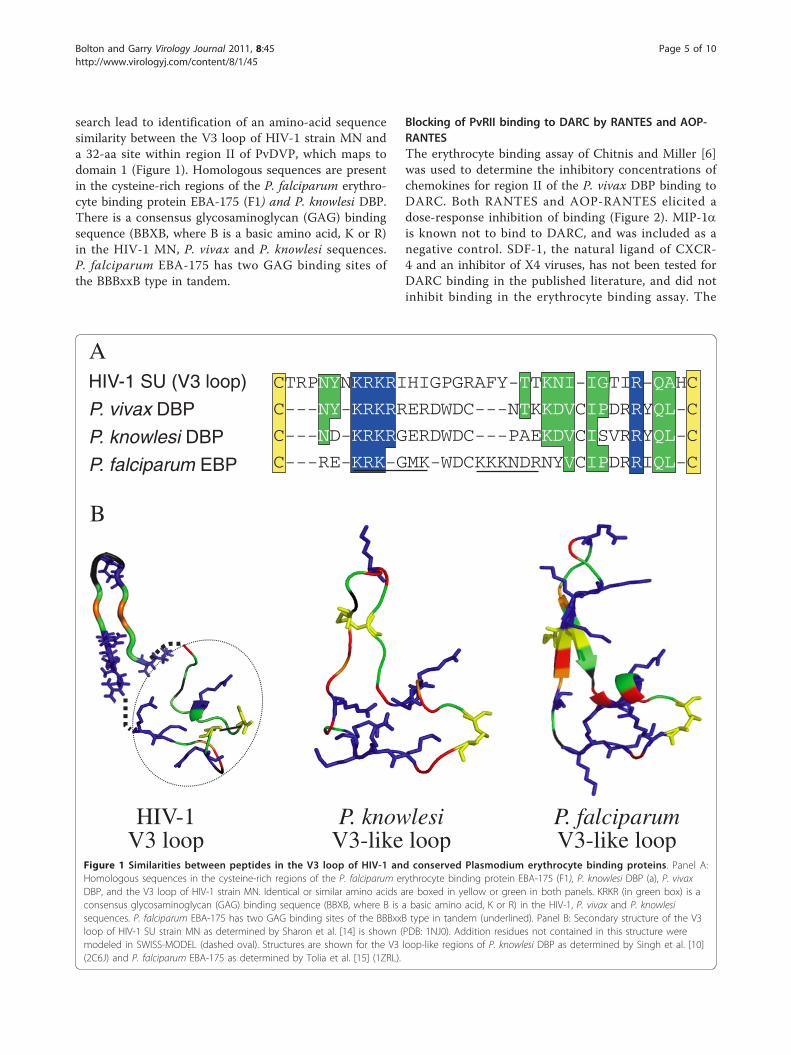
search lead to identification of an amino-acid sequencesimilarity between the V3 loop of HIV-1 strain MN anda 32-aa site within region II of PvDVP, which maps todomain 1 (Figure 1). Homologous sequences are presentin the cysteine-rich regions of the P. falciparum erythro-cyte binding protein EBA-175 (F1) and P. knowlesi DBP.There is a consensus glycosaminoglycan (GAG) bindingsequence (BBXB, where B is a basic amino acid, K or R)in the HIV-1 MN, P. vivax and P. knowlesi sequences.P. falciparum EBA-175 has two GAG binding sites ofthe BBBxxB type in tandem.
Blocking of PvRII binding to DARC by RANTES and AOP-RANTESThe erythrocyte binding assay of Chitnis and Miller [6]was used to determine the inhibitory concentrations ofchemokines for region II of the P. vivax DBP binding toDARC. Both RANTES and AOP-RANTES elicited adose-response inhibition of binding (Figure 2). MIP-1ais known not to bind to DARC, and was included as anegative control. SDF-1, the natural ligand of CXCR-4 and an inhibitor of X4 viruses, has not been tested forDARC binding in the published literature, and did notinhibit binding in the erythrocyte binding assay. The
A
B
HIV-1V3 loop
P. knowlesiV3-like loop
CTRPNYNKRKRIHIGPGRAFY-TTKNI-IGTIR-QAHC
C---ND-KRKRGERDWDC---PAEKDVCISVRRYQL-C
C---RE-KRK-GMK-WDCKKKNDRNYVCIPDRRIQL-C
HIV-1 SU (V3 loop)
P. vivax DBP
P. knowlesi DBP
P. falciparum EBP
C---NY-KRKRRERDWDC---NTKKDVCIPDRRYQL-C
P. falciparumV3-like loop
Figure 1 Similarities between peptides in the V3 loop of HIV-1 and conserved Plasmodium erythrocyte binding proteins. Panel A:Homologous sequences in the cysteine-rich regions of the P. falciparum erythrocyte binding protein EBA-175 (F1), P. knowlesi DBP (a), P. vivaxDBP, and the V3 loop of HIV-1 strain MN. Identical or similar amino acids are boxed in yellow or green in both panels. KRKR (in green box) is aconsensus glycosaminoglycan (GAG) binding sequence (BBXB, where B is a basic amino acid, K or R) in the HIV-1, P. vivax and P. knowlesisequences. P. falciparum EBA-175 has two GAG binding sites of the BBBxxB type in tandem (underlined). Panel B: Secondary structure of the V3loop of HIV-1 SU strain MN as determined by Sharon et al. [14] is shown (PDB: 1NJ0). Addition residues not contained in this structure weremodeled in SWISS-MODEL (dashed oval). Structures are shown for the V3 loop-like regions of P. knowlesi DBP as determined by Singh et al. [10](2C6J) and P. falciparum EBA-175 as determined by Tolia et al. [15] (1ZRL).
Bolton and Garry Virology Journal 2011, 8:45http://www.virologyj.com/content/8/1/45
Page 5 of 10

IC50 for RANTES and AOP-RANTES were 2.09 nM and1.51 nM, respectively. The difference in response asdetermined by a two-way ANOVA test was significantbetween RANTES and MIP-1a and between AOP-RANTES and MIP-1a, but not between RANTES andAOP-RANTES (p < 0.05).
Blocking of P. knowlesi invasion of DARC+ humanerythrocytes by RANTES and AOP-RANTESA standard erythrocyte invasion assay was used to deter-mine the chemokine inhibitory concentrations ofDARC-dependent invasion of human DARC+ erythro-cytes by P. knowlesi. Both RANTES and AOP-RANTESelicited a dose-response inhibition of invasion (Figure 3).MIP-1a was again used as a control. The IC50 ofRANTES and AOP-RANTES in the infection assay was0.053 nM and 0.062 nM, respectively. The IC50 for eachinhibitor in the invasion assay was more than a loglower than the IC50 in the erythrocyte binding assay.The difference in response as determined by a two-wayANOVA test was significant between RANTES andMIP-1a and between AOP-RANTES and MIP-1a, butnot between RANTES and AOP-RANTES (p < 0.05).
The V3-like peptide is necessary, but not sufficient forDARC bindingThe pHVDR22 plasmid expresses region II of the P. vivaxDBP and binds to DARC+ erythrocytes when expressedon the surface of COS-7 cells. Region II includes
12 conserved cysteine residues, C1-C12, and the 32 aminoacid V3-like peptide spans C1-C4. Deletion mutants lack-ing the V3-like peptide or adjacent sequences were madefrom pHVDR22 and tested for their ability to bind toDARC+ erythrocytes when expressed on COS-7 cells. Theexpression of each construct on the surface of COS-7 cellswas confirmed by immunofluorescence staining, and thenumber of COS-7 cells stained was 5-10% of the popula-tion. The same number of COS-7 cells transfected withthe parental pHVDR22 plasmid were stained and visua-lized by immunofluourescence.The pv22d32 construct that specifically deleted the
V3-like peptide completely failed to bind to DARC+erythrocytes in the erythrocyte binding assay (Figure 4A).This suggests that the DBP V3-like peptide is necessaryfor DARC binding. The deletion of all flankingsequences to the V3-like peptide, accomplished in thepv22suf32 mutant, also abrogated binding, showing thatthe DBP V3-like peptide is not sufficient for DARC bind-ing. This confirms that there are other areas of region IInecessary for binding. Truncation of the amino acidsflanking the DBP V3-like peptide toward the amino ter-minus, as accomplished in the pv22d5C construct, hadonly a small effect on binding. However, truncation ofthe region flanking the DBP V3-like peptide to the car-boxy terminus, in pv22d3C4, abrogated binding. Thissuggests that essential binding residues are located in theC-terminal, but not the N-terminal regions flanking theDBP V3-like peptide. To confirm the need for the DBPV3-like peptide in addition to the C-terminal flankingregion, truncation of the amino-terminal end of region IIup to and including the DBP V3-like peptide, in con-struct pv22d5C4, again abrogated binding.
RANTES
AOP-RANTES
MIP-1αSDF-1
120
100
80
60
40
20
0
-20
-40
Chemokine concentration (nM)
Bin
din
g in
hib
itio
n (
%)
0 0.1 1 10010 1000
Figure 2 Chemokine inhibition of PvRII binding to DARC+erythrocytes. The erythrocyte rosette assay of Chitnis and Miller [6]was used to quantify chemokine inhibition of PvRII Binding toDARC+ Erythrocytes. Binding was determined by subtracting thenumber of COS-7 cells expressing pvRII with rosettes of chemokine-treated DARC+ human erythrocytes (per 20 fields at 200Xmagnification) from the number with rosettes of untreatederythrocytes, and dividing by the number with rosettes of untreatederythrocytes. The data shown are the mean of three separateexperiments.
120
100
80
60
40
20
0
Chemokine concentration (nM)
Inva
sio
n in
hib
itio
n (
%)
RANTES
AOP-RANTES
MIP-1α
0 0.01 0.1 1 10010
Figure 3 Chemokine inhibition of P. knowlesi invasion of DARC+ erythrocytes. Inhibition of P. knowlesi invasion of DARC+erythrocytes was determined by subtracting the number ofchemokine-treated DARC+ human erythrocytes invaded by P.knowlesi merozoites (per 2000 erythrocytes) from the number ofuntreated DARC+ human erythrocytes invaded by P. knowlesimerozoites, and dividing by the number of untreated, invadederythrocytes.
Bolton and Garry Virology Journal 2011, 8:45http://www.virologyj.com/content/8/1/45
Page 6 of 10

A polycation sequence within the DBP V3-like Peptide isnecessary for DARC BindingTo determine if the polycation sequence in the DBP isnecessary for DARC binding, site directed mutagenesiswas used to introduce alanine substitutions for threepositively charged amino acids at K221, R224, and R227.The pv22KARA mutant contains these substitutions and
does not bind DARC+ erythrocytes when expressed onCOS-7 cells (Figure 4B). The six positively chargedamino acids at this site of PvRII create several possibleconsensus heparin binding sequences of the patternsBBXB or BBBXXB, where B is a basic amino acid and ×is any amino acid, including a basic one. To determinehow sensitive binding is to loss of charge at this the site,a single alanine substitution was made at K223 in thepv22KA construct. This mutant was capable of bindingto DARC+ erythrocytes as well as the wild typepHVDR22 protein.One other site in PvRII, at amino acids 364-373,
between C5 and C6, contains a polycationic site whichconforms to a consensus heparin binding sequence. Thepv22KAKA construct introduces two alanine substitu-tions for lysine residues in this second consensusheparin binding site at K367 and K370. The DBP regionII expressed from this construct is able to bind toDARC+ erythrocytes on the surface of COS-7 cells aswell as wild type pHVDR22.
The polycationic site has a conserved Role in the DBPprotein family for binding to Diverse ReceptorsStudies by Ranjan and Chitnis have identified a site inPvRII in the C-terminal flanking region to the DBP V3-like peptide, between C4-C7, that contain residuesnecessary for DARC binding [8]. This study also showedthat the C1-C4 region of the P. knowlesi b protein, amember of the DBP family that does not bind to DARC,was capable of substituting for the P. vivax C1-C4.Upon closer inspection, the polycationic site is well con-served in the DBP family, with great similarity betweenproteins that bind different receptors (Figure 4C). TheP. knowlesi a and g proteins have an identical consensusheparin binding site, but only a binds to DARC. To seeif the polycationic site may play a similar role inthe binding proteins of other members of the DBPfamily, the same three alanine substitutions found inpv22KARA were introduced by site directed mutagen-esis into the plasmids pHKADR22, pHKBDR22, andpHKGDR22. This yielded the constructs pkalphaKARA,pkbetaKARA, and pkgammaKARA, which contain theK221, R224, and R227 alanine substitution in the P.knowlesi a, b, and g genes, respectively. All three ofthese mutants failed to bind rhesus erythrocytes whenexpressed in COS-7 cells.
DiscussionHIV-1 binds to chemokine receptors such as CCR5 andCXCR4 using SU, and can be inhibited from in vivoinfection by mutation of the chemokine receptors or byincubation with chemokines, such as RANTES. Likewise,P. vivax uses its DBP to bind to DARC and can beinhibited by null mutations in the receptor or in vitro
pHVDR22
pv22d32
pv22suf32
pv22d3C4
pv22d5C1
pv22d5C5
pv22KARA
pv22KA
pv22KAKA
pkalphaKARA
pkbetaKARA
pkgammaKARA
BINDING
100%
0%
0%
0%
80%
0%
0%
100%
100%
0%
0%
0%
C1 C4 C5 C6 C12
YKRKRRERDW
YARKAREADW
YKRKRRERDW
YKRARRERDW
SVKKRLKGNF
SVKARLAGNF
A
C
B
DKRKRGERD
DARKAGEAD
NKRKRGTRD
NARARGTAD
DARKAGEAD
DKRKRGERD
Figure 4 Mutants of the region II of Erythrocyte BindingProteins. Panel A. P. vivax DBP region II is shown in blue withconserved cysteines C1, C4, C5, C6 and C12 shown. Deletionmutants in the are shown with the V3-like peptide (amino acids216-247, between C1-C4) highlighted in red. Primers flanking thissite, facing outward, were used to create pv22d32 (delete 32 aminoacids) by inverse PCR. The other mutants were created with primersfacing inward and containing restriction enzyme sites. Percentbinding is expressed as number of rosettes compared to pHVDR22.Panel B. Site directed mutagenesis using the StratageneQuickChange kit was used to make alanine substitutions within theconsensus heparin binding site of the V3-like peptide (R22KARA,R22KA), or another consensus site (R22KAKA) at amino acids 364-373, between conserved cysteines C5-C6. Panel C. Site directedmutagenesis was used to created the same KARA mutation in theconserved heparin binding site between C1-C4 of the P. knowlesi a,b and g proteins.
Bolton and Garry Virology Journal 2011, 8:45http://www.virologyj.com/content/8/1/45
Page 7 of 10

by MGSA or IL-8. Here, we show that the chemokine,RANTES, and its analog AOP-RANTES, known toblock HIV-1 SU binding to CCR5, also blocks P. vivaxDBP binding to DARC. This demonstrates that naturaland designed chemokine inhibitors can be cross-protec-tive to both pathogens, and may have importantimplications for drug and vaccine development inco-endemic areas.The overlap in chemokine inhibition of both HIV-
1 and Plasmodium infection supports a hypothesis thatSU and PvDBP have convergently evolved to mimicchemokines in such a way that the two proteins havestructural similarities. The N-terminus extracellulardomain of DARC is involved in binding to bothregion II of the PvDBP and to chemokines, just as theN-terminus of CCR5 is critical for SU and chemo-kine binding. In particular, negatively charged andsulfotyrosine residues in the CCR5 N-terminus andCXCR4 extracellular domain have important interac-tions with the C4/V3 stem of SU, and positively chargedresidues are implied to be important components of theSU chemokine receptor binding surface. Similarly, Pv-DBL or Pka-DBL have important interactions withsulphated tyrosine (Tyr 41) residue on DARC [9] Theresults of a homology search identifying an amino-acidsequence similarity between the V3 loop of HIV-1 strainMN and a 32-aa site within region II of PvDBP contain-ing a polycationic site (Figure 1). Other members of theEBP family share this homology or “V3-like peptide”.The crystal structure of the P. knowlesi DBL domain(Pka-DBL), which binds to DARC during infection ofhuman erythrocytes, shows that this structure is indeedsimilar, with disulfide bridges between C1 and C4 andbetween C2 and C3 forming a random coil structuredesignated domain 1 [10].To investigate the role of the V3-like peptide in DARC
binding we used an established erythrocyte binding assayand made mutants of the region II PvDBP expressionvector. Deletion of the 32-aa V3-like peptide in constructpv22d32, or deletion of the flanking regions in constructpv22suf32, abrogated binding to DARC, suggesting thatthe V3-like peptide was necessary but not sufficient forbinding (Figure 4A). In particular, the region between theconserved cysteines C4-C12 was necessary for binding asdemonstrated by binding of pv22d5C1 and nonbinding ofpv22d3C4, but the C4-C12 region was also not sufficientas shown by nonbinding of pv22d5C4. It is possible thatthe deletions we have made change the folding of thereceptor binding site, with the exception of the deletionof amino acids 198-216. Previous work by Ranjan andChitnis [8] using chimeras of region II between the P.vivax DBP and P. knowlesi b protein, which does notbind DARC but sialic acid, revealed the entire C4-C7 region of PvDBP region II is necessary for DARC
binding, which our data corroborate. Of note, their datashow that region C4-C7 is sufficient for binding, but thisdoes not mean that other regions are not involved bind-ing of the full-length protein. The authors of the chimericdata suggest that residues outside of C4-C7 influence thefine specificity of the DBL binding domain [8]. Thismight be comparable to the specificity of chemokinereceptor binding attributed to small changes in theV3 loop, which mimics the b hairpin structure in chemo-kines, while conserved portions of the SU molecule cre-ate the structural backbone of the chemokine receptorbinding surface [11].The polycationic site within the V3-like peptide is con-
served in the EBP family and contains consensus heparinbinding sequences of the patterns BBXB or BBBXXB,where B is a basic amino acid and × is any amino acid,including a basic one. It was previously shown that poly-anions inhibit DARC-binding by the P. knowlesi a pro-tein, and we have determined that this also to be true ofthe PvDBP region II (Bolton et al., in preparation). Wemade site-directed mutations to substitute alanines forpositively charged amino acids at K221, R224, and R227.This mutant, designated pv22KARA, did not bind. Suchminor changes make it less likely that this mutant doesnot bind due to folding error than to contributions theseresidues make directly to receptor binding. To deter-mimne if the polycationic site was sensitive to asingle alanine substitution, we created a mutation atK223 which did not change binding in pv22KA. Thismutant protein still contained consensus heparin bindingsequences and five positively charged residues at the site.We also mutated the only other polycationic site in thePvDBP region II that conforms to a consensus heparinbinding sequence at amino acids 364-373 by substitutingalanines at K367 and K370. pv22KAKA was still able tobind. These data show that the polycation site in the V3-like peptide is discretely involved in DARC binding andsuggest the multiple positive charges play a redundantrole at the site.Previous site-directed mutagenesis experiments have
identified residues Tyr 94, Asn 95, Lys 96, Arg 103, Leu168 and Ile 175 on domain 2 as required for recognitionof DARC on human erythrocytes [12-14]. Based on thecrystal structure of the Pka-DBL these residues lie close toa set of positively charged residues Lys 96, Lys 100, Arg103 and Lys 177 that have been suggested to interact withthe sulphate group on DARC Tyr 41 [10]. Mapping thepolycationic site we found to be sensitive to alanine substi-tuions onto the crystal structure shows that it is adjacentto the putative binding site residues and may provide suchan interaction with the sulphated Tyr 41 (Figure 5).The P. knowlesi a, b, and g proteins share the V3-like
loop and polycation site homology in region II withPvDBP, though only the a protein binds DARC. We
Bolton and Garry Virology Journal 2011, 8:45http://www.virologyj.com/content/8/1/45
Page 8 of 10

introduced similar alanine mutations into three posi-tively-charged amino acids of each of the three P. know-lesi EBPs at the polycationic site. In all 3 cases thiseliminated normal binding to rhesus erythrocytes. In thecase of the P. knowlesi a protein this reinforces the con-clusion that the site can contribute to DARC binding.The P. knowlesi b, and g proteins, however, don’t bindto DARC. The receptor for the P. knowlesi b protein issialic acid, which is negatively charged for which thepolycation site might contribute to a positively chargedbinding pocket. A chimera produced by Ranjan andChitnis [8] with the C1-C4 region exchanged betweenthe P. knowlesi b protein and P. vivax DBP bound toDARC on rhesus erythrocytes, without the removal ofsialic acid residues required for native P. vivax DBP tobind to rhesus DARC. It is possible that a closer homol-ogy between the polycationic site of P. knowlesi a and bproteins, each of which contain 5 cationic residues ver-sus the 6 cationic residues of the P. vivax DBP polyca-tionic site, allows for this change in specificity. Anotherchimera in the same study with the P. vivax polycationicsite is able to bind to rhesus erythrocytes in the samemanner as the P. knowlesi b protein, but only in the
presence of C4-C5 of the P. knowlesi b protein. Thisagain suggests that the homology of the polycationic sitewithin the EBP family may allow for a redundant func-tion in receptor binding, but the role of the polycationicsite is in conjunction with other residues in region IIwhich together allow for efficient receptor binding. Theresults presented here, in conjunction with previous stu-dies, indicate that the heparin binding site motif inmembers of the DBP family may form part of a con-served erythrocyte receptor binding pocket.
AcknowledgementsThis work was supported by National Institutes of Health grant RR018229.
Author details1Vaccine and Infectious Disease Institute, Fred Hutchinson Cancer ResearchCenter Division of Allergy and Infectious Diseases University of Washington1100 Fairview Avenue Seattle, Washington 98109 USA. 2Department ofMicrobiology and Immunology Tulane University 1430 Tulane Avenue NewOrleans, Louisiana 70112 USA.
Authors’ contributionsMJB performed the investigations described in this study. MJB and RFGconceived of the study, and RFG participated in its design and coordinationand helped to draft the manuscript. Both authors read and approved thefinal manuscript.
C16
K19R20
K21
K22
R40
R41
C29
C36
C45
Y94N95
K96R103
L168
I175
V3-like loopDomain 1
Domain 2
Domain 3
Figure 5 Three dimensional location of heparin binding motif in relation to known binding residues on the crystal structure ofrecombinant Pka-DBL. The crystal structure of the recombinant Pka-DBL that binds to human DARC is shown with previously describedbinding residues Tyr 94, Asn 95, Lys 96, Arg 103, Leu 168 and Ile 175 [10,12-14] highlighted in magenta in domain 2 of the molecule. Theheparin binding motif on the V3-like peptide is highlighted in yellow cysteines C1-C4 and blue for the basic residues (lysine and arginine) indomain 1 of the molecule.
Bolton and Garry Virology Journal 2011, 8:45http://www.virologyj.com/content/8/1/45
Page 9 of 10

Competing interestsThe authors declare that they have no competing interests.
Received: 29 November 2010 Accepted: 31 January 2011Published: 31 January 2011
References1. Horuk R, Chitnis CE, Darbonne WC, Colby TJ, Rybicki A, Hadley TJ, Miller LH:
A receptor for the malarial parasite Plasmodium vivax: the erythrocytechemokine receptor. Science 1993, 261(5125):1182-1184.
2. Simmons G, Wilkinson D, Reeves JD, Dittmar MT, Beddows S, Weber J,Carnegie G, Desselberger U, Gray PW, Weiss RA, Clapham PR: Primary,syncytium-inducing human immunodeficiency virus type 1 isolates aredual-tropic and most can use either Lestr or CCR5 as coreceptors forvirus entry. J Virol 1996, 70(12):8355-8360.
3. Liu R, Paxton WA, Choe S, Ceradini D, Martin SR, Horuk R, MacDonald ME,Stuhlmann H, Koup RA, Landau NR: Homozygous defect in HIV-1 coreceptor accounts for resistance of some multiply-exposedindividuals to HIV-1 infection. Cell 1996, 86(3):367-377.
4. Miller LH, Mason SJ, Clyde DF, McGinniss MH: The resistance factor toPlasmodium vivax in blacks. The Duffy-blood-group genotype, FyFy. NEngl J Med 1976, 295(6):302-304.
5. Cocchi F, DeVico AL, Garzino-Demo A, Arya SK, Gallo RC, Lusso P:Identification of RANTES, MIP-1 alpha, and MIP-1 beta as the major HIV-suppressive factors produced by CD8+ T cells. Science 1995,270(5243):1811-1815.
6. Chitnis CE, Miller LH: Identification of the erythrocyte binding domains ofPlasmodium vivax and Plasmodium knowlesi proteins involved inerythrocyte invasion. J Exp Med 1994, 180(2):497-506.
7. Chitnis CE, Chaudhuri A, Horuk R, Pogo AO, Miller LH: The domain on theDuffy blood group antigen for binding Plasmodium vivax and P.knowlesi malarial parasites to erythrocytes. J Exp Med 1996,184(4):1531-1536.
8. Ranjan A, Chitnis CE: Mapping regions containing binding residues withinfunctional domains of Plasmodium vivax and Plasmodium knowlesierythrocyte-binding proteins. PNAS 1999, 96(24):14067-14072.
9. Choe H, Moore MJ, Owens CM, Wright PL, Vasilieva N, Li W, Singh AP,Shakri R, Chitnis CE, Farzan M: Sulphated tyrosines mediate association ofchemokines and Plasmodium vivax Duffy binding protein with the Duffyantigen/receptor for chemokines (DARC). Molecular Microbiology 2005,55(5):1413-1422.
10. Singh SK, Hora R, Belrhali H, Chitnis CE, Sharma A: Structural basis forDuffy recognition by the malaria parasite Duffy-binding-like domain.Nature 2006, 439(7077):741-744.
11. Sharon M, Kessler N, Levy R, Zolla-Pasner S, Görlach M, Anglister J:Alternative comformations of HIV-1 V3 loops mimic β hairpins inchemokines, suggesting a mechanism for co-receptor specificity.Structure 2003, 11(2):225-236.
12. Singh SK, Singh AP, Pandey S, Yazdani SS, Chitnis CE, Sharma A: Definitionof structural elements in Plasmodium vivax and P. knowlesi Duffy-binding domains necessary for erythrocyte invasion. Biochemical Journal2003, 374(1):193-198.
13. VanBuskirk KM, Sevova E, Adams JH: Conserved residues in thePlasmodium vivax Duffy-binding protein ligand domain are critical forerythrocyte receptor recognition. PNAS 2004, 101(44):15754-15759.
14. Hans D, Pattnaik P, Bhattacharyya A, Shakri AR, Yazdani SS, Sharma M,Choe H, Farzan M, Chitnis CE: Mapping binding residues in thePlasmodium vivax domain that binds Duffy antigen during red cellinvasion. Molecular Microbiology 2005, 55(5):1423-1434.
15. Tolia NH, Enemark EJ, Sim BK, Joshua-Tor L: Structural basis for the EBA-175 erythrocyte invasion pathway of the malaria parasite Plasmodiumfalciparum. Cell 2005, 122(2):183-93.
doi:10.1186/1743-422X-8-45Cite this article as: Bolton and Garry: Sequence similarity between theerythrocyte binding domain 1 of the Plasmodium vivax Duffy bindingprotein and the V3 loop of HIV-1 strain MN reveals binding residues forthe Duffy Antigen Receptor for Chemokines. Virology Journal 2011 8:45.
Submit your next manuscript to BioMed Centraland take full advantage of:
• Convenient online submission
• Thorough peer review
• No space constraints or color figure charges
• Immediate publication on acceptance
• Inclusion in PubMed, CAS, Scopus and Google Scholar
• Research which is freely available for redistribution
Submit your manuscript at www.biomedcentral.com/submit
Bolton and Garry Virology Journal 2011, 8:45http://www.virologyj.com/content/8/1/45
Page 10 of 10