serbian journal of experimental and clinical research
TRANSCRIPT


1
Editor-in-Chief
Slobodan Janković
Co-Editors
Nebojša Arsenijević, Miodrag Lukić, Miodrag Stojković, Milovan Matović, Slobodan Arsenijević,
Nedeljko Manojlović, Vladimir Jakovljević, Mirjana Vukićević
Board of Editors
Ljiljana Vučković-Dekić, Institute for Oncology and Radiology of Serbia, Belgrade, Serbia
Dragić Banković, Faculty for Natural Sciences and Mathematics, University of Kragujevac, Kragujevac, Serbia
Zoran Stošić, Medical Faculty, University of Novi Sad, Novi Sad, Serbia
Petar Vuleković, Medical Faculty, University of Novi Sad, Novi Sad, Serbia
Philip Grammaticos, Professor Emeritus of Nuclear Medicine, Ermou 51, 546 23,
Th essaloniki, Macedonia, Greece
Stanislav Dubnička, Inst. of Physics Slovak Acad. Of Sci., Dubravska cesta 9, SK-84511
Bratislava, Slovak Republic
Luca Rosi, SAC Istituto Superiore di Sanita, Vaile Regina Elena 299-00161 Roma, Italy
Richard Gryglewski, Jagiellonian University, Department of Pharmacology, Krakow, Poland
Lawrence Tierney, Jr, MD, VA Medical Center San Francisco, CA, USA
Pravin J. Gupta, MD, D/9, Laxminagar, Nagpur – 440022 India
Winfried Neuhuber, Medical Faculty, University of Erlangen, Nuremberg, Germany
Editorial Staff
Ivan Jovanović, Gordana Radosavljević, Nemanja Zdravković
Vladislav Volarević
Management Team
Snezana Ivezic, Zoran Djokic, Milan Milojevic, Bojana Radojevic, Ana Miloradovic
Corrected by
Scientifi c Editing Service “American Journal Experts”
Design
PrstJezikIostaliPsi - Miljan Nedeljković
Medical Faculty, Kragujevac
Indexed in
EMBASE/Excerpta Medica, Index Copernicus, BioMedWorld, KoBSON, SCIndeks
Address:
Serbian Journal of Experimental and Clinical Research, Medical Faculty, University of Kragujevac
Svetozara Markovića 69, 34000 Kragujevac, PO Box 124
Serbia
e-mail: [email protected]
www.medf.kg.ac.rs/sjecr
SJECR is a member of WAME and COPE. SJECR is published at least twice yearly, circulation 300 issues Th e Journal is fi nan-
cially supported by Ministry of Science and Technological Development, Republic of Serbia
ISSN 1820 – 8665

2
Editorial / Editorijal
HOMOCYSTEINE: NEUROTOXICITY AND
MECHANISMS OF INDUCED HYPEREXCITABILITY ....................................................................................................................................3
Original Article / Orginalni naučni rad
THE EFFECT OF SEROTONERGIC LESIONS IN THE MEDIAL
PREFRONTAL CORTEX ON PSYCHOTOMIMETIC DRUGINDUCED
LOCOMOTOR HYPERACTIVITY AND PREPULSE INHIBITION IN RATS .........................................................................................11
Original Article / Orginalni naučni rad
STRUCTURAL INVESTIGATION OF DEXTROMETHORPHAN
USING MASS SPECTROMETRY AND
THERMAL ANALYSES COMBINED WITH MO CALCULATIONS ............................................................................................................21
Original Article / Orginalni naučni rad
EXPERIMENTAL MODELS OF DIABETES MELLITUS
EKSPERIMENTALNI MODELI DIJABETES MELITUSA ................................................................................................................................ 29
Review Article / Pregledni članak
DIAGNOSTICS AND THERAPY OF LEFT VENTRICULAR
HYPERTROPHY IN HAEMODIALYSIS PATIENTS ...........................................................................................................................................37
Letter To The Editor / Pismo uredniku
THE RELATIONSHIP BETWEEN SPORTS ENGAGEMENT,
BODY MASS INDEX AND PHYSICAL ABILITIES IN CHILDREN .............................................................................................................41
INSTRUCTION TO AUTHORS FOR MANUSCRIPT PREPARATION ..................................................................................................... 43
Table Of Contents
3
UDK 612.8:577.112.34 ; 616.853 / Ser J Exp Clin Res 2011; 12 (1): 3-9
HOMOCYSTEINE: NEUROTOXICITY AND MECHANISMSOF INDUCED HYPEREXCITABILITY
Stanojlovic Olivera, Hrncic Dragan, Rasic-Markovic Aleksandra and Djuric DraganInstitute of Medical Physiology “Richard Burian”, School of Medicine, University of Belgrade, Belgrade, Serbia
HOMOCISTEIN: NEUROTOKSIČNOST I MEHANIZMIINDUKOVANE HIPEREKSCITABILNOSTI
Stanojlović Olivera, Hrnčić Dragan, Rašić-Marković Aleksandra i Đurić DraganInstitut za Medicinsku fi ziologiju ”Rihard Burijan”, Medicinski fakultet, Univerzitet u Beogradu, Beograd, Srbija
Received / Primljen: 09. 02. 2011.
Correspondence to: Dragan M. Djurić, M.D. Ph.D. Institute of Medical Physiology “Richard Burian”, School of Medicine, University of Belgrade,
Višegradska 26/II, 11000 Belgrade, Serbia, Tel: +381-11-3607-112, Fax: +381-11-36 11 261, e-mail: [email protected]
ABSTRACT
Within the past four decades, investigators worldwide
have established that the amino acid homocysteine (Hcy) is
a potent, independent, novel and emerging risk factor for ar-
teriosclerosis. In addition, Hcy is considered a vasotoxic and
neurotoxic agent that interferes with fundamental biological
processes common to all living cells. Th e aim of this article is
to present data addressing central nervous system hyperex-
citability in experimental models of homocysteine-induced
seizures. Th ese results demonstrate that acute administra-
tion of homocysteine and homocysteine-related compounds
signifi cantly aff ects neuronal activity, electroencephalo-
graphic (EEG) recordings and behavioural responses in adult
Wistar male rats. It is suggested that hyperhomocysteinemia
may generate similar eff ects on human brain activity. Homo-
cysteine and D,L-homocysteine thiolactone are excitotoxic
and elicit seizures through stimulation of NMDA receptors.
In addition, they inhibit Na+/K+-ATPase activity in the cor-
tex, hippocampus and brain stem. Nitric oxide (NO) acts as
an anticonvulsant in D,L-homocysteine thiolactone-induced
seizures and prevents D,L-homocysteine thiolactone-induced
inhibition of Na+/K+-ATPase activity. EEG monitoring for the
eff ects of ethanol on homocysteine-induced epilepsy support
the idea that ethanol intake could represent one of the ex-
ogenous factors infl uencing brain excitability. Finally, it was
found that homocysteine thiolactone signifi cantly inhibits
acetylcholinesterase (AChE) activity in rat brain tissue.
Keywords: homocysteine, CNS, hyperexcitability, rat
SAŽETAK
Četiri decenije istraživačkog rada širom sveta dovele su do
ustanovljavanja semi-esencijalne sulfi hidrilne aminokiseline
homocisteina (Hcy) kao novog potentnog i nezavisnog faktora
rizika za arteriosklerozu, kao i vazotoksične i neurotoksične
supstance koja učestvuje u fundamentalnim biološkim pro-
cesima svojstvenim svim živim ćelijama. Cilj ovog rada je da
prikaže dostupna saznanja na temu hiperekscitabilnosti cen-
tralnog nervnog sistema u eksperimentalnom modelu homo-
cisteinom indukovene epilepsije. Rezultati pokazuju da akutna
administracija homocisteina i homocisteinu-srodnih supstanci
značajno utiče na nervnu aktivnost, elektroencefalografske
(ЕЕG) zapise i ponašanje odraslih mužjaka soja Wistar pacova,
a smatra se da hiperhomocisteinemija može imati slične efekte
i na aktivnost ljudskog mozga. Homocistein i D,L-homocistein
tiolakton su ekscitotoksični i izazivaju epileptične napade stimu-
lacijom NMDA receptora; oni indukuju značajnu inhibiciju ak-
tivnosti Na+/K+-ATPaze u korteksu, hipokampusu i moždanоm
stablu. Azot monoksid (NO) deluje kao antikonvulzant u D,L-
homocistein tiolakton indukovanim epileptičnim napadima i
vrši prevenciju D,L-homocistein tiolakton indukovane inhibicije
aktivnosti Na+/K+-ATPaze. EEG monitoring efekata etanola na
homocisteinom indukovanu epilepsiju podržava ideju da akut-
ni unos etanola može predstavljati jedan od faktora egzogenog
uticaja na moždanu ekscitabilnost. Takođe je utvrđeno da D, L-
homocistein tiolakton u značajnom procentu inhibira aktivnost
acetilholinesteraze u mozgu pacova.
Ključne reči: homocistein, CNS, hiperekscitabilnost, pacov
EDITORIAL EDITORIJAL EDITORIAL EDITORIJAL EDITORIAL EDITORIJAL
Abbreviations used:
AchE - acetylcholinesterase
BHMT - betaine-Hcy S-methyltransferase
CNS - central nervous system
EEG - electroencephalography
Hcy - homocysteine
NO - nitric oxide
SWDs - spike-wave discharges
tHcy - total plasma homocysteine
Korišćene skraćenice:
AchE – acetilholin esteraza
BHMT - betain-homocistein S-metiltransferaza
CNS – centralni nervni sistem
EEG - elektroencefalografija
Hcy - homocistein
NO – azot monoksid
SWDs – “šiljak pražnjenja”
tHcy – ukupni plazma homocistein

4
BACKGROUND
Within the past four decades, investigators worldwide
have established that the amino acid homocysteine (Hcy)
is a novel and independent risk factor for arteriosclerosis.
In addition, Hcy is a vasotoxic and neurotoxic agent that
interferes with the fundamental biological processes com-
mon to all living cells. Therefore, it has also been termed
the “cholesterol of the 21st century.”
Metabolism of homocysteine is coordinately regulated
to maintain a balance between remethylation and trans-
sulfuration pathways, which are critical to maintaining
low levels of this potentially cytotoxic sulphur containing
amino acid (1). Homocysteine belongs to a group of mol-
ecules known as cellular thiols and is considered a “bad”
thiol. Glutathione and cysteine, the most abundant cellular
thiols, are considered to be “good” thiols (2). In the methy-
lation pathway that occurs in all tissues, homocysteine
acquires a methyl group to form methionine in a vitamin
B12 dependent reaction, which is catalysed by methionine
synthase. The second substrate required by methionine
synthase is 5-methyltetrahydrofolate, which is formed by
the reduction of 5,10-methylenetetrahydrofolate. This re-
action is catalysed by the enzyme methylenetetrahydrofo-
late reductase (MTHFR). The TT genotype of this enzyme
causes thermolability and reduces enzyme activity, which
impairs the formation of 5-methyltetrahydrofolate. This
reduced enzyme activity explains why this genotype is as-
sociated with increased Hcy levels when the folate status is
relatively low. However, liver, kidney and the lens of the eye
have the ability to convert Hcy to L-methionine through
a vitamin B12-independent reaction catalysed by betaine-
Hcy S-methyltransferase (BHMT). The CNS lacks BHMT
and it is completely dependent on the folate and vitamin
B12 pathway for the conversion of Hcy to L-methionine.
Homocysteine condenses with serine to form cysta-
thionine in an irreversible reaction catalysed by the B6
containing enzyme cystathionine beta-synthase, which is
known as the transsulfuration pathway. Hcy catabolism re-
quires vitamin B6 and as a consequence, alterations in folic
acid and B vitamin status impairs Hcy biotransformation.
These alterations result in the synthesis of cysteine, taurine
and inorganic sulphates that are excreted in urine.
Elevation of homocysteine levels is known to lead to the
metabolic conversion and inadvertent elevation of homo-
cysteine thiolactone, which is a reactive thioester represent-
ing less than 1% of total plasma homocysteine. In all cell types,
from bacteria to human, homocysteine is metabolised to ho-
mocysteine thiolactone by methionyl-tRNA synthetase (3).
Homocysteine thiolactone causes lethality, growth retardation,
blisters and abnormalities in somite development by oxidative
stress, which is one of important mechanisms for its toxicity to
neuronal cells (4). The highly reactive and toxic homocysteine
metabolite, homocysteine thiolactone, can be produced in two
steps by enzymatic and/or non-enzymatic reactions in blood
serum. The ability to detoxify or eliminate homocysteine thi-
olactone is essential for biological integrity (3, 4).
Total plasma Hcy (tHcy) consists of a pool of free ho-
mocysteine, homocystine, Hcy-S-S-Cys disulphide, pro-
tein bound N- and S-linked Hcy as well as their oxidised
forms and Hcy-thiolactone (5-7). Under physiological
conditions, less than 1% of the total Hcy is present in a
free reduced form. Approximately 10–20% of total Hcy is
present in different oxidised forms, such as Hcy-Cys and
homocystine, which is a Hcy dimer. Plasma tHcy levels are
affected by genetic, physiologic (age and sex) and lifestyle
factors as well as various pathologic conditions (5, 6, 8).
Hyperhomocysteinemia is defined as an elevated plasma
total homocysteine (tHcy) concentration (>15 μM).
Elevated tHcy is a recognised risk factor for cardiovas-
cular disease (9) and has been linked to neurological diseas-
es during aging, such as cognitive declines, cerebrovascu-
lar disease and stroke, vascular dementia and Alzheimer’s
disease. In addition, elevated tHcy levels are linked with
pathological brain functioning, such as mental retarda-
tion, depression, schizophrenia and memory impairment.
Hcy is also a pro-thrombotic and pro-inflammatory factor,
vasodilatation impairing substance and an endoplasmatic
reticulum stress inducer (10, 11).
Mitrovic et al. (12) evaluated total plasma homocysteine
in patients with angiographically confirmed coronary ar-
tery disease. In addition, they investigated the effects of
homocysteine lowering therapy on endothelial function,
carotid wall thickness and myocardial perfusion. Their
study found that homocysteine levels decreased signifi-
cantly (34%) with folic acid therapy and that endothelium
function improved by 27% with this treatment. However,
carotid structure and myocardial perfusion did not show
any significant improvement in patients with confirmed
coronary artery disease (13). Djuric et al. (14, 15) also in-
vestigated the link between homocysteine and folic acid,
demonstrated folic acid induced coronary vasodilation
and decreased oxidative stress in the isolated rat heart.
HOMOCYSTEINE AND NEUROTOXICITY
Despite several theories, a complete understanding of
Hcy toxicity remains unclear. In particular, the hypothesis
that relates homocysteine to CNS dysfunction by way of
overt neurotoxicity is based on the neuroactive properties of
homocysteine. Elucidating the link between homocysteine
and CNS dysfunction is vital for improving the treatment
of homocysteine related CNS disorders. Hcy adversely af-
fects brain functioning (i.e., mental retardation, dementia
and memory impairment) and high tissue concentrations
cause oxidative stress and excitotoxicity in neurons (16).
When these symptoms are combined with homocystinuria,
patients often present with convulsions (17). These studies
provide evidence for a complex and multifaced relation-
ship between homocysteinemia and CNS disorders. Hcy is
an endogenous compound that is neurotoxic at supraphys-
iological concentrations and induces neuronal damage and
cell loss through excitotoxicity and apoptosis. These CNS

5
pathologies could be a direct consequence of the inability
of cerebral tissue to metabolise Hcy through the betaine
and transsulfuration pathways, thereby favouring Hcy ac-
cumulation in the nervous system (18). High brain concen-
trations of either Hcy or its oxidised derivatives have been
shown to alter neurotransmission (19). The accumulation
of homocysteine (at synapses or in the extracellular space)
increases intracellular S-adenosylhomocysteine (SAH),
which is a potent inhibitor of many methylation reactions
that are vital for neurological function including the O-
methylation of biogenic amines. Methylation of myelin
basic protein and reducing the synthesis of phosphatidyl
choline, which can lead to blood-brain barrier (BBB) dis-
ruption, are a few pathological processes that are possible
in the absence of normal methylation patterns (20).
It has been recently suggested that Hcy toxicity is a
consequence of its covalent binding to proteins, which in-
terferes with protein biosynthesis and decreases the nor-
mal physiological activity of proteins. This modification
of protein function is a process called homocysteinylation.
Transthyretin is a plasma protein that is modified through
homocysteinylation (3, 21). Other reports suggest that Hcy
exerts its toxicity through the induction of endoplasmic re-
ticulum (ER) stress. Increased intracellular Hcy concentra-
tions are associated with both the alteration of redox bal-
ance and post-translational protein modifications through
N- and S-homocysteinylation (22). Other studies suggest
that Hcy induces the expression of superoxide dismutase
in endothelial cells, which leads to the consumption of NO
and impairs endothelial vasorelaxation (23).
HOMOCYSTEINE AND SEIZURES
Experimental models of generalised clonic-tonic sei-
zures induced by metaphit (1-[1(3-isothiocyanatophenyl)-
cyclohexyl] piperidine) (24, 25) and lindane (γ-1,2,3,4,5,6-
hexachlorocyclohexane) (26, 27) are widely used for the
study of epilepsy and the preclinical evaluation of potential
antiepileptic treatments. In contrast to audiogenic epilep-
sy induced by metaphit with its typical and characteristic
three degrees of intensity, the lindane model is similar to
Hcy-induced epilepsy with a wide spectra of behavioural
manifestations (e.g., hot plate reaction and Kangaroo posi-
tion) incorporated into the rating scale from grades 0 - 4.
Almost four decades ago, Sprince et al. (28) described that
elevated levels of homocysteine arising from excess dietary
methionine may induce epilepsy and lethality. The fact that
increased homocysteine concentrations damage endothe-
lial structures and the blood coagulation system during
aging and in patients on antiepileptic drugs (29) justifies
the attention directed toward the examination of homo-
cysteine’s neural effects.
Furthermore, experimental models of epilepsy may be
induced by manipulations of γ-aminobuturic acid (e.g., bi-
cuculline, corasol, picrotoxin and benzylpenicillin sodium)
(30). In contrast, epilepsy can be induced by an opposite
mechanism such as by increasing cerebral excitatory neu-
rotransmission. There are several proposed mechanisms
by which exposure to excess D,L-homocysteine or D,L-ho-
mocysteine thiolactone induce seizures. The most reactive
natural amino acid is homocysteic monocarboxylic, which
is sulphur-containing and its homocysteic sulphinic acid
derivatives are excitatory amino acids that have been pro-
posed as candidates for brain excitatory neurotransmitters
(31). However, answering the question as to whether D,L-
homocysteine thiolactone should be included in the list of
epileptogenic factors according to behavioural and elec-
trographic responses will require additional studies and
future human therapeutic trials. Increased levels of Hcy
and its oxidised forms can provoke seizures by increasing
activation of certain neuronal receptors such as N-methyl
d-aspartate (NMDA), α-amino-3-hydroxy-5-methyl-4-
isoxazolepropionate (AMPA)/kainate ionotropic gluta-
mate receptors or group I and group III metabotropic glu-
tamate receptors (5). All of these glutamate receptors are
expressed in hippocampal pyramidal cells and may directly
induce or drive these cells over the threshold for excito-
toxic cell death. Overstimulation of these receptors trig-
gers cytoplasmic calcium pulses, Ca2+ influx and intraneu-
ronal calcium mobilisation in the presence of glycine (32).
Increased cytosolic Ca2+ concentrations affect enzyme
activities and the synthesis of nitric oxide, which is a retro-
grade messenger that overstimulates excitatory amino acid
receptors including glutamate, thereby lowering the con-
vulsive threshold (33). However, expression of the NMDA
receptor is not confined to neurons. Other cell types, in-
cluding endothelial cells from cerebral tissue, can express
this receptor. Free radicals induce the up-regulation of the
NR1 subunit of the NMDA receptor, thereby increasing
the susceptibility of cerebral endothelial cells to excitatory
amino acids, which can lead to blood brain barrier disrup-
tion (34). Microglia are also subject to the toxic effects
of Hcy (35). Hcy can induce convulsions in adults and in
immature experimental animals through modulating the
activity of metabotropic glutamate receptors (mGluRs)
(36). Kubova et al. (37) stated that the main epileptic phe-
nomenon in homocysteine thiolactone treated rats was
emprosthotonic, flexion seizures, which are observed in
young animals but never seen in rats older than 25 days.
Homocysteine was shown to elicit minor (predominantly
clonic) and major (generalised tonic-clonic) seizures dur-
ing ontogenesis in immature Wistar rats (36, 37). It seems
that Hcy exerts a direct excitatory effect comparable to the
action of glutamate (17). In addition, Hcy has been shown
to enhance either the release or uptake of other endoge-
nous excitatory amino acids (36).
Classical anticonvulsants such as phenytoin, carbam-
azepine and valproic acid lower plasma folate levels and sig-
nificantly increase homocysteine levels, which induces epilep-
togenesis and reduces the control of seizures in patients with
epilepsy (38). On the other hand, increasing excitability with
this native amino acid further contributes to epileptogenesis
and disturbs the balance between excitation and inhibition in

6
the brain. Following the work of Djuric’s study, Stanojlovic et
al. administered homocysteine (i.p.) to adult rats, and for the
first time, convulsive and non-convulsive seizures were re-
corded (39). This form of epilepsy was followed by two-wave
patterns of seizures with one replacing the other. It has been
suggested that D,L-homocysteine thiolactone may be consid-
ered as an excitatory metabolite, which is capable of becom-
ing a natural convulsant if accumulated to a great extent in
the brain (39). Hyperhomocysteinemia in awake adult male
Wistar rats induced a generalised seizure disorder character-
ised by recurrent unprovoked clonic-tonic convulsions and
absence-like seizures as well as epileptic electrical discharges.
In addition to a prolonged median latency to the first seizure,
the seizure incidence, median seizure episode severity and
median number of seizure episodes per rat were significantly
higher in all Hcy treated groups (39). Non-convulsive sta-
tus epilepticus can occur from a variety of causes, including
primarily generalised absence epilepsy, genetic origins (Wa-
kayama or tremor epileptic rats) or pharmacologically (peni-
cillin, pentylenetetrazole or γ-hydroxy-butyric acid) induced
models (40). Behavioural immobility during motor cortex
activation and the occurrence of generalised spike-wave ac-
tivity are among the most puzzling phenomena in absence
epilepsy. According to one of numerous hypotheses address-
ing the development of spike-wave discharges (SWDs), these
electrographic discharges may belong to the same class of
phenomena such as sleep spindles. Sleep spindals are nor-
mally generated sleep rhythms that transformation one, two
or more spindle waves into the spike component of the SWD
(41). Cortico-thalamo-cortical oscillatory network connec-
tions may play a pacemaker role in the electrogenesis of oscil-
lations and primarily represent the neurophysiological basis
underling the initiation and propagation of SWD (40).
In keeping with the well-known fact that rhythmic
bursts of spikes represent an electrophysiological marker
of a hyperexcitability, Folbergrova et al. (42) found a very
poor electroclinical correlation between electroencephalo-
graphic (EEG) patterns and motor phenomena in immature
rats. The epileptogenic process is closely associated with the
changes in neuronal synchronisation, and non-convulsive,
generalised epilepsy is characterised by brief episodes of un-
predictable and unresponsive behaviours with a sudden ar-
rest accompanied by SWDs. However, the spike-wave com-
plexes of SWDs have a unique shape. Bilateral, high-voltage,
synchronous, spindle-like electrical oscillations, which are
common phenomenon during paroxysmal EEG attacks,
are termed SWDs. These are associated with sudden mo-
tor immobility and minor clinical signs, such as the loss of
responsiveness with rhythmic twitches of vibrissae or cervi-
cofacial musculature and are seen after i.p. administration
of D,L-homocysteine thiolactone in adult rats. Interestingly,
absence-like animals showed no reaction to these different
stimuli (e.g., audiogenic, tactile).
Stanojlović et al. (39) found poor electro-clinical correla-
tions in Hcy treated rats. It is worth mentioning that electro-
graphic seizure discharges were absent even during motor
convulsions of grade 3 or 4. On the contrary, EEG seizures
without motor symptoms were regularly observed. These
aforementioned EEG signatures were distinguishable from
sleep spindles (10-16 Hz) in regard to their frequency, dura-
tion, morphology (sleep spindles are more stereotyped than
SWD waves) and moment of occurrence. For example, SWDs
occur during passive wakefulness, while sleep spindle-like os-
cillations occur during high amplitude delta activity (43). The
poor prognosis for the Hcy group of animals appears to be a
result of the combined systemic damage caused by continu-
ous seizure activity as well as the excitotoxic and neurotoxic
effects of homocysteine accompanied by the action of free
radicals, including oxidative brain damage (42, 44). In most of
the related studies, it is not quite clear whether the observed
effects are due to homocysteine itself or to homocysteine me-
tabolites. Notably, all of the above mentioned mechanisms in-
duce cell death (45) and result in a high mortality rate (100%).
The basic function of the Na+/K+-ATPase is to maintain
the high Na+ and K+ gradients across the plasma membrane
of animal cells and it is essential for the generation of the
membrane potential and maintenance of neuronal excit-
ability (2). In addition, it has been demonstrated that there
is a moderate inhibition of rat hippocampal Na+/K+-ATPase
activity by D,L-homocysteine, which is not observed in the
cortex and brain stem (46). In contrast, D,L-homocysteine
thiolactone strongly inhibits Na+/K+-ATPase activity in
cortex, hippocampus and the brain stem of rats, thereby
affecting the membrane potential with deleterious effects
for neurons. Considering that hyperhomocysteinemia in-
creases formation of superoxide and hydrogen peroxide
and that excitotoxicity indirectly provokes increased intra-
cellular free radicals production, it is feasible that oxidative
stress may also be associated with the CNS injury caused
by homocysteine and D,L-homocysteine thiolactone (47).
Otherwise, nitric oxide (NO) is a highly reactive sec-
ond messenger molecule synthesised in a number of tis-
sues. It serves a key role in interneuronal communica-
tions via modulating release of classical neurotransmitters
and influencing the excitability status of neurons (33). It
is produced from L-arginine by the action of a family of
enzymes known as NO synthases (NOS). Neural (nNOS)
and endothelial NOS (eNOS) are Ca2+/calmodulin-depen-
dent enzymes, while inducible NOS (iNOS) shows Ca2+-
independent properties. N-nitro-L-arginine methyl ester
(L-NAME) is a non-selective NOS inhibitor commonly
used to decrease NO levels. Recently, it has been deter-
mined that NO serves a role in the mechanisms underly-
ing D,L homocysteine thiolactone induced seizures. This
was demonstrated by testing the action of L- arginine (NO
precursor) and L-NAME (NOS inhibitor) on behavioural
and EEG manifestations of D,L homocysteine thiolactone
induced seizures (48). Following Djuric’s study, Hrncic et
al. (48) showed functional involvement of NO in the con-
vulsive activity of D,L-homocysteine thiolactone induced
seizures in adult rats. Pretreatment with L-NAME in a
dose–dependent manner increased seizure incidence and
severity and shortened latency time to the first seizure fol-
lowing injection with a subthreshold dose of D,L-homo-

7
cysteine thiolactone (5.5 mmol/kg, i.p.). Nitro indazole is
an inhibitor of neuronal nitric oxide synthase that attenu-
ates pilocarpine-induced seizures (49). NO has been shown
to modulate NMDA receptor activity by interacting with
the –SH group of its redox modulatory site via S-nitrosy-
lation. This modification results in the downregulation of
this receptor complex (19) and prevents the neurotoxic ef-
fects of an excessive Ca2+ influx during homocysteine in-
duced overstimulation of NMDA and mGluRs I receptors.
In addition, Kim et al. (50) demonstrated that NO ame-
liorates homocysteine’s adverse effects of S-nitrosylation
in cultured rat cortical neurons. Moreover, NO induces
a reduction of glutamate by activation of glial cells. Many
experimental studies have demonstrated co-localisation of
NOS and GABA and have suggested that basal NO levels
induce a depression of inhibition, while high concentra-
tions of NO increase GABA release. In contrast to homo-
cysteine, which increases oxidative stress by the produc-
tion of reactive oxygen species, NO can act as a neural
protector. This neuroprotective property is due to the
formation of S-nitroso-L-glutathione, which is an anti-
oxidant and NO scavenging molecule (51). However, the
role of NO in epileptogenesis has been studied in differ-
ent experimental models and reported results are highly
contradictory. Namely, the proconvulsive role of NO has
been demonstrated in the lindane model of convulsions
and several others (52). Thus, it seems that the activity
of NO depends on the animal strain, the seizure model
employed and the type and dose of drugs used in order to
modify cerebral NO levels.
Furthermore, it has been demonstrated that when L-
arginine is applied alone, it significantly increases the ac-
tivity of Na+/K+-ATPase activity in the hippocampus, the
cortex and the brain stem. However, when L-arginine is ap-
plied prior to D,L homocysteine thiolactone (8 mmol/kg),
this completely reverses its inhibitory effect (48). The same
holds true for its effects on Mg2+-ATPase activity in the rat
cortex and the brain stem. L-NAME increases Na+/K+-AT-
Pase activity in the cortex and the brain stem but not in the
hippocampus. When L-NAME was administered prior to
homocysteine thiolactone (5.5 mmol/kg), it increased the
activity of both Na+/K+-ATPase and Mg2+-ATPase in the
rat cortex and the hippocampus.
Changes in total spectral power density after ethanol
alone and together with D,L-homocysteine thiolactone in
adult rats were examined (53). Recording electrical activity
from the brain represents a measure of both brain function
and dysfunction. Ethanol is used as a social substance and is
the second most widely used psychoactive substance in the
world after caffeine. The influence of ethanol on the central
nervous system depends on the dose, drinking pattern, toler-
ance and other factors. While chronic ethanol consumption
is followed by a series of seizures during the withdrawal pe-
riod, acute ethanol intake exerts mainly inhibitory effects on
the CNS and is usually associated with an increase of seizure
threshold (54). Rasic-Markovic et al. (53) found that ethanol’s
action on electrographic pattern is biphasic, which is char-
acterised by potentiation of epileptiform activity in one dose
range and depression in another one. Low ethanol doses caus-
ing euphoria and behavioural arousal are associated with de-
synchronisation of the EEG, decreases in the mean amplitude
and increases in theta and alpha activity. In addition, ethanol
increases mean total spectral power density 15 min and 30
min after administration. Ethanol affects voltage-gated ion
channels, second messenger systems and a variety of differ-
ent neurotransmitter systems such as glycine, acetylcholine
as well as monoamines and neuropeptides systems (55). Two
major amino acid neurotransmitter systems, GABA and glu-
tamate, as well as aspartate are affected by ethanol. Acute ad-
ministration of ethanol inhibits NMDA-induced Ca2+ influx,
cyclic GMP production, neurotransmitter release and reduc-
es NMDA-evoked neurotoxicity (56, 57).
Pre-treatment with MK-801, which is a NMDA recep-
tor antagonist, showed a tendency to reduced the incidence
of convulsions, latency to the first seizure onset and the
severity of seizure episodes; however, there was no statisti-
cal significance when compared to the D,L-homocysteine
thiolactone treated group. Nevertheless, the median num-
ber of seizure episodes was significantly decreased by MK-
801 when compared to the D,L-homocysteine thiolactone
treated group. On the other hand, ifenprodil, which is an-
other type of NMDA receptor antagonist, decreased the
latency to the first seizure onset and increased the median
number of seizure episodes. The majority of seizure epi-
sodes in the ifenprodil (72.1%) and MK-801 (73.1%) groups
were significantly different compared to the D,L-homo-
cysteine thiolactone treated group (36.0%). Our findings
suggest that D,L-homocysteine thiolactone induces sei-
zures through the stimulation of NMDA receptors in the
central nervous system but other mechanisms (i.e., NO
signalling) may also be involved (58).
Finally, limited data exist in the literature regarding the
effects of homocysteine and D,L-homocysteine thiolactone
on the activity of the acetylcholinesterase (AChE) enzyme
in the blood, but practically no data exist regarding the in-
fluence of these compounds on this enzyme in the brain
and heart. Recent results showed a significant reduction in
AChE activity in all tissues obtained from rats treated with
D,L-homocysteine thiolactone compared to the enzyme
activity of the control group. In addition, these results also
showed that the blood enzyme activity was the lowest (12%)
after treatment, while the enzyme activity was slightly high-
er in the brain (27.8%) and heart samples (86.3%). Therefore,
it was concluded that D,L-homocysteine thiolactone signifi-
cantly inhibited AChE activity in the heart and brain tissue
but not in the blood of the rat (59).
Overall, these studies clearly demonstrate that acute admin-
istration of homocysteine and especially D,L-homocysteine
thiolactone elicit seizures in adult rats, affect neuronal activ-
ity, EEG recordings and behavioural responses. These effects
have been linked to the stimulation of NMDA receptors, inhi-
bition of the Na+/K+-ATPase, inhibition of AChE activity and
the functional involvement of NO during D,L-homocysteine
thiolactone induced seizures in adult rats.

8
ACKNOWLEDGEMENTS
This work was supported by the Ministry for Science
and Technological Development of Serbia, Grant No.
175032 and Grant No. 175043.
REFERENCES
1. Miller JW, Nadeau MR, Smith D, Selhub J. Vitamin
B-6 deficiency vs. folate deficiency: comparison of re-
sponses to methionine loading in rats. Am J Clin Nutr
1994; 59: 1033–39.
2. Mato JM, Lu SC. Homocysteine, the bad thiol. Hepatol-
ogy 2005; 41: 976-79.
3. Chwatko G, Jakubowski H. The determination of ho-
mocysteine thiolactone in human plasma. Anal Bio-
chem 2005; 15: 271-7.
4. Obeid R, Herrmann W. Mechanisms of homocysteine
neurotoxicity in neurodegenerative diseases with special
reference to dementia. FEBS Lett 2006; 580: 2994-05.
5. Jakubowski H. Homocysteine is a protein amino acid in
humans.Implications for homocysteine-linked disease.
J Biol Chem 2002; 277: 30425–28.
6. Jakubowski H. The pathophysiological hypothesis of
homocysteine thiolactone-mediated vascular disease. J
Physiol Pharmacol 2008; 59: 155–67.
7. Syardal A, Refsum H, Ueland PM. Determination of in
vivo protein binding of homocysteine and its relation to
free homocysteine in the liver and other tissues of the
rat. J Biol Chem 1986; 261: 3156–63.
8. De Bree A, Verschuren WMM, Kromhout D, Kluijt-
mans LAJ, Blom HJ. Homocysteine determinants and
the evidence to what extent homocysteine determines
the risk of coronary heart disease. Pharmacol Rev 2002;
54: 599–18.
9. Clarke R, Daly L, Robinson K, et al. Hyperhomo-
cysteinemia: an independent risk factor for vascular
disease. N Engl J Med 1991; 324: 1149– 55.
10. Troen AM. The central nervous system in animal mod-
els of hyperhomocysteinemia. Prog Neuropsychop-
harm Biol Psych 2005; 29: 1140-51.
11. Zou CG, Banerjee R. Homocysteine and redox signal-
ing. Antioxid Redox Signal 2005; 7: 547-559.
12. Mitrovic V, Djuric D, Petkovic D, Hamm C. Evaluation of
plasma total homocysteine in patients with angiographi-
cally confirmed coronary atherosclerosis: possible impact
on therapy and prognosis. Perfusion 2002; 15: 10-19.
13. Rieth A, Dill T, Deetjen A, Djuric D, Mitrovic V. Effects
of homocysteine-lowering therapy on endothelial func-
tion, carotid wall thickness and myocardial perfusion.
Acta Facultatis Medicae Naissensis 2006; 23: 179-184.
14. Djuric D, Vusanovic A, Jakovljevic V. The effects
of folic acid and nitric oxide synthase inhibition on
coronary flow and oxidative stress markers in isolated
rat heart. Molecular and Cellular Biochemistry 2007;
300(1-2): 177-183.
15. Djuric D, Jakovljevic V, Rasic-Markovic A, Djuric A,
Stanojlovic O. Homocysteine, folic acid and coronary
artery disease: possible impact on prognosis and thera-
py. Indian J Chest Dis Allied Sci 2008; 50: 39-48.
16. Reis EA, Zugno AI, Zugno AI, et al. Pretreatment with
vitamins E and C prevents the impairment of memory
caused by homocysteine administration in rats. Metab
Brain Disease 2002; 17: 211-17.
17. Wuerthele SE, Yasuda RP, Freed WJ, Hoffer BJ. The ef-
fect of local application of homocysteine on neuronal
activity in the central nervous system of the rat. Life Sci
1982; 31: 2683-91.
18. Finkelstein JD. The metabolism of homocysteine: path-
ways and regulation. Eur J Pediatr 1998; 157: S40–Z44
19. Lipton SA, Rosenberg PA. Excitatory amino acids as a
final common pathway for neurological disorders. N
Engl J Med 1994; 330: 613– 22.
20. Kamath AF, Chauhan AK, Kisucka J, et al. Elevated lev-
els of homocysteine compromise blood-brain barrier
integrity in mice. Blood 2006; 107: 591–93.
21. Hanyu N, Shimizu T, Yamauchi K, Okumura N, Hida-
ka H. Characterization of cysteine and homocysteine
bound to human serum transthyretin. Clin Chim Acta
2009; 403: 70–75.
22. Chigurupati S, Wei Z, Belal C, et al. The homocysteine-
inducible endoplasmic reticulum stress protein coun-
teracts calcium store depletion and induction of CCA-
AT enhancer-binding protein homologous protein in
a neurotoxin model of Parkinson disease. J Biol Chem
2009; 284: 18323–33.
23. Hucks D, Thuraisingham RC, Raftery MJ, Yagoob MM.
Homocysteine induced impairement of nitric oxide-de-
pendent vasorelaxation is reversible by the superoxide
dismutase mimetic TEMPOL. Nephrol Dial Transpl
2004; 19: 1999–05.
24. Stanojlovic O, Zivanovic D, Susic V. N-methyl-D-aspar-
tic acid and metaphit-induced audiogenic seizures in rat
model of seizure. Pharmacol Res 2000 42: 247–53.
25. Stanojlovic O, Hrncic D, Racic A, Loncar–Stevanovic
H, Djuric D, Susis V. Interaction of delta sleep-inducing
peptide and valproate on metaphit audiogenic seizure
model in rats. Cell Mol Neurobiol 2007; 27: 923- 32.
26. Vucevic D, Hrncic D, Radosavljevic T, et al. Correlation
between electroencephalographic and motor phenom-
ena in lindane – induced experimental epilepsy in rats.
Canad J Physiol Pharmacol 2008; 286: 173-179
27. Mladenovic D, Hrncic D, Vucevic D, et al. Ethanol sup-
pressed seizures in lindane-treated rats. Electroenceph-
alographic and behavioral studies. J Physiol Pharmacol
2007; 58:641-54.
28. Sprince H, Parker CM, Josephs JA. Homocysteine-in-
duced convulsions in the rat: Protection by homoserine,
serine, betaine, glycine and glucose. Agents Actions
1969; 1: 9-13.
29. Perla-Kajan J, Twardowski T, Jakubowski H. Mecha-
nisms of homocysteine toxicity in humans. Amino Ac-
ids 2007; 32:561-72.

9
30. Shandra AA, Godlevskii LS, Brusentsov AI, Karlyuga
VA. Effect of delta-sleep- inducing peptide on NMDA-
induced convulsive activity in rats. Neurosci Behav
Physiol 1998; 28: 694-97.
31. Thompson GA, Kilpatrick IC. The neurotransmitter
candidature of sulphur-containing excitatory amino
acids in the mammalian central nervous system. Phar-
macol Ther 1996; 72: 25– 36.
32. Zieminska E, Stafiej A, Lazarewicz J. Role of group I me-
tabotropic glutamate receptors and NMDA receptors in
homocysteine-evoked acute neurodegeneration of cultured
cerebellar granule neurons. Neurochem Int 2003; 43, 481-92.
33. Meldrum BS. The role of glutamate in epilepsy and
other CNS disorders. Neurology 1994; 44: 4-23.
34. Betzen C, White R, Zehendner CM, et al. Oxidative stress
upregulates the NMDA receptor on cardiovascular en-
dothelium. Free Radical Biol Med 2009; 47: 1212–20.
35. Zou CG, Zhao YS, Gao SY, et al. Homocysteine pro-
motes proliferation and activation of microglia. Neuro-
biol Aging 2010; 31: 2069–79.
36. Folbergrova J. Anticonvulsant action of both NMDA
and non-NMDA receptor antagonists against seizures
induced by homocysteine in immature rats. Exp Neurol
1997; 145: 442-50.
37. Kubova H, Folbergova J, Mares P. Seizures induced by
homocysteine in rats during ontogenesis. Epilepsia
1995, 36: 750-56.
38. Sener U, Zorlu Y, Karaguzel O, Ozdamar O, Coker I,
Topbas M. Effects of common anti-epileptic drug mono-
therapy on serum levels of homocysteine, vitamin B12,
folic acid and vitamin B6. Seizure 2006; 15: 79-85.
39. Stanojlovic O, Rasic-Markovic A, Hrncic D, et al. Two
types of seizures in homocysteine thiolactone-treated
adult rats, behavioral and electroencephalographic
study. Cell Mol Neurobiol 2009; 29: 329-39.
40. Coenen AML, Van Luijtelaar, ELJM. Genetic animal
models for absence epilepsy: a review of the WAG/Rij
strain of rats. Behav Genet 2003; 33: 635-55.
41. Kostopoulos GK. Spike-and-wave discharges of ab-
sence seizures as a transformation of sleep spindles: the
continuing development of a hypothesis. Clin Neuro-
physiol 2000; 111: S27-38.
42. Folbergrova J, Haugvicova R, Mares P. Seizures induced
by homocysteinic acid in immature rats are prevented by
group III metabotropic glutamate receptor agonist (R,S)-
4-phosphonophenylglycine. Exp Neurol 2002; 180: 46-54.
43. Pinault D, Vergnes M, Marescaux C. Medium-voltage
5-9 Hz oscillations give rise to spike-and-wave dis-
charges in a genetic model of absence epilepsy: in vivo
dual extracellular recording of thalamic relay and re-
ticular neurons. Neurosci 2001; 105: 181-201.
44. Sachdev PS. Homocysteine and brain atrophy. Prog Neu-
ropsychopharmacol Biol Psychiatry 2005; 29:1152-1161.
45. Undas A, Perla J, Lacinski M, Trzeciak W, Kazmierski R,
Jakubowski H. Autoantibodies against N-homocystei-
nylated proteins in humans: implications for athero-
sclerosis. Stroke 2004; 35: 1299-304.
46. Rasic-Markovic A, Stanojlovic O, Hrncic D, et al. The
activity of erythrocyte and brain Na+/K+ and Mg2+-
ATPases in rats subjected to acute homocysteine and
homocysteine thiolactone administration. Mol Cell
Biochem 2009; 327: 39–45.
47. Streck EL, Zugno AI, Tagliari B, Wannmacher C, Wa-
jner M, Wyse AT. Inhibition of Na+,K+-ATPase activ-
ity by the metabolites accumulating in homocystinuria.
Metabol Brain Dis 2002; 17: 83-91.
48. Hrncic D, Rasic-Markovic A, Krstic D, Macut D, Dju-
ric D, Stanojlović O. The role of nitric oxide in homo-
cysteine thiolactone-induced seizures in adult rats. Cel
Mol Neurobiol 2010; 30: 219-31
49. Van Leeuwen R, De Vries R, Dzoljic MR. 7-Nitro inda-
zole, an inhibitor of
neuronal nitric oxide synthase, attenuates pilocarpine- in-
duced seizures. Eur J Pharmacol 1995; 28:7211—7213
50. Kim JM, Lee H, Chang N. Hyperhomocysteinemia
due to shortterm folate deprivation is related to elec-
tron microscopic changes in the rat brain. J Nutr 2002;
132: 3418-21.
51. Rauhala P, Khaldi A, Mohanakumar KP, Chiueh CC.
Apparent role of hydroxyl radicals in oxidative brain in-
jury induced by sodium nitroprusside. Free Radic Biol
Med 1998; 24: 1065-73.
52. Hrncic D, Rasic-Markovic A, Djuric D, Susic V,
Stanojlovic O. The role of nitric oxide in convulsions
induced by lindane in rats. Food Chem Toxicol 2011;
In press.
53. Rasic-Markovic A, Djuric D, Hrncic D, et al. High dose
of ethanol decreases total spectral power density in
seizures induced by D,L- homocysteine thiolactone in
adult rats. Gen Physiol Biophys 2009; S28: 25–32.
54. Hillbom M, Pieninkeroinen I, Leone M. Seizures in al-
cohol-dependent patients: epidemiology, pathophysi-
ology and management. CNS Drugs 2003; 17: 1013-30.
55. Wang H, Jiang XH, Yang F, et al. Cyclin A transcrip-
tional suppression is the major mechanism mediating
homocysteine-induced endothelial cell growth inhibi-
tion. Blood 2002; 99: 939–45.
56. Simson PE, Criswell HE, Johnson KB, Hicks RE,
Breese GR. Ethanol inhibits NMDA-evoked electro-
physiological activity in vivo. J Pharmacol Exp Ther
1991; 257: 225-31.
57. Chandler LJ, Newsom H, Sumners C, Crews F.
Chronic ethanol exposure potentiates NMDA exci-
totoxicity in cerebral cortical neurons. J Neurochem
1993; 60: 1578-81.
58. Rašić-Marković A, Hrnčić D, Djurić D, Macut D,
Lončar-Stevanović H, Stanojlović O. The effect of N-
methyl-D-aspartate receptor antagonists on D,L-ho-
mocysteine thiolactone induced seizures in adult rats.
Acta Physiologica Hungarica 2011: 98 (1), 17–26.
59. Petrovic M, Fufanovic I, Elezovic I, Colovic M, Krstic
D, Jakovljevic V, Djuric D. The effect of homocysteine
thiolactone on acetylcholinesterase activity in rat brain,
blood and heart. Ser J Exp Clin Res 2010; 11 (1): 19-22.

10

11
THE EFFECT OF SEROTONERGIC LESIONS IN THE MEDIALPREFRONTAL CORTEX ON PSYCHOTOMIMETIC DRUGINDUCED
LOCOMOTOR HYPERACTIVITY AND PREPULSEINHIBITION IN RATS
Snezana Kusljic1,2 and Maarten van den Buuse 2,3
1Department of Nursing, University of Melbourne, Carlton, Victoria, Australia2Behavioural Neuroscience Laboratory, Mental Health Research Institute, Parkville, Victoria, Australia
3Department of Pharmacology, University of Melbourne, Carlton, Victoria, Australia
UDK 612.8:577.175.8 ; 616.895.8 / Ser J Exp Clin Res 2011; 12 (1): 11-19
Correspondence to: Dr. Snezana Kusljic, Department of Nursing, Melbourne School of Health Sciences, Level 5, 234 Queensberry Street,
Carlton VIC 3010 Melbourne Australia, Tel: +613 8344 9428, Fax: +613 9347 4375 , Email: [email protected]
ORIGINAL ARTICLE ORIGINALNI NAUČNI RAD ORIGINAL ARTICLE ORIGINALNI NAUČNI RAD
ABSTRACT:
While dysfunction of the prefrontal cortex has been re-
peatedly implicated in the pathophysiology of schizophrenia,
the role of serotonin in this brain region in schizophrenia is
unclear. We therefore examined the eff ects of local serotonin
depletion in the medial prefrontal cortex on psychotomi-
metic drug-induced locomotor hyperactivity and prepulse
inhibition, two animal models of aspects of schizophrenia.
Pentobarbital-anaesthetised (60 mg/kg, i.p.) male Sprague-
Dawley rats were stereotaxically micro-injected with 0.5
μl of a 5 μg/μl solution of the serotonin neurotoxin 5,7-di-
hydroxytryptamine into the medial prefrontal cortex. Two
weeks after the surgery, rats underwent behavioural testing.
When compared to sham-operated controls, rats with me-
Abbreviations used:
5,7-DHT- 5,7-dihydroxytryptamine
5-HT- 5-hydroxytryptamine, serotonin
5-HT1-7, A-F
- serotonin receptor subtypes
ANOVA- analysis of variance
D1-5
- dopamine receptors 1-5
DRN- dorsal raphe nucleus
GABA- γ-aminobutyric acid
HPLC- high pressure liquid chromatography
i.p.- intraperitoneal
MRN: median raphe nucleus
mPFC- medial prefrontal cortex
NMDA- N-methyl-D-aspartate
PPI- prepulse inhibition
PP- prepulse intensity
PP8- prepulse of 8 dB
s.c.- subcutaneous
SEM- standard error of the mean
VTA- ventral tegmental area
Received / Primljen: 18. 01. 2011. Accepted / Prihvaćen: 03. 03. 2011.
dial prefrontal cortical lesions did not show changes in either
psychotomimetic drug-induced locomotor hyperactivity or
prepulse inhibition. However, following the administration of
the serotonin neurotoxin into the medial prefrontal cortex,
the concentration of serotonin was reduced by 60%. Th ese re-
sults suggest that serotonin depletion in the medial prefron-
tal cortex does not lead to dysregulation of subcortical dop-
aminergic activity and does not cause aberrant responses to
environmental stimuli.
Keywords: schizophrenia, serotonin, medial prefrontal
cortex, prepulse inhibition
Running title: Serotonin, the medial prefrontal cortex
and behaviour

12
INTRODUCTION
Schizophrenia is a chronic and severe psychiatric dis-
order that generally occurs in late adolescence or early
adulthood. Approximately 1% of the population world-
wide is affected by schizophrenia, placing a heavy burden
on society, both in terms of emotional suffering and eco-
nomic loss [1]. Schizophrenia is the classic example of a
disorder that always has psychosis as one of its features
[2]. As psychotic episodes are extremely debilitating,
management and treatment aim to reduce and eliminate
these dramatic personality changes, consisting of irrita-
bility, confusion and paranoia associated with hallucina-
tions and delusions. Antipsychotic drugs are used to treat
nearly all forms of psychosis, including schizophrenia.
It has been generally accepted that the mechanism by
which antipsychotic drugs decrease hallucinations and
delusions is mediated at least in part by dopamine D2 re-
ceptor blockade [3-5]. Moreover, atypical antipsychotic
drugs, such as olanzapine, display a unique neurophar-
macological profile; they minimise psychoses by inter-
acting with a number of neurotransmitter and receptor
systems through binding at multiple receptor sites. Olan-
zapine has a high affinity for serotonin 5 HT2A
, dopamine,
cholinergic, histamine and α1-adrenergic receptors [6].
However, the most important mechanisms underlying
the clinical properties of atypical antipsychotic drugs ap-
pear to be mediated by interactions with the serotonin
5-HT2A
receptor subtype [7, 8].
Serotonin is one of the major neurotransmitters in the
human brain and plays a central role in the regulation of
a wide range of behaviours, such as mood, eating and the
stress response [9-11]. The serotonergic projections aris-
ing from the brainstem raphe nuclei form the largest and
most complex efferent system in the human brain [12, 13].
The axons of dorsal raphe nucleus (DRN) neurons contrib-
ute to the majority of the serotonergic innervation in the
frontal cortex, ventral hippocampus and striatal regions
[14, 15], while the axons of median raphe nucleus (MRN)
serotonergic neurons are more abundant in the dorsal hip-
pocampus and the cingulate cortex [16, 17]. The hypo-
thalamus, the substantia nigra and the nucleus accumbens
receive serotonergic innervation from both nuclei [18, 19].
This organisation of the serotonergic neuronal population
suggests that serotonin is involved in the regulation of dif-
ferent functional systems, such as the motor, limbic and
somatosensory systems [13]. Thus, it is not surprising that
atypical antipsychotic medications target multiple brain
serotonin receptor subtypes. As it is very difficult to assess
alterations of serotonergic transmission in the pathophysi-
ology of psychiatric disorders in the living human brain,
animal models are needed.
Animal models of psychiatric disorders, including
schizophrenia, rely on mimicking specific aspects or
symptoms associated with the disease [20-22]. The two
most widely used models are locomotor hyperactivity in-
duced by psychotomimetic drugs and prepulse inhibition.
Psychotomimetic drugs, such as amphetamine and phen-
cyclidine, can induce abnormal behaviours in animals and
mimic certain aspects of psychotic disorders in humans
[20, 23-25]. Amphetamine, an indirectly acting sympath-
omimetic, causes increased dopamine release from pre-
synaptic terminals [26], and hyperlocomotion induced by
amphetamine is dependent upon intact subcortical dop-
amine activity in the nucleus accumbens [27]. In contrast,
phencyclidine interferes with multiple neurotransmitter
systems [28]. Phencyclidine acts as a non-competitive
antagonist at the ion channel associated with the N-
methyl-D-aspartate (NMDA) glutamate receptor and
also indirectly facilitates dopaminergic and serotonergic
transmission [29]. Similar mechanisms are also activated
in humans by phencyclidine [30]. Prepulse inhibition of
the acoustic startle response is an operational measure
of sensorimotor gating that is disrupted in patients with
schizophrenia [31, 32] and in rats treated with drugs that
facilitate dopaminergic activity [33-35]. Furthermore,
prepulse inhibition is reduced in rats treated systemically
with serotonin releasers, such as fenfluramine, direct 5
HT1A
receptor agonists [36-38] and glutamate receptor
antagonists, such as phencyclidine [39]. The prepulse
inhibition-acoustic startle reflex model in rats offers a
unique opportunity to asses attentional and information
processing deficits in schizophrenia, as modulation of the
startle responses is similar among mammalian species
[40]. In animals, usually the whole body startle response
is measured after exposure to acoustic or tactile stimuli,
while in humans the eyeblink component of the startle
response is measured [40].
There is a growing body of evidence that suggests that
the hippocampus, amygdala and prefrontal cortex play an
important role in the pathogenesis of schizophrenia. The ac-
tivity of these brain regions may cause changes in subcorti-
cal dopaminergic activity and therefore lead to the inappro-
priate initiation of behavioural responses to external stimuli.
We have previously reported that serotonergic projections
into the hippocampus and amygdala are differentially in-
volved in the regulation of psychotomimetic drug-induced
locomotor hyperactivity and prepulse inhibition [41, 42]. As
serotonergic projections from both raphe nuclei innervate
the prefrontal cortex, in addition to the hippocampus and
amygdala, the aim of the present study was to determine
whether serotonergic lesions of the prefrontal cortex caused
behavioural changes similar to those produced by lesions of
the hippocampus and/or amygdala.
In humans, dysfunction of the prefrontal cortical ar-
eas, with which the medial prefrontal cortex of the rat
is comparable, is related to psychopathology of schizo-
phrenia and other psychiatric disorders (for a review,
see [43]). A wealth of evidence from studies in animals
and humans indicates that the medial prefrontal cortex
(mPFC) is a key component of the cortico-limbic-striatal
circuits that generate pathological emotional behaviour
[44, 45]. The various subdivisions of the mPFC appear to
serve separate and distinct functions. For example, ven-
13
tral regions of the mPFC (the prelimbic and the infralim-
bic cortices) have been associated with diverse emotional
and cognitive processes [43, 46, 47]. The ventral mPFC
is also of interest, as it has been strongly implicated in
the expression of behavioural and autonomic responses
to emotionally relevant stimuli [48]. As dysfunction of
the prefrontal cortex has been repeatedly implicated in
the pathophysiology of schizophrenia [49-52], studies in
the rat have focused on elucidating the role of this region
in paradigms such as locomotor activity and prepulse in-
hibition. Dopaminergic lesions and intra-mPFC infusion
of selective dopamine receptor antagonists have been
reported to disrupt prepulse inhibition [53, 54], whereas
the intra-mPFC infusion of amphetamine has been shown
to decrease systemic amphetamine-induced increases in
locomotor activity in the open field test [55]. However,
none of these studies have addressed the importance of
serotonergic innervation of the mPFC in neural circuitry
involved in the regulation of motor behaviour and pre-
pulse inhibition. Therefore, the present study investigated
the effects of local lesions of serotonergic projections into
the mPFC on psychotomimetic drug-induced locomotor
hyperactivity and prepulse inhibition.
MATERIALS AND METHODS
Experimental animals
A total of 25 male Sprague-Dawley rats (Department
of Pathology, University of Melbourne), weighing 250-
300 g at the time of surgery, were used in this study. The
animals were housed under standard conditions in groups
of two or three with free access to food and water. They
were maintained on a 12 h light/dark cycle (lights on at
0700 h) at a constant temperature of 21ºC. One week pri-
or to the surgical procedure, the animals were handled
each day over a five-day period. The experimental proto-
col and surgical procedures were approved by the Animal
Experimentation Ethics Committee of the University of
Melbourne, Australia.
Drugs and solutions
D-amphetamine sulphate (Sigma Chemical Co., St.
Louis, MO, USA) and phencyclidine HCl (PCP, Sigma)
were dissolved in a 0.9% saline solution and injected sub-
cutaneously (s.c.) into the nape of the neck. Desipramine
HCl (Sigma) was dissolved in distilled water and injected
intraperitoneally (i.p) 30 min prior to the neurotoxin mi-
croinjection. All doses are expressed as the weight of the
salt and were administered in an injection volume of 1 ml/
kg body weight. The serotonergic neurotoxin, 5,7-dihy-
droxytryptamine (5,7-DHT) (Sigma), was dissolved in 0.1%
ascorbic acid (BDH Chemicals, Kilsyth, VIC, Australia) in
saline to prevent oxidation of the neurotoxin. Carprofen
(50 mg/ml, Heriot AgVet, Rowille, VIC, Australia) was di-
luted in 0.9% saline to a dose of 5 mg/kg and injected s.c.
immediately after the surgical procedure.
Surgical procedure
The rats were pretreated with 20 mg/kg desipramine,
30 min prior to surgery, to prevent the destruction of no-
radrenergic neurons by 5,7-DHT [56]. The rats were sub-
sequently anaesthetised with sodium pentobarbitone (60
mg/kg i.p., Rhone Merieux, QLD, Australia). The rats were
mounted in a Kopf stereotaxic frame (David Kopf Instru-
ments, Tujunga, CA, USA) with the incisor bar set at –3.3
mm [57]. The skull surface was exposed, and a small hole
was drilled. A 25 gauge stainless-steel cannula, which was
attached to a 10 μl glass syringe and connected via polyeth-
ylene tubing mounted in an infusion pump (UltraMicro-
Pump, World Precision Instruments, Sarasota, FL, USA),
was lowered into the mPFC. With bregma set to zero and
the stereotaxic arm at 0º, the coordinates were as follows:
mPFC lesions (n=13 for behavioural experiments, n=2 for
histology): 3.2 mm anterior, 0.7 mm lateral and 4.5 mm
ventral to bregma. A volume of 0.5 μl of 5,7-DHT (5 μg/μl)
was infused over a period of 2 min on each side. Sham-op-
erated controls (n=10) underwent the same surgical pro-
cedure and received an equal volume of vehicle solution.
The injection volumes and rate of infusion were selected
to minimise non-specific damage at the site of injection.
Movement of the meniscus in the cannula was monitored
to ensure successful infusion. After infusion, the cannula
was left in place for a further 2 min to avoid backflow of
the solution up the injection path. After lesioning, the
skin was closed with silk-2 sutures (Cynamid, Baulkham
Hills, NSW, Australia), and the animals were administered
5 mg/kg of carprofen, a non-steroidal, anti-inflammatory
analgesic, to reduce post-operative inflammation and dis-
comfort. The rats were placed on a heated pad until they
recovered from the anaesthesia. After the surgery, the rats
were allowed to recover for two weeks, during which they
were handled regularly and health checks were made two
to three times a week.
Experimental design and apparatus
Behavioural tests were performed starting two weeks
after the surgery, and each session included random num-
bers of 5,7-DHT-lesioned rats and sham-operated rats.
Locomotor activity was monitored using eight automated
photocell cages (31 x 43 x 43 cm, h x w x 1, ENV-520, MED
Associates, St. Albans, VT, USA). The position of the rat at
any time was detected with sixteen evenly spaced infrared
sources and sensors on each of the four sides of the moni-
tor. The addition of a photobeam array above the subject
added a second plane of detection to the system to detect
rearing and vertical counts. This infrared beam array thus
defined an X, Y and Z coordinate map for the system. The
sensors detected the presence or absence of the infrared
beam at these coordinates. Every 50 msec, the software
checked for the presence or absence of the infrared beam
at each sensor, allowing for the very precise tracking of
the movement of a subject. Several types of behavioural
responses were recorded, including distance moved, am-
bulation, stereotypy and rearing. Ambulatory counts con-

14
sisted of consecutive interruptions of at least four beams
within a period of 500 ms. Small, repetitive beam breaks
within a virtual box of 4 x 4 beams around the rat were
recorded as stereotypic counts. Recordings of photocell
beam interruptions by the rat were taken every 5 min
and stored by the computer software. Three locomotor
activity tests were done after treatment with saline, 0.5
mg/kg of amphetamine or 2.5 mg/kg of phencyclidine ad-
ministered in a random order. These locomotor activity
tests were done with three to four day intervals to pre-
vent habituation due to repeated testing and to allow for
clearance of the drugs. Prior to any drug manipulation,
the rats were placed in the locomotor photocell cages for
30 min to establish baseline locomotor activity and allow
for habituation to the test environment. After 30 min of
spontaneous baseline activity, the rats were injected and
locomotor activity was recorded over a further 90 min,
generating a total session time of two hours. For the pur-
pose of this paper, locomotor activity data were expressed
as cumulative data from 30 min periods and presented as
a time course of distance moved during the 30 min before
injection and 90 min after injection.
After locomotor activity experiments, rats were tested
for prepulse inhibition. This was done using six automated
startle chambers (SR-LAB, San Diego Instruments, San
Diego, CA, USA) consisting of clear Plexiglas cylinders, 9
cm in diameter, resting on a platform inside a ventilated,
sound-attenuated and illuminated chamber. A speaker
mounted 24 cm above the cylinder produced both contin-
uous background noise at 70 dB and the various acoustic
stimuli. Whole-body startle responses of the animal in re-
sponse to acoustic stimuli caused vibrations of the Plexi-
glas cylinder, which were then converted into quantitative
responses by a piezoelectric accelerometer unit attached
beneath the platform. The percent prepulse inhibition was
calculated as 100 x {[pulse-alone trials – (prepulse + pulse
trials )] /(pulse-alone trials)} [31]. At least one day before
the prepulse inhibition testing, rats underwent a pretest
session where they were exposed to the testing cylinders
and the testing protocol for the first time. This session was
conducted to allow rats to habituate to the testing environ-
ment. Each rat was placed into the chamber for a 5 min
acclimation period with a 70 dB background noise level
that continued throughout the session. A single prepulse
inhibition session lasted for about 45 min and consisted
of high- and low-intensity stimulus combinations with a
continuous background noise of 70 dB. The session start-
ed and ended with a block of ten pulse-alone trials of 115
dB. These blocks, together with twenty pseudo-randomly
presented pulse-alone trials during the prepulse inhibition
protocol, were used to calculate the basal startle reactivity
and startle habituation. Prepulses were presented for 20 ms
and differed in intensity. Prepulse inhibition was assessed
by the random presentation of 115 dB pulses, ten each of
prepulse-2, -4, -8, 12 and –16 and ten ‘no-stimulus’ trials.
For example, prepulse-8 (PP8) is a 20-ms prepulse of 8 dB
above the background noise, i.e., 78 dB, followed 100 ms
later by a 40-ms 115 dB pulse [58]. The interval between
trials varied (10 - 37 s) to prevent conditioning of the re-
sponses. A microcomputer and an interface assembly that
controlled the delivery of acoustic stimuli digitised and re-
corded the readings.
Tissue preparation for histology and high pressure
liquid chromatography (HPLC)
At the end of the experiments, rats were killed by de-
capitation and the brains were removed from the skull.
The brains were placed on a cold plate. First, the frontal
cortex was dissected bilaterally [59], and then the mPFC
was dissected out. For the histological assessment of the
location of the injection sites, 20-μm-thick sections of the
medial prefrontal cortex of two mPFC-lesioned rats were
cut on a cryostat and mounted onto gelatin-coated glass
slides. The sections were then stained with cresyl violet
(ProSciTech, Thuringowa, QLD, Australia) and examined
microscopically to verify the location of the tips of the
infusion cannulas.
The HPLC measurements of the tissue serotonin (5-HT)
concentration were carried out in 23 animals. The dissected
structures were weighed and stored in Eppendorf tubes at
–80°C until the biochemical assays were performed. The tis-
sue samples were homogenised in 500 μl of 0.1 M perchlo-
ric acid by ultrasonication and centrifuged at 15,500 g for 5
min. A 50 μl aliquot of the supernatant was injected into a
high pressure liquid chromatography (HPLC) system to de-
termine the content of 5-HT (ng/mg tissue of wet weight).
The HPLC system consisted of a Waters Model 510 Solvent
delivery system, a Waters U6K injector, an Alphabond C18
125A 10 U 150* 3.9 mm column and a Column & Spectra-
Physics 970 D-A1 fluorescence spectrometer. The output
signal from the fluorescence detector was analysed with the
chromatography software package, 810 Baseline, version
3.31. The mobile phase used consisted of 9.8 g/l KH2PO
4, 1.0
g/l Na2EDTA, 5% acetonitrile and 1 ml/l triethylamine. The
pH of the mobile phase solution was adjusted to 3.0 with 1
M HCl. Subsequently, the solution was filtered and degassed
and delivered to the HPLC at a flow rate of 1 ml/min. Prior
to sample testing, the following standards for 5-HT were run
through the system: 12.5 ng/ml, 25 ng/ml, 50 ng/ml, 100 ng/
ml and 200 ng/ml. Calibration curves were constructed, and
the level of 5-HT in tissue samples was calculated relative
to these standards. Each run lasted 8 min and the retention
time for serotonin was 2.6 min.
Statistical analysis
Data were expressed as the mean ± the standard error
of the mean (SEM). All of the statistical analyses were per-
formed using the statistical software package SYSTAT 9.0
(SPSS Inc., Chicago, IL, USA). All of the data were analy-
sed using an analysis of variance (ANOVA) with repeated
measures where appropriate. In the locomotor activity ex-
periments, data were summed in 30-min blocks, and these
blocks were used to assess the main effects of the lesion
type (group), the treatment with amphetamine or phency-

15
clidine (time) and the interactions between these factors.
In this analysis, the time effect was a repeated-measures
factor. The baseline and the drug-induced locomotor activ-
ity were part of the data set analysed. In the prepulse inhi-
bition experiments, the factors were group and habituation
(four blocks of ten startle responses) or group and prepulse
(five different prepulse intensities), where habituation and
prepulse were repeated-measures factors. After calculating
ANOVAs for all of the surgery groups, subsequent pair-
wise ANOVAs were performed where needed. A ‘p-value’
of p<0.05 was considered to be statistically significant. For
HPLC measurements, a one-way ANOVA was used, fol-
lowed by the Bonferroni-corrected t-test comparison.
RESULTS
Histology: injection sites
Inspection of cresyl violet-stained brain sections from
two animals revealed that the tip of the infusion cannula
was situated within the boundaries of the mPFC, as delin-
eated by the Paxinos and Watson rat brain atlas [57].
HPLC: 5-HT depletion
The behavioural data of four mPFC-lesioned rats with
partial depletions of 5-HT (<50%), as measured by HPLC,
were excluded from the study. The final group size for the
analysis was n=10 for the sham-operated group and n=9
for the mPFC-lesioned group. In the mPFC-lesioned rats,
the local injection of 5,7 DHT caused a marked reduction
of the 5-HT concentration in the mPFC. After the mi-
croinjection of 5,7-DHT into the mPFC, the concentra-
tion of 5-HT was reduced by 60% (Figure 1). An ANOVA
revealed a significant reduction of the 5-HT concentra-
tion in the mPFC-lesioned rats compared to the controls
(F1,17
=92.3, p<0.001).
Figure 1
5-HT content in the mPFC after sham surgery or 5,7 DHT microinjection
into the mPFC. Th e data are expressed as the average 5-HT concentra-
tion (ng/mg of tissue wet weight) ± SEM. ***p<0.001 for the diff erence in
the 5-HT concentration between the mPFC-lesioned rats and the sham-
operated control rats as indicated by ANOVA.
Figure 2
Time course of the eff ects of a subcutaneous injection of saline, 0.5 mg/
kg of amphetamine or 2.5 mg/kg of phencyclidine on locomotor activity
in sham-operated rats and rats with 5,7 DHT-induced mPFC lesions. Lo-
comotor hyperactivity is expressed as the distance moved (cm) ± SEM for
sham-operated (n=10, ) and mPFC-lesioned rats (n=9, ). Th ere were no
signifi cant diff erences between the groups.

16
The effects of the microinjection of 5,7-DHT on
amphetamine- and phencyclidine-induced locomotor
hyperactivity
The locomotor hyperactivity caused by treatment
with amphetamine or phencyclidine was not signifi-
cantly different between sham-operated rats and mP-
FC-lesioned rats (Figure 2). After treatment with either
amphetamine or phencyclidine, there was an expected
main effect of time (F2,34
=10.4, p<0.001 and F2,34
=5.4,
p=0.01, respectively), reflecting the increases in activity
caused by these treatments. The lack of a main group
effect or a time x group interaction suggested that the
time course of the effects of either amphetamine or
phencyclidine was not altered after mPFC-lesions. Le-
sioned animals tended to show increased phencyclidine
responses (Figure 2, bottom panel); however, this was
seen in only two out of nine rats. The analysis of the
5-HT concentrations in these two rats did not reveal any
differences in the level of 5-HT depletion in comparison
with the other rats. After a saline injection, the locomo-
tor activity levels were very low, and there was no sig-
nificant difference between the lesioned group and the
sham-operated group (Figure 2).
The effects of the microinjection of 5,7-DHT on the
startle response, habituation and prepulse inhibition
The startle amplitude in the pulse-alone trials and the
habituation of the rats were not different between the mP-
FC-lesioned rats compared to the sham-operated controls
(Figure 3). An ANOVA revealed that there was no signifi-
cant main effect of group or of the habituation x group inter-
action. Figure 4 appeared to suggest that the mPFC-lesioned
rats showed an increase in prepulse inhibition at PP2 and
PP4 compared to the sham-operated controls; however, an
ANOVA indicated that there was no significant main effect
of group or of the prepulse x group interaction.
DISCUSSION
The behavioural assessment of rats with serotonergic
lesions of the mPFC, using the locomotor hyperactivity and
prepulse inhibition paradigms, revealed that there were no
differences between the lesioned and control groups. The
present findings suggest that normal regulation of locomo-
tor activity and prepulse inhibition is independent of sero-
tonin release from terminals in the mPFC.
Serotonin in the mPFC
and psychotomimetic drug action
As previously reported [41], it is likely that serotonin re-
lease from terminals in the dorsal hippocampus has an in-
hibitory effect on glutamatergic projections to the core of
the nucleus accumbens. Disruption of this inhibition may
lead to increased glutamatergic transmission in the nucleus
accumbens and enhancement of phencyclidine-induced hy-
perlocomotion. In addition, enhancement of glutamatergic
transmission has been shown to have an opposite effect on
amphetamine-induced hyperlocomotion [41].
It has been shown that phencyclidine enhances glu-
tamate release in the prefrontal cortex to compensate
for the blockade of NMDA receptors, which leads to the
overstimulation of postsynaptic non-NMDA glutamate
receptors [60, 61]. Additionally, the acute administra-
tion of phencyclidine promotes dopamine release in the
prefrontal cortex [62]. It has been reported that group
II metabotropic glutamate receptor agonists and non-
NMDA receptor antagonists diminish phencyclidine-
induced hyperlocomotion by preventing the responses
of pyramidal cells of the rat mPFC [60, 61]. We suggest
that serotonin depletion in the mPFC does not influence
phencyclidine-induced dopamine or glutamate release
in this brain region. Furthermore, our results suggest
that glutamatergic transmission in the nucleus accum-
bens is not altered after the manipulation of serotonin
release in the mPFC. We postulate that the effect of en-
Figure 3
Eff ect of sham surgery (n=10) or 5,7-DHT lesions of the mPFC (n=9) on
startle amplitude, startle habituation and prepulse inhibition. Th e top panel
illustrates the basal startle reactivity and the startle habituation. Th e data
are expressed as mean startle amplitudes ± SEM for each of the four blocks
of ten 115 dB pulses. Th e bottom panel illustrates the prepulse inhibition of
the acoustic startle response. Th e prepulse inhibition is expressed as the %
inhibition ± SEM at diff erent prepulse intensities. Th ere were no signifi cant
diff erences between the groups as indicated by ANOVA.

17
hanced glutamatergic transmission on the amphetamine
response depends on the balance of NMDA and non-
NMDA receptor activation interacting with dopamine
D1 and D
2 receptors. Both NMDA and non-NMDA
receptors in the nucleus accumbens are located at the
presynaptic level on glutamatergic and dopaminergic
terminals arising from the cortex, the hippocampus and
the midbrain, respectively (for a review see [63]). Our
results suggest that serotonin depletion in the mPFC
does not influence either dopaminergic or glutamatergic
transmission within the nucleus accumbens.
Serotonin in the mPFC and prepulse inhibition
Prepulse inhibition is thought to be mediated by a pre-
frontocortico-limbic-striato-pallidal circuit in which the
mPFC plays an important role [64]. Manipulations that
decrease dopamine levels in the mPFC disrupt prepulse
inhibition [53, 54], presumably by disinhibition of the de-
scending glutamatergic projections [64]. The mPFC sends
direct glutamatergic projections to the nucleus accumbens
and the ventral tegmental area (VTA) [65], from which do-
paminergic projections ascend to the nucleus accumbens.
Stimulation of the mPFC increases dopamine release in
the nucleus accumbens, probably via the VTA [66, 67]. It
is therefore possible that disinhibition of the mPFC gluta-
matergic output to the VTA increases dopamine release in
the nucleus accumbens and thereby reduces prepulse inhi-
bition [68]. Our results suggest that serotonin depletion in
the mPFC does not alter mPFC output neurons to either
the VTA or the nucleus accumbens, thus having no effect
on sensorimotor gating.
In conclusion, serotonin depletion in the mPFC does
not lead to the dysregulation of subcortical dopaminergic
activity and does not cause aberrant responses to environ-
mental stimuli. It is therefore clear that the behavioural ef-
fects of raphe lesions described in our initial studies [69,
70] are not mediated by serotonin depletion in the mPFC.
REFERENCES
1. Awad, A.G. and L.N. Voruganti, Impact of atypical an-
tipsychotics on quality of life in patients with schizo-
phrenia. CNS Drugs, 2004. 18(13): p. 877-893.
2. Harrison, P.J., The neuropathology of schizophrenia.
Brain, 1999. 122: p. 593-624.
3. Carlsson, A. and M. Lindqvist, Effect of chlorpromaz-
ine or haloperidol on formation of 3-methoxytyramine
and normetanephrine in mouse brain. Acta Pharmacol
Toxicol, 1963. 20: p. 140-144.
4. Josselyn, S.A., R. Miller, and R.J. Beninger, Behavioral
effects of clozapine and dopamine receptor subtypes.
Neurosci Biobehav Rev, 1997. 21(5): p. 531-558.
5. Peroutka, S.J. and S.H. Synder, Relationship of neuro-
leptic drug effects at brain dopamine, serotonin, alpha-
adrenergic, and histamine receptors to clinical potency.
Am J Psychiatry, 1980. 137(12): p. 1518-1522.
6. Bymaster, F., et al., Olanzapine: a basic science update.
Br J Psychiatry Suppl, 1999(37): p. 36-40.
7. Carlsson, A., N. Waters, and M.L. Carlsson, Neurotrans-
mitter interactions in schizophrenia--therapeutic impli-
cations. Biol Psychiatry, 1999. 46(10): p. 1388-1395.
8. Weiner, D.M., et al., 5-hydroxytryptamine2A receptor
inverse agonists as antipsychotics. J Pharmacol Exp
Ther, 2001. 299(1): p. 268-276.
9. Costall, B. and R.J. Naylor, Serotonin and psychiatric
disorders. A key to new therapeutic approaches. Arz-
neimittelforschung, 1992. 42(2A): p. 246-249.
10. Meston, C.M. and B.B. Gorzalka, Psychoactive drugs
and human sexual behavior: the role of serotonergic
activity. J Psychoactive Drugs, 1992. 24(1): p. 1-40.
11. Schlundt, D.G., et al., A sequential behavioral analysis
of craving sweets in obese women. Addict Behav, 1993.
18(1): p. 67-80.
12. Azmitia, E.C. and P.M. Whitaker-Azmitia, Anatomy, cell
biology, and plasticity of the serotonergic system. Neu-
ropsychopharmacological implications for the actions of
psychotropic drugs, in Psychopharmacology: the fourth
generation of progress, F.E. Bloom and D.J. Kupfer, Edi-
tors. 1995, Raven Press: New York. p. 443-449.
13. Hornung, J.P., The human raphe nuclei and the serotoner-
gic system. J Chem Neuroanat, 2003. 26(4): p. 331-343.
14. Adell, A. and R.D. Myers, Selective destruction of mid-
brain raphe nuclei by 5,7-DHT: is brain 5-HT involved
in alcohol drinking in Sprague-Dawley rats? Brain Res,
1995. 693(1-2): p. 70-79.
15. McQuade, R. and T. Sharp, Functional mapping of dor-
sal and median raphe 5-hydroxytryptamine pathways in
forebrain of the rat using microdialysis. J Neurochem,
1997. 69(2): p. 791-796.
16. Mokler, D.J., et al., Serotonin neuronal release from
dorsal hippocampus following electrical stimulation of
the dorsal and median raphe nuclei in conscious rats.
Hippocampus, 1998. 8(3): p. 262-273.
17. Thomas, H., et al., Lesion of the median raphe nucleus:
a combined behavioral and microdialysis study in rats.
Pharmacol Biochem Behav, 2000. 65(1): p. 15-21.
18. Abi-Dargham, A., et al., The role of serotonin in the
pathophysiology and treatment of schizophrenia. J
Neuropsych Clin Neurosci, 1997. 9(1): p. 1-17.
19. Kapur, S. and G. Remington, Serotonin-dopamine in-
teraction and its relevance to schizophrenia. Am J Psy-
chiatry, 1996. 153(4): p. 466-476.
20. Geyer, M.A. and A. Markou, Animal models of psy-
chiatric disorders, in Psychopharmacology: the fourth
generation of progress, F. Bloom and D. Kupfer, Editors.
1995, Raven Press: New York. p. 787-798.
21. Koch, M., Can animal models help to understand human
diseases? Commentary on Swerdlow et al., ‘Animal mod-
els of deficient sensorimotor gating: what we know, what
we think we know, and what we hope to know soon’. Be-
hav Pharmacol, 2000. 11(3-4): p. 205-207.
22. Matthysse, S., Animal models in psychiatric research.
Prog Brain Res, 1986. 65: p. 259-270.

18
23. Ellenbroek, B.A., Pre-attentive processing and schizo-
phrenia: animal studies. Psychopharmacology (Berl),
2004. 174(1): p. 65-74.
24. Ellenbroek, B.A. and A.R. Cools, Animal models with
construct validity for schizophrenia. Behav Pharmacol,
1990. 1(6): p. 469-490.
25. Ellenbroek, B.A. and A.R. Cools, Animal models for
the negative symptoms of schizophrenia. Behav Phar-
macol, 2000. 11(3-4): p. 223-233.
26. Seiden, L.S., K.E. Sabol, and G.A. Ricaurte, Amphet-
amine: effects on catecholamine systems and behavior.
Annu Rev Pharmacol Toxicol, 1993. 33: p. 639-677.
27. Kelly, P.H., P.W. Seviour, and S.D. Iversen, Amphet-
amine and apomorphine responses in the rat following
6-OHDA lesions of the nucleus accumbens septi and
corpus striatum. Brain Res, 1975. 94(3): p. 507-522.
28. Contreras, P.C., et al., Phencyclidine. Physiological actions,
interactions with excitatory amino acids and endogenous
ligands. Mol Neurobiol, 1987. 1(3): p. 191-211.
29. Javitt, D.C. and S.R. Zukin, Recent advances in the
phencyclidine model of schizophrenia. Am J Psychia-
try, 1991. 148(10): p. 1301-1308.
30. Pradhan, S.N., Phencyclidine (PCP): some human stud-
ies. Neurosci Biobehav Rev, 1984. 8(4): p. 493-501.
31. Geyer, M.A., et al., Startle response models of senso-
rimotor gating and habituation deficits in schizophre-
nia. Brain Res Bull, 1990. 25(3): p. 485-498.
32. Swerdlow, N.R., et al., Seroquel, clozapine and chlorpro-
mazine restore sensorimotor gating in ketamine-treated
rats. Psychopharmacology (Berl), 1998. 140(1): p. 75-80.
33. Mansbach, R.S., M.A. Geyer, and D.L. Braff, Dopamin-
ergic stimulation disrupts sensorimotor gating in the rat.
Psychopharmacology (Berl), 1988. 94(4): p. 507-514.
34. Swerdlow, N.R., D.L. Braff, and M.A. Geyer, GABAer-
gic projection from nucleus accumbens to ventral pal-
lidum mediates dopamine-induced sensorimotor gat-
ing deficits of acoustic startle in rats. Brain Res, 1990.
532(1-2): p. 146-150.
35. Wan, F.J., M.A. Geyer, and N.R. Swerdlow, Presynaptic
dopamine-glutamate interactions in the nucleus ac-
cumbens regulate sensorimotor gating. Psychophar-
macology (Berl), 1995. 120(4): p. 433-441.
36. Mansbach, R.S., D.L. Braff, and M.A. Geyer, Prepulse
inhibition of the acoustic startle response is disrupted
by N-ethyl-3,4-methylenedioxyamphetamine (MDEA)
in the rat. Eur J Pharmacol, 1989. 167(1): p. 49-55.
37. Rigdon, G.C. and J.K. Weatherspoon, 5-Hydroxytryptam-
ine 1a receptor agonists block prepulse inhibition of
acoustic startle reflex. J Pharmacol Exp Ther, 1992.
263(2): p. 486-493.
38. Sipes, T.A. and M.A. Geyer, Multiple serotonin receptor sub-
types modulate prepulse inhibition of the startle response in
rats. Neuropharmacology, 1994. 33(3-4): p. 441-448.
39. Mansbach, R.S. and M.A. Geyer, Effects of phencycli-
dine and phencyclidine biologs on sensorimotor gating
in the rat. Neuropsychopharmacology, 1989. 2(4): p.
299-308.
40. Braff, D. and M. Geyer, Sensorimotor gating and the
neurobiology of schizophrenia: human and animal
model studies, in Schizophrenia: Scientific Progress, S.
Schulz and C. Tamminga, Editors. 1989, Oxford Uni-
versity Press: Oxford. p. 124-137.
41. Kusljic, S. and M. van den Buuse, Functional dissocia-
tion between serotonergic pathways in dorsal and ven-
tral hippocampus in psychotomimetic drug-induced
locomotor hyperactivity and prepulse inhibition in
rats. Eur J Neurosci, 2004. 20(12): p. 3424-3432.
42. Kusljic, S. and M. van den Buuse, Differential involve-
ment of 5-HT projections within the amygdala in
prepulse inhibition but not in psychotomimetic drug-
induced hyperlocomotion. Behav Brain Res, 2006.
168(1): p. 74-82.
43. Heidbreder, C.A. and H.J. Groenewegen, The me-
dial prefrontal cortex in the rat: evidence for a dor-
so-ventral distinction based upon functional and
anatomical characteristics. Neurosci Biobehav Rev,
2003. 27(6): p. 555-579.
44. Drevets, W.C., et al., Subgenual prefrontal cortex abnormali-
ties in mood disorders. Nature, 1997. 386(6627): p. 824-827.
45. Mayberg, H.S., et al., Reciprocal limbic-cortical func-
tion and negative mood: converging PET findings in
depression and normal sadness. Am J Psychiatry, 1999.
156(5): p. 675-682.
46. Kolb, B., Functions of the frontal cortex of the rat: a
comparative review. Brain Res, 1984. 320(1): p. 65-98.
47. Vertes, R.P., Differential projections of the infralimbic and
prelimbic cortex in the rat. Synapse, 2004. 51(1): p. 32-58.
48. Hajos, M., et al., In vivo inhibition of neuronal activity
in the rat ventromedial prefrontal cortex by midbrain-
raphe nuclei: role of 5-HT1A receptors. Neuropharma-
cology, 2003. 45(1): p. 72-81.
49. Grace, A.A., Phasic versus tonic dopamine release and
the modulation of dopamine system responsivity: a hy-
pothesis for the etiology of schizophrenia. Neurosci-
ence, 1991. 41(1): p. 1-24.
50. Laruelle, M., et al., Selective abnormalities of prefrontal
serotonergic receptors in schizophrenia. A postmortem
study. Arch Gen Psychiatry, 1993. 50(10): p. 810-818.
51. Weinberger, D.R., K.F. Berman, and R.F. Zec, Physi-
ologic dysfunction of dorsolateral prefrontal cortex
in schizophrenia. I. Regional cerebral blood flow evi-
dence. Arch Gen Psychiatry, 1986. 43(2): p. 114-124.
52. Weinberger, D.R. and B.K. Lipska, Cortical maldevel-
opment, anti-psychotic drugs, and schizophrenia: a
search for common ground. Schizophrenia Res, 1995.
16(2): p. 87-110.
53. Ellenbroek, B.A., S. Budde, and A.R. Cools, Prepulse
inhibition and latent inhibition: the role of dopamine
in the medial prefrontal cortex. Neuroscience, 1996.
75(2): p. 535-542.
54. Koch, M. and M. Bubser, Deficient sensorimotor gat-
ing after 6-hydroxydopamine lesion of the rat medial
prefrontal cortex is reversed by haloperidol. Eur J Neu-
rosci, 1994. 6(12): p. 1837-1845.

19
55. Lacroix, L., et al., Effects of local infusions of dopaminer-
gic drugs into the medial prefrontal cortex of rats on latent
inhibition, prepulse inhibition and amphetamine induced
activity. Behav Brain Res, 2000. 107(1-2): p. 111-121.
56. Jonsson, G., Chemical neurotoxins as denervation tools in
neurobiology. Ann. Rev. Neurosci., 1980. 3: p. 169-187.
57. Paxinos, G. and C. Watson, The rat brain in stereotaxic
co-ordinates, 5th edition. 5th ed. 2005, New York: El-
sevier Academic press.
58. Van den Buuse, M., Deficient prepulse inhibition of
acoustic startle in Hooded-Wistar rats compared with
Sprague-Dawley rats. Clin Exp Pharmacol Physiol,
2003. 30: p. 254-261.
59. Gispen, W.H., P. Schotman, and E.R. De Kloet, Brain
RNA and hypophysectomy: a topographical study.
Neuroendocrinology, 1972. 9: p. 285-296.
60. Moghaddam, B., et al., Activation of glutamatergic neu-
rotransmission by ketamine: a novel step in the path-
way from NMDA receptor blockade to dopaminergic
and cognitive disruptions associated with the prefron-
tal cortex. J Neurosci, 1997. 17(8): p. 2921-2927.
61. Moghaddam, B. and B.W. Adams, Reversal of phencycli-
dine effects by a group II metabotropic glutamate receptor
agonist in rats. Science, 1998. 281(5381): p. 1349-1352.
62. Jentsch, J.D., et al., Alpha-noradrenergic receptor mod-
ulation of the phencyclidine- and delta9-tetrahydro-
cannabinol-induced increases in dopamine utilization
in rat prefrontal cortex. Synapse, 1998. 28(1): p. 21-26.
63. Tarazi, F.I. and R.J. Baldessarini, Regional localization
of dopamine and ionotropic glutamate receptor sub-
types in striatolimbic brain regions. J Neurosci Res,
1999. 55(4): p. 401-410.
64. Swerdlow, N.R., M.A. Geyer, and D.L. Braff, Neural cir-
cuit regulation of prepulse inhibition of startle in the
rat: current knowledge and future challenges. Psychop-
harmacology (Berl), 2001. 156(2-3): p. 194-215.
65. Sesack, S.R. and V.M. Pickel, Prefrontal cortical effer-
ents in the rat synapse on unlabeled neuronal targets
of catecholamine terminals in the nucleus accumbens
septi and on dopamine neurons in the ventral tegmen-
tal area. J Comp Neurol, 1992. 320(2): p. 145-160.
66. Karreman, M. and B. Moghaddam, The prefrontal cor-
tex regulates the basal release of dopamine in the lim-
bic striatum: an effect mediated by ventral tegmental
area. J Neurochem, 1996. 66(2): p. 589-598.
67. Taber, M.T., S. Das, and H.C. Fibiger, Cortical regula-
tion of subcortical dopamine release: mediation via the
ventral tegmental area. J Neurochem, 1995. 65(3): p.
1407-1410.
68. Koch, M., The neurobiology of startle. Prog Neurobiol,
1999. 59(2): p. 107-128.
69. Kusljic, S., et al., Brain serotonin depletion by lesions
of the median raphe nucleus enhances the psychotomi-
metic action of phencyclidine, but not dizocilpine (MK-
801), in rats. Brain Res, 2005. 1049(2): p. 217-226.
70. Kusljic, S., D.L. Copolov, and M. van den Buuse, Differ-
ential role of serotonergic projections arising from the
dorsal and median raphe nuclei in locomotor hyperac-
tivity and prepulse inhibition. Neuropsychopharmacol-
ogy, 2003. 28(12): p. 2138-2147.

20
21
ORIGINAL ARTICLE ORIGINALNI NAUČNI RAD ORIGINAL ARTICLE ORIGINALNI NAUČNI RAD
STRUCTURAL INVESTIGATION OF DEXTROMETHORPHANUSING MASS SPECTROMETRY AND THERMAL
ANALYSES COMBINEDWITH MO
CALCULATIONS
M.A. Zayed*1 , M.A. Fahmey 2, M.F. Hawash2, S.A.M. Abdallah3
1 Chemistry Department, Faculty of Science, Cairo University, 12316 Giza, Egypt.2 Nuclear Physics Dept., N.R.C., Atomic Energy Authority 13759, Cairo, Egypt.
3 University of Workers, 10 Aswan, A.R. Egypt
UDK 615.233.074 / Ser J Exp Clin Res 2011; 12 (1): 21-28
Correspondence to: [email protected], Tel: 00202-22728437, 002-010577665.
ABSTRACT
(C18
H25
NO, mole mass = 271) was investigated using
thermal analyses (TA) measurements (TG/DTG and DSC)
compared with EI mass spectral (MS) electron impact frag-
mentation at 70 eV of electron energy. Semi-empirical MO
calculations using the PM-3 procedure were performed with
dextromethorphan (DMP) as a neutral molecule and the cor-
responding positively charged ion. Th ese included molecular
geometry (bond length), bond order, bond strain, charge dis-
tribution on diff erent atoms, heat of formation, and ionisation
energy. Th e mass spectral fragmentation pathways and ther-
mal analyses decomposition were proposed and compared
with each other to select the most suitable scheme. In electron
ionisation (EI) mass spectral fragmentation, the initial rup-
Received / Primljen: 10. 03. 2011. Accepted / Prihvaćen: 07. 04. 2011.
ture is due to C3H
7N (bridge) loss followed by C
9H
11O. In TA,
the primary loss is due to loss of C3H
7N Bridge + HBr (after
H2O loss of crystallisation). TA revealed a high response of
the drug to the temperature variation with very fast rate. It
decomposed in several sequential steps in temperature range
from 100ºC to 600ºC. Th e initial thermal decomposition was
similar to that obtained using MS. So, it is possible to select
the proper pathway using both techniques. Th is comparison
was successfully confi rmed by MO calculation. Th e structural-
reactivity eff ect on metabolism and the biological eff ect com-
pared with codeine are discussed.
Keywords: Dextromethorphan structure, Mass Spec-
trometry, Th ermal analyses, MO calculation
INTRODUCTION
Dextromethorphan (DMP, methoxy-methyl morph-
inan) is an antitussive agent used in many nonprescrip-
tion cough and cold medications [1]. DM has an IUPAC
name (+)-3-methoxy-17-methyl-(9α, 13α, 14α)- morph-
inan (C18
H25
NO, m/z=271) and is a synthetic product that
is chemically related to codeine [2]. The drug is a safe, oral
antitussive that is widely available [3 – 6]. The structural for-
mula and numbering system of the drug is given in Fig. 1.
A number of methods have been reported to measure
DM in biological fluids, including high-performance liquid
chromatography (HPLC) [7, 8], gas chromatography (GC)
[9, 10] and gas chromatography–mass spectrometry (GC–
MS) [11-14, 1]. The latter technique is highly sensitive [5].
Rapid advances in biological sciences have led to an
increased demand for chemical and structural informa-
tion from biological systems. Mass spectrometry plays a
pivotal role in the structural characterisation of biological
molecules [15]. The technique is important because it pro-
vides a substantial amount of structural information using
a small amount of sample. Moreover, the techniques of-
fer comparative advantages in speed and productivity for
pharmaceutical analysis [16]. In contrast, thermal analysis
delivers extremely sensitive measurements of heat change,
which can be applied on a broad scale with pharmaceuti-
cal development. These methods provide unique informa-
tion relating to thermodynamic data of the system studied
[17]. The increasing use of the combined techniques in TA
could provide more specific information and thus facilities
a more rapid interpretation of the curves obtained [17].
In the electron ionisation (EI) mass spectra, the frag-
mentation consists of competitive and consecutive unimo-
lecular fragmentation [18]. The fragmentation of the ionised
molecule depends mainly on their internal energy [19]. The
thermo-gravimetric TG/DTG analysis provides quantitative
information on weight losses due to decomposition and/or
evaporation of low molecular weight fragments as a function
of time and temperature. In conjunction with mass spectro-
metric analysis [20-22], the nature of the released fragments

22
EXPERIMENTAL
Mass spectrometry (MS)
The electron ionisation (EI) mass spectrum of DMP
was obtained using a Shimadzu GC-MS-Qp 1000 PX qua-
druple mass spectrometer with an electron multiplier de-
tector equipped with the GC-MS data system. The direct
probe for solid material was used in this study. The sample
was put into a glass sample micro vial by a needle (˜ 1 μg
max). The vial was installed on the tip of the DP contain-
ing the heating cable and was inserted into the evacuated
ion source. The sample was ionised by an electron beam
emitted from the filament with the generated ions being
effectively introduced into the analyser by the focusing and
extractor lens systems. The MS was continuously scanned,
and the spectra obtained were stored. The electron ioni-
sation mass spectra were obtained at an ionising energy
value of 70 eV, ionisation current of 60 μA and a vacuum
greater than 10-6 torr.
Th ermal analysis (TA)
The thermal analyses of DMP were made using a con-
ventional thermal analyser (Shimadzu system of DTA-50
and 30 series TG-50). The mass loss for 5 mg of sample and
the heat response to changes in the sample were measured
from room temperature up to 600ºC. The heating rate in an
inert argon atmosphere was 10ºC min-1. These instruments
were calibrated using indium metal as a thermal stable ma-
terial. The reproducibility of the instrument reading was de-
termined by repeating each experiment more than twice.
Quantum chemical calculations
The MO calculations were performed using a semi-
empirical molecular orbital calculation. The method used
in these computations was the parametric PM-3 method
described by Stewart [29]. The geometry of all stable spe-
cies studied was completely optimised with respect to all
geometrical variables using the Eigen vector following (EF)
routine [30]. The program was run under the molecular
orbital calculation package MOPAC2000 by Stewart [31]
for microcomputers.
RESULTS AND DISCUSSION
The chemistry and reactivity of pharmaceutical drugs
are of great interest because of their importance in treating
various diseases. Knowledge of the thermal decomposition
mechanism of the drug is very important to understand
the chemical processes in biological systems. It is difficult
to establish the exact major fragmentation pathway in EI
using conventional MS. However, the combination of ex-
perimental techniques (TA and MS) and MO calculation is
very important to understand the following topics:
Dextromethorphan drug (DMP)
Figure 1. Structure and numbering system of DMP for, C, N and O skelton.
may be deduced, thus greatly facilitating the interpretation of
thermal degradation processes. In contrast, the computation-
al quantum chemistry could provide additional information
about the atoms and bonds, which could be used successfully
in an interpretation of experimental results [23]. The applica-
tion of the computational quantum chemistry in addition to
experimental results (MS and TA) gives valuable information
about the atoms and bonds, which helps in the description
and prediction of the primary fragmentation site of cleavage
and subsequent ones [24-28].
Although the literature is wealthy in information related
to the biological activities of DMP and its metabolites in vivo
and in vitro [3-6], there seems to be a lack of any correlation
between chemical behaviour and its electronic structure.
The main aim of this work was to perform an experi-
mental and theoretical investigation of DM using thermal
analyses (TA) and EI mass spectral (MS) fragmentation at
70 eV. In addition, MO calculations were performed using
the PM3 procedure on the neutral molecule and charged
molecule to investigate geometrical parameters (bond
length, bond order, bond strain, heats of formation ionisa-
tion energy) and charge distribution.
The calculations correlated the experimental results
[TA and MS] to obtained information regarding the sta-
bility of the drug and the prediction of the site of primary
fragmentation and subsequent ones. This study could be
helpful in establishing a quantitative and qualitative struc-
ture–activity relationship for the drug, which is the main
objective of this work. It is worth mentioning that there is
scant literature concerning such a comparative study.
Moreover, the authors tried to correlate and discuss the
structure reactivity and relationship between DM and Co-
deine [26], which are chemically related to each other [2]
and which affect the O and OH on MS and TA and MO
parameters. In addition, they attempted to find the metab-
olite structures in the mass spectrum and correlated the
biological behaviour of the two drugs.

23
1 -The primary site fragmentation process and its major
fragmentation pathways in both techniques.
2 -The stability of the drug as a neutral molecule in the
solid-state phase and molecular ion in the gas phase.
3 -Selection of the most probable decomposition pathway
in both TA and MS.
Th ermal analysis (TA)
The TG/DTA curves of DMP (Fig. 2) were displayed be-
tween 25ºC and 600ºC. The drug has the general formula
C18
H25
NO.HBr.H2O in solid-state phase with a molecular
weight (MW) of 370.3 and melting point (m.p.) 125 ºC.
It is clear from the thermal survey of the drug that there
are four mass losses in the TGA (Table 1). The first one
appeared at 100-150ºC with a mass loss % of 4.1, which
may be attributed to the loss of water present in the drug
(cal. mass loss % = 4.8). The second mass loss was approxi-
mately 32.5% and occurred at 150-350ºC. The third one
was 54.57% and occurred at 350-450ºC. The final one was
5.68% and occurred at 450-600ºC, which may be attributed
to the loss of the remainder. These mass losses were ob-
served at the three main peaks at values: 120.8ºC, 287.2ºC
and 378.6ºC in DTG, respectively. These mass losses also
appeared as two endothermic peaks in the DTA. The first
one appeared at 110.77ºC, requiring an energy E1= +242.6
J/g, and the second one appeared at 362ºC, requiring an
energy E2 = + 356.75 J/g. The fragmentation of this com-
pound and its chemical changes occurring after this ther-
mal degradation appeared at 555.56ºC as a very strong exo-
thermic peak requiring an energy E3 = 513.5 J/g.
Mass spectral behaviour of dextromethorphan (MS)
The electron impact (EI) mass spectrum for DMP drug
at 70 eV was recorded and investigated. A typical mass
spectrum (barograph) of the drug is shown in Fig. 3.
Scheme 1 shows the proposed principal fragmentation
pathways of DMP following electron impact at 70 eV. In
addition, prominent ions and their relative intensities with
some common ions formed from codeine [26] are listed
Fig.2a : TGA curve of Dextromethorphan Hydrobromide
Fig.2b : Dr.TGA of Dextromethorphan Hydrobromide
Fig 2 C: DTA of Dextromethorphan Hydrobromide
Table 1: Structure identifi cation of Dextromethorphan Hydrobromide
TGA DTA
Wt. loss% Temp.range oC Peak temp. oC Descripion E J/g
4.10 100-150 118.77 Endothermic +242.6
32.50 150-350 367.6 endothermic +365.75
54.57 350-450 555.56 St.exothermic peak -613.5
5.68
ΣWt.loss %=96.85450-600
*Dr. TGA gives three main peaks temperatures at 120.58, 287.23 and 378.62 oC.

24
Fig. (3): Mass spectrum of DMP at 70 eV.
Dextromethorphan Codeine
m/z R.I. Structure Process m/z Process R.I.
271 31.6 C18
H25
NO+ M+ 299 M+ 100%
270 20.3 C18
H24
NO+ [M – 1]+ 298 [M – 1]+ 14.3
214 26.6 C15
H18
O+ [M – C3H
7N]+ 242 [M–C
3H
7N+] 9.3
150 83.0 C9H
12NO+ [M – C
9H
13]+
82 12.5 C6H
10+ [M – C
3H
7N – C
9H
8O]+
59 100% C3H
9N+ [M – C
15H
16O]+
Table 2: Th e prominent ions in the mass spectrum of DMP drug in comparison with codeine drug [26].
Scheme (1): Mass spectral fargmentation pathways of Dextromethorphan

25
in Table 2. The mass spectrum of DMP showed that the
molecular ion (C18
H25
NO+, m/z = 271) signal represented
approximately 30% of the base peak (= 100%) at m/z = 59
[C3H9N+], indicating a moderate stability of the molecular
ion in gas phase compared with codeine [26].
The main fragmentation pathway for DMP after ionisa-
tion at 8.83 eV consists of three principal pathways (paths
1-3) as rationalised in scheme 1. The formation of the frag-
ment ion at signal m/z = 214 (path 1, scheme 1) with R.I.
= 26.6% is mainly due to the formation of [M-C3H
7N]+ as
a loss of the C3H
7N bridge by disruption of the C
2–C
18 and
C10
–C17
bonds. It is worth mentioning that this fragment
ion, C15
H18
O+ (M-C3H
7), was formed directly from the
molecular ion. This formation was confirmed by the full
scan positive ion mass spectrum using tandem mass spec-
trometry (MS/MS) [6] as a prominent peak. Subsequent
cleavage of the C7–C
2 and C
1–C
10 bonds (ring B) occurred
forming the C6H
10+ fragment ion (m/z = 82, R.I. = 12.5%)
(path 1 scheme 1).
Two important possible modes of fragmentation (paths
2 and 3 scheme 1) are due to the cleavage of the two bonds
ß to the nitrogen atom (i.e., cleavage of C1–C
10 and C
9–C
10
competitively forming two important fragment ions in the
mass spectrum.) First, cleavage of the C1–C
10 bond and for-
mation of the most prominent fragment (the base peak) at
m/z = 59 (path 2) of formula C3H
9N. Also, cleavage of the
C9–C
10 bond formed the second prominent peak at m/z =
150 (R.I. = 83.0%) with the structure C9H
12NO (path 3).
Structural determinations for the compounds of un-
known structure are generally based upon information
obtained through the use of a variety of modern instru-
ments. The instrumental methods usually include mass
spectrometric studies, and the techniques are well known
to organic chemists and scientists [32].
It is known that DMP is chemically related to codeine
[2] but that it differs in biological effect. DMP is an an-
titussive agent used in many nonprescription cough and
cold medications [1]. Codeine has an analgesic effect [26].
Biotransformation (metabolites) in vivo and in vitro of the
two drugs has been investigated by many investigators
[3–6]. Metabolism studies often lead to compounds with
unknown structures that are related to the material under
study. The chemical structure and metabolic pathway of
DMP were rationalised [5] in Fig. 3.
The metabolism of DMP is primarily by o-demethyla-
tion to dextrophan. DMP is also metabolised to 3-meth-
oxymorphinan and 3-hydroxymorphinan.
It was noted that from mass spectrum of DMP, the sig-
nal due to C2H
4 loss was observed with minor abundance
m/z = 243, R.I. = 1.21, which may be due to the formation
of hydroxymorphinan.
Comparing the fragmentation pathway of DMP to co-
deine [26], one can notice that the two drugs are similar in
the initial processes (i.e., loss of the bridge C3H
7N and the
ring opening of the two bonds ßto the nitrogen atom). The
subsequent fragmentation differed because of the presence
of different substituent groups (O and OH).
Computation
Molecular orbital calculations gave valuable informa-
tion regarding the structure and reactivity of the mole-
cules and molecular ions. The computational data sup-
ports the experimental data. The parameters calculated
using the MO calculation include geometry, bond order,
bond strain, charge distribution, heat of formation and
ionisation energy.
The investigation of the molecular structure of DMP
with the common formula C18
H25
NO was of interest in this
work, and this investigation aimed to predict the weakest
bond cleavage and the stability of the neutral molecule by
TA and the charged molecular by MS.
Table 3 shows the comparison of computed bond
length, bond order and bond strain using the PM3 method
for neutral and molecular cationic forms. Table 4 shows a
comparison of computed partial charges of neutral and
charged species. The computations reveal some impor-
tant results.
1 The charge density localised on the nitrogen atom in-
creased from -0.076 to 0.489 from a neutral to charged
atom, which reveals that the electron disruption upon
ionisation at 8.83 eV occurs in the nitrogen atom. No
appreciable change in the charge on the other atom was
due to ionisation of the molecule.
2 C2–C
18 is the lowest bond order for neutral molecule
(0.964) and charged cationic form (0.967).
Correlation of TA, MS and MO-calculations.
DMP is an essential cough and cold medication [1].
The drug is chemically related to codeine [2]. As indicated
previously [32], a determination of initial bond cleavage is
an important first step.
The scope of this investigation was restricted to a
search or prediction and discerned the features of initial
bond disruption during the course of fragmentation of
DMP. Empirical observations indicate that the course of
subsequent fragmentation is determined to a large extent
by the initial bond disruption of the molecular ion in MS
[34]. It is quite acceptable to say that the computational
quantum chemistry can provide additional data, which
can be used successfully to interpret both TA and MS
experimental results. These theoretical data are particu-
larly valuable for mass spectral scientists. These scientists
study gas-phase species, which can be handled much
more easily by quantum chemistry [33].
The mass spectrum of DMP in gas-phase ion revealed
three competitive processes (1 – 3, scheme 1).
PM3 calculation (Table 3) revealed that C10
–N17
was
the weakest bond in the charged system (weakest bond
order at 0.961, large bond length = 1.484 Aº and bond
strain). Moreover, the weakness of this bond (C10
–N17
)
was due to the presence of a lone pair on the nitrogen. Ta-
ble 3 shows that the electron disruption upon ionisation
occurred from the nitrogen, which increased the weak-

26
ness of the bond. After an easy disruption of this bond,
the subsequent bond is C2–C
18 (bond order = 0.967, bond
length = 1.54 7Aº and bond strain = 0.178 K cal mol-1). Dis-
ruption of these two bonds (C10–N17
and C2–C
18) formed
a fragment ion at m/z = 214 by loss C3H
7N (bridge) from
the molecular ion. The subsequent loss of C9H
11O (rupture
of the B ring) formed the fragment ion C6H
10 at m/z = 82,
which can lead to a loss of H2 to form the fragment ion
C6H
8 at m/z = 80 (process 1, scheme 1). One can conclude
that process 1 is the principal one for DMP, which can help
to interpret TA decomposition.
The primary TA decomposition of DMP (wt loss =
4.1%) between 100 ºC and 150ºC was mainly due to loss
of water. It was followed by a mass loss of 32.5% between
150 ºC and 350ºC. On the basis of the MO calculation
(Table 2), C2–C
18 was the first bond cleavage of neutral
system having the lowest bond order at 0.964 with a long
bond length (1.545 ºA) and large bond strain (0.118 k cal
mol-1) followed by C10
–N17
(bond order = 0.972, bond
length = 1.503 Aº and bond strain = 0.226 k cal mol-1).
As a result of the disruption of these two bonds, C2–C18
and C10
–N17
lost the bridge C3H
7N. The wt loss of 32.5%
resulted from the bridge and HBr molecule (C3H
7N +
HBr). The subsequent third mass loss of 54.57% oc-
curred at 350-450ºC and may be attributed to the loss of
OCH3C
6H
4CH
2CH
2 (i.e., C
9H
11O) via the cleavage of the
B-ring (C2–C
7 and C
1–C
10).
The final mass loss of 5.68%, which occurred at temper-
ature range 450-600ºC, may be attributed to the loss of the
remainder of the drug molecule. It is possible to rationalise
the thermal degradation scheme 2 as follows.
The dextromethorphan (DMP-HBr) neutral compound
is stable up to 150ºC before being fragmented by the loss
of the bridge C3H
7N + HBr, whereas codeine fragments at
160oC [26]. In contrast, the molecular ion of DMP is RI =
30%, while the molecular ion of codeine is considered the
base peak (RI = 100%). This high stability of codeine may
be due to the presence of OH and O atoms in its skeleton.
This work provides further insights into utility of ex-
perimental TA and MS techniques and a theoretical in-
vestigation with MO calculation using the semi-empirical
PM3 procedure on DMP. From the practical and theoreti-
cal techniques, we concluded that the primary fragmen-
tation of DMP is initiated by the loss of C3H
7N + HBr in
BondBond length (Å) Bond order Bond strain (k Cal/mol)
Neutral Ionic Neutral Ionic Neutral Ionic
C1-C2 1.541 1.542 0.965 0.970 0.206 0.189
Cl-C6 1.525 1.531 0.984 0.983 0.049 0.208
Cl-C10 1.533 1.542 0.973 0.962 0.049 0.101
C2-C3 1.548 1.547 0.969 0.971 0.457 0.639
C2-C7 1.518 1.520 0.969 0.974 0.252 0.290
C3-C4 1.519 1.519 0.993 0.996 0.034 0.071
C4-C5 1.518 1.515 0.992 0.997 0.107 0.004
C5-C6 1.526 1.522 0.987 0.992 0.174 0.095
C7-C8 1.403 1.405 1.376 1.365 0.082 0.140
C7-C14 1.395 1.393 1.424 1.442 0.084 0.097
C8-C9 1.493 1.491 0.990 0.997 0.035 0.058
C8-C11 1.396 1.397 1.409 1.401 0.044 0.047
C9-C10 1.533 1.535 0.971 0.959 0.073 0.115
C11-C12 1.388 1.387 1.429 1.444 0.007 0.003
C12-C13 1.389 1.401 1.373 1.360 0.015 0.010
C13-C14 1.400 1.405 1.371 1.351 0.012 0.015
C13-C15 1.382 1.372 1.029 1.067 0.038 0.038
C15-C16 1.406 1.409 0.987 0.980 0.008 0.009
C10-N17 1.503 1.484 0.972 0.961 0.226 0.192
C2-C18 1.545 1.547 0.964 0.967 0.118 0.178
C18-C19 1.524 1.525 0.990 0.980 0.075 0.031
N17-C20 1.480 1.450 0.997 1.013 0.057 0.063
C19-N17 1.492 1.460 0.980 0.994 0.092 0.059
Table 3: Comparison of computed bond length, bond order and bond strain (k Cal mol-1) using PM3 method for neutral and molecular cation.

27
TA and C3H
7N in MS. MO-calculation confirmed these
processes. Subsequent cleavage in both techniques is due
to the cleavage of the B ring and loss of the neutral frag-
ment C9H
11O. DMP can be completely dissociated be-
tween 150 and 600ºC in TA, while in MS, the molecule
AtomPartial Charge
Neutral cation
C1 - 0.073 - 0.072
C2 0.013 0.014
C3 - 0.107 - 0.106
C4 - 0.093 - 0.097
C5 - 0.091 - 0.096
C6 - 0.098 - 0.094
C7 - 0.019 - 0.047
C8 - 0.102 - 0.174
C9 - 0.080 - 0.050
C10 - 0.048 - 0.144
C11 - 0.073 - 0.042
C12 - 0.181 - 0.164
C13 0.087 0.124
C14 - 0.150 - 0.142
O15 - 0.189 - 0.177
C16 0.051 0.047
N17 - 0.076 0.489
C18 - 0.095 - 0.116
C19 - 0.077 - 0.169
C20 - 0.094 - 0.198
Table 4: Comparison of computed partial charge using PM3 procedure
for neutral and molecular ion for DMP drug.
Scheme (2) : Th ermal Decomposition of
Dextrometharphan Hydrobromide Drug
can be fragmented after a few electron volts above the
ionisation energy of 8.83 eV. Theoretical calculation can
help to select the most probable fragmentation pathway
in both techniques.
Signal at m/z = 243 and R.I. = 1.2% may have the struc-
ture C16
H21
NO (hydroxymorphinan structure), which is
the metabolite of DMP by the loss of CH2 + CH
2.
Compared with codeine, we noticed the following oc-
currences due to the presence of O and OH atoms.
1 -DMP is less stable than codeine in TA (decomposes
10ºC before codeine).
2 -The stability of the molecular ion of DMP in gas phase
(MS) is about 30% compared with codeine (100%).
3 -The biological behaviour of DMP is significantly dif-
ferent from codeine due to the presence of two excess
O-atoms in the skeleton of the latter, which may help its
transport to the brain and its differential effect.
REFERENCES
1. Kim JY, Suh SI, K. Paeng In-JMK. Determination of
Dextromethorphan and its Metabolite Dextrorphan
in Human Hair by Gas Chromatography–Mass Spec-
trometry. Chromatographia 2004; 60: 703-707.
2. Intelligence Bulletin, DXM (Dextromethorphan), Prod-
uct no. 2004; L0424-029.
3. Lutz U, Volkel W, Lutz RW, Lutz WK. Microbial Transforma-
tion of Dextromethorphan by Cunninghamella blakesleeana
AS 3.153 J. Chromatography B 2004; 813: 217-225.
4. Arellano C, Philibert C, Dane à Yakan EN, Vachoux C,
Lacombe O, Woodley J, Houin G. Validation of a liquid
chromatography-mass spectrometry method to assess
the metabolism of dextromethorphan in rat everted gut
sacs. J. Chromatography B 2005; 819: 105-113.
5. Bagheri H, Es-haghi A, Rouini MR. Sol-gel-based solid-
phase microextraction and gas chromatography-mass
spectrometry determination of dextromethorphan and
dextrorphan in human plasma. J. Chromatography B
2005; 818: 147-157.
6. Constanzer ML, Cavez-Eng CM, Fu I, Woolf EJ, Matusze-
wski BK. Determination of dextromethorphan and
its metabolite dextrorphan in human urine using high
performance liquid chromatography with atmospheric
pressure chemical ionization tandem mass spectrome-
try: a study of selectivity of a tandem mass spectrometric
assay. J. Chromatography B 2005; 816: 297-308.
7. Bendriss EK, Markoglou N, Wainer IW. High-perfor-
mance liquid chromatography assay for simultaneous
determination of dextromethorphan. J. Cromatogra-
phy B 2001; 754: 2009-215.
8. Hendrickson HP, Gurley BJ, Wessinger WD. Determi-
nation of dextromethorphan and its metabolites in rat
serum by liquid-liquid extraction and liquid chroma-
tography with fluorescence detection. .J. Chromatogra-
phy B 2003; 260: 261-268.

28
9. Salsali M, Coutts RT, Baker GB. Analysis of dextror-
phan, a metabolite of dextromethorphan, using gas
chromatography with electron-capture detection
- role of CYP2C9, CYP2C19, CYP2D6, and CYP3A
J.Pharmacol. Toxicol 1999; 41: 143-146.
10. Wu YJ, Cheng YY, Zeng S, Ma MM. Determination of
dextromethorphan and its metabolite dextrorphan in
human urine by capillary gas chromatography without
derivatization. J. Chrpmatography B 2003; 784: 219-224.
11. Eichhold TH, Quijano M, Seibel WL, Wehmeyer KR.
Highly sensitive high-performance liquid chromato-
graphic-tandem mass spectrometric method for the
analysis of dextromethorphan in human plasma. J.
Chromatography B 1997; 698: 147-154.
12. Mccauley-Myers DL, Eichhold TH, Hoke II SH. Rapid
bioanalytical determination of dextromethorphan in
canine plasma by dilute-and-shoot preparation com-
bined with one minute per sample LC-MS/MS analysis
to optimize formulations for drug delivery. J. Pharm.
Biomed. Anal. 2000; 2: 825-835.
13. Bolden RD, Hoke II SH, Eichhold TH, Wehmeyer
KR. Semi-automated liquid--liquid back-extraction
in a 96-well format to decrease sample preparation
time for the determination of dextromethorphan and
dextrorphan in human plasma. J. Chromatography B
2002; 772(1): 1-10.
14. Vengurlekar SS, Heitkamp J, McCush F, Velagaleti PR,
Brisson JH, Bramer SL. A sensitive LC-MS/MS assay for
the determination of dextromethorphan and metabo-
lites in human urine--application for drug interaction
studies assessing potential CYP3A and CYP2D6 inhibi-
tion. J. Pharm. Biomed. Anal. 2002; 30(1): 113-124.
15. Ed: Larson BS, Meewen CN. Mass Spectrometry of
Biological Materials. Marcel Dakker, Inc, New York,
Bqsel, Hong Kong 1998.
16. Kerns EH, Rourich RA, Volk KJ, Lee MS. Buspirone
metabolite structure profile using a standard liquid
chromatographic-mass spectrometric protocol. J.
Chromatogr. B 1997; 698: 133-145.
17. Giron D, Goldbrown G. PSTT1, 1998.
18. Levsen K. Fundamental Aspects of Organic Mass Spec-
trometry. Verlag Chemie, Weinheim, New York 1978.
19. Bourcier S, Hoppilliard Y. Fragmentation mecha-
nisms of protonated benzylamines. Electrospray
ionisation-tandem mass spectrometry study and ab
initio molecular orbital calculations. Eur. J. Mass
Spectrom 2003; 9: 351-360.
20. Fahmey MA, Zayed MA, Keshek YH. 2001 Compara-
tive study on the fragmentation of some simple pheno-
lic compounds using mass spectrometry and thermal
analyses. Therm. Chem. Acta. 2003; 366: 183-188.
21. Fahmey MA, Zayed MA. Phenolic-iodine redox prod-
ucts. Mass spectrometry, thermal analysis and other
Physicochemical Methods of Analyses. J. Therm. Anal.
Calor. 2002; 67: 163-165.
22. Fahmey MA, Zayed MA, El-Shobaky HG. Study of
some phenolic-iodine redox polymeric products by
thermal analyses and mass spectrometry. J. Therm.
Anal Calor. 2005; 82: 137-142.
23. Mayer I, Gömöry Á. Semiempirical quantum chemical
method for predicting mass spectrometric fragmenta-
tions. J. Molec. Struct. Theochem. 1994; 311: 331-341.
24. Zayed MA, Fahmey MA, Hawash MF. Investigation of
malomananilide and its dinitro-isomers using thermal
analyses, mass spectrometry and semi-empirical MO
calculations. Egypt. J. Chem. 2005; 48(1): 43-57.
25. Zayed MA, Fahmey MA, Hawash MF. Investigation of
diazepam drug using thermal analyses, mass spectra
and semi-empirical MO calculations. Spectrochimica
Acta, part A 2005; 61:799-805.
26. Zayed MA, Hawash MF, Fahmey MA. Structure inves-
tigation of codeine drug using mass spectra, thermal
analyses and semi-empirical MO calculations. Spectro-
chemica Acta A 2006; 64: 363-371.
27. Zayed MA, Fahmey MA, Hawash MF, El-Habeeb AA.
Mass spectrometric investigation of buspirone drug in
comparison with thermal analyses and molecular orbital
calculations. Spectrochemica Acta A 2007; 67: 522-530.
28. Zayed MA, Hawash MF, Fahmey MA, El Haeeb AA.
Structure investigation of Sertraline drug and its iodine
prodcut using mass spectrometry, thermal analyses
and MO-Calculations. Spectrochemica Acta, Part A
2007; 68: 970-978.
29. Stewart JJP. Optimization of parameters for semiem-
pirical methods. J Comp Chem. 2004; 10 (2): 209-220.
30. Baker J. An algorithm for the location of transition
states. J. Com. Chem. 2004; 7 (4): 385-395.
31. Stewart JJP. “MOPAC 2000” Fujitsu Limited, Tokyo,
Japan 1999.
32. Horning EC. Advances in Mass Spectrometry, Ed: J.F.J.
Todd. 3, 1985.
33. Mayer I, Gömöry Á. Semiempirical quantum chemical
method for predicting mass spectrometric fragmenta-
tions. Journal of Molecular Structure 1994; 311: 331-341.
34. Loew G, Chadwick M, Smith D. Applications of molecu-
lar orbital theory to the interpretation of mass spectra.
Prediction of primary fragmentation sites in organic
molecules. Org Mass Spectrom 1973; 7: 1241-1251.

29
UDK 616.379-008.64 / Ser J Exp Clin Res 2011; 12 (1): 29-35
Correspondence to: Jelena Pantic, Address: Janka Veselinovica 3/3, 11000 Beograd
Tel: 064/1550001, 011/3861942, E-mail: [email protected]
EXPERIMENTAL MODELS OFDIABETES MELLITUS
Jelena Pantic1, Vladislav Volarevic1, Aleksandar Djukic1
1Faculty of Medicine, University of Kragujevac
EKSPERIMENTALNI MODELIDIJABETES MELITUSA
Jelena Pantić1, Vladislav Volarević1, Aleksandar Đukić1
1Medicinski fakultet, Univerzitet u Kragujevcu
APSTRAKT
Early studies on pancreatectomised dogs confi rmed the
central role of the pancreas in the homeostasis of glycemia and
resulted in the discovery of insulin. Today, hundreds of diff erent
animal models are used in experimental studies of diabetes. Th e
aim of this review is to present experimental models of type 1
and type 2 diabetes. In preparing for this review, we searched the
electronic databases Medline, Highwire, and Hinari. Th e majority of the experiments are conducted on rodent
models (mice and rats). Selective inbreeding resulted in the devel-
opment of numerous models with pathogenic characteristics and
the manifestation of type 1 and 2 diabetes and the related phe-
notypes of obesity and insulin resistance. In addition to analyzing
the pathogenic mechanisms of the disease and its complications,
these models are used to evaluate new treatment solutions as well
as the transplantation of beta cells and disease prevention. New
animal models have been created using techniques based in mo-
lecular biology and genetic engineering: transgenic, knockout and
tissue-specifi c knockout models. Th ese are very powerful methods,
which may lead to exciting results in the future.
Key words: animal models, experimental diabetes, mouse, rat
SAŽETAK
Prve studije rađene na psima kojima je prethodno uk-
lonjen pankreas potvrdile su centralnu ulogu pankreasa u
homeostazi glikemije i doprineli otkriću insulina. Danas,
veliki broj različitih eksperimentalnih modela se koristi u
istraživanjima u oblasti dijabetesa. Cilj ovog revijskog rada
je da predstavi neke od ovih modela, koristeći kao literatu-
ru originalne i revijske radove elektronskih baza Medline, Highwire i Hinari.
Većina laboratorija koristi selektivnim inbridingom dobi-
jene miševe i pacove, kao i transgene i nokaut životinje dobi-
jene genetskim inženjeringom, koje zbog svojih karakteristika
(npr. gojaznost, rezistencija na insulin) predstavljaju idealne
eksperimentalne modele za dijabetes tip 1 ili 2. Ovi eksperi-
mentalni modeli koriste se kako za ispitivanje patogeneze
dijabetesa tako i za ispitivanja novih terapijskih sredstava u
lečenju šećerne bolesti.
Ključne reči: animalni modeli, eksperimentalni dijabe-
tes, miš, pacov
Received / Primljen: 27. 07. 2010. Accepted / Prihvaćen: 03. 03. 2011.
ORIGINAL ARTICLE ORIGINALNI NAUČNI RAD ORIGINAL ARTICLE ORIGINALNI NAUČNI RAD

30
port (the role of glucokinase) and induces numerous le-
sions in the beta-cell DNA chain (3-5). It can selectively
bind to glucose-transport protein 2 (GLUT2), which is
predominantly expressed on the membranes of pancre-
atic beta-cells (3).
Toxicity of the streptozotocin on beta-cells is complex
and includes genetic and non-genetic mechanisms (6). Ni-
tric oxide (NO) and other free radicals influenced by strep-
tozotocin result in spontaneous lesions of the DNA chain
(7). Damaged DNA chains activate poly-(ADP-ribose)
polymerase (PARP) as a reparatory mechanism, which in
case of over-activation may have proapoptotic effects (8).
PARP knockout mice were shown to be resistant for strep-
tozotocin-induced diabetes (8).
In addition, the toxicity of streptozotocin is associated
with a drastic decrease of insulin reserves within beta cells,
resulting from the fast catabolism of nicotinamide-ade-
nine-dinucleotide+ (NAD+), a substrate for PARP activa-
tion (9). Decrease of NAD+ and ATP+ provokes significant
depletion of insulin secretion as well as other basic pro-
cesses inside the cell. While excessive activation of PARP
represents a proapoptotic stimulus, the reduction of nec-
essary cell energy results in cellular necrotic death.
A single high dose of streptozotocin provokes diabetes
in rodents, possibly as a result of the direct toxic effect.
Multiple low-dose streptozotocin-induced diabetes (when
streptozotocin is administered in lower doses (40 mg/kg)
during five consecutive days) is an experimental model
of type 1 diabetes and immune-mediated insulitis and is
widely used for studying the immunological background
of this disease (10).
Partial pancreatectomy
The partially pancreatectomised rat was one of the first
experimental models of diabetes (11). The surgical removal
of 90% of the pancreatic tissue resulted in a slow develop-
ment of hyperglycemia over the course of 12 weeks to 20
months (12). The period of post-surgical euglycemia is fol-
lowed by the development of chronic hyperglycemia as a
consequence of a further failure of the remaining beta cells.
The first stage of disease refers to glucose intolerance
followed by a compensatory hyper-function of beta-cells,
the exhaustion of the remaining beta cells and their further
breakdown 14 weeks after surgery (12). Altered beta cell
gene expression is associated with a decreased production
of islet amylin polypeptide (IAPP), transcription factors,
ionic canals and pumps, including GLUT2, glucokinase,
mitochondrial glycerophosphate dehydrogenase (mGPD)
and pyruvate carboxylase (13). A reduction of insulin se-
cretion follows even in conditions of high concentrations
of glucose and even after application of insulin secret-
agogues (14).
Histopathological findings demonstrate hypertrophy
and fibrosis of pancreatic islets related to the level of hy-
perglycemia (13). Insulitis, demonstrated as the infiltra-
tion of inflammatory cells, may be depressed, thus slowing
down the process of cell degeneration (15).
INTRODUCTION
Since 1880, when von Mering and Minkowski removed
the pancreas from a dog displaying symptoms of diabetes
mellitus (1), many experiments have been conducted on
mice, rats, rabbits, and dogs, which were all used as experi-
mental models for this disease.
The rodents (mice and rats) are most frequently used
in laboratory research of type 1 and type 2 diabetes be-
cause they are biologically and genetically similar to peo-
ple, their reproductive potential is high, their life span
is short, and they are cheap and easy to handle (1). The
main drawback of these models is that sometimes they
are unable to successfully simulate pathological processes
occurring in humans; in those situations, the use of cats,
dogs, pigs or primates is justifiable.
Models of experimental diabetes are:
1. induced (experimental)
2. spontaneous (genetically induced) or
3. transgenic and knock-out models (resulting from ge-
netic engineering).
Hyperglycemia is induced by:
1. surgical method (partial or total pancreatectomy)
2. non-surgical method (using a diabetogenic toxin in ex-
perimental animals) (Table 1) (2).
MATERIALS AND METHODS
We searched the electronic databases Medline, High-
wire, and Hinari. Selected papers, both original and re-
view, have been summarized and cited with regard to their
scientific relevance for this research field.
EXPERIMENTAL MODELS OF
TYPE 1 DIABETES MELLITUS
A) Induced models
Multiple low-dose streptozotocin-induced diabetes
Streptozotocin (STZ) is a derivative of nitrosourea
that has been isolated from Streptomyces achromogenes.
It is a strong alkylating agent that affects glucose trans-
Toxin Diabetogenic effect
Streptozotocin Hyperglycemia, glucose intolerance
Alloxan Hyperglycemia, glucose intolerance
Chlorozotocin Hyperglycemia, glucose intolerance
Dithizone Hyperglycemia, glucose intolerance
Vacor Hyperglycemia
Oxine Hyperglycemia
Methylnitrosurea Glucose intolerance
Cyproheptadine Glucose intolerance
Table 1. Toxins with diabetogenic eff ect

31
The disorder is autoimmune, and insulitis is characterized by
the infiltration of T lymphocytes, B lymphocytes, macrophag-
es and NK cells (23). Typical for this species is extreme lym-
phopenia associated with a decreased number of T lympho-
cytes with an elevated expression of ADP-ribosyltransferase 2
(ART2); this has not been registered in the human population
(24). A transfusion of histocompatible T ART2+ lymphocytes
prevents spontaneous hyperglycemia in this model (24).
Recent studies show that BB rat diabetic syndrome is a
complex, polygenic disease that may share additional sus-
ceptibility genes aside from MHC class II with human type
1 diabetes (25). The expression of two main susceptibil-
ity genes is responsible for the development of diabetes in
the BB rat: MHC (RT1) class II u haplotype (Iddm1) and
the Gimap5 (GTPase immunity associated protein family
member 5) gene, a key genetic factor for lymphopenia in
spontaneous BB rat diabetes (Iddm2) (25).
EXPERIMENTAL MODELS OF
TYPE 2 DIABETES
A) Induced models
C57BL/6J mouse fed a high-fat diet
This model was originally introduced by Surwit et al. in
1988 as a robust model of impaired glucose tolerance (IGT)
and early type 2 diabetes (26). C57BL/6J mice fed a high-fat
diet (58% energy by fat, a “Western diet”) developed hyper-
glycemia, hyperinsulinemia, hyperlipidemia and increased
adiposity (27). High-fat diet results in insulin resistance with
compensatory hyperinsulinemia. After one week on the diet,
baseline plasma glucose and insulin are significantly elevated
and intravenous glucose tolerance test (IVGTT) shows re-
duced glucose elimination and impaired insulin secretion
(28). The model thus shows two important pathophysiologi-
cal characteristics for impaired glucose tolerance (IGT) and
type 2 diabetes: insulin resistance and islet dysfunction.
In this model, the weight gain is due primarily to an
increase in mesenteric adiposity. In high-fat diet-fed mice,
energy intake is higher and metabolic efficiency index is
lower compared to normal diet-fed mice (28). Despite
obesity, plasma leptin levels are significantly lower than in
the control group in the absence of hyperphagia (29). In-
creased serum lipid levels develop concomitantly with the
development of hyperglycemia, thus deteriorating insulin
resistance and producing lipid deposits in non-adipose
cells, including beta cells and skeletal muscle cells (30).
Microscopically, a high-fat diet induces hypertrophy of
pancreatic islets (31).
This model is suitable for examining novel therapeutic
interventions. The dipeptidyl peptidase-IV (DPP-IV) in-
hibitor is efficient in improving glucose tolerance and insu-
lin secretion in the high-fat diet-fed mouse model (28).
Partial pancreatectomy as a surgical method of in-
ducing diabetes was described above.
B) Spontaneous models
The most frequently used spontaneous models of type
1 diabetes are the non-obese diabetic (NOD) mouse and
the bio-breeding (BB) rat. These two strains are the result
of laboratory inbreeding (keen cross-breeding) of a large
number of generations; they develop the disease spontane-
ously, similar to type 1 human diabetes.
NOD mouse
The NOD mouse is an inbred, homozygous strain dis-
covered in 1974 in Shionogi Research Laboratories in Osaka,
Japan. It was a result of keen cross-breeding from normogly-
cemic strains used for cataract research (Jcl-ICR mouse) and
bred in sterile conditions, free from species-specific patho-
gens (16). The NOD mouse developed insulitis detectable by
electronic microscopy after 2 weeks, and within 4-5 weeks it
becomes visible under standard microscopy (17). The infiltra-
tion is mainly characterized by the presence of a large number
of CD 4+ T lymphocytes, while CD 8+ T and B lymphocytes,
as well as NK cells are not so numerous (18). Insulitis is pro-
gressive and accompanied by the destruction of beta cells and
a decrease of insulin levels in serum. In contrast to the disease
in the human population, these strains have mild ketoacido-
sis and can survive for weeks without insulin substitution (1).
Incidence of this disease differs between genders (90% among
females and 60% among males) (1).
Similar to the human population, the main autoantigenes
in pathogenesis of this disease, recognized by secreted anti-
bodies, are insulin, glutamic acid decarboxylase (GAD), and
tyrosine phosphatase (ICA512, known as IA-2) (19).
The genetic background of diabetes in this model highlights
the role of a major histocompatibile complex (MHC) class II
gene that codes for molecule I-Ag7 (20-21). The expression of
MHC molecule class I refers to proteins Kd and Db (22). An al-
teration of INS gene expression, controlling the expression of
insulin in the thymus and the deletion of T reactive lympho-
cytes in the case of central tolerance, is related to an initiation
of the autoimmune process (22). A similar situation is observed
with the cytotoxic T lymphocyte antigen 4 (CTLA-4) gene,
which codes for a negative signal molecule for CD8+ T cells
and controls the activation and expansion of T lymphocytes
(22). The protein tyrosine phosphatase non-receptor type 22
(PTPN22) gene, associated with autoimmune diabetes, codes
for tyrosine phosphatase (LYP or PEP), a negative regulator
of T cells (22). Variations of genes coding for IL2 and CD25
FoxP3+ that a key role in maintaining immune homeostasis are
common for both human and NOD mice (22).
BB rat
The BB rat was discovered in 1974 within the commercial
campaign of the Company Bio Breeding Laboratories, Ot-
tawa. It develops diabetes spontaneously (decrease of body
weight, polyuria, polydipsia, hyperglycemia and insulopenia)
during puberty—approximately the 12th week of life (1). Simi-
lar to the human population, the BB rat develops serious/fatal
ketoacidosis unless insulin substitution is administered. (1)

32
B Spontaneous models
Goto Kakizaki Rat
The Goto Kakizaki (GK) rat is skinny, diabetic rat de-
veloped in Japan in 1975 from Wistar species (6). Both
insulin resistance and impaired insulin secretion are pres-
ent in this model (32). GK rats have a decreased numbers
of beta cells at birth, which is probably the consequence
of their apoptosis during embryonic development (16-18
day). However, GK rats are still normoglycemic and with-
out marked changes in islet morphology at birth (33). The
characteristics of diabetes type 2 in this model are mild
hyperglycemia (9 mmol/l) in the fourth week and the in-
crease of basal insulin secretion and insulin resistance fol-
lowed by a decrease in glucose tolerance (34). GK rats are
resistant to food intake restrictions (35).
Starting from the 8th week, insulin islets show signs of fi-
brosis with a marked elevation of hyperglycemia (36). In addi-
tion, the expression of CD38 and GLUT2 proteins is reduced
in GK islets (37-38). Microscopically, numerous macrophages
(major histocompatibility complex class II+ and CD68+) and
granulocytes were found in and around pancreatic islets (39).
Elevated islet IL-1β activity in GK rat promotes cytokine and
chemokine expression, leading to the recruitment of innate
immune cells (40). Treatment of GK rats with IL-1 receptor
antagonists decreases hyperglycemia, reduces the proinsulin/
insulin ratio and improves insulin sensitivity (40).
The most important factor for the disorder of insulin se-
cretion is the dysfunction of glucose metabolism connected
with a deficiency of enzymes that control oxidative glycolysis
(6). The GK rat develops some features that can be compared
with the complications of diabetes seen in humans (1).
Zucker Fatty and Zucker Fatty Diabetic Rat
The Zucker Fatty (ZF) rat has a defect of a gene that
codes for leptin receptors (fa/fa), resulting in the develop-
ment of obesity and hypertension with associated renal
and cardiovascular disease (6). The Zucker Fatty Diabetic
Rat (ZDF) has the identical genetic mutation and, addi-
tionally, a mutation which leads to the spontaneous de-
velopment of hyperglycemia in males during the 7th-10th
week (41). The females become hyperglycemic only after
high-fat diet, and it is supposed that this additional muta-
tion is expressed in beta cells (42).
Leptin suppresses insulin secretion and controls food
intake. The fa/fa mutation of the leptin receptor results in
insulin resistance with severe glucose intolerance (6). ZF
and ZDF rats are hyperphagic due to the reduction in lep-
tin signalling that results in obesity (6). These animals, in
adulthood, become extremely dislipemic with increased
concentration of free fatty acids, cholesterol, and triglycer-
ides in plasma with a consequent development of diabetes
mellitus complications (cardiovascular, renal, and periph-
eral neuropathy) (6, 43-44).
Pancreatic islets in adults show increased beta cell ac-
tivity, with marked characteristics of a prediabetic state (6).
After the onset of diabetes, pancreatic islets become irreg-
ular as a result of hyperplasia, hypertrophy and infiltration
of inflammatory cells (45). An almost complete absence of
beta cells was detected by the 14th week of age (45).
Reduced expression of mGDP and pyruvate carboxy-
lase activity was noticed in this model (46).
Otsuka Long Evans Tokushima Fatty Rat
The Otsuka Long Evans Tokushima Fatty Rat (OLETF)
is derived by inbreeding from the glucose-intolerant Long-
Evans colony of rats (47). These animals develop hypergly-
cemia slowly, by 40 weeks of age, with greater incidence in
males than in females (1). Adult animals are mildly obese
and hyperglycemic, with increased appetite and renal
glomerulosclerosis. These changes are the result of a dele-
tion of the gene for cholecystokinin 1 receptors. Diet and
exercise prevent type 2 diabetes in this model (48).
Histopathologic changes inside the islets during the 8th-
10th week show an infiltration of inflammatory cells followed
by tissue damage. Beta cell hyperplasia and proliferation oc-
cur between the 20th and 40th weeks, while atrophy and fi-
brosis of the islets start after the 40th week (47). From the
72nd week, inflammatory cytokines (tumour necrosis factor-
(TNF- ), interleukin-1β (IL-1β) and interleukin-6 (IL-6)) are
detectable by immunohistochemical method (49).
Psammomys obesus
The Psammomys obesus is rat from North America
and the Middle East. It usually feeds on a low-calorie diet
and demonstrates the strong effect of a high-calorie diet by
developing hyperglycemia within 4-7 days (6).
Pathogenesis of the disorders goes through four typi-
cal stages: normoglycemia and normoinsulinemia, nor-
moglycemia with hyperinsulinemia (indicating insulin
resistance), hyperglycemia with hyperinsulinemia, and hy-
perglycemia with hypoinsulinemia (50). The last stage is
characterized by marked apoptosis and a reduction of beta
cell mass (51). Returning to a low-calorie diet results in the
recovery of beta cells, indicating that the irreversible loss of
beta cell mass is a late event (52).
TRANSGENIC AND KNOCKOUT MODELS
OF DIABETES MELLITUS
In experimental diabetes, numerous transgenic and
knockout models are being used. These models have been
created by genetic manipulations using either transgene
insertion and/or targeted gene-deletion approaches.
Still, these techniques have some limitations. Some
homozygotes may die in the womb, which makes studies
in adults impossible. Several genes could function dif-
ferently during embryogenesis and in adulthood. Finally,
some models’ gene expression is altered in soft tissues, and
therefore it is impossible to analyze their function in the
desired tissue. Tissue-specific knockout models for diabe-
tes, in which the gene being studied is only knocked out in
specific tissues, were created to address this issue.

33
REFERENCES
1. Rees DA, Alcolado JC. Animal models of diabetes mel-
litus. Diabetic medicine 2005; 22: 359-370.
2. Wilson GL, LeDoux SP. The Role of Chemicals in the Etiology
of Diabetes Mellitus. Toxicologic Pathology 1989; 17(2): 357.
3. Wang Z, Gleichmann H. GLUT2 in pancreatic islets: cru-
cial target molecule in diabetes induced with multiple low-
doses of streptozotocin in mice. Diabetes 1998; 47: 50-56.
4. Zahner D, MalaisseWJ. Kinetic behaviour of liver glu-
cokinase in diabetes, I, Alteration in streptozotocin-
diabetic rats. Diabetes Res 1990; 14: 101-108.
5. Bolzan AD, Bianchi MS. Genotoxicity of streptozoto-
cin. Mutat Res 2002; 512: 121-134.
6. Nugent DA, Smith DM, Jones HB. A Rewiew of Islet of
Langerhans Degeneration in Rodent Models of Type 2
Diabetes. Toxicologic Pathology 2008; 36(4): 529-551.
7. Wada R, Yagihasi S. Nitric oxide generation and poly
(ADP ribose) polymerase activation precede beta-cell
death in rats with a single high-dose injection of strep-
tozotocin. Virchows Arch 2004; 444: 375-82.
8. Peiper AA, Brat DJ, Krug DK, Watkins CC, Gupta
A, BlackshawB S, Verma A, Wang ZQ, Snyder SH.
Poly(ADP-ribose) polymerase-deficient mice are pro-
tected from streptozotocin-induced diabetes. Proc Natl
Acad Sci USA 1999; 96: 3059-62.
9. Cardinal JW, Allan DJ, Cameron DR. Poly(ADP-ribose)
polymerase activation determines strain sensitivity to
streptozotocin-induced β cell death in inbred mice. J
Mol Endocrinol 1999; 22: 65-70.
10. Mensah-Brown EP, Stosic Grujicic S, Maksimovic D,
Jasima A, Lukic ML. Downregulation of apoptosis in the
target tissue prevents low-dose streptozotocin-induced
autoimmune diabetes. Mol Immunol 2002; 38: 941-946.
The first knockout models of insulin resistance aimed
at the disruption of major molecules in insulin recep-
tor (IR) signaling. Heterozygous IR knockout mice show
50% of IR expression, which is sufficient for the mainte-
nance of physiological blood glucose contentrations (53).
In contrast, homozygous IR-deficient mice rapidly develop
diabetic ketoacidosis and die within 3-7 days after birth,
showing the central role of IR in glucose metabolism (54).
Aside from IR knockouts, various molecules of the signal
transduction pathway initiated by IR have been disrupted,
such as insulin receptor substrates (IRS-1, IRS-2, IRS-3)
(55-58). Insulin resistance and insulin secretory defects
have also been presented by a generation of mice deficient
in beta cell glucokinase (GK). The heterozygous beta cell
GK knockout mouse is characterised by decreased insu-
lin secretion in response to glucose (59). The homozy-
gous knockout mouse model with a disrupted gene for the
glucose transporter 4 (GLUT4), which mediates glucose
transport in muscle and adipose tissue in response to insu-
lin, is characterised by mild changes in glucose hemostasis,
growth retardation, reduction of fat and cardiac hypertro-
phy (60). The important physiological role of peroxisome
proliferator-activator receptors (PPARα and PPARγ) was
deduced from findings identifying the PPARs as primary
targets of tiasolidinedione insulin sensitizers and fibrates
that have been used in the successful treatment of diabetes
and dyslipidemia. Diverse knockout models have been cre-
ated to study the function of single PPARs isoforms (61-
63). Similar to other mouse models, a transgenic human
islet amyloid polypeptide-expressing rat has been recently
developed (64). This model shows pancreatic amyloidosis,
insulin resistance, and reduced β-cell mass with resulting
hyperglycemia (64). Some of the transgenic/knockout and
tissue-specific knockout models are presented in Table 2.
Mouse/rat model Phenotype Reference
IR +/- Mild insulin resistance 53, 54
IR -/- Severe hyperglycemia, neonatal death from ketoacidosis 54
IRS-1 -/- Mild insulin resistance, normoglycemia,
elevated serume triglycerides, hypertension, growth retardation
55
IRS-2 -/- Insulin resistance, hyperglycemia, reduced beta-cell mass 56
IRS-1 -/- IRS-3 -/- Hyperglycemia, lipatrophy, decreased plasma levels of leptin, hyperinsulinemia 57
IRS-1+/- IR +/- Insulin resistance, beta-cell hyperplasia 58
GK -/- Hyperglycemia, perinatal death 59
GK +/- Glucose intolerance 59
GLUT4 -/- Mild changes in glucose hemostasis, growth retardation, reduction of fat,
cardiac hypertrophy
60
PPARγ +/- Mild insulin resistance 61
PPARγ2 -/- Insulin resistance, reduction of white adipose tissue 62
PPARα -/- No phenotypical defect without pharmacological intervention 63
hIAPP +/+ rat Decreased proliferation and increased apoptosis of beta cell,
hyaline replacement of islet cells
64
homozygous (-/-), heterozygous (-/+) Table 2. Transgenic and knockout models of Diabetes mellitus

34
model (C57BL/6J) of non-insulin-dependent diabetes
mellitus. Metabolism 1993; 42: 1405-1409.
28. Winzell MS, Ahren B. The high-fat diet-fed mouse A
model for studying mechanisms and treatment of im-
pared glucose tolerance and type 2 diabetes. Diabetes
2004; 53(3): S215-S219.
29. Parekh PI, Petro AE, Tiller JM et al. Reversal of diet-
induced obesity and diabetes in C57BL/6J mice. Me-
tabolism 1998; 47: 1089-1096.
30. Shimabukuro M, Zhou YT, Moshe L, Unger RH. Fatty
acid-induced β cell apoptosis: a link between obesity and
diabetes. Proc Natl Acad Sci USA 1998; 95: 2498-502.
31. Hull RL, Kodama K, Utzschneider KM, Carr DB, Pri-
geon RL, Kahn SE. Dietary fat-induced obesity in mice
results in beta cell hyperplasia but not increased insu-
lin release: evidence for specificity of impaired beta cell
adaptation. Diabetologia 2005; 48: 1350-1358.
32. Bisbis S, Bailbe D, Tormo MA, Picarel-Blanchot F, Der-
ouet M, Simon J, Portha B. Insulin resistance in the GK
rat: decreased receptor number but normal kinase ac-
tivity in liver. Am J Physiol 1993; 265: E807-813.
33. Portha B, Giroix MH, Serradas P, Gangrenau MN, Mo-
vassat J, Rajas, F, Bailbe D, Plachot C, Mithieux G, &
Marie, JC. (2001). Beta-cell function and viability in the
spontaneously diabetic GK rat: information from the
GK/Par colony. Diabetes 2001; 50: S89-93.
34. Ostenson CG, Khan A, Abdel-Halim SM, Guenifi A,
Suzuki K, Goto Y, Efendic S. Abnormal insulin se-
cretion and glucose metabolism in pancreatic islets
from the spontaneously diabetic GK rat. Diabetolo-
gia 1993; 36: 3-8.
35. Alvarez, C, Bailbe D, Picarel-Blanchot F, Bertin E,
Pascual-Leone AM, Portha B. Effect of early dietary re-
striction on insulin action and secretion in the GK rat,
a spontaneous model of NIDDM. Am J Physiol Endo-
crinol Metab 2000; 278 : E1097-103.
36. Guenifi A, Abdel-Halmi SM, Hoog A, Falkmer S, Os-
tenson CG. Preserved beta-cell density in the endo-
crine pancreas of young, spontaneously diabetic Goto-
Kakizaki (GK) rats. Pancreas 1995; 10: 148-153.
37. Matsuoka T, Kajimoto Y, Watada H, Umayahara Y,
Kubota M, Kawamori R, Yamasaki Y, Kamada T. Expres-
sion of CD38 gene, but not of mitochondrial glycerol-
3-phosphate dehydrogenase, is impaired in pancreatic
islets of GK rats. Biochem Biophys Res Commun 1995;
214: 239-246.
38. Ohneda M, Johnson JH, Inman LR, Chen L, Suzuki
K, Goto Y, Alam T, Ravazzola M, Ocri L, Unger RH.
GLUT2 expression and function in the beta-cells of GK
rats with NIDDM. Dissociation between reductions in
glucose transport and glucose-stimulated insulin secre-
tion. Diabetes 1993; 42: 1065-1072.
39. Homo-Delarche F, Calderari S, Irminder JC, Gangn-
erau MN, Coulaud J, Rickenbach K, Dolz M, Halban
P, Portha B. Islet inflammation and fibrosis in a spon-
taneous model of type 2 the GK rat. Diabetes 2006;
55: 1625-1633.
11. Lewis JT, Foglia VG, Rodriguez RR. The effects of ste-
roids on the incidence of diabetes in rats after subtotal
pancreatectomy. Endocrinology 1950; 46: 111-21.
12. Yomemura Y, Takashima T, Matsuki N, Hagino S, Sawa
T, Takashima S, Miwa K, Miyazaki I. The pathogenesis
of diabetes produced by subtotal pancreatectomyp,
with special reference to the cell kinetics of the islet
cells. Nippon Geka Gakkai Zasshi 1984; 85: 356-62.
13. Laybutt DR, Glandt M, Xu G, Ahn YB, Trivedi N, Bon-
ner-Weir S, Weir GC. Critical reduction in beta-cell
mass results in two distinct outcomes over time. Adap-
tion with impaired glucose tolerance or decompensated
diabetes. J Biol Chem 2003; 278: 2997-3005.
14. Rossetti L, Shulman GI, Zawalich W, DeFronzo RA. Ef-
fect of chronic hyperglycaemia on in vivo insulin se-
cretion in partially pancreatectomized rat.J Clin Invest
1887; 80: 1037-1044.
15. Lampeter EF, Tubes M, Klemens C, Brocker U, Fri-
emann J, Kolb-Bachofen V, Gries FA, Kolb H. Insulitis
and islet-cell antibody formation in rats with experi-
mentally reduced beta-cell mass. Diabetologia 1995;
38: 1397-04.
16. Hanafusa T, Miyagawa J, Nakajima H, Tomita K, Kuwa-
jima M, Matsuzawa Y, Tarui S. The NOD mouse. Dia-
betes Research and Clinical Practice 1994; 24: 307-311.
17. Fijino-Kurihara H, Fujita H, Hakura $, Nonaka K, Tarui
S. Morphological aspects of non-obese diabetic (NOD)
mice. Virchows Arch 1885; 149: 107-120.
18. Miyazaki A, Hanafusa T, Yamada K. Predominance of
T lymphocytes in pancreatic islets and spleen of predi-
abetic non-obese diabetic (NOD) mice: a longitudinal
study. Clin Exp Immunol 1985; 60: 622-630.
19. Wong FS, Janeway CA Jr. Insulin-dependent diabetes
mellitus and its animal models. Curr Opin in Immunol
1999; 11: 643-647.
20. Murphy K, Travers P, Walport M. Janeways Immunobi-
ology. Garland Science 2008; Chapter 14: 600-609.
21. Serreze DV, Leiter EH. Genetic and pathogenic basis of
autoimmune diabetes in NOD mice. Curr Opin in Im-
munol 1994; 6: 900-906.
22. Wicker LS, Clark J, Fraser HI et all. Type 1 diabetes
genes and pathways shared by humans and NOD mice.
Journal of Autoimmunity 2005; 25: 29-33.
23. Lally FJ, Ratcliff H, Bone AJ. Apoptosis and disease pro-
gression in the spontaneosly diabetic BB/S rat. Diabe-
tologia 2001; 44: 320-324.
24. Bortel R, Waite DJ, Whalen BJ, Todd D, Leif JH, Lesma
E et all. Levels of Art2+ cells but not soluble Art 2 pro-
tein corelate with expression of autoimune diabetes in
the BB rat. Autoimmunity 2001; 33: 199-211.
25. Wallis RH, Wang K. Type 1 Diabetes in the BB rat: A
Polygenic Disease. Diabetes 2009; 58(4): 1077-11.
26. Surwit RS, Kuhn CM, Cochrane C, McCubbin JA, Fe-
inglos MN. Diet-induced type 2 diabetes in C57BL/6J
mice. Diabetes 1988; 37: 1163-1167.
27. Rebuffe-Scrive M, Surwit RS, Feinglos MN et al. Re-
gional fat distribution and metabolism in a new mouse

35
40. Ehses JA, Lacraz G, Giroix MH, Schmidlin F, Cou-
laud J, Kassis N, Irminger JC, Kergoat M, Portha
B, Homo-Delarche F, Donath MY. IL-1 antagonism
reduces hyperglycemia and tissue inflammation in
the type 2 diabetic GK rat. PNAS 2009;
106: 13998-14003.
41. Peterson RG, Shaw WN, Neel MA, Little LA, Eichberg J.
(1990). Zucker diabetic fatty rat as a model for non-insulin
dependent diabetes mellitus. ILAR J 1990; 32: 16-19.
42. Coresttis JP, Sparks, JD, Peterson, RG, Smith, RL, &
Sparks, CE. Effect of dietary fat on the development
of non-insulin dependent diabetes mellitus in obese
Zucker diabetic fatty male and female rats. Atheroscle-
rosis 1999; 148: 231-41.
43. Lee Y, Hirose H, Zhou YT, Esser V, McGarry JD, Unger
RH. Increased lipogenic capacity of the islets of obese
rats: a role in the pathogenesis of NIDDM. Diabetes
1997; 46: 408-13.
44. Erdely A, Freshour G, Maddox, D, Olson, J, Samsell L,
Baylis C. Renal disease in rats with type 2 diabetes is as-
sociated with decreased renal nitric oxide production.
Diabetologia 2004; 47: 1672-6.
45. Finegood DT, McArthur, MD, Kojwang D, Thomas,
MJ, Topp BG, Leonard, T, Buckingham RE. β-Cell
mass dynamics in Zucker diabetic fatty rats. Rosigli-
tazone prevents the rise in net cell death. Diabetes
2001; 50: 1021-9.
46. MacDonald MJ, Tang J, Polonsky KS. Low mitochon-
drial glycerol phosphate carboxylase in pancreatic
islets of Zucker diabetic fatty rats. Diabetes 1996; 45:
1326-1330.
47. Kawano K, Hirashima T, Mori S, Saitoh Y, Kurosumi M,
Natori T. Spontaneous long-term hyperglycaemic rat
with diabetic complications Otsuka Long-Evans Tokush-
ima Fatty (OLETF) strain. Diabetes 1992; 41: 1422-8.
48. Moran TH, Bi S. Hyperphagia and obesity of OLETF
rats lacking CCK1 receptors: developmental aspects.
Dev Psychobiol 2006; 48: 360-7.
49. Jia D, Taguchi, M, Otsuki M. (2005). Synthetic protease
inhibitor camostat prevents and reverses dyslipidemia
insulin secretory defects, and histological abnormali-
ties of the pancreas in genetically obese and diabetic
rats. Metabolism 2005; 54: 619-27.
50. Lewandowski PA, Cameron-Smith D, Jackson CJ, Kul-
tys ER, Collier GR. The role of lipogenesis in the de-
velopment of obesity and diabetes in Israeli sand rats
(Psammomys obesus). J Nutr 1998; 128: 1984-8.
51. Bar-On H, Ben-Sasson R, Ziv E, Arar N, Shafrir E. Ir-
reversibility of nutritionally induced NIDDM in Psam-
momys obesus is related to beta-cell apoptosis. Pancre-
as 1999; 18: 259-265..
52. Kaiser N, Yuli M, Uckaya G, Oprescu A, Berthault
MF, Kargar C, Donath M, Erol C, Ktorza A. Dynamic
changes in β-cell mass and pancreatic insulin dur-
ing the evolution of nutrition-dependent diabetes
in Psammomys obesus. Impact of glycemic control.
Diabetes 2005b; 54: 138-145.
53. Plum L, Wunderlich FT, Baudler S, Krone W, Brun-
ing JC. Transgenic and Knockout Mice in Diabe-
tes Research: Novel Insights into Pathophysiology,
Limitations, and Perspectives. Physiology 2005;
20(3): 152-161.
54. Accili D, Drago J, Lee EJ, Johnson MD, Cool MH, Salva-
tore P, Asico LD, Jose PA, Taylor SI, Westphal H. Early
neonatal death in mice homozygous for a null allele of
the insulin receptor gene. Nat Genet 1996; 12: 106–109.
55. Abe H, Yamada N, Kamata K, Kuwaki T, Shimada M,
Osuga J, Shionoiri F, Yahagi N, Kadowaki T, Tamemoto
H, Ishibashi S, Yazaki Y, Makuuchi M. Hypertension,
hypertriglyceridemia, and impaired endothelium-de-
pendent vascular relaxation in mice lacking insulin re-
ceptor substrate-1. J Clin Invest 1998; 101: 1784–1788.
56. Previs SF, Withers DJ, Ren JM, White MF, Shulman GI.
Contrasting effects of IRS-1 versus IRS-2 gene disrup-
tion on carbohydrate and lipid metabolism in vivo. J
Biol Chem 2000; 275: 38990–38994.
57. Laustsen PG, Michael MD, Crute BE, Cohen SE, Ueki
K, Kulkarni RN, Keller SR, Lienhard GE, and Kahn CR.
Lipoatrophic diabetes in Irs1(–/–)/Irs3(–/–) double
knockout mice. Genes Dev 2002; 16: 3213–3222.
58. Bruning JC, Winnay J, Bonner-Weir S, Taylor SI, Ac-
cili D, and Kahn CR. Development of a novel polygen-
ic model of NIDDM in mice heterozygous for IR and
IRS-1 null alleles. Cell 1997; 88: 561–572.
59. Grupe A, Hutlgren B, Ma YH, Bauer M, Stewart TA.
Transgenic knockouts reveal a critical requirement for
pancreatic P cell glucokinase in maintaining glucose
homeostasis. Cell 1995; 83- 69-78.
60. Katz EB, Stenbit AE, Hatton K, DePinho R, and Char-
ron MJ. Cardiac and adipose tissue abnormalities but
not diabetes in mice deficient in GLUT4. Nature 1995;
377: 151–155.
61. Kubota N, Terauchi Y, Miki H, Tamemoto H, Yamauchi
T, Komeda K, Satoh S, Nakano R, Ishii C, Sugiyama
T, Eto K, Tsubamoto Y, Okuno A, Murakami K, Seki-
hara H, Hasegawa G, Naito M, Toyoshima Y, Tanaka
S, Shiota K, Kitamura T, Fujita T, Ezaki O, Aizawa S,
Kadowaki T. PPAR mediates high-fat diet-induced ad-
ipocyte hypertrophy and insulin resistance. Mol Cell
1999; 4: 597–609.
62. Zhang J, Fu M, Cui T, Xiong C, Xu K, Zhong W, Xiao Y,
Floyd D, Liang J, Li E, Song Q, Chen YE. Selective dis-
ruption of PPAR 2 impairs the development of adipose
tissue and insulin sensitivity. Proc Natl Acad Sci USA
2004; 101: 10703–10708.
63. Djouadi F, Weinheimer CJ, Saffitz JE, Pitchford C, Bas-
tin J, Gonzalez FJ, and Kelly DP. A gender-related defect
in lipid metabolism and glucose homeostasis in peroxi-
some proliferator-activated receptor -deficient mice. J
Clin Invest 1998; 102: 1083–1091.
64. Butler, AE, Jang, J, Gurlo T, Carty MD, Soeller WC,
& Butler, PC. Diabetes due to a progressive defect in
β-cell mass in rats transgenic for human islet amyloid
polypeptide (HIP rat). Diabetes 2004; 53: 1509-1516.

36

37
REVIEW ARTICLE PREGLEDNI ČLANAK REVIEW ARTICLE PREGLEDNI ČLANAK
UDK 616.61-78-06 ; 616.1 / Ser J Exp Clin Res 2011; 12 (1): 37-40
Correspondence: Doc. Dr Dejan Petrovic, Clinic for Urology and Nephrology, Center for Nephrology and Dialysis, Clinical Center “Kragujevac”, Kragujevac,
Zmaj Jovina 30, 34000 Kragujevac, Tel.: +381 34 370302, Fax: +381 34 300380, e-mail: [email protected]
DIAGNOSTICS AND THERAPY OF LEFT VENTRICULARHYPERTROPHY IN HAEMODIALYSIS PATIENTS
Dejan Petrovic1, Nikola Jagic2, Vladimir Miloradovic3, Aleksandra Nikolic3, Biljana Stojimirovic4
1Clinic for Urology and Nephrology, Center for Nephrology and Dialysis, Clinical Center “Kragujevac”, Kragujevac 2Center for Radiology Diagnostics, Department for Interventional Radiology, Clinical Center “Kragujevac”,Kragujevac
3Clinic for Internal Medicine, Clinical Center “Kragujevac”, Kragujevac4Institut for Urology and Nephrology, Clinic for Nephrology, Clinical Center of Serbia, Belgrade, Serbia
DIJAGNOSTIKA I LEČENJE HIPERTROFIJE LEVE KOMORE KOD BOLESNIKA NA HEMODIJALIZI
Dejan Petrović1, Nikola Jagić2, Vladimir Miloradović3, Aleksandra Nikolić3,Biljana Stojimirović4
1Klinika za urologiju i nefrologiju, Centar za nefrologiju i dijalizu, KC Kragujevac, Kragujevac2Centar za radiologiju, Odsek interventne radiologije, KC “Kragujevac”, Kragujevac
3Klinika za internu medicinu, KC “Kragujevac”, Kragujevac4Institut za urologiju i nefrologiju, Klinika za nefrologiju, KC Srbije, Beograd, Srbija
SAŽETAK
Kardiovaskularne bolesti su vodeći uzrok smrti bole-
snika koji se leče hemodijalizom. Stopa kardiovaskular-
nog mortaliteta kod ovih bolesnika iznosi približno 9%
godišnje, a među kardiovaskularnim bolestima najveća
je prevalencija hipertrofije leve komore, ishemijske bolesti
srca i srčane slabosti. Hipertrofiju leve komore ima 75-
80% bolesnika koji se leče hemodijalizom. Najznačajniji
faktori rizika za razvoj hipertrofije leve komore su: hiper-
tenzija, arterioskleroza, sekundarna aortna stenoza, ane-
mija, povećan volumen ekstracelularne tečnosti i povećan
protok krvi kroz vaskularni pristup za hemodijalizu. Hip-
ertrofija leve komore je prisutna ako je na ehokardiograf-
skom pregledu indeks mase leve komore veći od 131 g/m2
kod muškaraca i veći od 100 g/m2 kod žena. Hipertrofija
leve komore je faktor rizika za nepovoljan ishod bolesnika
koji se leče hemodijalizom. Izdvajanje bolesnika koji im-
aju povećan rizik za razvoj hipertrofije leve komore, pra-
vovremena primena odgovarajućeg lečenja, ostvarivanje
i održavanje ciljnih vrednosti faktora rizika usporavaju
razvoj i dovode do regresije postojeće hipertrofije leve ko-
more, do smanjenja stope kardiovaskularnog morbiditeta
i mortaliteta, i poboljšanja kvaliteta života bolesnika koji
se leče hemodijalizom.
Ključne reči: faktori rizika, hipertrofi ja leve komore, ehokar-
diografi ja, hemodijaliza
ABSTRACT
Cardiovascular diseases present a leading cause of death
in patients treated with haemodialysis. Th e rate of cardiovas-
cular mortality in this population is approximately 9% on an
annual basis, with left ventricular hypertrophy, ischemic heart
diseases and heart failure having the highest rates of mortality.
Left ventricular hypertrophy is present in 75-80 % of haemo-
dialysis-treated patients. Th e most important risk factors for
the progression of left ventricular hypertrophy are: hyperten-
sion, arteriosclerosis, secondary aortic stenosis, anaemia, in-
creased volume of extracellular fl uid and increased blood fl ow
through the vascular access for haemodialysis. Left ventricular
hypertrophy is present when the left ventricular mass index on
echocardiography exceeds 131 g/m2 in males and 100 g/m2 in
females. Left ventricular hypertrophy is a risk factor of unfa-
vourable outcome in patients treated with haemodialysis. Th e
identifi cation of patients with increased risk of progression
of left ventricular hypertrophy, the timely implementation of
adequate treatment, and the realisation and maintenance of
targeted values of risk factors decelerates the progression of the
hypertrophy and leads to the regression of existing left ventric-
ular hypertrophy, the reduction of cardiovascular morbidity
and mortality rates and the improvement of the quality of life
of patients treated with haemodialysis.
Key words: risk factors, left ventricular hypertrophy,
echocardiography, haemodialysis
Received / Primljen: 10. 01. 2011. Accepted / Prihvaćen: 03. 04. 2011.

38
Diagnostics of left ventricular hypertrophy
Morphological and functional disorders of the left
ventricle in haemodialysis patients include systolic disor-
der, concentric hypertrophy, eccentric hypertrophy, left
ventricle dilation and diastolic dysfunction. The systolic
function of the left ventricle is disturbed when echocar-
diography reveals that the fractional shortening of the left
ventricle (FSLV) is ≤ 25% and that the ejection fraction
of the left ventricle (EFLV) is ≤ 50% [12-14]. Concentric
hypertrophy of the left ventricle is defined by a left ven-
tricle mass index– (LVMi) > 131 g/m2 in males and >100
g/m2 in females and a relative wall thickness of the left
ventricle (RWT) > 45% [12-14]. Eccentric hypertrophy of
the left ventricle is characterised by an increased left ven-
tricle mass index (LVMi > 131 g/m2 in males and >100 g/
m2 in females) and an RWT ≤ 45% [12-14]. An echochar-
diographic estimation of the left ventricle function in di-
astole is based upon the measurement of the velocity of
early (E) and late (A) components of blood flow through
the mitral valve and their relative ratio E/A. There are
three types of left ventricle diastolic dysfunction: relax-
ation disorder [VmaxE/VmaxA < 1,0; E wave deceleration
time (DTE) > 250 ms; and isovolumetric relaxation of the
left ventricle (IVRT) > 100 ms]; pseudonormalisation and
restriction disturbance [VmaxE/VmaxA > 1,6; DTE < 150
ms; and IVRT < 60 ms] [15]. Cardiac Magnetic Resonance
Imaging (CMRI) is the golden standard for remodelling
of the left ventricle and for the estimation of myocardial
interstitium fibrosis in haemodialysis patients [16].
Clinical importance of left ventricular hypertrophy
Left ventricle hypertrophy is accompanied by disordered
diastolic function (50-60% of haemodialysis patients), by
ventricle arrhythmias (dispersion interval (QTd) > 50 ms),
by de novo ischemic heart disease (in patients with LVMi >
160 g/m2) and by unfavourable outcomes in haemodialysis
patients [17-19]. A high left ventricle mass index (LVMi >
120 g/m2 ) and an LV mass/volume ratio (LVMi/iEDV) > 2,2
g/ml are predictors of a poor outcome in dialysis patients
with a normal LV volume (iEDV ≤ 90 ml/m2) and a normal
systolic function (FS > 25%, EF > 50%) [20, 21].
Therapy of left ventricular hypertrophy
Well-timed risk factor detection and adequate therapy
help regression of left ventricle hypertrophy in haemodialy-
sis patients [22-25]. Results of large, well-controlled clini-
cal trials stress that the management of anaemia and blood
pressure control, the correction of metabolic disorders of
calcium, phosphate, vitamin D and parathormone (parathy-
roid hormone), as well as the individualisation of treatment
of haemodialysis as the most important factors in the suc-
cessful management of left ventricle hypertrophy [22-25].
Anaemia is an important cause of left ventricle hy-
pertrophy, and a decrease in haemoglobin of 10 g/l is
INTRODUCTION
Cardiovascular diseases present a leading cause of
death in patients treated with haemodialysis [1, 2]. The
rate of cardiovascular mortality in this population is ap-
proximately 9% on an annual basis, with left ventricular
hypertrophy, ischemic heart diseases and heart failure hav-
ing the highest rates of mortality [1, 2].
METHODS
For this study, the Medline database was used. The fol-
lowing key words were used for searching: risk factors, left
ventricular hypertrophy, echocardiography, and haemo-
dialysis. About 40.000 references were found dealing with
these types of problems. Systematic review articles and
well-controlled clinical studies were singled out. Editor’s
letters and uncontrolled clinical studies were not used.
Systematic review articles were used and analysed by the
National Kidney Foundation Kidney Disease Outcome
Quality Initiative (NKF KDOQI), which confirmed the va-
lidity and quality of the selected references.
RESULTS
Aetiopathogenesis of left ventricle hypertrophy
The most important risk factors of left ventricle hyper-
trophy in haemodialysis patients are hypertension, arterio-
sclerosis, adult aortic stenosis, anaemia, increased blood flow
through the vascular access for haemodialysis and increased
volume of extracellular fluid [3, 4]. Pressure overloads of the
left ventricle due to hypertension, aortic stenosis and ar-
teriosclerosis of blood vessels can cause thickening of the
left ventricle wall without the enlargement of the ventricle
diameter (concentric left ventricle hypertrophy) [5, 6]. Vol-
ume overloads of the left ventricle due to increased sodium
and water intake, anaemia and increased blood flow through
the vascular access for haemodialysis (QAV
≥ 1000 ml/min)
cause the ventricle wall to become thickened and cause an
enlargement of ventricle diameter (eccentric left ventricle
hypertrophy) [5, 6]. The development of left ventricle hyper-
trophy and fibrosis of the myocardial interstitium can also
be affected by factors that do not depend on the afterload
and preload, such as microinflammation, oxidative stress,
the activation of the intracardial renin-angiotensin system-
RAS, the vitamin D deficiency and uncontrolled secondary
hyperparathyroidism, scheme 1 [7-10]. Left ventricle hyper-
trophy goes through two phases. Adaptive hypertrophy is
actually the response to increased tension stress to the left
ventricle wall and has a protective function. When volume
and pressure overload the left ventricle to the point of car-
diac muscle failure, adaptive hypertrophy becomes mal-
adaptive left ventricle hypertrophy, with myocyte loss, heart
failure and, eventually, death [11].
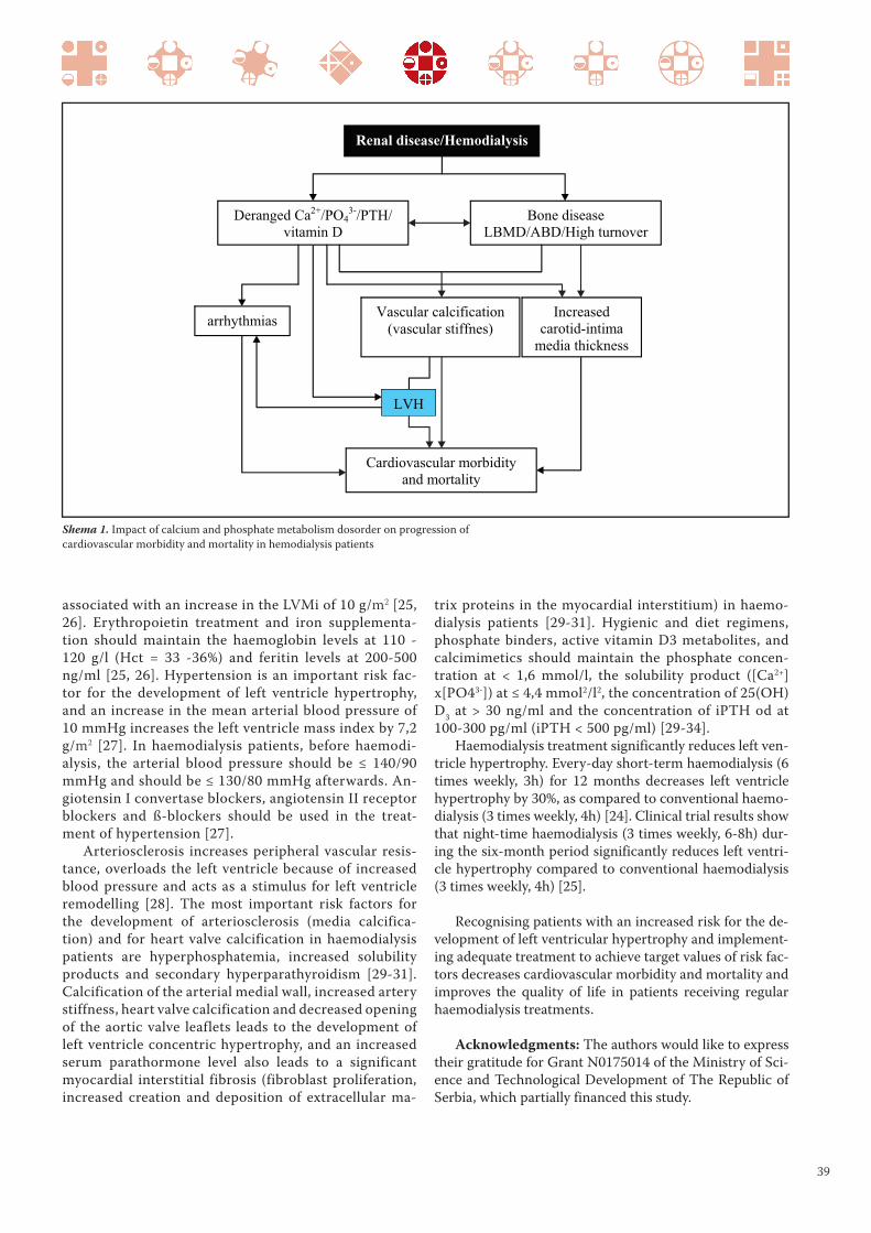
39
trix proteins in the myocardial interstitium) in haemo-
dialysis patients [29-31]. Hygienic and diet regimens,
phosphate binders, active vitamin D3 metabolites, and
calcimimetics should maintain the phosphate concen-
tration at < 1,6 mmol/l, the solubility product ([Ca2+]
x[PO43-]) at ≤ 4,4 mmol2/l2, the concentration of 25(OH)
D3 at > 30 ng/ml and the concentration of iPTH od at
100-300 pg/ml (iPTH < 500 pg/ml) [29-34].
Haemodialysis treatment significantly reduces left ven-
tricle hypertrophy. Every-day short-term haemodialysis (6
times weekly, 3h) for 12 months decreases left ventricle
hypertrophy by 30%, as compared to conventional haemo-
dialysis (3 times weekly, 4h) [24]. Clinical trial results show
that night-time haemodialysis (3 times weekly, 6-8h) dur-
ing the six-month period significantly reduces left ventri-
cle hypertrophy compared to conventional haemodialysis
(3 times weekly, 4h) [25].
Recognising patients with an increased risk for the de-
velopment of left ventricular hypertrophy and implement-
ing adequate treatment to achieve target values of risk fac-
tors decreases cardiovascular morbidity and mortality and
improves the quality of life in patients receiving regular
haemodialysis treatments.
Acknowledgments: The authors would like to express
their gratitude for Grant N0175014 of the Ministry of Sci-
ence and Technological Development of The Republic of
Serbia, which partially financed this study.
associated with an increase in the LVMi of 10 g/m2 [25,
26]. Erythropoietin treatment and iron supplementa-
tion should maintain the haemoglobin levels at 110 -
120 g/l (Hct = 33 -36%) and feritin levels at 200-500
ng/ml [25, 26]. Hypertension is an important risk fac-
tor for the development of left ventricle hypertrophy,
and an increase in the mean arterial blood pressure of
10 mmHg increases the left ventricle mass index by 7,2
g/m2 [27]. In haemodialysis patients, before haemodi-
alysis, the arterial blood pressure should be ≤ 140/90
mmHg and should be ≤ 130/80 mmHg afterwards. An-
giotensin I convertase blockers, angiotensin II receptor
blockers and ß-blockers should be used in the treat-
ment of hypertension [27].
Arteriosclerosis increases peripheral vascular resis-
tance, overloads the left ventricle because of increased
blood pressure and acts as a stimulus for left ventricle
remodelling [28]. The most important risk factors for
the development of arteriosclerosis (media calcifica-
tion) and for heart valve calcification in haemodialysis
patients are hyperphosphatemia, increased solubility
products and secondary hyperparathyroidism [29-31].
Calcification of the arterial medial wall, increased artery
stiffness, heart valve calcification and decreased opening
of the aortic valve leaflets leads to the development of
left ventricle concentric hypertrophy, and an increased
serum parathormone level also leads to a significant
myocardial interstitial fibrosis (fibroblast proliferation,
increased creation and deposition of extracellular ma-
Shema 1. Impact of calcium and phosphate metabolism dosorder on progression of
cardiovascular morbidity and mortality in hemodialysis patients

40
18. Petrović D, Miloradović V, Poskurica M, Stojimirović
B. Diagnostics and treatment of ischemic heart dis-
ease in hemodialysis patients. Vojnosanit Pregl 2009;
66(11): 897-903.
19. Voroneanu L, Covic A. Arrythmias in hemodialysis pa-
tients. J Nephrol 2009; 22(6): 716-25.
20. Foley RN, Parfrey PS, Harnett JD, Kent GM, Murray
DC, Barre PE. The prognostic importance of left ven-
tricular geometry in uremic cardiomyopathy. J Am Soc
Nephrol 1995; 5(12): 2024-31.
21. Petrović D, Jagić N, Miloradović V, Stojimirović B. Left ven-
tricular hypertrophy - risk factor for poor outcome in he-
modialysis patients. Ser J Exp Clin Res 2008; 9(4): 129-35.
22. De Bie MK, van Dam B, Gaasbeek A, van Buren M, van
Erven L, Bax JJ, et al. The current status of interven-
tions aiming at reducing sudden cardiac death in dialy-
sis patients. Eur Heart J 2009; 30(13): 1559-64.
23. Henrich WL. Optimal Cardiovascular Therapy for Pa-
tients with ESRD over the Next Several Years. Clin J
Am Soc Nephrol 2009; 4(Suppl 1): 106-9.
24. Hampl H, Riedel E. Cardiac Disease in the Dialysis Pa-
tient: Good, Better, Best Clinical Practice. Blood Purif
2009; 27(1): 99-113.
25. Glassock RJ, Pecoits-Filho R, Barberato SH. Left Ven-
tricular Mass in Chronic Kidney Disease and ERSD.
Clin J Am Soc Nephrol 2009; 4(Suppl 1): 79-91.
26. Stojimirović B, Petrović D, Obrenović R. Left ventric-
ular hypertrophy in patients on hemodialysis: impor-
tance of anaemia. Med Rev 2007; LX (Suppl 2): 155-9.
27. Lynn KL. Hypertension and Survival in Hemodialysis
Patients. Semin Dial 2004; 17(4): 270-4.
28. London GM. Arterial function in renal failure. Nephrol
Dial Transplant 1998; 13(Suppl 4): 12 5.
29. Petrović D, Stojimirović B. Secondary hiperparathy-
roidism - risk factor for development of cardiovascular
complications in hemodialysis patients. Med Rev 2010;
LXIII(9-10): (in press).
30. Petrović D, Obrenović R, Stojimirović B. Risk Factors
for Aortic Valve Calcification in Patients on Regular
Hemodialysis. Int J Artif Organs 2009; 32(3): 173-9.
31. Hörl WH. The clinical consequences of secondary hy-
perparathyroidism: focus on clinical outcomes. Neph-
rol Dial Transplant 2004; 19(Suppl 5): 2-8.
32. Messa P, Macario F, Yaqoob M, Bouman K, Braun J,
von Albertini B, et al. The OPTIMA Study: Assessing a
New Cinacalcet (Sensipar/Mimpara) Treatment Algo-
rithm for Secondary Hyperparathyroidism. Clin J Am
Soc Nephrol 2008; 3(1): 36-45.
33. Valdivieslo JM, Ayus JC. Role of vitamin D receptor
activators on cardiovascular risk. Kidney Int 2008;
74(Suppl 111): 44-9.
34. Artaza JN, Mehrotra R, Norris KC. Vitamin D and the
Cardiovascular System. Clin J Am Soc Nephrol 2009;
4(9): 1515-22.
REFERENCES
1. Parfrey PS. Cardiac disease in dialysis patients: diagnosis,
burden of disease, prognosis, risk factors and manage-
ment. Nephrol Dial Transplant 2000; 15(Suppl 5): 58 68.
2. Petrović D, Stojimirović B. Cardiovascular morbidity
and mortality in hemodialysis patients - epidemiologi-
cal analysis. Vojnosanit Pregl 2008; 65(12): 893-900.
3. Zoccali C, Mallamaci F, Tripepi G. Novel Cardiovascu-
lar Risk Factors in End-Stage Renal Disease. J Am Soc
Nephrol 2004; 15(Suppl 1): 77-80.
4. Petrović D, Jagić N, Miloradović V, Stojimirović B.
Non-tradicional risk factors for development of cardio-
vascular complications in haemodialysis patients. Ser J
Exp Clin Res 2009; 10(3): 95-102.
5. London GM. Left ventricular alterations and end-
stage renal disease. Nephrol Dial Transplant 2002;
17(Suppl 1): 29-36.
6. London GM. Cardiovascular Disease in Chronic Renal Fail-
ure: Patophysiologic Aspects. Semin Dial 2003; 16(2): 85-94.
7. Raggi P, Kleerekoper M. Contribution of Bone and
Mineral Abnormalities to Cardiovascular Disease in
Patients with Chronic Kidney Disease. Clin J Am Soc
Nephrol 2008; 3(3): 836-43.
8. Valdivieslo JM, Ayus JC. Role of vitamin D receptor
activators on cardiovascular risk. Kidney Int 2008;
74(Suppl 111): 44-9.
9. Eddington H, Klara PA. The association of chronic kid-
ney disease - mineral bone disorder and cardiovascular
risk. J Ren Car 2010; 36(Suppl 1): 61-7.
10. Cozzolino M, Ketteler M, Zehnder D. The vitamin D
system: a crosstalk between the heart and kidney. Eur J
Heart Fail 2010; 12(10): 1031-41.
11. Rigatto C, Parfrey PS. Uraemic Cardiomyopathy: an Over-
load Cardiomyopathy. J Clin Basic Cardiol 2001; 4(2): 93-5.
12. Parfrey PS, Collingwood P, Foley RN, Bahrle A. Left
ventricular disorders detected by M-meode echocar-
diography in chronic uraemia. Nephrol Dial Transplant
1996; 11(7): 1328-31.
13. Middleton RJ, Parfrey PS, Foley RN. Left Ventricular
Hypertrophy in the Renal Patient. J Am Soc Nephrol
2001; 12(5): 1079-84.
14. McIntyre C.W., John S.G., Jefferies H.J. Advances in the
cardiovascular assessment of patients with chronic kid-
ney disease. NDT Plus, 2008; 1(6): 383-91.
15. Cohen-Solal A. Left ventricular diastolic dysfunction:
pathophysiology, diagnosis and treatment. Nephrol
Dial Transplant 1998; 13(Suppl 4): 3-5.
16. Sood MM, Pauly RP, Rigatto C, Komenda P. Left ven-
tricular dysfunction in the haemodialysis population.
NDT Plus 2008; 1(4): 199-205.
17. Johnston N, Dargie H, Jardine A. Diagnosis and treat-
ment of coronary artery disease in patients with chron-
ic kidney disease. Heart 2008; 94(8): 1080-8.

41
UDK 613.71-053.6 / Ser J Exp Clin Res 2011; 12 (1): 41-42
Correspondence: Predrag Lazarevic, Assistant trainee in Physical Education, Faculty of Medicine, University of Kragujevac
Svetozara Markovica 69, 34000 Kragujevac, Republic of Serbia, Tel. +381 64 877 66 68, E-mail: [email protected]
THE RELATIONSHIP BETWEEN SPORTS ENGAGEMENT,BODY MASS INDEX AND PHYSICAL
ABILITIES IN CHILDREN
Predrag Lazarevic1, Vladimir Zivkovic1, Milena Vuletic1, Nevena Barudzic1 and Dejan Cubrilo1
1Department for Physical Education, Faculty of Medicine, University of Kragujevac, Kragujevac
ABSTRACT
Hypokinesia, which is a consequence of modern technologi-
cal development, is now one of the three biggest health risk fac-
tors. Whereas physical inactivity and obesity in adults is subject
of concern, these factors in children presage an even greater pub-
lic health problem because many health-related problems and
life-threatening diseases begin in childhood and adolescence.
It has been noted that regular physical activity modulates
vascular endothelial function via an eff ect on basal nitric ox-
ide (NO) production, thus leading to a signifi cant reduction
in the incidence of cardiovascular disease (1). Exercise can
also increase energy expenditure and can create a negative
energy balance. Conversely, scientifi c data regarding the ef-
fect of school-based physical activity interventions on body
LETTER TO THE EDITOR PISMO UREDNIKU LETTER TO THE EDITOR PISMO UREDNIKU
Received / Primljen: 21. 05. 2010. Accepted / Prihvaćen: 11. 04. 2011.
mass index (BMI) in children are confl icting (2, 3). Moreover,
there is insuffi cient clinically useful scientifi c data on BMI in
children and its possible association with motor skills. With
both sexes, motor abilities improve from physical develop-
ment in adolescents, and regular sports training infl uences
fi tness and motor abilities.
Th e aim of our study was to evaluate the morphological
status and physical abilities of 13-year-old children in order
to investigate the relationship between sports engagement,
BMI and physical abilities. Th e results of our study should
give coaches important parameters in the selection of young
athletes, as well as provide them with information that may
be useful in training and programming assignments.
MATERIALS AND METHODS
The research was carried out with a group of 67 boys
from an elementary school. The boys were 12-13 years old
and had differing sports training experiences.
After an assessment of morphological characteristics
(height, weight, BMI), the children were subjected to a bat-
tery of motor tests designed to test speed and coordination
(Jumping and going through Swedish boxes, slalom with 3
balls, hand tapping, 50-m sprint, 30-m flying start sprint
and 20-m slalom sprint (4)).
RESULTS AND DISCUSSION
The average BMI of the children who participated in
the study was 19.2±3.4. After transforming BMI data to
appropriate percentiles according to the formulas pro-
posed by the National Center for Health Statistics of
United States (5), we classified each subject into one of
four groups: underweight (athletes vs. non-athletes: 7.7%
vs. 17.8%), healthy weight (69.2% vs. 46.4%), overweight
(12.8% vs. 21.4%) and obese (10.2% vs. 14.3%). Although
BMI is thought to measure excess weight rather than ex-
cess body fat, if appropriate cut-off points are used, a high
BMI level is a moderately sensitive and very specific indi-
cator of excess adiposity among children (6).
Since hypokinesis in children and adolescents is thought
to be a growing problem, especially in urban places, it is
encouraging that 58.2% of the boys in the study stated
that they participated in a sport activity. To determine if
sport activity influences morphological status of young 13-
year-old boys, we applied a contingency analysis (χ2-test)
and found no significant difference between children who
train for a sport and those that do not. These results are in
agreement with many other studies that have found that, in
contrast to biological growth, the absence of involvement
in sports activities in children is not the main factor that
influences their morphological status and physical fitness.
Moreover, an analysis of morpho-functional skills of young

42
REFERENCES
1. Green DJ, Maiorana A, O’Driscoll G, Taylor R. Effect of
exercise training on endothelium-derived nitric oxide
function in humans. J Physiol 2004; 561:1–25.
2. Lazaar N, Aucouturier J, Ratel S, Rance M, Meyer M,
Duché P. Effect of physical activity intervention on body
composition in young children: influence of body mass in-
dex status and gender. Acta Paediatr 2007; 96(9): 315-20.
3. Harris KC, Kuramoto LK, Schulzer M, Retallack JE. Ef-
fect of school-based physical activity interventions on
body mass index in children: a meta-analysis. CMAJ
2009; 180(7):719-26.
4. Perić D. Operationalisation of research in sports and phys-
ical education. Beograd: Politop-p, 1994. (In Serbian)
5. National Center for Health Statistics of United States (2009):
CDC Growth Charts. www. cdc.gov/childrens_BMI
6. Freedman DS, Ogden CL, Berenson GS, Horlick M.
Body mass index and body fatness in childhood. Curr
Opin Clin Nutr Metab Care 2005; 8(6): 618-23.
7. Čubrilo D: Systemic effects of redox balance distur-
bance caused by the intensive training of young soccer
players, PhD dissertation, Medical faculty, University of
Kragujevac, Serbia, 2009.
8. Manić G. Multivariant differencies of some biome-
chanical dimensions of elementary school children in
relation to amount of fat tissue. Kinesiologica 2007; 1:
44-48. (in Croatian)
teens and premature soccer players (14 and 15 vs. 16 and
17 year olds) revealed that the aerobic capacity (VO2 max)
and BMI significantly changed with age, but not with the
training experience of the examined athletes (7).
Regarding the influence of morphological status of
13-year-old boys on motor performance, the results of
our study suggest that morphological status, estimated
by BMI, is an important predictor of physical abilities in
children. This finding is in agreement with the results of
other studies (8). Discriminative analysis (Kruskal-Wallis
test, Table 1) showed that subjects with different morpho-
logical status had significantly different performance in 5
out of the 6 tests performed, of which two were designed
to test speed and three tested coordination. However, ca-
nonical discrimination, used to determine the influence
of morphological status on latent motor function, showed
significant differences only in coordination tests.
Regarding the influence of sports engagement on motor
performance in our participants, a Man-Whitney test (Table
2) showed that, compared with those that did not train, boys
who train for a sport had significantly better results only in
2 motor tests, which were confirmed by factor analysis to
be valid tests of coordination. These results suggest that the
morphological status of 13-year-old children better predicts
coordination performance than speed performance.
The results of our study suggest that involvement of
adolescents in sport activities has much less influence on
motor abilities than that of natural biological growth and
maturation. The age at which the boys in this study were
tested falls within the first phase of adolescence, which is
characterised by sudden and intermittent skeletal matu-
ration without being followed by adequate skeletal mus-
cle development. That is one of the reasons why boys of
this age experience a fall in coordination performance.
In other words, motor development in 13-year-old boys
does not depend on training experience but instead relies
on genetics and acceleration of growth.
It should be emphasised that the small number of
participants in this study limited our ability to make any
significant anthropological generalisations. However, the
sample size did not affect the ability to address the main
aim of this study, which was to assess the predictive value
of morphological status and involvement in sports as pa-
rameters that affect motor performance.
Table 1. Mean values of results that subjects with diff erent morphological status achieved on motor tests.
Variable UnderweightHealthy
weightOverweight Obese Sig.
Jumping and going through
Swedish boxes (sec)15.60 15.37 17.59 20.23 .000*
Hand tapping (taps) 60.00 69.00 73.09 73.75 .040*
Slalom with 3 balls(sec) 28.65 22.26 21.61 28.29 .004*
50-m sprint (sec) 9.90 9.15 9.47 10.28 .002*
30-m flying start sprint (sec) 5.82 5.37 5,53 6.01 .008*
20-m slalom sprint (sec) 5.03 4.77 4.97 5.05 .204
VariableAth-
letes
Non-
athletesSig.
Jumping and going through
Swedish boxes (sec)15.85 16.58 .097
Hand tapping (taps) 69.28 64.07 .018*
Slalom with 3 balls (sec) 22.04 25.86 .004*
50-m sprint (sec) 9.24 9.49 .088
30-m flying start sprint (sec) 5.38 5.50 .065
20-m slalom sprint (sec) 4.76 4.88 .132
Table 2. Mean values of results that boys who train or do not train
some sport achieved on motor tests.

43
Serbian Journal of Experimental and Clinical Research
is a peer-reviewed, general biomedical journal. It publishes
original basic and clinical research, clinical practice arti-
cles, critical reviews, case reports, evaluations of scientific
methods, works dealing with ethical and social aspects of
biomedicine as well as letters to the editor, reports of asso-
ciation activities, book reviews, news in biomedicine, and
any other article and information concerned with practice
and research in biomedicine, written in the English.
Original manuscripts will be accepted with the under-
standing that they are solely contributed to the Journal.
The papers will be not accepted if they contain the mate-
rial that has already been published or has been submitted
or accepted for publication elsewhere, except of prelimi-
nary reports, such as an abstract, poster or press report
presented at a professional or scientific meetings and not
exceeding 400 words. Any previous publication in such
form must be disclosed in a footnote. In rare exceptions
a secondary publication will acceptable, but authors are
required to contact Editor-in-chief before submission of
such manuscript. the Journal is devoted to the Guidelines
on Good Publication Practice as established by Commit-
tee on Publication Ethics-COPE (posted at www.publica-
tionethics.org.uk).
Manuscripts are prepared in accordance with „Uni-
form Requirements for Manuscripts submitted to Biomedi-
cal Journals“ developed by the International Committee of
Medical Journal Editors. Consult a current version of the
instructions, which has been published in several journals
(for example: Ann Intern Med 1997;126:36-47) and posted at
www.icmje.org, and a recent issue of the Journal in prepar-
ing your manuscript. For articles of randomized controlled
trials authors should refer to the „Consort statement“ (www.
consort-statement.org). Manuscripts must be accompanied
by a cover letter, signed by all authors, with a statement that
the manuscript has been read and approved by them, and not
published, submitted or accepted elsewhere. Manuscripts,
which are accepted for publication in the Journal, become the
property of the Journal, and may not be published anywhere
else without written permission from the publisher.
Serbian Journal of Experimental and Clinical Research
is owned and published by Medical Faculty University of
Kragujevac. However, Editors have full academic freedom
and authority for determining the content of the journal, ac-
cording to their scientific, professional and ethical judgment.
Editorial policy and decision making follow procedures
which are endeavoring to ensure scientific credibility of pub-
lished content, confidentiality and integrity of auth ors, re-
viewers, and review process, protection of patients’ rights to
privacy and disclosing of conflict of interests. For difficulties
which might appear in the Journal content such as errors in
published articles or scientific concerns about research find-
ings, appropriate handling is provided. The requirements for
the content, which appears on the Journal internet site or
Supplements, are, in general, the same as for the master ver-
sion. Advertising which appears in the Journal or its internet
site is not allowed to influence editorial decisions.
Ad dress ma nu scripts to:
Serbian Journal of Experimental and
Clinical Research
The Me di cal Fa culty Kra gu je vac
P.O. Box 124, Sve to za ra Mar ko vi ca 69
34000 Kra gu je vac, Ser bia
Tel. +381 (0)34 30 68 00;
Tfx. +381 (0)34 30 68 00 ext. 112
E-mail: [email protected]
Manuscript can also be submitted to web address of
journal: www.medf.kg.ac.rs/journal
MA NU SCRIPT
Original and two anonymous copies of a ma nu script,
typed do u ble-spa ced thro ug ho ut (in clu ding re fe ren ces, ta-
bles, fi gu re le gends and fo ot no tes) on A4 (21 cm x 29,7 cm)
pa per with wi de mar gins, should be submitted for consid-
eration for publication in Serbian Journal of Experimen-
tal and Clinical Research. Use Ti mes New Ro man font, 12
pt. Ma nu script sho uld be sent al so on an IBM com pa ti ble
INSTRUCTION TO AUTHORSFOR MANUSCRIPT PREPARATION

44
floppy disc (3.5”), writ ten as Word file (version 2.0 or la ter),
or via E-mail to the edi tor (see abo ve for ad dress) as fi le
at tac hment. For papers that are accepted, Serbian Journal
of Experimental and Clinical Research obligatory requires
authors to provide an identical, electronic copy in appro-
priate textual and graphic format.
The ma nu script of original, scinetific articles should
be ar ran ged as fol lowing: Ti tle pa ge, Ab stract, In tro duc-
tion, Pa ti ents and met hods/Ma te rial and met hods, Re-
sults, Di scus sion, Ac know led ge ments, Re fe ren ces, Ta bles,
Fi gu re le gends and Fi gu res. The sections of other papers
should be arranged according to the type of the article.
Each ma nu script com po nent (The Ti tle pa ge, etc.)
should be gins on a se pa ra te pa ge. All pa ges should be num-
be red con se cu ti vely be gin ning with the ti tle pa ge.
All me a su re ments, ex cept blood pres su re, should be re-
por ted in the System In ter na ti o nal (SI) units and, if ne ces-
sary, in con ven ti o nal units, too (in pa rent he ses). Ge ne ric
na mes should be used for drugs. Brand na mes may be in-
ser ted in pa rent he ses.
Aut hors are advi sed to re tain ex tra co pi es of the ma nu-
script. Serbian Journal of Experimental and Clinical Research
is not re spon si ble for the loss of ma nu scripts in the mail.
TI TLE PA GE
The Ti tle pa ge con ta ins the ti tle, full na mes of all the
aut hors, na mes and full lo ca tion of the de part ment and in-
sti tu tion whe re work was per for med, ab bre vi a ti ons used,
and the na me of cor re spon ding aut hor.
The ti tle of the ar tic le should be con ci se but in for ma-
ti ve, and in clu de ani mal spe ci es if ap pro pri a te. A sub ti tle
could be ad ded if ne ces sary.
A list of ab bre vi a ti ons used in the pa per, if any, should
be in clu ded. The ab bre vi a ti ons should be listed alp ha be ti-
cally, and fol lo wed by an ex pla na ti on of what they stand for.
In ge ne ral, the use of ab bre vi a ti ons is di sco u ra ged un less
they are es sen tial for im pro ving the re a da bi lity of the text.
The na me, te lep ho ne num ber, fax num ber, and exact
po stal ad dress of the aut hor to whom com mu ni ca ti ons
and re prints sho uld be sent are typed et the end of the
ti tle pa ge.
AB STRACT
An ab stract of less than 250 words should con ci sely sta-
te the ob jec ti ve, fin dings, and con clu si ons of the stu di es
de scri bed in the ma nu script. The ab stract do es not con-
tain ab bre vi a ti ons, fo ot no tes or re fe ren ces.
Be low the ab stract, 3 to 8 keywords or short phra ses
are pro vi ded for in de xing pur po ses. The use of words from
Medline thesaurus is recommended.
IN TRO DUC TION
The in tro duc tion is con ci se, and sta tes the re a son and
spe ci fic pur po se of the study.
PA TI ENTS AND MET HODS/MA TE RIAL
AND MET HODS
The se lec tion of pa ti ents or ex pe ri men tal ani mals, in-
clu ding con trols, should be de scri bed. Pa ti ents’ na mes and
ho spi tal num bers are not used.
Met hods should be de scri bed in suf fi ci ent de tail to per mit
eva lu a tion and du pli ca tion of the work by ot her in ve sti ga tors.
When re por ting ex pe ri ments on hu man su bjects, it
should be in di ca ted whet her the pro ce du res fol lo wed we re
in ac cor dan ce with et hi cal stan dards of the Com mit tee on
hu man ex pe ri men ta ti on (or Ethics Committee) of the in sti-
tu tion in which they we re do ne and in ac cor dan ce with the
Hel sin ki Dec la ra tion. Ha zar do us pro ce du res or che mi cals,
if used, should be de scri bed in de ta ils, in clu ding the sa-
fety pre ca u ti ons ob ser ved. When ap pro pri a te, a sta te ment
should be in clu ded ve rifying that the ca re of la bo ra tory ani-
mals fol lo wed ac cep ted stan dards.
Sta ti sti cal met hods used should be outli ned.
RE SULTS
Re sults should be cle ar and con ci se, and in clu de a mi-
ni mum num ber of ta bles and fi gu res ne ces sary for pro per
pre sen ta tion.
DI SCUS SION
An ex ha u sti ve re vi ew of li te ra tu re is not ne ces sary. The
ma jor fin dings sho uld be di scus sed in re la tion to ot her pu-
blis hed work. At tempts sho uld be ma de to ex pla in dif fe-
ren ces bet we en the re sults of the pre sent study and tho se
of the ot hers. The hypot he sis and spe cu la ti ve sta te ments
sho uld be cle arly iden ti fied. The Di scus sion sec tion sho uld
not be a re sta te ment of re sults, and new re sults sho uld not
be in tro du ced in the di scus sion.
ACKNOWLEDGMENTS
This section gives possibility to list all persons who
contributed to the work or prepared the manuscript, but
did not meet the criteria for authorship. Financial and ma-
terial support, if existed, could be also emphasized in this
section.
RE FE REN CES
Re fe ren ces should be iden ti fied in the text by Ara bic
nu me rals in pa rent he ses. They should be num be red con-
se cu ti vely, as they ap pe ared in the text. Per so nal com mu ni-
ca ti ons and un pu blis hed ob ser va ti ons should not be ci ted
in the re fe ren ce list, but may be men ti o ned in the text in
pa rent he ses. Ab bre vi a ti ons of jo ur nals should con form to
tho se in In dex Serbian Journal of Experimental and Clini-
cal Research. The style and pun ctu a tion should con form to
the Serbian Journal of Experimental and Clinical Research
style re qu i re ments. The fol lo wing are exam ples:

45
Ar tic le: (all aut hors are li sted if the re are six or fe wer;
ot her wi se only the first three are li sted fol lo wed by “et al.”)
12. Tal ley NJ, Zin sme i ster AR, Schleck CD, Mel ton LJ.
Dyspep sia and dyspep tic sub gro ups: a po pu la tion-ba sed
study. Ga stro en te ro logy 1992; 102: 1259-68.
Bo ok: 17. Sher lock S. Di se a ses of the li ver and bi li ary
system. 8th ed. Ox ford: Blac kwell Sc Publ, 1989.
Chap ter or ar tic le in a bo ok: 24. Tri er JJ. Ce li ac sprue.
In: Sle i sen ger MH, For dtran JS, eds. Ga stro-in te sti nal di se-
a se. 4th ed. Phi la delp hia: WB Sa un ders Co, 1989: 1134-52.
The aut hors are re spon si ble for the exac tness of re-
fe ren ce da ta.
For other types of references, style and interpunction,
the authors should refer to a recent issue of Serbian Jour-
nal of Experimental and Clinical Research or contact the
editorial staff.
Non-English citation should be preferably translated to
English language adding at the end in the brackets native
langauage source, e.g. (in Serbian). Citation in old lan-
guage recognised in medicine (eg. Latin, Greek) should be
left in their own. For internet soruces add at the end in
small bracckets ULR address and date of access, eg. (Ac-
cessed in Sep 2007 at www.medf.kg.ac.yu). If available, in-
stead of ULR cite DOI code e.g. (doi: 10.1111/j.1442-2042
.2007.01834.x)
TA BLES
Ta bles should be typed on se pa ra te she ets with ta ble
num bers (Ara bic) and ti tle abo ve the ta ble and ex pla na tory
no tes, if any, be low the ta ble.
FI GU RES AND FI GU RE LE GENDS
All il lu stra ti ons (pho to graphs, graphs, di a grams) will
be con si de red as fi gu res, and num be red con se cu ti vely in
Ara bic nu me rals. The num ber of fi gu res in clu ded should
be the le ast re qu i red to con vey the mes sa ge of the pa per,
and no fi gu re should du pli ca te the da ta pre sented in the
ta bles or text. Fi gu res should not ha ve ti tles. Let ters, nu-
me rals and symbols must be cle ar, in pro por tion to each
ot her, and lar ge eno ugh to be readable when re du ced for
pu bli ca tion. Fi gu res should be sub mit ted as ne ar to the ir
prin ted si ze as pos si ble. Fi gu res are re pro du ced in one of
the fol lo wing width si zes: 8 cm, 12 cm or 17 cm, and with
a ma xi mal length of 20 cm. Le gends for fi gu res sho uld be
gi ven on se pa ra te pa ges.
If mag ni fi ca tion is sig ni fi cant (pho to mic ro graphs)
it should be in di ca ted by a ca li bra tion bar on the print,
not by a mag ni fi ca tion fac tor in the fi gu re le gend. The
length of the bar should be in di ca ted on the fi gu re or in
the fi gu re le gend.
Two com ple te sets of high qu a lity un mo un ted glossy
prints should be sub mit ted in two se pa ra te en ve lo pes, and
shi el ded by an ap pro pri a te card bo ard. The backs of sin-
gle or gro u ped il lu stra ti ons (pla tes) should be ar the first
aut hors last na me, fi gu re num ber, and an ar row in di ca ting
the top. This in for ma tion should be pen ci led in lightly or
pla ced on a typed self-ad he si ve la bel in or der to pre vent
mar king the front sur fa ce of the il lu stra tion.
Pho to graphs of iden ti fi a ble pa ti ents must be ac com pa-
nied by writ ten per mis sion from the pa ti ent.
For fi gu res pu blis hed pre vi o usly the ori gi nal so ur ce
should be ac know led ged, and writ ten per mis sion from the
copyright hol der to re pro du ce it sub mit ted.
Co lor prints are ava i la ble by re qu est at the aut hors
ex pen se.
LET TERS TO THE EDI TOR
Both let ters con cer ning and tho se not con cer ning
the ar tic les that ha ve been pu blis hed in Serbian Journal
of Experimental and Clinical Research will be con si de-
red for pu bli ca tion. They may con tain one ta ble or fi gu-
re and up to fi ve re fe ren ces.
PRO OFS
All ma nu scripts will be ca re fully re vi sed by the pu-
blis her desk edi tor. Only in ca se of ex ten si ve cor rec ti ons
will the ma nu script be re tur ned to the aut hors for fi nal
ap pro val. In or der to speed up pu bli ca tion no pro of will
be sent to the aut hors, but will be read by the edi tor and
the desk edi tor.

46
CIP – Каталогизација у публикацији
Народна библиотека Србије, Београд
61
SERBIAN Journal of Experimental and Clinical Research
editor - in - chief Slobodan
Janković. Vol. 9, no. 1 (2008) -
Kragujevac (Svetozara Markovića 69):
Medical faculty, 2008 - (Kragujevac: Medical faculty). - 29 cm
Je nastavak: Medicus (Kragujevac) = ISSN 1450 – 7994
ISSN 1820 – 8665 = Serbian Journal of
Experimental and Clinical Research
COBISS.SR-ID 149695244
