serinehydroxymethyltransferaseanchors denovo ... ·...
TRANSCRIPT

Serine Hydroxymethyltransferase Anchors de NovoThymidylate Synthesis Pathway to Nuclear Lamina for DNASynthesis*□S
Received for publication, December 13, 2011, and in revised form, January 9, 2012 Published, JBC Papers in Press, January 10, 2012, DOI 10.1074/jbc.M111.333120
Donald D. Anderson‡, Collynn F. Woeller‡§, En-Pei Chiang¶�, Barry Shane�, and Patrick J. Stover‡§1
From the §Division of Nutritional Sciences and the ‡Graduate Field of Biochemistry, Molecular and Cell Biology, Cornell University,Ithaca, New York 14853, the ¶Department of Food Science and Biotechnology, National Chung Hsing University, Taichung,Taiwan, and the �Department of Nutritional Sciences and Toxicology, University of California, Berkeley, California 94720
Background: De novo thymidylate biosynthesis occurs in the nucleus.Results: This pathway forms a multienzyme complex that is associated with the nuclear lamina and the DNA replicationmachinery.Conclusion: De novo thymidylate biosynthesis occurs at the sites of DNA replication and is enriched at sites of replicationinitiation.Significance: The compartmentation of thymidylate biosynthesis at the replication fork may permit regulation of dUTP incor-poration into DNA.
The de novo thymidylate biosynthetic pathway inmammaliancells translocates to the nucleus for DNA replication and repairand consists of the enzymes serine hydroxymethyltransferase 1and 2� (SHMT1 and SHMT2�), thymidylate synthase, anddihydrofolate reductase. In this study, we demonstrate that thispathway forms a multienzyme complex that is associated withthe nuclear lamina. SHMT1 or SHMT2� is required for co-lo-calization of dihydrofolate reductase, SHMT, and thymidylatesynthase to the nuclear lamina, indicating that SHMT serves asscaffold protein that is essential for complex formation. Themetabolic complex is enriched at sites of DNA replication initi-ation and associated with proliferating cell nuclear antigen andother components of the DNA replication machinery. Thesedata provide a mechanism for previous studies demonstratingthat SHMT expression is rate-limiting for de novo thymidylatesynthesis and indicate that de novo thymidylate biosynthesisoccurs at replication forks.
Regulation of cellular dTTP synthesis is essential for DNAreplication and genome stability in the nucleus (1) and mito-chondria (2). Depressedde novo thymidylate synthesis resultingfrom folate deficiency or anti-folate treatment results indeoxyuridine misincorporation into mitochondrial DNA (2)and nuclear DNA leading to genome instability (3). Tetrahy-drofolate (THF)2 is a metabolic cofactor that carries and acti-vates single carbons for the synthesis of purine and thymidine
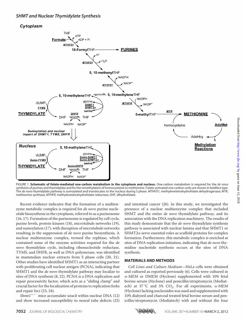
nucleotides and for homocysteine remethylation tomethionine(4). Folate-mediated one-carbon metabolism is compartmen-talized in the mitochondria, nucleus, and cytoplasm of eukary-otic cells (5). The enzymes that constitute the de novo thymidy-late pathway include SHMT1, SHMT2�, thymidylate synthase(TYMS), and dihydrofolate reductase (DHFR). Methylene-THF generated by SHMT is the one-carbon donor for theTYMS-catalyzed conversion of dUMP to thymidylate, generat-ing dihydrofolate. DHFR catalyzes the NADPH-dependentreduction of dihydrofolate to regenerate THF for subsequentcycles ofde novo thymidylate synthesis (Fig. 1). SHMT1,TYMS,and DHFR have been localized to the nucleus, and their trans-location is mediated by post-translational modification withthe small ubiquitin-like modifier (SUMO) (6, 7). SHMT1nuclear translocation is cell cycle-dependent and occurs duringthe S and G2/M phases and in response to UV damage (7–9). Inmice, nuclear localization of the de novo thymidylate synthesispathway is required to minimize uracil misincorporation intonuclear DNA (10). Intact purified nuclei from mouse liverexhibit de novo thymidylate synthesis activity, whereas nucleidisrupted by sonication lack this activity, indicating that theformation of a multienzyme complex may be required for thepathway to function (6).Previous studies in cell culture and mouse models have
shown that SHMT1expression determinesde novo thymidylatesynthesis activity, indicating that this enzyme is limiting forde novo thymidylate synthesis in vivo (11, 12). In mammals,there are two SHMT isozymes encoded by distinct genes (13–15). SHMT1 encodes the cytoplasmic/nuclear isozyme(SHMT1), and SHMT2 encodes the mitochondrial (SHMT2)and the cytoplasmic/nuclear (SHMT2�) isoform through alter-native promoter use (6, 13–15). This second SHMT2 transcriptencodes SHMT2�, which provides functional redundancy withSHMT1 in the de novo thymidylate synthesis pathway.De novothymidylate biosynthesis activity is reduced by 75% in nucleiisolated from Shmt1�/� mice (6).
* This work was supported, in whole or in part, by National Institutes of HealthGrants DK58144 and HD059120 (to P. J. S.).
□S This article contains supplemental Figs. S1–S8 and Tables S1–S5.1 To whom correspondence should be addressed: 315 Savage Hall, Division of
Nutritional Sciences, Cornell University, Ithaca, NY 14853. Tel.: 607-255-9751; Fax: 607-255-1033; E-mail: [email protected].
2 The abbreviations used are: THF, tetrahydrofolate; SHMT, serine hydroxy-methyltransferase; TYMS, thymidylate synthase; DHFR, dihydrofolatereductase; SUMO, small ubiquitin-like modifier; PCNA, proliferating cellnuclear antigen; SV40 LT, SV40 large T antigen; MEM, minimum essentialmedium; TAP, tandem affinity purification.
THE JOURNAL OF BIOLOGICAL CHEMISTRY VOL. 287, NO. 10, pp. 7051–7062, March 2, 2012© 2012 by The American Society for Biochemistry and Molecular Biology, Inc. Published in the U.S.A.
MARCH 2, 2012 • VOLUME 287 • NUMBER 10 JOURNAL OF BIOLOGICAL CHEMISTRY 7051
by guest on June 13, 2018http://w
ww
.jbc.org/D
ownloaded from
Recent evidence indicates that the formation of a multien-zyme metabolic complex is required for de novo purine nucle-otide biosynthesis in the cytoplasm, referred to as a purinosome(16, 17). Formation of the purinosome is regulated by cell cycle,purine levels, protein kinases (18), microtubule networks (19),and sumoylation (17), with disruption ofmicrotubule networksresulting in the suppression of de novo purine biosynthesis. Anuclear multienzyme complex, termed the replitase, whichcontained some of the enzyme activities required for the denovo thymidylate cycle, including ribonucleotide reductase,TYMS, and DHFR, as well as DNA polymerase, was identifiedin mammalian nuclear extracts from S phase cells (20, 21).Other studies have identified SHMT1 as an interacting partnerwith proliferating cell nuclear antigen (PCNA), indicating thatSHMT1 and the de novo thymidylate pathway may localize tosites of DNA synthesis (8, 22). PCNA is a DNA replication andrepair processivity factor, which acts as a “sliding clamp” andcrucial factor for the localization of proteins to replication forksand repair foci (23, 24).Shmt1�/� mice accumulate uracil within nuclear DNA (12)
and show increased susceptibility to neural tube defects (25)
and intestinal cancer (26). In this study, we investigated thepresence of a nuclear multienzyme complex that includedSHMT and the entire de novo thymidylate pathway, and itsassociation with the DNA replicationmachinery. The results ofthis study demonstrate that the de novo thymidylate synthesispathway is associated with nuclear lamina and that SHMT1 orSHMT2� serve essential roles as scaffold proteins for complexformation. Furthermore, this metabolic complex is enriched atsites of DNA replication initiation, indicating that de novo thy-midine nucleotide synthesis occurs at the sites of DNAsynthesis.
MATERIALS AND METHODS
Cell Lines and Culture Medium—HeLa cells were obtainedand cultured as reported previously (6). Cells were cultured in�-MEM or DMEM (Hyclone) supplemented with 10% fetalbovine serum (Hyclone) and penicillin/streptomycin (Mediat-ech) at 37 °C and 5% CO2. For all experiments, �-MEM(Hyclone) lacking nucleosideswas used and supplementedwith10% dialyzed and charcoal treated fetal bovine serum and pen-icillin/streptomycin (Mediatech) with and without the four
FIGURE 1. Schematic of folate-mediated one-carbon metabolism in the cytoplasm and nucleus. One-carbon metabolism is required for the de novosynthesis of purines and thymidylate and for the remethylation of homocysteine to methionine. Folate-activated one-carbon units are shown in boldface type.The de novo thymidylate pathway is sumoylated and translocates to the nucleus during S phase. MTHFD1, methylenetetrahydrofolate dehydrogenase; MTR,methionine synthase; MTHFR, methylenetetrahydrofolate reductase; DHF, dihydrofolate.
SHMT and Nuclear Thymidylate Synthesis
7052 JOURNAL OF BIOLOGICAL CHEMISTRY VOLUME 287 • NUMBER 10 • MARCH 2, 2012
by guest on June 13, 2018http://w
ww
.jbc.org/D
ownloaded from

canonical deoxyribonucleosides, each at a concentration of 10mg/liter. Cells were maintained at 37 °C and 5% CO2 for 2 pas-sages over 1 week prior to transfection. The SH-SY5Y humanneuroblastoma cell line has been described previously (27).Cells were cultured in �-MEM with 10% dialyzed fetal bovineserum for all experiments. The expression of a dominant neg-ative SHMT1 protein, DN2-SHMT1, was induced in cell linesby the addition of 1 �g/ml tetracycline for aminimum of 4 daysprior to experimentation as described previously for SH-SY5Y-FDH cell lines (28).Generation of Human DN2-SHMT1-expressing Cell Lines—
Generation of the pet28a-DN2-SHMT1 cDNA was describedpreviously (29). This DN2-SHMT1 cDNA was cloned into theKpnI and BamHI (New England Biolabs) site of thepcDNA4/TO/myc-His C vector (Invitrogen), generatingpcDNA4-DN2-SHMT1. Stable SH-SY5Y cell lines expressingpcDNA6/TR vector (Invitrogen) and pcDNA4-DN2-SHMT1were generated. SH-SY5Y cells were electroporated with 20 �gof plasmid DNA (pcDNA4-DN2-SHMT1 and pcDNA6/TRvector) at 0.22 kV and 950 microfarads (Bio-Rad Gene-Pulser)and then cultured with �-MEM for 48 h. The medium wasreplaced with �-MEM containing blasticidin (10 �g/ml) andZeocin (100�g/ml; Invitrogen) to select for cells that integratedthe plasmid. Individual colonies resistant to Zeocin treatmentwere selected and expanded using �-MEM containing blastici-din and Zeocin, and clonal lines expressing human DN2-SHMT1 protein were screened by Western blot analyses.Vectors and Transfection Procedures—The pCMV6-AC-
DHFR-GFP, pCMV6-AC-TYMS-GFP, phiYFP-SHMT1, andTagRFP-N-SHMT2� vectors were previously described (6).PmKate2-Lamin B1was purchased fromEvrogen. The pBABE-Puro-SV40 LT vectorwas deposited inAddgene byDr. ThomasRoberts and purchased from Addgene (Addgene plasmid13970). pNTAPB and pCMV-FLAG-MAT-Tag-1 were ob-tained from Stratagene and Sigma, respectively. PCR was usedto create adapters on SHMT1 cDNA for ligation. The forwardprimers for both pNTAPB-SHMT1 and pCMV-FLAG-SHMT1-MAT-Tag-1 were 5�-ATATAAGCTTATGACGAT-GCCAGTCAAC-3�, where the underline indicates a HindIII(New England Biolabs Inc.) restriction site. The reverse primerfor pNTAPB-SHMT1 was 5�-ATATCTCGAGGAAGTCAG-GCAGGCCAGG-3�, where the underline indicates an XhoI(New England Biolabs Inc.) restriction site. The reverse primerfor pCMV-FLAG-SHMT1-MAT-Tag-1 was 5�-ATAT-AGATCTGAAGTCAGGCAGGCCAGG-3�, where the under-line indicates a BglII (NewEngland Biolabs Inc.) restriction site.PCRs were conducted as follows: 95 °C for 45 s, 55 °C for 45 s,and 72 °C for 2min. Following PCR, products were gel-purifiedusing the QIAquick gel extraction kit (Qiagen). Vectors andPCR products were digested with their respective restrictionenzymes as per themanufacturer’s protocol. Ligation was com-pleted using T4DNA ligase (Invitrogen) as per themanufactur-er’s protocol. All transfection procedureswere completed usingKit R (Lonza) for a Nucleofector II (Lonza) as per the manufac-turer’s instructions.Confocal Microscopy—Following nucleofection, cells were
incubated at 37 °C under 5% CO2 for 24 h. For visualization ofnuclei in cells not transfected with pmKate2-Lamin B1,
DRAQ5 (Alexis Biochemicals) was used per themanufacturer’sinstructions. Confocal fluorescence microscopy (Leica TCSSP2 system) was used to image all cells at the Cornell Micro-scope and Imaging Facility.Cell Cycle Synchronization and Analysis—HeLa cells at 60%
confluence were arrested at various cell cycle stages using 30�M lovastatin (Sigma) (for G1 phase), 2 mM hydroxyurea(Sigma) (for S phase), or 100 ng/ml nocodazole (Sigma) (forG2/M phase) following transfection. Preparation for fluores-cence-activated cell sorting (FACS) was described previously(8). FACS analysis was performed at the Biomedical SciencesFlow Cytometry Core Laboratory, Cornell University.Immunoblotting—SHMT1, SHMT2, TYMS, DHFR, and
GAPDH immunoblots were performed as described previously(6, 7). Antibodies directed toward PCNA and the SV40 LTwerepurchased from Abcam. Mouse anti-PCNA and mouse anti-SV40 large T antigen were diluted in 5% nonfat dry milk(Carnation) containing 1% Nonidet P-40 (U.S. Biologicals) at aconcentration of 1 �g/ml and 1:1000, respectively. Goat anti-mouse HRP (Pierce) secondary antibodies were used at a dilu-tion of 1:10,000. SuperSignal West Pico Chemiluminescentsubstrate was used for visualization (Pierce).Immunoprecipitation—Immunoprecipitation was con-
ducted using the Dynabeads Protein G immunoprecipitationkit (Invitrogen). For whole cell immunoprecipitation, HeLacells were lysed using mammalian protein extraction reagent(Pierce) supplemented with 2 mM �-mercaptoethanol (Calbi-ochem), 0.1 mM EDTA (Fisher), 1 mM PMSF (Alexis Biochem-icals), and 1:1000 protease inhibitor mixture (Sigma). Forimmunoprecipitations from nuclear and cytosolic extracts,nuclei were purified using the Active Motif Nuclear extract kitper the manufacturer’s protocol. Antibodies directed towardthe following proteins were purchased from Abcam: DNApolymerase �, uracil DNA glycosylase 2 (UNG2), PCNA, andDHFR. Lamin A/C, Lamin B1, DNA polymerase �, lysine dem-ethylase 1 (KDM1), and replication factor C activator 1 (RFC1)antibodies were purchased from Santa Cruz Biotechnology,Inc. (Santa Cruz, CA). Non-immune IgG was purchased fromPierce. SHMT1 antibody was obtained as described previously(8). 1 mg of total protein per sample was incubated with 5 �g ofantibody overnight at 4 °C. The beads were collected andwashed four times with PBS, 0.1% Tween 20 (Fisher). For laminA/C and lamin B1 immunoprecipitations, washes containedeither 150 mM NaCl or 650 mM NaCl in PBS, 0.1% Tween 20.For all immunoprecipitations, 40 �l of SDS-PAGE samplebuffer were added to the beads to elute the protein with heatingat 100 °C.SHMT1 Tandem Affinity Purification—pNTAPB-SHMT1
was transfected into HeLa cells as described above. For the iso-lation of TAP-tagged SHMT1 and co-eluting proteins, theInterplay Mammalian TAP System kit (Stratagene) was usedfollowing the manufacturer’s protocol. For isolation of FLAG-SHMT1-MAT and co-eluting proteins for peptide sequencing,pCMV-FLAG-SHMT1-MAT-Tag-1 was transfected as de-scribed above. Approximately 4� 108 HeLa cells were used pergroup. The groupswere as follows: empty pCMV-FLAG-MAT-Tag-1 control, pCMV-FLAG-SHMT1-MAT-Tag-1 S phase-synchronized cells, and UV-treated cells. UV treatment was
SHMT and Nuclear Thymidylate Synthesis
MARCH 2, 2012 • VOLUME 287 • NUMBER 10 JOURNAL OF BIOLOGICAL CHEMISTRY 7053
by guest on June 13, 2018http://w
ww
.jbc.org/D
ownloaded from

accomplished as previously reported (9). 24 h following treat-ments, cells were washed with 20 ml of PBS, 1 mM PMSF threetimes. Cells were scraped into 5 ml of PBS, 1 mM PMSF andcollected in 50-ml conical tubes (Corning Glass). Cells werepelleted at 1000 rpm for 5min and suspended in 15ml of swell-ing buffer (25 mM HEPES (Fisher), pH 7.8, 1.5 mM MgCl2(Fisher), 10 mM KCl (Fisher), 0.1% Nonidet P-40 (U.S. Biologi-cals), 1.0 mM PMSF (Alexis Biochemicals), 1:100 proteaseinhibitor (Sigma)) and incubated on ice for 10 min. Cells werehomogenizedwith 25 strokes of a B-typeDounce homogenizer,and cell lysis was verified by trypan blue (Fisher) exclusion. Thehomogenate was then filtered (70-�m mesh; BD Bioscience)and centrifuged at 4000 rpm for 5 min to collect nuclei. Nucleiwere washed with 2� swelling buffer and pelleted by centrifu-gation. Nuclei were suspended in 1ml of ice-cold sodium phos-phate (Fisher), pH 8.0, 75mMNaCl, 1mMPMSF, 1:100 proteaseinhibitor for histidine tag isolation (Sigma). MgCl2 was addedto a concentration of 2 mM. Benzonase (Sigma) was added at aconcentration of 250 units/ml sample for removal of DNA andincubated for 30 min on ice. Following benzonase treatment,samples were centrifuged at 14,000 rpm for 15 min. The super-natant was collected, and FLAG-SHMT1-MAT and co-elutingproteins were purified using Invitrogen His-TALONmagneticbeads per the manufacturer’s instructions. Following elution,the eluates were combined and diluted with 1 volume of a solu-tion containing 100 mM Tris, pH 7.4, 2 mM EDTA, and a 1:100dilution of protease inhibitor mixture. FLAG immunoprecipi-tation was conducted using FLAG affinity gel (Sigma) per themanufacturer’s instructions. The eluate from the FLAG immu-noprecipitation was then subjected to protein precipitationusing the ND protein precipitation kit (National Diagnostics)per the manufacturer’s protocol. The protein pellet was sus-pended in 20 �l of 20 mM Tris, pH 7.5, and 20 �l of 2� SDS-PAGE loading buffer (Bio-Rad) and heated at 95 °C for 10 min.Mass Spectrometry—Tandem affinity-purified samples from
S phase-blocked and UV-treated cells were run on a 12% poly-acrylamide gel and were stained with Novex colloidal bluestaining reagent (Invitrogen). The lanes were excised and cutinto nine fractions of �7 mm by 5-mm areas and sent to theHarvard Microchemistry Facility for peptide digestion, purifi-cation, and sequencing. Each excised fractionwas reduced, car-boxyamidomethylated, and digestedwith trypsin. Peptide iden-tification of each digestion mixture was performed bymicrocapillary reversed-phaseHPLCnanoelectrospray tandemmass spectrometry on an LTQ-Orbitrap Velos or XL massspectrometer (Thermo Fisher Scientific, San Jose, CA). TheOrbitrap repetitively surveyed an m/z range from 395 to 1600,whereas data-dependent MS/MS spectra on the 20 (Velos) or10 (XL) most abundant ions in each survey scan were acquiredin the linear ion trap. MS/MS spectra were acquired with rela-tive collision energy of 30%, 2.5-Da isolation width, and recur-ring ions were dynamically excluded for 60 s. Preliminarysequencing of peptides was facilitated with the SEQUEST algo-rithm with a 30 ppm mass tolerance against a species-specific(human) subset of the UniProt Knowledgebase. With a customversion of the Harvard Proteomics Browser Suite (ThermoFisher Scientific), peptide spectrum matches were acceptedwith mass error of �2.5 ppm and score thresholds to attain an
estimated false discovery rate of �1%, using a reverse decoydatabase strategy. Gene ontology term enrichment was per-formed using DAVID with the total set of proteins with a pep-tide count of �3 and a ratio of experimental group peptidecounts to control group peptide counts of �1 (30, 31).Metabolic Isotope Tracer Studies—Metabolic isotope tracer
studies were completed as described previously (32).Tandem Chromatin Immunoprecipitation—Clones 1, 2, and
3 of pBABE-Puro-SV40 LT-expressing HeLa cells that wereselected using puromycin were used for this experiment.Approximately 2 � 108 cells were used per clone. Each platewas washed three times with ice-cold PBS. The bifunctionalcross-linker dimethyl 3,3�-dithiobispropionimidate (Pierce)was suspended at a concentration of 5mM in PBS at pH 8.0, and20 ml per plate was added. Cells were incubated at 4 °C for 30min and thenwashed twice with PBS. 20ml of ice-cold quench-ing buffer (100 mM Tris, pH 8.0, 150 mM NaCl) was added perplate, and the cells were incubated for 10min at 4 °C. Each platewas washed three times with PBS at room temperature. Eachplate was treated with a 1% formaldehyde (Fisher) solution inPBS and then incubated at room temperature for 10 min. Toquench formaldehyde cross-linking, 3 ml of 1.375 M glycine(Fisher) were added. Plates were washed three times with ice-cold PBS supplemented with 0.5 mM PMSF. Cells were scrapedinto 5 ml of ice-cold PBS containing 0.5 mM PMSF and centri-fuged at 1000 rpm for 5 min at 4 °C. The pelleted cells wereresuspended in 10 volumes of swelling buffer and then sub-jected to Dounce homogenization and centrifugation asreported above. Pelleted nuclei were resuspended in 5 ml ofsonication buffer (50 mM Hepes, pH 7.9, 140 mM NaCl, 1 mM
EDTA, 1% Triton X-100, 0.1% sodium deoxycholate, 0.1% SDS,0.5 mM PMSF, and 1:1000 protease inhibitor mixture). Nucleiwere sonicated using a 3-mm microtip probe on a BransonSonifier 150 at 80%with 10-s pulses, 10 times, with 1-min incu-bations on ice between pulses. Sonicated nuclei were centri-fuged at 14,000 rpm for 15min at 4 °C, and the supernatant wassupplemented with 1 �g/ml sonicated salmon sperm DNA(Invitrogen) and bovine serum albumin (Sigma) at 1 mg/ml.The lysate was precleared using 50 �l of Invitrogen Protein GDynabeads for 2 h at 4 °C, beads were collected on a magnet,and the supernatant was removed. Samples were divided into1-ml aliquots for immunoprecipitation. 5�g of PCNA (Abcam)antibodywas incubated for 12 hper 1-ml aliquot at 4 °C. 50�l ofInvitrogen Protein G Dynabeads were added, and the solutionwas incubated for 1 h at 4 °C. The beads were collected with amagnet and washed twice with 1 ml of sonication buffer. Thebeads were washed twice with 1 ml of sonication buffer con-taining 500 mM NaCl and then twice with 1 ml of a solutioncontaining 20mMTris, pH 8.0, 1 mM EDTA, 250mM LiCl, 0.5%Nonidet P-40, 0.5% sodium deoxycholate, 0.5 mM PMSF, andprotease inhibitor mixture. Two more washes were performedwith 1 ml of TE buffer. 50 �l of 10 mM DTT was then added tothe beads, which were then incubated at 37 °C for 30 min. Theeluate was removed, and that step was repeated. The eluateswere then combined and diluted 40� with sonication buffer.10% of the sample was kept for input. The sample was thenaliquoted into 1-ml fractions for TYMS, DHFR, SHMT1, andIgG control immunoprecipitations. 5 �g of each antibody was
SHMT and Nuclear Thymidylate Synthesis
7054 JOURNAL OF BIOLOGICAL CHEMISTRY VOLUME 287 • NUMBER 10 • MARCH 2, 2012
by guest on June 13, 2018http://w
ww
.jbc.org/D
ownloaded from

used, and samples were incubated for 12 h at 4 °C. 50 �l ofInvitrogen Protein G Dynabeads were added and incubated for1 h at 4 °C. Washes were performed as above. To elute theproteins, 200 �l of a solution containing 50 mM Tris, pH 8.0, 1mM EDTA, 1% SDS, and 50 mM NaHCO3 was added, and thebeads were heated to 65 °C for 10 min. This step was repeated,and eluates were combined, giving a 400-�l final volume. Theinput and samples were then treated with 21�l of 4 MNaCl andincubated for 12 h at 65 °C. 2 �l of RNase A (5 mg/ml) (Rock-land Immunochemicals, Inc.) was added to each sample, fol-lowed by incubation at 37 °C for 1 h. EDTAwas then added to aconcentration of 5 mM. 2 �l of proteinase K (10 mg/ml) (Rock-land Immunochemicals, Inc.) was then added, and sampleswere incubated for 2 h at 42 °C. Samples were extracted withphenol/chloroform/isoamyl alcohol (Sigma) once and thenwith chloroform/isoamyl alcohol (Sigma). 1 �l of glycogen(Sigma) (20mg/ml)was added, followed by the addition of 40�lof 3 M sodium acetate (Fisher) and 1 ml of 100% ethanol(Pharmco-AAPER). Samples were vortexed and precipitatedfor 12 h at�20 °C, followed by centrifugation at 14,000 rpm for30min. The pellets were washed with 75% ethanol. Centrifuga-tion was repeated, and the pellet was allowed to dry at roomtemperature. Samples were then suspended in 100 �l of 10 mM
Tris, pH 7.5, for real-time PCR analysis.Real-time PCR—Forward and reverse primers surrounding
the SV40 origin of replication were 5�-CAGCAGGCAGAAG-TATGCAAAGCA-3� and 5�-TACTTCTGGAATAGCTCA-GAGGCCGA-3�, respectively. To amplify the pBABE-Purovector region, the forward and reverse primers were 5�-ACA-GAGTTCTTGAAGTGGTGGCCT-3� and 5�-TGGTTT-GTTTGCCGGATCAAGAGC-3�, respectively. To amplify thelarge T-antigen insert, the forward and reverse primers were5�-ACTCCACACAGGCATAGAGTGTCT-3� and 5�-CCCA-CCTGGCAAACTTTCCTCAAT-3�, respectively. Real-timePCR analysis was performed using the Quantifast SYBR GreenPCR kit (Qiagen). PCR products were quantified using anApplied Biosystems 7500 real-time PCR system.
RESULTS
SHMT1, SHMT2�, DHFR, and TYMS Are Present in Nucleiduring S and G2/M Phases—SHMT1, SHMT2�, TYMS, andDHFR have been localized previously to the nucleus and cyto-plasm of human and mouse cell lines (6, 8, 10). Nuclear local-ization of SHMT1 and SHMT2� in mouse embryonic fibro-blasts and human cell lines (6, 8) is restricted to the S andG2/Mphases of the cell cycle and in response toUVdamage (9). In thisstudy, the nuclear localization of TYMS and DHFR was deter-mined as a function of cell cycle. As seen previously for SHMT1,DHFR (supplemental Fig. S1) andTYMS (supplemental Fig. S2)localized to the nucleus during S andG2/Mphases but not inG1phase. These data confirm that all of the enzymes necessary forde novo thymidylate biosynthesis are presentwithin the nucleusduring S and G2/M phases during DNA replication and repairbut are restricted to the cytoplasm in G1.Identification of SHMT1-interacting Proteins in Nuclear
Extracts—Intact purified nuclei from mouse liver can converttritium-labeled serine and dUMP to tritium-labeled thymidy-late, but this activity is lost in sonicated nuclei, indicating that
nuclear integrity and multienzyme complex formation may benecessary for de novo thymidylate synthesis (6). To determine ifthe de novo thymidylate biosynthesis pathway is present innuclei within a multienzyme complex, SHMT1 tandem affinitypurification and SHMT1 co-precipitation experiments wereperformed on benzonase-treated nuclear extracts isolated fromS phase cells and cells treated with UV (supplemental Fig. S3).Over 833 proteins were identified as SHMT1-interacting pro-teins. The list was refined by excluding proteinswith fewer thanthree peptides identified, and the remaining proteins weregrouped by gene ontology term enrichment using the bioinfor-matics tool DAVID (Table 1 and supplemental Tables S1–S5).Multiple proteins involved in nucleotide metabolism and DNAreplication and repair were identified, as well as 80 lamin-inter-acting proteins. The majority of the identified proteins knownto be involved in DNA replication and repair, as well as lamin-interacting proteins, were found in both S phase andUV-treated samples, although some of these interactions wereidentified only in one of those two exposures (Table 1).Validation of SHMT1-interacting Partners—DNA replica-
tion occurs on nucleoskeletal structures (34) and disruptionof the nuclear lamina results in DNA replication arrests (35,36). Several proteins involved in DNA replication are lamin-binding proteins, including PCNA, which acts as a proces-sivity factor for DNA replication (37). SHMT1 has beenidentified previously as a PCNA-interacting partner (8), and
TABLE 1Subsets of proteins identified using tandem affinity purification andMS/MSA complete listing of proteins identified is available in supplemental Tables S1–S5.
SHMT and Nuclear Thymidylate Synthesis
MARCH 2, 2012 • VOLUME 287 • NUMBER 10 JOURNAL OF BIOLOGICAL CHEMISTRY 7055
by guest on June 13, 2018http://w
ww
.jbc.org/D
ownloaded from

this interaction was confirmed in this tandem affinity puri-fication experiment (supplemental Fig. S3 and Table 1). Tovalidate SHMT1 as a lamin-interacting partner, co-immuno-precipitation experiments using benzonase-treated nuclearextracts were performed with antibodies against eitherlamin A/C or lamin B1. SHMT1 co-precipitated with laminA/C and lamin B1 antibodies under stringent (650 mMNaCl)and less stringent conditions (150 mM NaCl) (Fig. 2A). Nei-ther TYMS nor DHFR co-precipitated with lamin proteinsunder these conditions.Other SHMT1-interacting partners identified in Table 1
were validated by co-immunoprecipitation followed by immu-noblotting against the interacting protein. SHMT1, TYMS, andDHFR all co-precipitated with PCNA in nuclear extracts (sup-plemental Fig. S4, A–C) but not in cytoplasmic extracts (sup-plemental Fig. S4, A and B). Lysine demethylase 1 (KDM1),which has been recently identified as a folate-binding protein(38), immunoprecipitated with anti-SHMT1 antibodies (sup-plemental Fig. S4D). Other proteins involved in DNA replica-tion and repair, includingDNApolymerases � and � (POLDandPOLE respectively), replication factor C activator 1 (RFC1), anduracil DNA glycosylase 2 (UNG2) (supplemental Fig. S4E) co-immunoprecipitated with SHMT1. In total, eight validationswere performed, and no false positives were identified.
SHMT1 Interaction with TYMS and DHFR Is DNA-dependent—Neither TYMS nor DHFR were identified asSHMT1- or lamin-interacting proteins by tandem affinity puri-fication. The ability of nuclear SHMT1 to interact with TYMSand DHFR in the absence of DNA digestion was determined byco-precipitation with SHMT1 in small scale tandem affinitypurification (Fig. 2B) and SHMT1 immunoprecipitation (Fig.2C). In the tandem affinity purification, both TYMS and DHFRco-precipitatedwith SHMT1 in the absence of benzonase treat-ment (Fig. 2B). However, neither TYMS nor DHFR co-precip-itated with SHMT1 in nuclear extracts treated with benzonase,whereas in the absence of benzonase treatment, co-immuno-precipitates contained TYMS and DHFR (Fig. 2C).Co-localization of de Novo Thymidylate Biosynthesis Path-
way with Lamin B1—The co-localization of the de novo thymi-dylate biosynthesis pathway with the nuclear lamina was inves-tigated by confocal microscopy. HeLa cells transfected withpCMV6-AC-DHFR-GFP, phiYFP-N-SHMT1, and mKate2-lamin B1 expression plasmids exhibited co-localization oflamin B1, SHMT1, and DHFR fusion proteins in filament-likelinear structures and clusters in the S and G2/M phases of thecell cycle (Fig. 2D). To control for potential artifacts resultingfrom lamin B1 fusion protein expression, confocal microscopywas performed onHeLa cells following transfection of pCMV6-
FIGURE 2. SHMT1 is a nuclear lamin-associated protein. A, both lamin A/C and lamin B1 co-immunoprecipitations from benzonase-treated nuclear extractscontained SHMT1 even in high salt conditions (650 mM NaCl), whereas neither contained TYMS or DHFR even under low salt conditions (150 mM NaCl).B, pNTAPb-SHMT1 and pNTAPb empty vectors were transfected into HeLa cells, and tandem affinity purification was performed using streptavidin andcalmodulin resins. SHMT1, TYMS, and DHFR were detected in pNTAPb-SHMT1 transfections in samples that were not treated with nucleases. C, the DNAdependence of SHMT1, TYMS, and DHFR interactions were determined by SHMT1 immunoprecipitation in purified nuclear extracts treated with or withoutbenzonase. TYMS and DHFR only immunoprecipitated with SHMT1 in samples lacking benzonase treatment. D, the interaction of SHMT1, lamin B1, and DHFRwas investigated by confocal microscopy following transfection of cDNAs encoding GFP-DHFR, SHMT1-YFP, and mKate-lamin B1 fusion proteins in cellsblocked in G1 (30 �M lovastatin), S phase (1 mM hydroxyurea), and G2/M (100 ng/ml nocodazole) phases of the cell cycle. Co-localization of DHFR and SHMT1with lamin B1 is concomitant with nuclear localization of DHFR and SHMT1 during S and G2/M but not during G1 phases of the cell cycle. IP,immunoprecipitation.
SHMT and Nuclear Thymidylate Synthesis
7056 JOURNAL OF BIOLOGICAL CHEMISTRY VOLUME 287 • NUMBER 10 • MARCH 2, 2012
by guest on June 13, 2018http://w
ww
.jbc.org/D
ownloaded from

AC-DHFR-GFP (supplemental Fig. S5A), pCMV6-AC-TYMS-GFP (supplemental Fig. S5, B and C), or phiYFP-N-SHMT1(supplemental Fig. S5C) plasmids. All three fusion proteins thatconstitute the de novo thymidylate biosynthesis cycle co-local-ized as linear structures within nuclei, demonstrating that theformation of these structures was not of the result of lamin B1overexpression.Formation of Thymidylate Biosynthesis Complex Is
Nucleotide-independent—Previous studies have demonstratedthat the assembly of the purinosome, a cytoplasmic multien-zyme complex comprising the de novo purine biosynthesisenzymes, is regulated by purine levels within the cell. The effectof nucleosides on the formation of the de novo thymidylatebiosynthesis pathway complex in nuclei was determined inHeLa cells cultured in medium lacking all nucleosides(DMEM), lacking only deoxyuridine (dU), lacking only thymi-dine (dT), or replete with all nucleosides (�-MEM) followingtransfection with the plasmids pCMV6-AC-TYMS-GFP, phi-YFP-N-SHMT1, and mKate2-lamin B1 (Fig. 3). The presenceor absence of nucleosides in the culturemedia did not affect theco-localization of SHMT1, TYMS, and lamin B1 as determinedby confocal microscopy, demonstrating that formation of theobserved linear structures is independent of nucleosideavailability.SHMT1 and SHMT2� Are Required for DHFR, TYMS, and
Lamin B1 Co-localization—The lamin-binding property ofSHMT1 and the lack of interactions among TYMS and DHFRand lamins suggested that SHMT1 and/or SHMT2� may func-
tion as scaffold proteins required for DHFR and TYMS local-ization to the lamina and assembly of the de novo thymidylatesynthesis pathway complex. Therefore, the co-localization ofTYMS and DHFR with nuclear lamins was investigated in theabsence of SHMT. DHFR-GFP and mKate-lamin B1 (supple-mental Fig. S6A) or TYMS-GFP and mKate-lamin B1 (Fig. 4A)co-localized in HeLa cells transfected with scrambled siRNA in95% of the observed cells, but co-localization was reduced to 50and 75%, respectively, in cells treated with SHMT2 siRNA.Lamin B1 and DHFR or TYMS co-localizing structures wereobserved in only 5 and 14%, respectively, of cells treated withSHMT1 siRNA and were absent in SHMT1 and SHMT2siRNA-treated samples. Immunoblotting was performedagainst SHMT1 and SHMT2 to ensure that the knockdown ofthose enzymes had occurred (Fig. 4B and supplemental Fig.S6B). These data demonstrate that SHMT1 and SHMT2� func-tion as scaffolds that are required for TYMS and DHFR co-lo-calization with lamin B1.TYMS,DHFR, and SHMT2DoNotAffect SHMT1and Lamin
B1 Co-localization—The effect of TYMS, DHFR, and SHMT2expression on SHMT1 co-localizationwith lamin B1was inves-tigated inHeLa cells expressing SHMT1-YFP andmKate-laminB1 (supplemental Fig. S7A). Lamin B1 and SHMT1 co-localizedindependent of reduced TYMS, DHFR, or SHMT2 expression,which was achieved by siRNA treatment. Immunoblotting wasperformed against TYMS, DHFR, and SHMT2 to ensure that theknockdown of those enzymes had occurred (supplemental Fig.S7B). These data demonstrate that SHMT1 is essential for the de
FIGURE 3. SHMT1 and TYMS form linear lamin B1 co-localizing structures independent of nucleoside availability. The cDNAs encoding YFP-SHMT1,TYMS-GFP, and mKate-lamin B1 fusion proteins were transfected into cells cultured in medium containing nucleosides (�-MEM), medium without nucleosides(DMEM), and �-MEM that lacks either deoxyuridine (dU) or thymidine (dT) and visualized by confocal microscopy. The co-localization of SHMT1 and TYMS withlamin B1 was independent of nucleosides in the culture medium.
SHMT and Nuclear Thymidylate Synthesis
MARCH 2, 2012 • VOLUME 287 • NUMBER 10 JOURNAL OF BIOLOGICAL CHEMISTRY 7057
by guest on June 13, 2018http://w
ww
.jbc.org/D
ownloaded from

novo thymidylate synthesis pathway complex formation and thatSHMT1 anchors the de novo thymidylate pathway to lamin B1.SHMT2� Functions as Scaffold Protein Independent of
SHMT1—Previous studies have demonstrated that SHMT1and SHMT2� are functionally redundant in nuclear de novothymidylate synthesis (6). The dependence of the SHMT2�-lamin interaction on SHMT1, TYMS, and DHFR expression
was investigated in HeLa cells expressing SHMT2�-RFP andmKate-lamin B1 fusion proteins (supplemental Fig. S8A).SHMT2� and lamin B1 co-localized independent of TYMS,DHFR, and SHMT1 expression. Immunoblotting was per-formed against TYMS, DHFR, and SHMT1 to ensure that theknockdown of those enzymes had occurred (supplemental Fig.S8B). These data demonstrate that SHMT2� co-localizes with
FIGURE 4. SHMT1 and SHMT2 are required for TYMS and lamin B1 co-localization. A, cDNAs encoding GFP-TYMS, mKate-lamin B1 fusion proteins, andsiRNAs, including scrambled (MOCK), SHMT1, SHMT2, or both SHMT1 and SHMT2, were transfected into HeLa cells. Cells were blocked in S phase usinghydroxyurea (2 mM) and visualized with confocal microscopy. For each siRNA treatment, 100 cells were counted. The presence of DHFR and lamin B1co-localizing structures occurred in 95 � 4, 14 � 4.5, 75 � 5.7, and 0 � 0.6% of cells transfected with scrambled, SHMT1, SHMT2, and both SHMT1 and SHMT2siRNAs, respectively. Error is expressed in S.D. (n 3). B, immunoblotting was performed on siRNA-treated samples to ensure knockdown of SHMT1 and SHMT2.GAPDH was visualized as a loading control.
SHMT and Nuclear Thymidylate Synthesis
7058 JOURNAL OF BIOLOGICAL CHEMISTRY VOLUME 287 • NUMBER 10 • MARCH 2, 2012
by guest on June 13, 2018http://w
ww
.jbc.org/D
ownloaded from

lamin B1 even in the absence of SHMT1, further supporting thefunctional redundancy of SHMT2� and SHMT1 in nuclear denovo thymidylate synthesis.SHMT1 Scaffold Function Can Determine de Novo Thymidy-
late Synthesis Capacity—Previous studies of SHMT1-deficientmice (12) and cell culture models (11, 32) have demonstratedstrong correlations between SHMT expression and de novothymidylate synthesis capacity, although enzyme kinetic stud-ies do not indicate that SHMT1 is catalytically rate-limiting inthe de novo thymidylate biosynthesis pathway (39). To distin-guish between the catalytic and scaffold functions of SHMT1 innuclear de novo thymidylate biosynthesis, the effect of express-ing a dominant negative SHMT1 protein in SH-SY5Y cells thatexpress endogenous SHMT1 on de novo thymidylate synthesiswas determined. SHMT is a tetramer, best described as a dimerof obligate dimers, in which amino acid residues from eachmonomer in the obligate dimer contribute to both active sitesin the dimer (29, 40). Previously, we have described the gener-ation of a dominant negative SHMT1 protein, termed DN2-SHMT1(Y82A/Y83F/K257Q), that dimerizes with wild typeSHMT1, creating a stable protein that lacks catalytic activity inboth active sites of the dimer (29). Tyr-82 is required for hydro-phobic stacking with the p-aminobenzoylglutamate moiety oftetrahydrofolate, and the Y82A mutation eliminates folatebinding. The side chains of Tyr-83 and Lys-257 are required forSHMT1 catalytic activity. Expression of DN2-SHMT1 in cellsexpressing endogenous SHMT1 results in the formation ofinactive SHMT1 tetramers and a decrease in the specific activ-ity of SHMT1 in the cell. Co-expression of YFP-DN2-SHMT1andmKate-lamin B fusion proteins resulted in the formation ofSHMT1-lamin B linear structures that were indistinguishablefrom structures resulting from coexpression of wild-type YFP-SHMT1 and mKATE-lamin B1 (Fig. 5A), indicating that themutations in DN2-SHMT1 did not impair lamin bindingactivity.To determine the effect of DN2-SHMT1 expression on
SHMT1 activity in cells with respect to de novo thymidine bio-synthesis and homocysteine remethylation, metabolic isotopetracer experiments were performed using L-[2H3]serine andSH-SY5Y cells that express DN2-SHMT1 from a tet-induciblepromoter (28). In this assay, the methylene-THF that is gener-ated by SHMT from L-[2H3]serine is incorporated into methi-onine or thymidine with the donated one-carbon unit retainingtwo deuterium atoms (CD2) that were present on the hydroxy-methyl group of serine (Fig. 1). Alternatively, if [L-2H3]serineenters the mitochondria, the hydroxymethyl group will bereleased frommitochondria as formate containing a single deu-teriumatom (CD1) and enter themethylene-THFpoolwith onedeuterium atom (CD1), which can be incorporated into methi-onine or thymidylate (Fig. 1). CD2-methylene-THF can also beconverted to CD1 methylene-THF through its reversible con-version to methenyl-THF through the activity of MTHFD1,which was identified to interact with SHMT1 in the nucleusboth during S phase and in response toUV (supplemental TableS2).The effect ofDN2-SHMT1 expression onCD2 enrichment in
thymidine in DNA and on serine and methionine in cellularprotein was determined following the culture of SH-SY5Y with
L-[2H3]serine for 8 days. Fig. 5B shows that 81.3–86.6% of theisotopically labeled serine used for protein synthesis was unme-tabolized because it retained both deuteriums on theC3 carbonin SH-SY5Y cells and SH-SY5Y-DN2-SHMT1 cells in the pres-ence and absence of tetracycline. If the methionine and thymi-dylate one-carbon units were only derived from SHMT1, themass �2 species of these metabolites should be �80% of thelabeled species. Expression of DN2-SHMT1 suppressed CD2incorporation intomethionine by 60%, consistent with a loss of
FIGURE 5. Dominant negative SHMT1 (DN2-SHMT1) localizes with laminB1 and enhances SHMT1 activity for de novo thymidylate biosynthesis.A, cDNAs encoding mKate-lamin B1 and either YFP-SHMT1 or YFP-DN2-SHMT1fusion proteins were expressed in HeLa cells, and co-localization of the pro-teins was determined by confocal microscopy. Both SHMT1 and DN2-SHMT1colocalize with lamin B1. B, the effect of tetracycline (Tet)-inducible expres-sion of DN2-SHMT1 on SHMT1 activity in de novo thymidylate biosynthesiswas determined in SH-SY5Y cells. Cellular protein and DNA were isolated fromcells cultured for 8 days in defined medium containing L-[2,3,3-2H3]serine.Isotopic enrichment of the one carbon derived from L-[2,3,3-2H3]serine intocellular protein pools was determined by the detection of labeled Met anddehydroalanine (DHA). Dehydroalanine is formed from serine during thederivatization procedure. Enrichment of [2,3,3-2H3]serine in thymidine (dT)was determined by analysis of nuclear DNA. All values are expressed as thepercentage of L-[2,3,3-2H3]serine-derived carbons that contain two deute-rium atoms (CD2) in the target compound (the ratio of carbons containingtwo deuterium atoms in the target compound divided by the total number ofcarbons that contain one or two deuterium atoms � 100). Two independentexperiments were performed with duplicate measurements for each sample,and identical values were obtained within each experiment. The results fromone experiment are shown. The data demonstrate that tet-inducible expres-sion of DN2-SHMT1 decreased SHMT1 specific activity in homocysteine rem-ethylation to methionine in the cytoplasm by 60%, whereas DN2-SHMT1expression enhances SHMT1 specific activity in de novo thymidylate biosyn-thesis by 45% C, immunoblotting was performed on SH-SY5Y-DN2-SHMT1cells in a tet-inducible system. The SH-SY5Y-DN2-SHMT1 cells exhibitedincreased levels of total SHMT1 in response to tetracycline. These cells wereused for isotopic tracer studies. GAPDH was used as a loading control.
SHMT and Nuclear Thymidylate Synthesis
MARCH 2, 2012 • VOLUME 287 • NUMBER 10 JOURNAL OF BIOLOGICAL CHEMISTRY 7059
by guest on June 13, 2018http://w
ww
.jbc.org/D
ownloaded from
SHMT1 specific activity in the cytoplasm. Interestingly, CD2incorporation into thymidine within nuclear DNA wasincreased by 82%, indicating that DN2-SHMT1 expressionincreased the flux of SHMT1-derived one-carbon units intothymidylate, presumably by recruiting wild type SHMT1 sub-units to the lamina within the SHMT1 tetramer. These resultsdemonstrate that the lamin-binding activity and scaffold func-tion of SHMT1 contribute more than its catalytic activity to denovo thymidylate biosynthesis in this cell line (Fig. 5B).De Novo Thymidylate Pathway Is Associated with Episomal
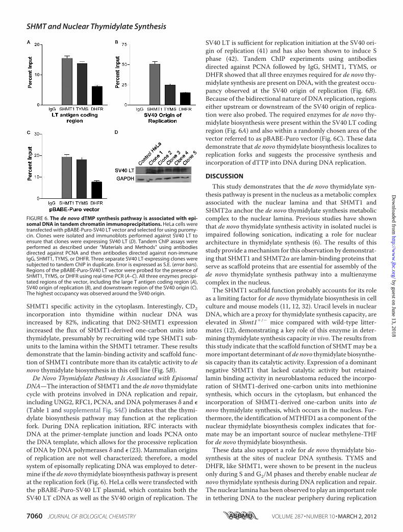
DNA—The interaction of SHMT1 and the de novo thymidylatecycle with proteins involved in DNA replication and repair,including UNG2, RFC1, PCNA, and DNA polymerases � and �(Table 1 and supplemental Fig. S4E) indicates that the thymi-dylate biosynthesis pathway may function at the replicationfork. During DNA replication initiation, RFC interacts withDNA at the primer-template junction and loads PCNA ontothe DNA template, which allows for the processive replicationof DNA by DNA polymerases � and � (23). Mammalian originsof replication are not well characterized; therefore, a modelsystem of episomally replicating DNA was employed to deter-mine if the de novo thymidylate biosynthesis pathway is presentat the replication fork (Fig. 6). HeLa cells were transfected withthe pBABE-Puro-SV40 LT plasmid, which contains both theSV40 LT cDNA as well as the SV40 origin of replication. The
SV40 LT is sufficient for replication initiation at the SV40 ori-gin of replication (41) and has also been shown to induce Sphase (42). Tandem ChIP experiments using antibodiesdirected against PCNA followed by IgG, SHMT1, TYMS, orDHFR showed that all three enzymes required for de novo thy-midylate synthesis are present on DNA, with the greatest occu-pancy observed at the SV40 origin of replication (Fig. 6B).Because of the bidirectional nature of DNA replication, regionseither upstream or downstream of the SV40 origin of replica-tion were also probed. The required enzymes for de novo thy-midylate biosynthesis were present within the SV40 LT codingregion (Fig. 6A) and also within a randomly chosen area of thevector referred to as pBABE-Puro vector (Fig. 6C). These datademonstrate that de novo thymidylate biosynthesis localizes toreplication forks and suggests the processive synthesis andincorporation of dTTP into DNA during DNA replication.
DISCUSSION
This study demonstrates that the de novo thymidylate syn-thesis pathway is present in the nucleus as ametabolic complexassociated with the nuclear lamina and that SHMT1 andSHMT2� anchor the de novo thymidylate synthesis metaboliccomplex to the nuclear lamina. Previous studies have shownthat de novo thymidylate synthesis activity in isolated nuclei isimpaired following sonication, indicating a role for nucleararchitecture in thymidylate synthesis (6). The results of thisstudy provide amechanism for this observation by demonstrat-ing that SHMT1 and SHMT2� are lamin-binding proteins thatserve as scaffold proteins that are essential for assembly of thede novo thymidylate synthesis pathway into a multienzymecomplex in the nucleus.The SHMT1 scaffold function probably accounts for its role
as a limiting factor for de novo thymidylate biosynthesis in cellculture and mouse models (11, 12, 32). Uracil levels in nuclearDNA, which are a proxy for thymidylate synthesis capacity, areelevated in Shmt1�/� mice compared with wild-type litter-mates (12), demonstrating a key role of this enzyme in deter-mining thymidylate synthesis capacity in vivo. The results fromthis study indicate that the scaffold function of SHMTmay be amore important determinant ofde novo thymidylate biosynthe-sis capacity than its catalytic activity. Expression of a dominantnegative SHMT1 that lacked catalytic activity but retainedlamin binding activity in neuroblastoma reduced the incorpo-ration of SHMT1-derived one-carbon units into methioninesynthesis, which occurs in the cytoplasm, but enhanced theincorporation of SHMT1-derived one-carbon units into denovo thymidylate synthesis, which occurs in the nucleus. Fur-thermore, the identification ofMTHFD1 as a component of thenuclear thymidylate biosynthesis complex indicates that for-mate may be an important source of nuclear methylene-THFfor de novo thymidylate biosynthesis.These data also support a role for de novo thymidylate bio-
synthesis at the sites of nuclear DNA synthesis. TYMS andDHFR, like SHMT1, were shown to be present in the nucleusonly during S and G2/M phases and thereby enable nuclear denovo thymidylate synthesis during DNA replication and repair.The nuclear lamina has been observed to play an important rolein tethering DNA to the nuclear periphery during replication
FIGURE 6. The de novo dTMP synthesis pathway is associated with epi-somal DNA in tandem chromatin immunoprecipitations. HeLa cells weretransfected with pBABE-Puro-SV40 LT vector and selected for using puromy-cin. Clones were isolated and immunoblots performed against SV40 LT toensure that clones were expressing SV40 LT (D). Tandem ChIP assays wereperformed as described under “Materials and Methods” using antibodiesdirected against PCNA and then antibodies directed against non-immuneIgG, SHMT1, TYMS, or DHFR. Three separate SV40 LT-expressing clones weresubjected to tandem ChIP in duplicate. Error is expressed as S.E. (error bars).Regions of the pBABE-Puro-SV40 LT vector were probed for the presence ofSHMT1, TYMS, or DHFR using real-time PCR (A–C). All three enzymes precipi-tated regions of the vector, including the large T antigen coding region (A),SV40 origin of replication (B), and downstream region of the SV40 origin (C).The highest occupancy was observed around the SV40 origin.
SHMT and Nuclear Thymidylate Synthesis
7060 JOURNAL OF BIOLOGICAL CHEMISTRY VOLUME 287 • NUMBER 10 • MARCH 2, 2012
by guest on June 13, 2018http://w
ww
.jbc.org/D
ownloaded from

and repair, and in this study, it is shown to support the forma-tion of the de novo thymidylate synthesis multienzyme com-plex. The results of this study also indicate that de novo thymi-dylate biosynthesis occurs at the sites of DNA synthesis and isassociated with the DNA replication machinery. SHMT1 wasshown to interactwith PCNA,RFC1, and theDNApolymerases� and �, and PCNAwas shown to interact with SHMT1, TYMS,and DHFR. SHMT1, TYMS, and DHFR proteins are associatedwith replicating DNA and are enriched at sites of DNA replica-tion initiation.Thymidine nucleotides are unique from the other nucleo-
tides required for DNA replication. They are not essential dur-ing DNA replication, because dUTP can be incorporated intoDNA in lieu of dTTP. Furthermore, although all other nucleo-tide synthesis occurs in the cytoplasm, we have shown that thede novo thymidylate biosynthesis pathway localizes to thenucleus and that its presence in the nucleus is essential to pre-vent uracil accumulation in nuclear DNA (10). The finding thatSHMT1 is an essential scaffold for the formation of the de novothymidylate biosynthesis multicomplex accounts for the sensi-tivity of Shmt�/� mice to increased uracil misincorporation inDNA, increased risk of intestinal cancer (26), and neural tubedefects (25). Furthermore, the nuclear compartmentation of denovo thymidylate biosynthesis at the replication fork may per-mit the regulation of dUTP incorporation into DNA, asopposed to its misincorporation in DNA, for the regulation oftranscriptional networks and biological pathways, as has beendemonstrated in Drosophila (33, 43).
REFERENCES1. Samsonoff, W. A., Reston, J., McKee, M., O’Connor, B., Galivan, J., Maley,
G., andMaley, F. (1997) Intracellular location of thymidylate synthase andits state of phosphorylation. J. Biol. Chem. 272, 13281–13285
2. Anderson, D. D., Quintero, C. M., and Stover, P. J. (2011) Identification ofa de novo thymidylate biosynthesis pathway inmammalianmitochondria.Proc. Natl. Acad. Sci. U.S.A. 108, 15163–15168
3. Blount, B. C., Mack, M. M., Wehr, C. M., MacGregor, J. T., Hiatt, R. A.,Wang, G., Wickramasinghe, S. N., Everson, R. B., and Ames, B. N. (1997)Folate deficiency causes uracil misincorporation into human DNA andchromosome breakage. Implications for cancer and neuronal damage.Proc. Natl. Acad. Sci. U.S.A. 94, 3290–3295
4. Fox, J. T., and Stover, P. J. (2008) Folate-mediated one-carbonmetabolism.Vitam. Horm. 79, 1–44
5. Tibbetts, A. S., and Appling, D. R. (2010) Compartmentalization of mam-malian folate-mediated one-carbon metabolism. Annu. Rev. Nutr. 30,57–81
6. Anderson, D. D., and Stover, P. J. (2009) SHMT1 and SHMT2 are func-tionally redundant in nuclear de novo thymidylate biosynthesis. PLoS One4, e5839
7. Anderson, D. D., Woeller, C. F., and Stover, P. J. (2007) Small ubiquitin-like modifier-1 (SUMO-1) modification of thymidylate synthase and di-hydrofolate reductase. Clin. Chem. Lab. Med. 45, 1760–1763
8. Woeller, C. F., Anderson, D. D., Szebenyi, D. M., and Stover, P. J. (2007)Evidence for small ubiquitin-like modifier-dependent nuclear import ofthe thymidylate biosynthesis pathway. J. Biol. Chem. 282, 17623–17631
9. Fox, J. T., Shin, W. K., Caudill, M. A., and Stover, P. J. (2009) A UV-responsive internal ribosome entry site enhances serine hydroxymethyl-transferase 1 expression for DNA damage repair. J. Biol. Chem. 284,31097–31108
10. Macfarlane, A. J., Anderson, D. D., Flodby, P., Perry, C. A., Allen, R. H.,Stabler, S. P., and Stover, P. J. (2011) Nuclear localization of de novothymidylate biosynthesis pathway is required to prevent uracil accumula-tion in DNA. J. Biol. Chem. 286, 44015–44022
11. Oppenheim, E. W., Adelman, C., Liu, X., and Stover, P. J. (2001) Heavychain ferritin enhances serine hydroxymethyltransferase expression andde novo thymidine biosynthesis. J. Biol. Chem. 276, 19855–19861
12. MacFarlane, A. J., Liu, X., Perry, C. A., Flodby, P., Allen, R. H., Stabler, S. P.,and Stover, P. J. (2008) Cytoplasmic serine hydroxymethyltransferase reg-ulates the metabolic partitioning of methylenetetrahydrofolate but is notessential in mice. J. Biol. Chem. 283, 25846–25853
13. Garrow, T. A., Brenner, A. A., Whitehead, V. M., Chen, X. N., Duncan,R. G., Korenberg, J. R., and Shane, B. (1993) Cloning of human cDNAsencoding mitochondrial and cytosolic serine hydroxymethyltransferasesand chromosomal localization. J. Biol. Chem. 268, 11910–11916
14. Stover, P. J., Chen, L.H., Suh, J. R., Stover, D.M., Keyomarsi, K., and Shane,B. (1997) Molecular cloning, characterization, and regulation of the hu-man mitochondrial serine hydroxymethyltransferase gene. J. Biol. Chem.272, 1842–1848
15. Girgis, S., Nasrallah, I.M., Suh, J. R., Oppenheim, E., Zanetti, K. A.,Mastri,M. G., and Stover, P. J. (1998) Molecular cloning, characterization andalternative splicing of the human cytoplasmic serine hydroxymethyltrans-ferase gene. Gene 210, 315–324
16. An, S., Kumar, R., Sheets, E. D., and Benkovic, S. J. (2008) Reversiblecompartmentalization of de novo purine biosynthetic complexes in livingcells. Science 320, 103–106
17. Field, M. S., Anderson, D. D., and Stover, P. J. (2011) Mthfs is an essentialgene in mice and a component of the purinosome. Front. Genet. 2, 1–13
18. An, S., Kyoung, M., Allen, J. J., Shokat, K. M., and Benkovic, S. J. (2010)Dynamic regulation of a metabolic multienzyme complex by protein ki-nase CK2. J. Biol. Chem. 285, 11093–11099
19. An, S., Deng, Y., Tomsho, J. W., Kyoung, M., and Benkovic, S. J. (2010)Microtubule-assisted mechanism for functional metabolic macromolec-ular complex formation. Proc. Natl. Acad. Sci. U.S.A. 107, 12872–12876
20. Prem veer Reddy, G., and Pardee, A. B. (1980) Multienzyme complex formetabolic channeling in mammalian DNA replication. Proc. Natl. Acad.Sci. U.S.A. 77, 3312–3316
21. Noguchi, H., Prem veer Reddy, G., and Pardee, A. B. (1983) Rapid incor-poration of label from ribonucleoside disphosphates into DNA by a cell-free high molecular weight fraction from animal cell nuclei. Cell 32,443–451
22. Li, S., Armstrong, C. M., Bertin, N., Ge, H., Milstein, S., Boxem, M., Vida-lain, P. O., Han, J. D., Chesneau, A., Hao, T., Goldberg, D. S., Li, N., Mar-tinez, M., Rual, J. F., Lamesch, P., Xu, L., Tewari, M., Wong, S. L., Zhang,L. V., Berriz, G. F., Jacotot, L., Vaglio, P., Reboul, J., Hirozane-Kishikawa,T., Li, Q., Gabel, H. W., Elewa, A., Baumgartner, B., Rose, D. J., Yu, H.,Bosak, S., Sequerra, R., Fraser, A., Mango, S. E., Saxton,W.M., Strome, S.,Van Den Heuvel, S., Piano, F., Vandenhaute, J., Sardet, C., Gerstein, M.,Doucette-Stamm, L., Gunsalus, K. C., Harper, J. W., Cusick, M. E., Roth,F. P., Hill, D. E., and Vidal, M. (2004) Amap of the interactome network ofthe metazoan C. elegans. Science 303, 540–543
23. Moldovan, G. L., Pfander, B., and Jentsch, S. (2007) PCNA, the maestro ofthe replication fork. Cell 129, 665–679
24. Ulrich, H. D. (2009) Regulating post-translational modifications of theeukaryotic replication clamp PCNA. DNA Repair 8, 461–469
25. Beaudin, A. E., Abarinov, E. V., Noden, D.M., Perry, C. A., Chu, S., Stabler,S. P., Allen, R. H., and Stover, P. J. (2011) Shmt1 and de novo thymidylatebiosynthesis underlie folate-responsive neural tube defects in mice.Am. J.Clin. Nutr. 93, 789–798
26. Macfarlane, A. J., Perry, C. A., McEntee, M. F., Lin, D. M., and Stover, P. J.(2011) Shmt1 heterozygosity impairs folate-dependent thymidylate syn-thesis capacity and modifies risk of Apc(min)-mediated intestinal cancerrisk. Cancer Res. 71, 2098–2107
27. Girgis, S., Suh, J. R., Jolivet, J., and Stover, P. J. (1997) 5-Formyltetrahydro-folate regulates homocysteine remethylation in human neuroblastoma.J. Biol. Chem. 272, 4729–4734
28. Anguera,M. C., Field,M. S., Perry, C., Ghandour, H., Chiang, E. P., Selhub,J., Shane, B., and Stover, P. J. (2006) Regulation of folate-mediated one-carbon metabolism by 10-formyltetrahydrofolate dehydrogenase. J. Biol.Chem. 281, 18335–18342
29. Zanetti, K. A., and Stover, P. J. (2003) Pyridoxal phosphate inhibits dy-namic subunit interchange among serine hydroxymethyltransferase te-
SHMT and Nuclear Thymidylate Synthesis
MARCH 2, 2012 • VOLUME 287 • NUMBER 10 JOURNAL OF BIOLOGICAL CHEMISTRY 7061
by guest on June 13, 2018http://w
ww
.jbc.org/D
ownloaded from

tramers. J. Biol. Chem. 278, 10142–1014930. Huang da, W., Sherman, B. T., and Lempicki, R. A. (2009) Systematic and
integrative analysis of large gene lists using DAVID bioinformatics re-sources. Nat. Protoc. 4, 44–57
31. Huang da, W., Sherman, B. T., and Lempicki, R. A. (2009) Bioinformaticsenrichment tools. Paths toward the comprehensive functional analysis oflarge gene lists. Nucleic Acids Res. 37, 1–13
32. Herbig, K., Chiang, E. P., Lee, L. R., Hills, J., Shane, B., and Stover, P. J.(2002) Cytoplasmic serine hydroxymethyltransferase mediates competi-tion between folate-dependent deoxyribonucleotide and S-adenosylme-thionine biosyntheses. J. Biol. Chem. 277, 38381–38389
33. Deutsch, W. A., and Spiering, A. L. (1982) A new pathway expressedduring a distinct stage of Drosophila development for the removal ofdUMP residues in DNA. J. Biol. Chem. 257, 3366–3368
34. Hozak, P., Hassan, A. B., Jackson, D. A., and Cook, P. R. (1993) Visualiza-tion of replication factories attached to nucleoskeleton. Cell 73, 361–373
35. Spann, T. P., Moir, R. D., Goldman, A. E., Stick, R., and Goldman, R. D.(1997) Disruption of nuclear lamin organization alters the distribution ofreplication factors and inhibits DNA synthesis. J. Cell Biol. 136,1201–1212
36. Moir, R. D., Spann, T. P., Herrmann, H., and Goldman, R. D. (2000) Dis-ruption of nuclear lamin organization blocks the elongation phase ofDNAreplication. J. Cell Biol. 149, 1179–1192
37. Shumaker, D. K., Solimando, L., Sengupta, K., Shimi, T., Adam, S. A.,Grunwald, A., Strelkov, S. V., Aebi, U., Cardoso,M.C., andGoldman, R.D.
(2008) The highly conserved nuclear lamin Ig-fold binds to PCNA. Its rolein DNA replication. J. Cell Biol. 181, 269–280
38. Luka, Z., Moss, F., Loukachevitch, L. V., Bornhop, D. J., and Wagner, C.(2011) Histone demethylase LSD1 is a folate-binding protein. Biochemis-try 50, 4750–4756
39. Reed,M. C., Nijhout, H. F., Neuhouser,M. L., Gregory, J. F., 3rd, Shane, B.,James, S. J., Boynton, A., and Ulrich, C. M. (2006) A mathematical modelgives insights into nutritional and genetic aspects of folate-mediated one-carbon metabolism. J. Nutr. 136, 2653–2661
40. Szebenyi, D.M., Liu, X., Kriksunov, I. A., Stover, P. J., andThiel, D. J. (2000)Structure of a murine cytoplasmic serine hydroxymethyltransferase qui-nonoid ternary complex. Evidence for asymmetric obligate dimers. Bio-chemistry 39, 13313–13323
41. Abdel-Aziz, W., Malkas, L. H., Wills, P. W., and Hickey, R. J. (2003) TheDNA synthesome. Its potential as a novel in vitro model system for study-ing S phase-specific anticancer agents. Crit. Rev. Oncol. Hematol. 48,19–33
42. Ahuja, D., Saenz-Robles, M. T., and Pipas, J. M. (2005) SV40 large T anti-gen targets multiple cellular pathways to elicit cellular transformation.Oncogene 24, 7729–7745
43. Bekesi, A., Pukancsik, M., Muha, V., Zagyva, I., Leveles, I., Hunyadi-Gu-lyas, E., Klement, E., Medzihradszky, K. F., Kele, Z., Erdei, A., Felfoldi, F.,Konya, E., and Vertessy, B. G. (2007) A novel fruitfly protein under devel-opmental control degrades uracil-DNA. Biochem. Biophys. Res. Commun.355, 643–648
SHMT and Nuclear Thymidylate Synthesis
7062 JOURNAL OF BIOLOGICAL CHEMISTRY VOLUME 287 • NUMBER 10 • MARCH 2, 2012
by guest on June 13, 2018http://w
ww
.jbc.org/D
ownloaded from

StoverDonald D. Anderson, Collynn F. Woeller, En-Pei Chiang, Barry Shane and Patrick J.
Pathway to Nuclear Lamina for DNA Synthesis Thymidylate Synthesisde NovoSerine Hydroxymethyltransferase Anchors
doi: 10.1074/jbc.M111.333120 originally published online January 10, 20122012, 287:7051-7062.J. Biol. Chem.
10.1074/jbc.M111.333120Access the most updated version of this article at doi:
Alerts:
When a correction for this article is posted•
When this article is cited•
to choose from all of JBC's e-mail alertsClick here
Supplemental material:
http://www.jbc.org/content/suppl/2012/01/10/M111.333120.DC1
http://www.jbc.org/content/287/10/7051.full.html#ref-list-1
This article cites 43 references, 26 of which can be accessed free at
by guest on June 13, 2018http://w
ww
.jbc.org/D
ownloaded from