serum 25-hydroxyvitamin d levels throughout pregnancy: a
TRANSCRIPT
7:5 698–707Y Agudelo-Zapata et al. Serum 25OHD levels throughout pregnancy
RESEARCH
Serum 25-hydroxyvitamin D levels throughout pregnancy: a longitudinal study in healthy and preeclamptic pregnant womenYessica Agudelo-Zapata1, Luis Miguel Maldonado-Acosta1, Héctor Fabio Sandoval-Alzate2, Natalia Elvira Poveda3, María Fernanda Garcés3, Jonathan Alexander Cortés-Vásquez3, Andrés Felipe Linares-Vaca3, Carlos Alejandro Mancera-Rodríguez3, Shahar Alexandra Perea-Ariza3, Karen Yuliana Ramírez-Iriarte3, Camilo Andrés Castro-Saldarriaga3, Juan Manuel Arteaga-Diaz1, Roberto Franco-Vega1, Edith Ángel–Müller4, Arturo José Parada-Baños4 and Jorge E Caminos3
1Division of Endocrinology, Department of Internal Medicine, School of Medicine, Universidad Nacional de Colombia, Bogotá, Colombia2Department of Human Nutrition, School of Medicine, Universidad Nacional de Colombia, Bogotá, Colombia3Department of Physiology, School of Medicine, Universidad Nacional de Colombia, Bogotá, Colombia4Department of Obstetrics and Gynecology, School of Medicine, Universidad Nacional de Colombia, Bogotá, Colombia
Correspondence should be addressed to J E Caminos: [email protected]
Abstract
Background: Worldwide there is a high prevalence of 25-hydroxyvitamin D (25OHD)
deficiency and has been associated with adverse outcomes during pregnancy.
Objective: This is a nested, case–control study in a longitudinal cohort to compare the
serum 25OHD levels and other biomarkers throughout pregnancy in a group of 20
preeclamptic women and 61 healthy pregnant women. An additional group of 29 healthy
non-pregnant women were also studied during the two phases of the menstrual cycle.
Results: Mean 25OHD levels in non-pregnant women were 31.9 ng/mL and 34.9 ng/mL
during follicular and luteal phase, respectively (P < 0.01). Mean serum 25OHD levels
in healthy pregnant women were 26.5, 30.1 and 31.9 ng/mL, at first, second and third
trimester, respectively (P < 0.001). The first trimester levels of 25OHD were lower than
those of healthy non-pregnant women (P < 0.001), showing a significant recovery at third
trimester. In the group of healthy pregnant women, the 25OHD levels were 25.7 ng/mL
and 27.2 ng/mL at 3 and 6 months postpartum, respectively; both values were lower than
those observed in the non-pregnant women (P < 0.001). In preeclamptic women, 25OHD
serum levels were similar to those of healthy pregnant women; nevertheless, they
remained almost unchanged throughout pregnancy.
Conclusion: There were no significant differences between healthy and preeclamptic
pregnant women in terms of 25OHD levels throughout the pregnancy. Serum 25OHD
levels in non-pregnant women were higher during luteal phase compared with follicular
phase. The 25OHD levels of non-pregnant women tended to be higher than those of
pregnant women.
Introduction
There is a high prevalence of 25-hydroxyvitamin D (25OHD) deficiency during pregnancy (1). In the United States, a prevalence of 33% has been reported, while in
countries such as Turkey, the prevalence reaches 90% (1). 25OHD deficiency has been associated with adverse outcomes such as preeclampsia (2). This association is
-18-0055
Key Words
f 25 hydroxyvitamin D
f pregnancy
f preeclampsia
f menstrual cycle
Endocrine Connections(2018) 7, 698–707
ID: 18-0055
7 5
This work is licensed under a Creative Commons Attribution-NonCommercial-NoDerivatives 4.0 International License.
https://doi.org/10.1530/EC-18-0055http://www.endocrineconnections.org ©2018 The authors
Published by Bioscientifica Ltd
Downloaded from Bioscientifica.com at 02/28/2022 11:47:44PMvia free access
Y Agudelo-Zapata et al. Serum 25OHD levels throughout pregnancy
6997:5
supported by the fact that pregnant women with low serum 25OHD levels could have an increased activity of the renin–angiotensin–aldosterone system; an increase in pro-inflammatory cytokines and a decrease in the calcium absorption (3). Additionally, leptin has been shown to be a hormone that rises significantly during preeclampsia, and several authors in observational studies have described a correlation between elevated serum leptin levels and low serum 25OHD levels in the general population (4).
Moreover, Zabul et al. proposes that vitamin D3 might acts as a competitive inhibitor of placental cytochrome P450scc preventing the production of lipid peroxides or excess of progesterone synthesis, both of which may contribute to the etiopathogenesis of preeclampsia (5).
The World Health Organization does not recommend the universal screening of serum 25OHD levels during pregnancy but in their vitamin D supplementation guidelines, they suggest that when the 25OHD levels are less than 20 ng/mL, supplementation should be provided (6).
The objective of this study is to compare the serum 25OHD levels in a group of healthy pregnant women during the first, second and third trimester of pregnancy, as well as 3 and 6 months postpartum, with a group of pregnant women with preeclampsia. In addition, serum 25OHD levels were determined in healthy non-pregnant women in the follicular and luteal phases of the menstrual cycle. Serum 25OHD levels were correlated with anthropometric, biochemical and hormonal parameters, such as leptin.
Methods
Patients and study design
This study was approved by the Ethics Committee of the School of Medicine of the Universidad Nacional de Colombia and was conducted according to the Helsinki Declaration. Patient’s recruitment was performed by the Department of Obstetrics and Gynecology and Physiology of the School of Medicine of the Universidad Nacional de Colombia at the Hospital de Engativá in Bogotá Colombia, between January 2012 and December 2014. All patients in the study signed an informed consent.
In this nested case–control study in a longitudinal cohort of 450 pregnant women, 81 were included. In this cohort, pregnant women were recruited before the 14 weeks of gestation and the inclusion criteria were normal single pregnancy, BMI between 18 kg/m2 and 30 kg/m2.
The exclusion criteria were the presence of any important medical comorbidity such as history of diabetes mellitus, gestational diabetes, past or present history of thyroid disease, vascular disease, chronic hypertension, renal disease, polycystic ovary syndrome, use of corticosteroids, β-blockers, β-agonists and other drugs that could affect metabolism.
During the pregnancy (since 14th week of gestational age), they were provided supplements as recommended by the Colombian Guidelines: carbonate calcium 600 mg 2 times per day, folic acid 1 mg per day and ferrous sulfate 300 mg per day (7).
Additionally, in the present study, the calcium supplementation provided to pregnant women was not adjusted to calcium status or nutrition and the calcium intake of the pregnant women was unknown.
Twenty pregnant women out of 450 developed preeclampsia (cases) and 61 healthy pregnant women were selected as controls.
The group of healthy pregnant women had no medical or obstetric complications during pregnancy or delivery and was followed during the three trimesters of pregnancy as well as 3 and 6 months postpartum.
Preeclampsia was defined by the criteria of the American College of Gynecology and Obstetrics, these criteria include features such as blood pressure ≥140/90 mmHg, examined twice with an interval of at least 4 h after 20 weeks of gestation (in a woman with previously normal blood pressure) and proteinuria >300 mg by collection of 24-h urine (8). Severity features include thrombocytopenia, impaired liver function, progressive renal insufficiency, pulmonary edema, new-onset cerebral or visual disturbances (8). In this study, pregnant women with preeclampsia without severity features were studied.
Additionally, a group of 29 healthy non-pregnant women with regular menstrual cycles, without use of hormonal contraceptive methods, medications or supplements was recruited from the Universidad Nacional de Colombia. Serum 25OHD levels and other biochemical parameters were studied during the follicular (days 3–5) and luteal (days 20–22) phases of their menstrual cycle.
Laboratory test
Blood samples were collected in 5.0 mL BD Vacutainer serum tubes from a forearm vein between 07:00 h and 08:00 h after an overnight fasting. The blood samples were centrifuged at 3500 g for 10 min at 4°C. Serum samples
This work is licensed under a Creative Commons Attribution-NonCommercial-NoDerivatives 4.0 International License.
https://doi.org/10.1530/EC-18-0055http://www.endocrineconnections.org ©2018 The authors
Published by Bioscientifica Ltd
Downloaded from Bioscientifica.com at 02/28/2022 11:47:44PMvia free access
Y Agudelo-Zapata et al. Serum 25OHD levels throughout pregnancy
7007:5
were immediately transferred and stored in aliquots at −80°C until biochemical and hormonal analysis were performed. The 25OHD-deficient pregnant women were not treated because the biochemical analyses were carried out long after the patients completed their pregnancy.
Serum leptin levels measurement
Serum leptin levels were quantified using a commercially available Human ELISA Kit, according to the manufacturer’s recommendations (Invitrogen, catalog number KAC2281). The intra-assay and inter-assay variation coefficients were <3.9 and <5.3%, respectively, and the sensitivity of the assay was less than <3.5 pg/mL and the leptin ELISA kit shows no cross – reactivity with other cytokines tested. Also, serum leptin concentrations were analyzed in duplicate for each patient.
Serum 25OHD levels measurement
Serum 25OHD levels were quantified by chemiluminescence immunoassay by commercially available kit, according to the manufacturer’s protocols (LIAISON 25 OH Vitamin D TOTAL Assay REF 310600).
Statistical analysis
Statistical analysis was performed using software Python with Scipy and Matplotlib libraries. The normality of the variables was defined by Kolmogorov–Smirnov test. Data with normal distribution were reported as mean ± standard deviation (s.d.), while data with non-normal distribution were reported as median and interquartile range (IQR). Statistical differences between the anthropometric, hormonal and biochemical variables were established with non-parametric tests: Friedman test, Wilcoxon or Mann–Whitney.
We also studied the correlation between 25OHD and leptin levels. Univariate correlations were assessed in the groups by Spearman’s partial correlation coefficient. Statistical values are presented as *P < 0.05, **P < 0.01 and ***P < 0.001. Statistical significance was assumed with P < 0.05.
Results
Non-pregnant women
Twenty-nine non-pregnant women were characterized. Samples were obtained during the follicular and luteal
phases of the menstrual cycle and confirmed by measuring serum progesterone that was statistically different between the two phases (Table 1). Non-pregnant women had normal weight and normal blood pressure, and serum leptin levels were lower during the follicular phase of the menstrual cycle as shown in (Table 1).
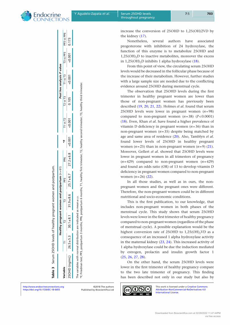
The serum 25OHD levels changed during the menstrual cycle and were lower in the follicular phase compared with the luteal phase (31.9 ng/mL (±6.0) vs 34.9 ng/mL (±8.6), P < 0.01) (Table 1).
Healthy pregnant women
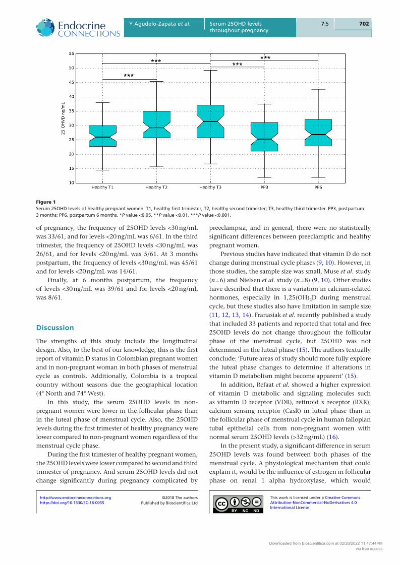
Sixty-one healthy pregnant women were characterized (Table 2). Mean serum 25OHD levels in healthy pregnant women were 26.5 ng/mL (±5.27), 30.1 ng/mL (±8.1) and 31.9 ng/mL (±8.6), at first, second and third trimester, respectively (P < 0.001) (Table 2).
When serum 25OHD levels were compared between the first and second trimester of healthy pregnancy, a statistically significant increase was observed (26.5 ng/mL (±5.3) vs 30.1 ng/mL (±8.1), P < 0.001), as well as a statistically significant increase was observed between the first and third trimester (26.5 ng/mL (±5.3) vs 31.9 ng/mL (±8.6), P < 0.001) (Fig. 1 and Table 3).
Also, serum 25OHD levels were lower in the first trimester of the healthy pregnant women compared to the levels of non-pregnant women in both phases of menstrual cycle (Fig. 2 and Table 4).
Table 1 Non-pregnant women characteristics.
Non-pregnant women characteristics
Follicular phase
(n = 29)
Luteal phase
(n = 29)
P value
Age (years) median (IQR)
21 (18–22) NA NA
BMI (kg/m2) mean ± s.d.
21.9 ± 2.0 NA NA
SBP (mmHg) mean ± s.d.
108 ± 7 NA NA
DBP (mmHg) mean ± s.d.
72 ± 7 NA NA
Progesterone (ng/mL) median (IQR)
0.5 (0.3–0.7) 7.1 (4.7–11.1) <0.001
Leptin (ng/mL) median (IQR)
15.7 (14.9–17.5) 22.3 (21.0–27.3) <0.001
25OHVD (ng/mL) mean ± s.d.
31.9 ± 6.0 34.9 ± 8.6 <0.01
The non-normal variables indicated as median (IQR), the variables with normal distribution are indicated as mean ± s.d.DBP, diastolic blood pressure; NA, non-applicable; SBP, systolic blood pressure.
This work is licensed under a Creative Commons Attribution-NonCommercial-NoDerivatives 4.0 International License.
https://doi.org/10.1530/EC-18-0055http://www.endocrineconnections.org ©2018 The authors
Published by Bioscientifica Ltd
Downloaded from Bioscientifica.com at 02/28/2022 11:47:44PMvia free access
Y Agudelo-Zapata et al. Serum 25OHD levels throughout pregnancy
7017:5
Three months after delivery, the mean serum 25OHD levels was 25.7 ng/mL (±6.7), and at 6 months was 27.2 ng/mL (±6.8). Postpartum levels were significantly lower compared to those of the third trimester (Fig. 1 and Table 3).
No statistically significant differences were found when serum 25OHD levels of the first trimester of healthy pregnancy were compared with the levels of 3 months postpartum (26.5 ng/mL (±5.3) vs 25.7 ng/mL (±6.7) P = 0.41), neither differences with the levels 6 months postpartum (26.5 ng/mL (±5.3) vs 27.2 ng/mL (±6.8) P = 0.50).
And regardless of the menstrual cycle phase, serum 25OHD levels were higher in non-pregnant women compared to 3 and 6 months postpartum (Fig. 2 and Table 4).
Pregnant women with preeclampsia
Twenty pregnant women who developed preeclampsia (without severity features) were studied. These patients had significant differences in blood pressure, BMI and leptin levels compared to healthy pregnant women (Table 2).
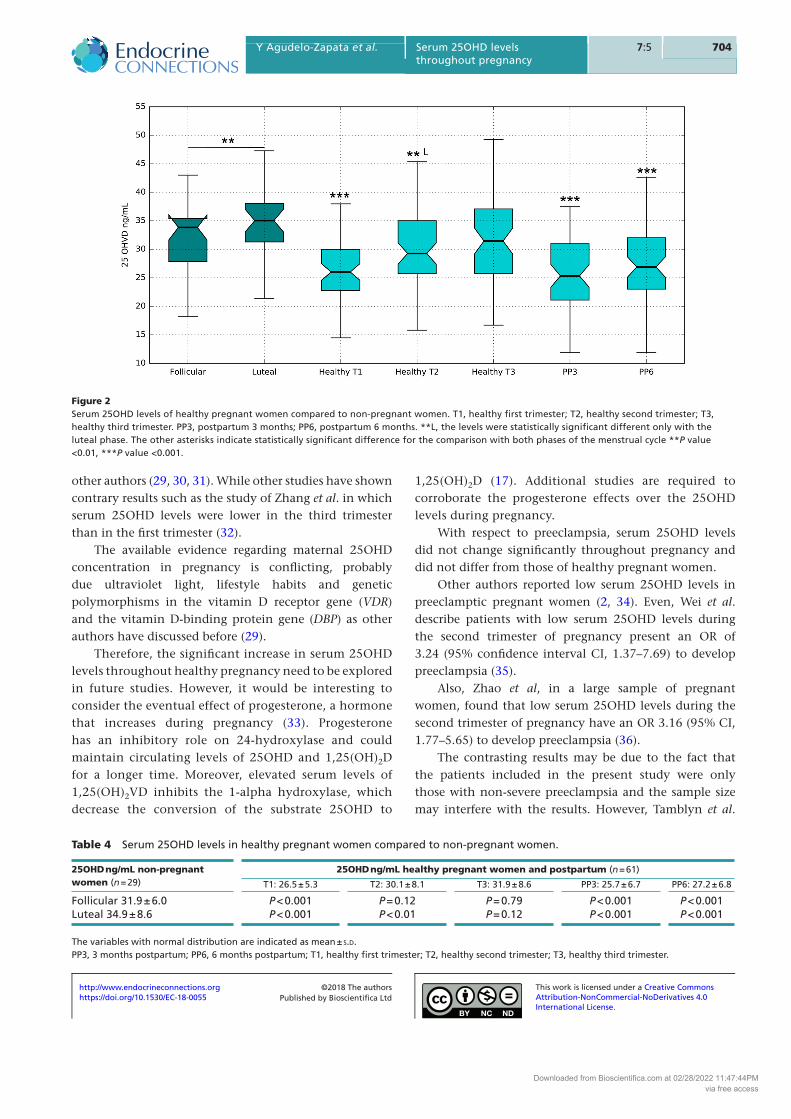
Mean serum 25OHD levels in preeclamptic women were 28.3 ng/mL (±8.1), 29.6 ng/mL (±6.4) and 29.3 ng/mL (±7.5) during the first, second and third trimester, respectively (P = 1) (Table 2).
Healthy pregnancy compared with preeclampsia
There were no statistically significant differences between serum 25OHD levels of preeclamptic pregnant women compared to healthy pregnant women during the first, second and third trimester of pregnancy (Fig. 3 and Table 1).
Correlation with leptin
During healthy pregnancy, there were no statistically significant correlations between leptin and 25OHD, and the correlations indexes were first trimester (r = 0.073, P = 0.574), second (r = −0.126; P = 0.333) and third trimester (r = −0.177; P = 0.173).
There were no statistically significant correlations between 25OHD and leptin in women who developed preeclampsia neither in the first (r = −0.101; P = 0.670), second (r = −0.043; P = 0.857) nor third trimester (r = 0.2804; P = 0.231).
Frequencies of 25OHD levels <20 ng/mL and <30 ng/mL in healthy pregnancy
In the first trimester of healthy pregnancy, the frequency of 25OHD levels <30 ng/mL was 46/61, and for levels <20 ng/mL was 6/61. During the second healthy trimester Ta
ble
2
Clin
ical
par
amet
ers
and
25O
HV
D s
eru
m le
vels
of
hea
lth
y p
reg
nan
t w
om
en a
nd
pre
ecla
mp
tic
wo
men
.
Vari
ab
leFi
rst
trim
est
er
(T1)
Seco
nd
tri
mest
er
(T2)
Thir
d t
rim
est
er
(T3)
Hea
lth
y p
reg
nan
tPr
eecl
amti
cP
valu
eH
ealt
hy
pre
gn
ant
Pree
clam
tic
P va
lue
Hea
lth
y p
reg
nan
tPr
eecl
amti
cP
valu
e
Ag
e (y
ears
) m
edia
n (
IQR
)23
(19
–30)
20 (
18–2
6)0.
134
NA
NA
NA
NA
NA
NA
Ges
tati
on
al a
ge
(wee
ks)
med
ian
(IQ
R)
12.1
(11
–13.
6)12
.3 (
11.2
–13.
4)0.
969
24 (
24.2
–24.
6)24
.3 (
24.1
–24.
5)0.
434
34.5
(34
.2–3
5.4)
34.9
(34
.2–3
5.5)
0.39
5
BM
I (kg
/m2 )
mea
n ±
s.d.
22.6
± 2
.422
.6 ±
2.4
<0.
001
24.4
± 2
.526
.4 ±
2.8
<0.
001
26.3
± 2
.329
.4 ±
2.7
<0.
001
SBP
(mm
Hg
) m
ean
± s.
d.
95 ±
595
± 5
<0.
001
93 ±
10
104
± 9
<0.
001
98 ±
10
109
± 1
<0.
001
DB
P (m
mH
g)
mea
n ±
s.d.
60 (
60–6
4)68
(60
–70)
<0.
0560
(58
–60)
63 (
60–7
0)<
0.01
62 (
60–7
0)60
(60
–70)
0.40
45Le
pti
n (
ng
/mL)
med
ian
(I
QR
)20
.5 (
17.2
–21.
5)32
.6 (
28.9
–38.
0)<
0.00
125
.4 (
20.2
–27.
3)61
.1 (
51.0
–77.
7)<
0.00
135
.1 (
28.6
–42.
3)87
.9 (
24.3
–18.
5)<
0.00
1
25-O
HV
D (
ng
/mL)
m
ean
± s.
d.
26.5
± 5
.3
28.3
± 8
.1
0.56
1 30
.1 ±
8.1
29
.6 ±
6.4
0.
952
31.9
± 8
.6
29.3
± 7
.5
0.35
8
The
no
n-n
orm
al v
aria
ble
s in
dic
ated
as
med
ian
(IQ
R).
Th
e va
riab
les
wit
h n
orm
al d
istr
ibu
tio
n a
re in
dic
ated
as
mea
n ±
s.d.
DB
P, d
iast
olic
blo
od
pre
ssu
re, N
A, n
on
-ap
plic
able
; SB
P, s
ysto
lic b
loo
d p
ress
ure
.
This work is licensed under a Creative Commons Attribution-NonCommercial-NoDerivatives 4.0 International License.
https://doi.org/10.1530/EC-18-0055http://www.endocrineconnections.org ©2018 The authors
Published by Bioscientifica Ltd
Downloaded from Bioscientifica.com at 02/28/2022 11:47:44PMvia free access
Y Agudelo-Zapata et al. Serum 25OHD levels throughout pregnancy
7027:5
of pregnancy, the frequency of 25OHD levels <30 ng/mL was 33/61, and for levels <20 ng/mL was 6/61. In the third trimester, the frequency of 25OHD levels <30 ng/mL was 26/61, and for levels <20 ng/mL was 5/61. At 3 months postpartum, the frequency of levels <30 ng/mL was 45/61 and for levels <20 ng/mL was 14/61.
Finally, at 6 months postpartum, the frequency of levels <30 ng/mL was 39/61 and for levels <20 ng/mL was 8/61.
Discussion
The strengths of this study include the longitudinal design. Also, to the best of our knowledge, this is the first report of vitamin D status in Colombian pregnant women and in non-pregnant woman in both phases of menstrual cycle as controls. Additionally, Colombia is a tropical country without seasons due the geographical location (4° North and 74° West).
In this study, the serum 25OHD levels in non-pregnant women were lower in the follicular phase than in the luteal phase of menstrual cycle. Also, the 25OHD levels during the first trimester of healthy pregnancy were lower compared to non-pregnant women regardless of the menstrual cycle phase.
During the first trimester of healthy pregnant women, the 25OHD levels were lower compared to second and third trimester of pregnancy. And serum 25OHD levels did not change significantly during pregnancy complicated by
preeclampsia, and in general, there were no statistically significant differences between preeclamptic and healthy pregnant women.
Previous studies have indicated that vitamin D do not change during menstrual cycle phases (9, 10). However, in those studies, the sample size was small, Muse et al. study (n = 6) and Nielsen et al. study (n = 8) (9, 10). Other studies have described that there is a variation in calcium-related hormones, especially in 1,25(OH)2D during menstrual cycle, but these studies also have limitation in sample size (11, 12, 13, 14). Franasiak et al. recently published a study that included 33 patients and reported that total and free 25OHD levels do not change throughout the follicular phase of the menstrual cycle, but 25OHD was not determined in the luteal phase (15). The authors textually conclude: ‘Future areas of study should more fully explore the luteal phase changes to determine if alterations in vitamin D metabolism might become apparent’ (15).
In addition, Refaat et al. showed a higher expression of vitamin D metabolic and signaling molecules such as vitamin D receptor (VDR), retinoid x receptor (RXR), calcium sensing receptor (CasR) in luteal phase than in the follicular phase of menstrual cycle in human fallopian tubal epithelial cells from non-pregnant women with normal serum 25OHD levels (>32 ng/mL) (16).
In the present study, a significant difference in serum 25OHD levels was found between both phases of the menstrual cycle. A physiological mechanism that could explain it, would be the influence of estrogen in follicular phase on renal 1 alpha hydroxylase, which would
Figure 1Serum 25OHD levels of healthy pregnant women. T1, healthy first trimester; T2, healthy second trimester; T3, healthy third trimester. PP3, postpartum 3 months; PP6, postpartum 6 months. *P value <0.05, **P value <0.01, ***P value <0.001.
This work is licensed under a Creative Commons Attribution-NonCommercial-NoDerivatives 4.0 International License.
https://doi.org/10.1530/EC-18-0055http://www.endocrineconnections.org ©2018 The authors
Published by Bioscientifica Ltd
Downloaded from Bioscientifica.com at 02/28/2022 11:47:44PMvia free access
Y Agudelo-Zapata et al. Serum 25OHD levels throughout pregnancy
7037:5
increase the conversion of 25OHD to 1,25(OH)2VD by the kidney (17).
Nonetheless, several authors have associated progesterone with inhibition of 24 hydroxylase, the function of this enzyme is to metabolize 25OHD and 1,25(OH)2D to inactive metabolites, moreover the excess in 1,25(OH)2D inhibits 1 alpha hydroxylase (18).
From this point of view, the circulating serum 25OHD levels would be decreased in the follicular phase because of the increase of their metabolism. However, further studies with a large sample size are needed due to the conflicting evidence around 25OHD during menstrual cycle.
The observation that 25OHD levels during the first trimester in healthy pregnant women are lower than those of non-pregnant women has previously been described (19, 20, 21, 22). Holmes et al. found that serum 25OHD levels were lower in pregnant women (n = 98) compared to non-pregnant women (n = 38) (P < 0.0001) (18). Even, Khan et al. have found a higher prevalence of vitamin D deficiency in pregnant women (n = 36) than in non-pregnant women (n = 35) despite being matched by age and same area of residence (20). Also, Tamblyn et al. found lower levels of 25OHD in healthy pregnant women (n = 25) than in non-pregnant women (n = 9) (21). Moreover, Gellert et al. showed that 25OHD levels were lower in pregnant women in all trimesters of pregnancy (n = 429) compared to non-pregnant women (n = 429) and found an odds ratio (OR) of 13 to develop vitamin D deficiency in pregnant women compared to non-pregnant women (n = 26) (22).
In all those studies, as well as in ours, the non-pregnant women and the pregnant ones were different. Therefore, the non-pregnant women could be in different nutritional and socio-economic conditions.
This is the first publication, to our knowledge, that includes non-pregnant women in both phases of the menstrual cycle. This study shows that serum 25OHD levels were lower in the first trimester of healthy pregnancy compared to non-pregnant women (regardless of the phase of menstrual cycle). A possible explanation would be the highest conversion rate of 25OHD to 1,25(OH)2VD as a consequence of an increased 1 alpha hydroxylase activity in the maternal kidney (23, 24). This increased activity of 1 alpha hydroxylase could be due the induction mediated by estrogen, prolactin and insulin growth factor 1 (25, 26, 27, 28).
On the other hand, the serum 25OHD levels were lower in the first trimester of healthy pregnancy compare to the two late trimester of pregnancy. This finding has been described not only in our study but also by Ta
ble
3
Seru
m 2
5OH
D le
vels
of
hea
lth
y p
reg
nan
t w
om
en a
nd
po
stp
artu
m.
Vari
ab
leH
ealt
hy p
reg
nan
t w
om
en
an
d p
ost
part
um
(n
= 6
1) *F
Po
st h
oc
an
aly
sis
P v
alu
e
T1T2
T3PP
3PP
6T1
vs
T2T2
vs
T3T1
vs
T3T3
vs
PP3
PP3
vs P
P6
25O
HD
(n
g/m
L)26
.5 ±
5.3
30.1
± 8
.131
.9 ±
8.6
25.7
± 6
.727
.2 ±
6.8
<0.
001
<0.
001
0.10
5<
0.00
1<
0.00
10.
173
The
vari
able
s w
ith
no
rmal
dis
trib
uti
on
are
ind
icat
ed a
s m
ean
± s.
d.
*F, F
ried
man
tes
t; P
P3, p
ost
par
tum
3 m
on
ths;
PP6
, po
stp
artu
m 6
mo
nth
s; T
1, h
ealt
hy
firs
t tr
imes
ter;
T2,
hea
lth
y se
con
d t
rim
este
r; T
3, h
ealt
hy
thir
d t
rim
este
r.
This work is licensed under a Creative Commons Attribution-NonCommercial-NoDerivatives 4.0 International License.
https://doi.org/10.1530/EC-18-0055http://www.endocrineconnections.org ©2018 The authors
Published by Bioscientifica Ltd
Downloaded from Bioscientifica.com at 02/28/2022 11:47:44PMvia free access
Y Agudelo-Zapata et al. Serum 25OHD levels throughout pregnancy
7047:5
other authors (29, 30, 31). While other studies have shown contrary results such as the study of Zhang et al. in which serum 25OHD levels were lower in the third trimester than in the first trimester (32).
The available evidence regarding maternal 25OHD concentration in pregnancy is conflicting, probably due ultraviolet light, lifestyle habits and genetic polymorphisms in the vitamin D receptor gene (VDR) and the vitamin D-binding protein gene (DBP) as other authors have discussed before (29).
Therefore, the significant increase in serum 25OHD levels throughout healthy pregnancy need to be explored in future studies. However, it would be interesting to consider the eventual effect of progesterone, a hormone that increases during pregnancy (33). Progesterone has an inhibitory role on 24-hydroxylase and could maintain circulating levels of 25OHD and 1,25(OH)2D for a longer time. Moreover, elevated serum levels of 1,25(OH)2VD inhibits the 1-alpha hydroxylase, which decrease the conversion of the substrate 25OHD to
1,25(OH)2D (17). Additional studies are required to corroborate the progesterone effects over the 25OHD levels during pregnancy.
With respect to preeclampsia, serum 25OHD levels did not change significantly throughout pregnancy and did not differ from those of healthy pregnant women.
Other authors reported low serum 25OHD levels in preeclamptic pregnant women (2, 34). Even, Wei et al. describe patients with low serum 25OHD levels during the second trimester of pregnancy present an OR of 3.24 (95% confidence interval CI, 1.37–7.69) to develop preeclampsia (35).
Also, Zhao et al, in a large sample of pregnant women, found that low serum 25OHD levels during the second trimester of pregnancy have an OR 3.16 (95% CI, 1.77–5.65) to develop preeclampsia (36).
The contrasting results may be due to the fact that the patients included in the present study were only those with non-severe preeclampsia and the sample size may interfere with the results. However, Tamblyn et al.
Figure 2Serum 25OHD levels of healthy pregnant women compared to non-pregnant women. T1, healthy first trimester; T2, healthy second trimester; T3, healthy third trimester. PP3, postpartum 3 months; PP6, postpartum 6 months. **L, the levels were statistically significant different only with the luteal phase. The other asterisks indicate statistically significant difference for the comparison with both phases of the menstrual cycle **P value <0.01, ***P value <0.001.
Table 4 Serum 25OHD levels in healthy pregnant women compared to non-pregnant women.
25OHD ng/mL non-pregnant women (n = 29)
25OHD ng/mL healthy pregnant women and postpartum (n = 61)
T1: 26.5 ± 5.3 T2: 30.1 ± 8.1 T3: 31.9 ± 8.6 PP3: 25.7 ± 6.7 PP6: 27.2 ± 6.8
Follicular 31.9 ± 6.0 P < 0.001 P = 0.12 P = 0.79 P < 0.001 P < 0.001Luteal 34.9 ± 8.6 P < 0.001 P < 0.01 P = 0.12 P < 0.001 P < 0.001
The variables with normal distribution are indicated as mean ± s.d.PP3, 3 months postpartum; PP6, 6 months postpartum; T1, healthy first trimester; T2, healthy second trimester; T3, healthy third trimester.
This work is licensed under a Creative Commons Attribution-NonCommercial-NoDerivatives 4.0 International License.
https://doi.org/10.1530/EC-18-0055http://www.endocrineconnections.org ©2018 The authors
Published by Bioscientifica Ltd
Downloaded from Bioscientifica.com at 02/28/2022 11:47:44PMvia free access
Y Agudelo-Zapata et al. Serum 25OHD levels throughout pregnancy
7057:5
in a similar sample size also did not find a statistically significant difference in serum 25OHD levels between healthy and preeclamptic pregnant women (23).
Finally, the frequency in the first trimester of pregnancy of serum 25OHD levels lower than 30 ng/mL (Endocrine Society cutoff point for inadequate serum 25OHD levels) was 46/61. Additionally, the frequency of levels lower than 20 ng/mL (cut-off point IOM for inadequate serum 25OHD levels) was 6/61. These data are similar to those reported in Brazil by Figueiredo et al. who reported a prevalence of levels below 30 ng/mL around 70.4% and levels below 20 ng/mL of 16.1% (37).
In contrast to these results, other countries such as Turkey reported a prevalence of serum 25OHD levels below 20 ng/mL in about 90% (1).
The limitations of this study include a small sample size and lack of data about sun exposure and food intake. Those limitations may make results less generalizable to other population.
Conclusion
Pregnant women are a population group with a higher risk of 25OHD insufficiency. There were no significant differences between healthy and preeclamptic women in terms of 25OHD levels throughout pregnancy. Serum 25OHD levels in non-pregnant women were higher during luteal phase compared with follicular phase. Regardless
of the phase of menstrual cycle, the 25OHD levels of non-pregnant women tended to be higher than those of pregnant women. The differences were found during the first and second trimester, while there were no differences during the third trimester.
Finally, no correlation between 25OHD levels and leptin levels was found.
Declaration of interestThe authors declare that there is no conflict of interest that could be perceived as prejudicing the impartiality of the research reported.
FundingThe authors acknowledge the grant support from Dirección Nacional de Investigaciones and Vicerrectoria de Investigaciones of the Universidad Nacional de Colombia, COLCIENCIAS Cod. 110154531660.
AcknowledgementsThe authors acknowledge Dr Adarve and Dr Cohen for their comments.
References 1 De-Regil LM, Palacios C, Lombardo LK & Pena-Rosas JP. Vitamin
D supplementation for women during pregnancy. Cochrane Database of Systematic Reviews 2016 1 CD008873. (https://doi.org/10.1002/14651858.CD008873.pub3)
2 Hyppönen E, Cavadino A, Williams D, Fraser A, Vereczkey A, Fraser WD, Bánhidy F, Lawlor D & Czeizel AE. Vitamin D and pre-eclampsia: original data, systematic review and meta-analysis. Annals of Nutrition and Metabolism 2013 63 331–340. (https://doi.org/10.1159/000358338)
Figure 3Serum 25OHD levels of health pregnant women compared to preeclamptic women. T, trimester. Comparisons among preeclamptic women: Friedman test = 1, Analysis post hoc (preeclamptic T1 vs preeclamptic T2) P = 0.47; (preeclamptic T2 vs preeclamptic T3) P = 1; (preeclamptic T1 vs preeclamptic T3) P = 0.71.
This work is licensed under a Creative Commons Attribution-NonCommercial-NoDerivatives 4.0 International License.
https://doi.org/10.1530/EC-18-0055http://www.endocrineconnections.org ©2018 The authors
Published by Bioscientifica Ltd
Downloaded from Bioscientifica.com at 02/28/2022 11:47:44PMvia free access
Y Agudelo-Zapata et al. Serum 25OHD levels throughout pregnancy
7067:5
3 Barrera D, Díaz L, Noyola-Martínez N & Halhali A. Vitamin D and inflammatory cytokines in healthy and preeclamptic pregnancies. Nutrients 2015 7 6465–6490. (https://doi.org/10.3390/nu7085293)
4 Hajimohammadi M, Shab-Bidar S & Neyestani TR. Vitamin D and serum leptin: a systematic review and meta-analysis of observational studies and randomized controlled trials. European Journal of Clinical Nutrition 2017 71 1144–1153. (https://doi.org/10.1038/ejcn.2016.245)
5 Zabul P, Wozniak M, Slominski AT, Preis K, Gorska M, Korozan M, Wieruszewski J, Zmijewski MA, Zabul E, Tuckey R, et al. A proposed molecular mechanism of high-dose vitamin D3 supplementation in prevention and treatment of preeclampsia. International Journal of Molecular Sciences 2015 916 13043–13064. (https://doi.org/10.3390/ijms160613043)
6 WHO. Guideline: Vitamin D Supplementation in Pregnant Women. Geneva, Switzerland: World Health Organization, 2012. (available at: https://www.ncbi.nlm.nih.gov/books/NBK310615/)
7 Centro Nacional de Investigación en Evidencia y Tecnologías en Salud CINETS. Guía de Práctica Clínica para la prevención, detección temprana y tratamiento de las complicaciones del embarazo, parto o puerperio. 2013 – Guías No. 11-15. Bogotá. Colombia: Ministerio de Salud y Protección Social - Colciencias, 2013. (available at: https://www.minsalud.gov.co/sites/rid/Lists/BibliotecaDigital/RIDE/INEC/IETS/Guía.completa.Embarazo.Parto.2013.pdf)
8 American College of Obstetricians and Gynecologist; Task Force on Hypertension in pregnancy. Hypertension in pregnancy. Report of the American College of Obstetricians and Gynecologists’ Task Force on Hypertension in Pregnancy. Obstetrics and Gynecology 2013 122 1122–1131. (https://doi.org/10.1097/01.AOG.0000437382.03963.88)
9 Muse KN, Manolagas SC, Deftos LJ, Alexander N & Yen SS. Calcium-regulating hormones across the menstrual cycle. Journal of Clinical Endocrinology and Metabolism 1986 62 1313–1316. (https://doi.org/10.1210/jcem-62-6-1313)
10 Nielsen HK, Brixen K, Bouillon R & Mosekilde L. Changes in biochemical markers of osteoblastic activity during the menstrual cycle. Journal of Clinical Endocrinology and Metabolism 1990 70 1431–1437. (https://doi.org/10.1210/jcem-70-5-1431)
11 Thys-Jacobs S & Alvir MJ. Calcium-regulating hormones across the menstrual cycle: evidence of a secondary hyperparathyroidism in women with PMS. Journal of Clinical Endocrinology and Metabolism 1995 80 2227–2232. (https://doi.org/10.1210/jcem.80.7.7608284)
12 Gray TK, McAdoo T, Hatley L, Lester GE & Thierry M. Fluctuation of serum concentration of 1,25-dihydroxyvitamin D3 during the menstrual cycle. American Journal of Obstetrics and Gynecology 1982 144 880–884.
13 Tjellesen L, Christiansen C, Hummer L & Larsen NE. Unchanged biochemical indices of bone turnover despite fluctuations in 1,25-dihydroxyvitamin D during the menstrual cycle. Acta Endocrinologica 1983 102 476–480. (https://doi.org/10.1530/acta.0.1020476)
14 Baran DT, Whyte MP, Haussler MR, Deftos LJ, Slatopolsky E & Avioli LV. Effect of the menstrual cycle on calcium-regulating hormones in the normal young woman. Journal of Clinical Endocrinology and Metabolism 1980 50 377–379. (https://doi.org/10.1210/jcem-50-2-377)
15 Franasiak JM, Wang X, Molinaro TA, Green K, Sun W, Werner MD, Juneau CR & Scott RT. Free vitamin D does not vary through the follicular phase of the menstrual cycle. Endocrine 2016 53 322–326. (https://doi.org/10.1007/s12020-016-0946-1)
16 Refaat B, Ahmad J, Idris S, Kamfar F, Ashshi AM, Batwa SA & Malibary FA. Characterisation of vitamin D-related molecules and calcium-sensing receptor in human Fallopian tube during the menstrual cycle and in ectopic pregnancy. Cell and Tissue Research 2017 368 201–213. (https://doi.org/10.1007/s00441-016-2519-2)
17 Pitkin RM, Reynolds WA, Williams GA & Hargis GK. Calcium-regulating hormones during the menstrual cycle. Journal of Clinical Endocrinology and Metabolism 1978 47 626–632. (https://doi.org/10.1210/jcem-47-3-626)
18 Rodriguez GC, Turbov J, Rosales R, Yoo J, Hunn J, Zappia KJ, Lund K, Barry CP, Rodriguez IV, Pike JW, et al. Progestins inhibit calcitriol-induced CYP24A1 and synergistically inhibit ovarian cancer cell viability : an opportunity for chemoprevention. Gynecologic Oncology 2016 143 159–167. (https://doi.org/10.1016/j.ygyno.2016.04.022)
19 Holmes VA, Barnes MS, Alexander HD, McFaul P & Wallace JM. Vitamin D deficiency and insufficiency in pregnant women: a longitudinal study. British Journal of Nutrition 2009 102 876–881. (https://doi.org/10.1017/S0007114509297236)
20 Khan FR, Ahmad T, Hussain R & Bhutta ZA. Vitamin D status and periodontal disease among pregnant and non-pregnant women in an underdeveloped district of Pakistan. Journal of International Society of Preventive and Community Dentistry 2016 6 234–239. (https://doi.org/10.4103/2231-0762.183111)
21 Tamblyn J, Jenkinson C, Larner D, Hewison M & Kilby M. Serum and urine vitamin D metabolite analysis in early preeclampsia. Endocrine Connections 2017 7 199–210. (https://doi.org/10.1530/EC-17-0308)
22 Gellert S, Ströhle A, Bitterlich N & Hahn A. Higher prevalence of vitamin D deficiency in German pregnant women compared to non-pregnant women. Archives of Gynecology and Obstetrics 2017 296 43–51. (https://doi.org/10.1007/s00404-017-4398-5)
23 Tamblyn JA, Susarla R, Jenkinson C, Jeffery LE, Ohizua O, Chun RF, Chan SY, Kilby MD & Hewison M. Dysregulation of maternal and placental vitamin D metabolism in preeclampsia. Placenta 2017 50 70–77. (https://doi.org/10.1016/j.placenta.2016.12.019)
24 Kumar R, Cohen WR, Silva P & Epstein FH. Elevated 1,25-dihydroxyvitamin D plasma levels in normal human pregnancy and lactation. Journal of Clinical Investigation 1979 63 342–324. (https://doi.org/10.1172/JCI109308)
25 Boass A, Garner SC, Schultz VL & Toverud SU. Regulation of serum calcitriol by serum ionized calcium in rats during pregnancy and lactation. Journal of Bone and Mineral Research 1997 12 909–914. (https://doi.org/10.1359/jbmr.1997.12.6.909)
26 Spanos E, Barrett D, MacIntyre I, Pike JW, Safilian EF & Haussler MR. Effect of growth hormone on vitamin D metabolism. Nature 1978 273 246–247.
27 Spanos E, Colston KW, Evans IM, Galante LS, Macauley SJ & Macintyre I. Effect of prolactin on vitamin D metabolism. Molecular and Cellular Endocrinology 1976 5 163–167.
28 Garabedian M, Holick MF, Deluca HF & Boyle IT. Control of 25-hydroxycholecalciferol metabolism by parathyroid glands. PNAS 1972 69 1673–1676.
29 Lundqvist A, Sandstrom H, Stenlund H, Johansson I & Hultdin J. Vitamin D status during pregnancy: a longitudinal study in Swedish Women from early pregnancy to seven months postpartum. PLoS ONE 2016 11 e0150385. (https://doi.org/10.1371/journal.pone.0150385)
30 Milman N, Hvas A-M & Bergholt T. Vitamin D status during normal pregnancy and postpartum. A longitudinal study in 141 Danish women. Journal of Perinatal Medicine 2011 40 57–61. (https://doi.org/10.1515/JPM.2011.120)
31 Choi R, Kim S, Yoo H, Cho YY, Kim SW, Chung JH, Oh SY & Lee SY. High prevalence of vitamin D deficiency in pregnant korean women: the first trimester and the winter season as risk factors for vitamin D deficiency. Nutrients 2015 7 3427–3448. (https://doi.org/10.3390/nu7053427)
32 Zhang JY, Lucey AJ, Horgan R, Kenny LC & Kiely M. Impact of pregnancy on vitamin D status: a longitudinal study. British Journal of Nutrition 2014 112 1081–1087. (https://doi.org/10.1017/S0007114514001883)
33 Batra S, Bengtsson LP, Grundsell H & Sjöberg NO. Levels of free and protein-bound progesterone in plasma during late pregnancy. Journal
This work is licensed under a Creative Commons Attribution-NonCommercial-NoDerivatives 4.0 International License.
https://doi.org/10.1530/EC-18-0055http://www.endocrineconnections.org ©2018 The authors
Published by Bioscientifica Ltd
Downloaded from Bioscientifica.com at 02/28/2022 11:47:44PMvia free access
Y Agudelo-Zapata et al. Serum 25OHD levels throughout pregnancy
7077:5
of Clinical Endocrinology and Metabolism 1976 42 1041–1047. (https://doi.org/10.1210/jcem-42-6-1041)
34 Baca KM, Simhan HN, Platt RW & Bodnar LM. Low maternal 25-hydroxyvitamin D concentration increases the risk of severe and mild preeclampsia. Annals of Epidemiology 2016 26 853.e1–857.e1. (https://doi.org/10.1016/j.annepidem.2016.09.015)
35 Wei SQ, Audibert F, Hidiroglou N, Sarafin K, Julien P, Wu Y, Luo ZC & Fraser WD. Longitudinal vitamin D status in pregnancy and the risk of pre-eclampsia. BJOG 2012 119 832–839. (https://doi.org/10.1111/j.1471-0528.2012.03307.x)
36 Zhao X, Fang R, Yu R, Chen D, Zhao J & Xiao J. Maternal vitamin D status in the late second trimester and the risk of severe preeclampsia in Southeastern China. Nutrients 2017 9 E138. (https://doi.org/10.3390/nu9020138)
37 Figueiredo ACC, Cocate PG, Adegboye ARA, Franco-Sena AB, Farias DR, de Castro MBT, Brito A, Allen LH, Mokhtar RR, Holick MF, et al. Changes in plasma concentrations of 25-hydroxyvitamin D and 1,25-dihydroxyvitamin D during pregnancy: a Brazilian cohort. European Journal of Nutrition 2017 57 1059–1072. (https://doi.org/10.1007/s00394-017-1389-z)
Received in final form 29 March 2018Accepted 17 April 2018Accepted Preprint published online 17 April 2018
This work is licensed under a Creative Commons Attribution-NonCommercial-NoDerivatives 4.0 International License.
https://doi.org/10.1530/EC-18-0055http://www.endocrineconnections.org ©2018 The authors
Published by Bioscientifica Ltd
Downloaded from Bioscientifica.com at 02/28/2022 11:47:44PMvia free access