setaria viborg, 1795 in south africa. ii. setaria scalprum (von
TRANSCRIPT
Oncierstepoort Journal of Veterinary Research, 70:7- 13 (2003)
Studies on the genus Setaria Viborg, 1795 in South Africa. II. Setaria scalprum (Von Linstow, 1908) and Setaria saegeri (Le Van Hoa, 1961)
R. WATERMEYER', J. BOOMKER' and J.F. PUTIERIL2
ABSTRACT
WATERMEYER, R., BOOMKER, J. & PUTTER ILL, J.F. 2003. Studies on the genus Setaria Viborg, 1795 In Sooth Africa. II. Setaria scalprum (Von Unstow, 1908) and Setaria saegeri (Le Van Hoa, 1961). Onderstepoort Journal of Veterinary Research, 70:7-13
Setaria scalprum (Von Unstow, 1908) and Setaria saegeri (Le Van Hoa, 1961) are dosely related filarid species that occur in the smaller antelope of Africa. Material previously coIleded from common duiker, Sylvicapra grimmia, steenbok, Raphicerus campestris and grysbok, Raphicerus melanotis, from several localities in the northem and eastem regions of South Africa was re-examined and measurements of adult worms were compared with those given in the original descriptions of the species. Scanning electron microscopy of the anterior and posterior regions of the female worms confirmed the validity of the two species. Differences in the postdeirid, ventral transverse bands and bosses on the cuticle of the male specimens were also observed. Setaria saegeri in common duiker and grysbok is a new parasite record for these hosts.
Keywords: Filarids, Setaria saegeri, Setaria scalprum, South African wildlife
INTRODUCTION
Various Setaria species have been recorded from wildlife in Africa, amongst which are Setaria scalprum (Von linstow, 1908), described from staenbok, Raphicerus campestris and Setaria saegeri (La Van Hoa, 1961) from common duiker, Sylvicapra grimmia. These two filarids are very similar and the possibility of misidentification of either species is possible, as stated by Le Van Hoa (1961) and Desset (1966). The description by Yeh (1959) of S. scalprum was, amongst others, from steenbok from Grahamstown, Eastem Cape Province, South Africa. Setaria saegeri has been recorded from com-
Department of Veterinary Tropical Diseases, University of Pretoria, Private Bag X04, Onderstepoort, 0110 South Africa
2 Division of Pathology, Electron Microscope Unit, Onderstepoort Veterinary Institute, Private Bag X05, Onderstepoort, 0110 South Africa
Accepted for publication 7 October 2002-Editor
man duiker in other parts of Africa, but no records of this species in South Africa could be found in the literature. Detailed scanning electron microscopic (SEM) studies on the morphological characteristics of Setaria species have been conducted by various workers, but mainly on species that occur in domesticated animals. There appears to be a paucity of information regarding SEM studies of Setaria spp. of wild animals of Africa.
During surveys of the helminth parasites of South African wildlife, specimens of the genus Setaria were collected from various artiodactylids, including common duiker (Boomker, Du Plessis & Boomker 1983; Boomker, Horak & De Vos 1986; Boomker, Keep & Horak 1987; Boomker, Horak & Maclvor 1989) and grysbok, Raphicerus melanotis, (Boomker et a/. 1989). Ortlepp (1961) and Boomker et al. (1987, 1989) recorded Setaria cae/um and S. scalprum from common duiker. Subsequent records of Setaria spp. from common duiker (Boomker et at.
7
Studies on the genus Setaria Viborg, 1795 in South A/rica. II
1983. 1986) and grysbok (Boomker ef al. 1989) were identified only to the genus level. The SEM appearance, together with the measurements of S. scalprum and S. saegeri, are presented here and compared with the findings of Yeh (1959), Le Van Hoa (1961), and Desset (1966).
MATERIALS AND METHODS
The specimens originated from the helminthological collection of one of us (J.B.), currently housed in the Department of Veterinary Tropical Diseases, University of Pretoria, as well as the National Collection of Animal Helminths (NCAH). The following specimens were examined: 25 females from common duiker from the Weza Forest Nature Reserve, KwaZu lu-Natal (WFNR); three males from the same host from Uitenhage, Eastern Cape Province; one female from grysbok, tram the latter locality; five males and 19 females from common duiker from the farm Riekerts Laager, Limpopo Province; len females from common duiker from Malelane, Kruger National Park (KNP); one female from steenbok from Nwashitsumbe, KNP; five males and 28 females from steenbok from Stel1enbosch, Western Cape Province; two males and ten females from common duiker from Ondangua, Namibia, NCAH No. S2246 and three females from common duiker from Ndumu Nature Reserve, KwaZulu-Natal, NCAH No. 52336.
The nematodes were cleared in lactophenol and examined under a compound microscope using differential interference illumination. Measurements were obtained from camera lucida drawings of the material , and are given in millimetres in Tables 1 and 2. Specimens for scanning electron microscopy, which had been preserved in 70 % ethanol, had a segment of the head and tail removed prior to further processing. Samples were re-hydrated to distilled water after which they were post-fixed in 4% glutaraldehyde and 1% osmium tetroxide. Specimens were dehydrated through graded ethanol and critical point dried from 100% ethanol to carbon dioxide. Each dried head and tail segment was individually mounted onto a conical brass SEM viewing stub and sputter coated with gold. Samples were viewed and micrographed using a Hitachi S-2500 scanning electron microscope operated at 8 kV.
RESULTS AND DISCUSSION
Of the 112 helminths examined, 65 out of n from common duiker from the different localities as well
8
as the one nematode from grysbok from Uitenhage proved to be S. saegeri. The 12 specimens from common duiker from Ondangua, Namibia were not suitable for identification. The 34 helminths from steenbok from the KNP and Stellenbosch were identified as S. scalprum.
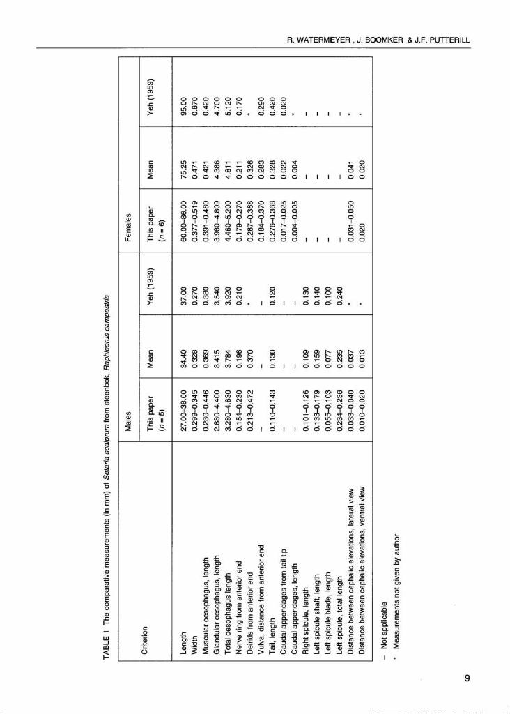
In Tables 1 and 2 the measurements of S. scalprum and S. saegeri are compared with those of Yeh (1959) and Le Van Hoa (1961), respectively.
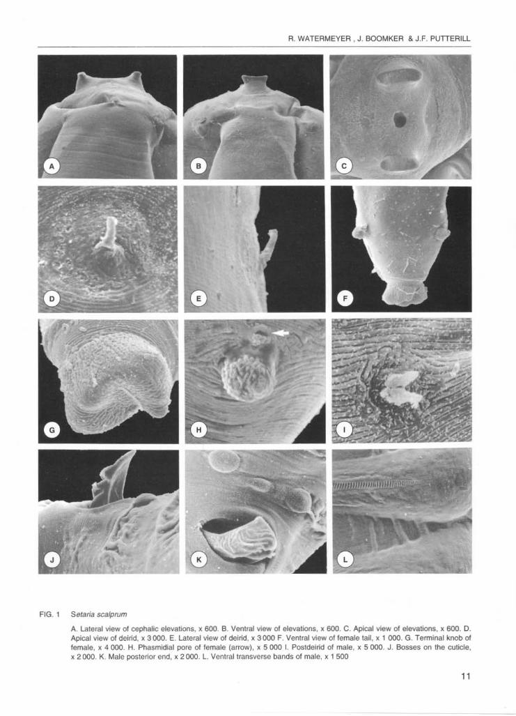
Setaria scalprum from steenbok examined in this study generally corresponded closely to the description of Yeh (1959) , except for being slightly smaller. The majority of measurements of the South African S. saegeri were similar to those recorded by Le Van Hoa (1961) . However, the following differences were apparent: female specimens had a shorter oesophagus, the deirids were closer to the anterior end, and the tail was longer than that recorded by Le Van Hoa (1961) . The scanning electron microscopical appearance of S. scalprum and S. saegeri are presented in Fig. 1 and 2.
It was evident that the two species are morphologically distinct, as recorded by Le Van Hoa (1961) and Desset (1966) . In lateral view, the cephalic elevations of S. scalprum are short, stub·like projections whereas those of S. saegeri are prominent, long, tooth-like structures (Fig. 1 A and 2A). In ventral view the elevations of S. scalprum have a wide base with the sides tapering down gradually towards the peribuccal crown whereas those of S. saegeri have a rounded base with the sides almost parallel (Fig. 18 and 28). In apical view, the mouth opening of S. scalprum is round and is surrounded by a slightly raised peribuccal crown. The elevations are elongated in a do'rsoventral plane. The mouth of S. saegeri is oval in shape and the elevations are smaller and rounded with diverging tips (Fig. 1C and 2C). Furthermore, the deirids of S. scalprum are single whereas those of S. saegeri are double (Fig. 10, E and 20, E) and the caudal appendages of S. scalprum are larger than those of S. saegeri (Fig. 1 F and 2F). Yeh (1959) described the terminal button on the posterior extremity of S. scalprum females as a small knob, often ill-defined and Le Van Hoa (1961) only mentions the tail length of S. sa8geri. Desset (1966) describes the terminal buttons of the two species as more or less tuberculated in S. saegeri and smooth in S. scalprum and her illustrations are the same as those of Le Van Hoa (1961). This is contradictory to our findings in that the terminal button of S. scalprum is bluntly rounded and bifid whereas that of S. saegeri
Ma
les
Crr
terlo
n T
his
pape
r M
ean
(n::
5)
Leng
th
27
.00
-38
.00
34
.40
Wid
th
0.2
99
-0.3
45
0.
328
Mus
cula
r oe
soph
agus
, len
gth
0.23
0--0
.446
0.
369
Gla
ndul
ar o
esop
hagu
s, l
engt
h 2.
880-
---4.
400
3.41
5
Tot
al o
esop
hagu
s le
ngth
3
.260
----4
.630
3.
764
Ner
ve r
ing
from
ant
erio
r en
d 0
.15
4-0
.23
0
0.19
6
Dei
rids
from
anl
erio
r en
d 0
.21
3-0
.47
2
0.37
0
Vul
va.
dist
ance
Iro
m a
nter
ior
end
--
Tai
l, le
ngth
0
.11
0-0
.14
3
0.13
0
Cau
dal a
ppen
dage
s Ir
om t
ail l
ip
--
Cau
dal a
ppen
dage
s, l
engt
h -
-R
ight
spi
cule
, le
ngth
0
.10
1-0
.12
6
0.10
9
Left
spic
ule
shaf
t, le
ngth
0.
133-
{).1
79
0.15
9
Left
spic
ule
blad
e,
leng
th
0.0
55
-0.1
03
0.
077
Left
spic
ule
, tot
al l
engt
h 0
.23
4-0
.23
6
0.23
5
Dis
tanc
e be
twee
n ce
phal
ic e
leva
tions
, la
tera
l vi
ew
0
.03
3-0
.04
0
0.03
7
Dis
tanc
e be
twee
n ce
phal
ic e
leva
tions
, ve
ntra
l vi
ew
0.0
10
-0.0
20
0.
013
'"
Fem
ales
Yeh
(19
59)
Thi
s pa
per
(n::
6)
37.0
0 6
0.0
0-e
6.0
0
0.27
0 0
.37
7-0
.51
9
0.38
0 0
.39
1-0
.48
0
3.54
0 3
.980
--4.
609
3.92
0 4.
460-
-5.2
00
0.21
0 0
.17
9-0
.27
0
· 0
.26
7-0
.36
6
-0
.16
4-0
.37
0
0.12
0 0
.27
6-0
.36
6
-0
.01
7-0
.02
5
-0
.00
4-0
.00
5
0.1
30
-0
.140
-
0.1
00
-0
.240
-
· 0
.03
1-0
.05
0
· 0
.020
Mea
n
75.2
5
0.47
1
0.42
1
4.36
6
4.61
1
0.21
1
0.32
6
0.26
3
0.32
8
0.02
2
0.00
4
- - - - 0.0
41
0.0
20
Yeh
(19
59)
95.0
0
0.67
0
0.42
0
4.70
0
5.12
0
0.17
0
· 0.29
0
0.42
0
0.02
0
· - - - - · ·
~ ~ m
~
~ ~ m
~
~ ~ ~ ~ ~ ~
~ ~ F

o
Ma
les
Cri
teri
on
Thi
s p
ap
er
Gre
y d
uik
er
(n"'
5 )
Ran
ge
Mea
n
Len
gth
26
.00
-33.
00
29.0
0
Wid
th
0.3
00
-0.3
70
0.3
30
Mus
cula
r oe
soph
agus
, le
ngth
0
.26
0-0
.500
0.
410
Gla
nd
ula
r o
eso
ph
ag
us
, le
ng
th
2.6
00
-5.1
80
4.21
0
To
la!
oe
sop
ha
gu
s le
ng
lh
3.0
50
-5.6
80
4.6
70
Ne
rve
rin
g fr
om
an
teri
or e
nd
0.
180-
(1.2
50
0.
200
Del
rlds
from
ant
erio
r en
d 0.
450-
0.53
0 0.
500
Vul
va,
dist
ance
fro
m a
nter
ior
end
--
Tal
t, le
ng
th
0.1
20
-0.1
40
0
.120
Ca
ud
al
ap
pe
nd
ag
es
fro
m t
an t
ip
--
Cau
dal
appo
ndag
es,
leng
th
--
Rig
ht s
picu
lo,
leng
th
0.10
0-0
.13
0
0.11
0
left
spi
cule
sha
ft, le
ngth
0
.18
8-0
.20
7
0.1
97
left
spi
cule
bla
de, l
engt
h 0
.06
2-0
.064
0
.063
left
spi
cule
, to
tal l
engt
h 0
.25
2-0
.26
9
0.2£
0
Dis
tanc
e be
twee
n ce
phal
ic e
leva
tions
, la
tera
l vie
w
0.0
30
-0.0
40
0
.030
Dis
tanc
e be
twee
n ce
phal
ic e
leva
tions
, ve
ntra
l vie
w
0.0
20
0.0
20
Fem
ales
Le V
an H
oa (
1961
) T
his
pape
r
Gre
y d
uik
er
Gry
sbok
(n
" I
I
30.
00
37
.00
0.2
30
0.3
90
0.37
0 0.
510
4.7
00
5.1
50
5.07
0 5.
660
0.1
80
0.17
0
0.47
5 0.
340
-0.
250
0.14
0 0.
320
-0.
020
-0.
002
0.1
20
-·
-·
-0.
230
-
· 0
.040
· 0
.020
Gre
y du
ikar
(n
" 1
9)
Ran
ge
Mea
n
44.0
0-7
6.00
6
6.2
8
0.3
60
-0.5
60
0.4
70
0.2
80-0
.590
0
.47
0
4.57
0-5.
660
5.2
30
S.l
ElO
--e.1
40
5.71
0
0.1
80-0
.200
0.
180
O.3
1O-C
.450
0.
340
0.2
30-0
.350
0.
280
0.3
00-0
.480
0
.380
0.0
20-0
.030
0
.020
0.0
02-0
.003
0.
002
--
--
--
--
0.0
40
-0.0
50
0.
040
0.02
0 0
.02
0
La V
an H
oe
(19
61)
Gre
y do
lker
70.
00
0.47
0
0.48
0 6
.120
6.6
00
0.20
0
o.sOO
0,
300
0.2
30
· · - - - - · ·
i- g " • ~ ~ o • ~ • I ~ o· I ~ ~ =
R. WATERMEYER , J. BOOMKER & J.F. PUTTERILL
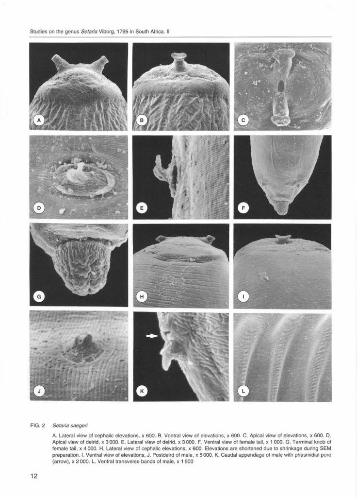
FIG . 1 Setaria scalpn;m
A. LaleraJ view of cephalic elevations, Ie 600. B. Venlral view of elevations, Ie 600. C. Apical view of elevalions. Ie 600. D. Apical view of deirid, Ie 3000. E. Lateral view 01 deirid, Ie 3000 F. Ventral view o//emale tall , Ie 1 000. G. Terminal knob oj /emale, Ie 4000. H. Phasmidial pore o/Iemale (arrow), Ie 5 000 I. Postdelrid of male. Ie 5000. J. Bosses on the cuticle, Ie 2000. K. Male posterior end, Ie 2000. l. Ventral transverse bands of male, Ie 1 500
11
Studies on ItIe genus Setaria Viborg, 1795 in South Africa. II
FIG. 2 Setaria saegeri
12
A. lateral view 01 cephalic elevations, )I; 600. 6 . Ventral view of elevations, )I; 600. C. Apical view of elevations, )I; 600. O. Apical view of deilid, )I; 3000. E. lateral view of deirid, )I; 3000. F. Ventral view of female tail , )I; 1000. G. Terminal knob of female tail , )I; 4000. H. lateral view of cephalic elevations, )I; 600. Elevations are shortened due to shrinkage during SEM preparation. I. Ventral view 01 etevations, J. Postdeird of male, )I; 5000. K. Caudal appendage 01 male with phasmidial pore (arrow), )I; 2000. l. Ventral transverse bands of male, )I; 1 500
I
I
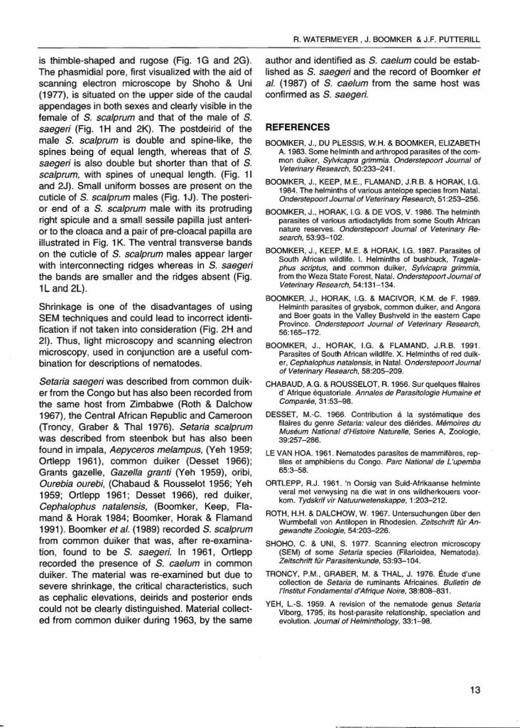
is thimble-shaped and rugose (Fig. 1G and 2G). The phasmidial pore, first visualized with the aid of scanning electron microscope by Shoho & Uni (1977), is situated on the upper side of the caudal appendages in both sexes and clearly visible in the female of S. scalprum and that of the male of S. saegeri (Fig. 1 H and 2K). The postdeirid of the male S. scalprum is double and spine-like, the spines being of equal length, whereas that of S. saegeri is also double but shorter than that of S. scalprum, with spines of unequal length. (Fig. 11 and 2J) . Small uniform bosses are present on the cuticle of S. scalprum males (Fig. 1J). The posterior end of a S. scalprum male with its protruding right spicule and a small sessile papilla just anterior to the cloaca and a pair of pre-cloacal papilla are illustrated in Fig. 1 K. The ventral transverse bands on the cuticle of S. scalprum males appear larger with interconnecting ridges whereas in S. saegeri the bands are smaller and the ridges absent (Fig. 1L and 2L).
Shrinkage is one of the disadvantages of using SEM techniques and could lead to incorrect identification if not taken into consideration (Fig. 2H and 21) . Thus, light microscopy and scanning electron microscopy, used in conjunction are a useful combination for descriptions of nematodes.
Setaria saeger; was described from common duiker from the Congo but has also been recorded from the same host from Zimbabwe (Roth & Dalchow 1967) , the Central African Republic and Cameroon (Troncy, Graber & Thai 1976). Setaria scalprum was described from steenbok but has also been found in impala, Aepyceros melampus, (Yeh 1959; Ortlepp 1961), common duiker (Desset 1966) ; Grants gazelle, Gazella granti (Yeh 1959), oribi, Ourebia ourebi, (Chabaud & Rousselot 1956; Yeh 1959; Ortlepp 1961 ; Desset 1966) , red duiker, Cephalophus natalensis, (Boomker, Keep, Flamand & Horak 1984; Boomker, Horak & Flamand 1991). Boomker et al. (1989) recorded S. scalprum from common duiker that was, after re-examination, found to be S. saegeri. In 1961 , Ortlepp recorded the presence of S. cae/um in common duiker. The material was re-examined but due to severe shrinkage, the critical characteristics, such as cephalic elevations, deirids and posterior ends could not be clearly distinguished. Material collected from common duiker during 1963, by the same
A. WATEAMEYEA , J. BOOMKER & J.F. PUTTEAlll
author and identified as S. cae/um could be established as S. saegeri and the record of Boomker et al. (1987) of S. cae/um from the same host was confirmed as S. saegeri.
REFERENCES BOOMKER, J., DU PLESSIS, W.H. & BOOMKER, ELIZABETH
A. 1983. Some helminth and arthropod parasites of the common duiker, SyMcaprs grimmia. Onderstepoort Journal of Veterinary Research, 50:233-241.
BOOMKER, J., KEEP. M.E., FlAMAND. J.R.B. & HORAK, I.G. 1984. The helminlhs of various antelope species lrom Natal. Onderstepoorf Journa/ of Veterinary Research, 51 :253-256.
BOOMKER, J .• HOAAK, I.G. & DE vos, V. 1986. The helminth parasites 01 various artiodactylids from some South African nature reserves. Onders/epoorf Journal of Veterinary Research, 53:93-102.
BOOMKER, J., KEEP, M.E. & HORAK, LG. 1987. Parasites 01 South African wildlife. I. Helminths of bushbock, Trege/aphus scrip/us, and common duiker, Sy/vicapra grimmia, from the Weza State Forest, Natal. Onderstepoorf Journal of Veterinary Research, 54:131- 134.
BOOMKER, J., HORAK. I.G. & MACIVOR, K.M. de F. 1989. Helminth parasites of grysbok, common duiker. and Angora and Boer goats In ItIe Valley Bushveld in the eastern cape Province. Dnderstepoor1 Journal of Veterinary Research, 56:165-172.
BOOMKER, J ., HOAAK, LG. & FLAMANO, J.R.B. 1991 . Parasites 01 Souttl African wildlife. X. Helminths 01 red dulker, Cephalophus natalens/s, ln Natal. Onderstepoorf Joumal of Veterinary Reseerch, 58:205-209.
CHABAUO, A.G. & ROUSSElOT, R. 1956. Sur quelques fllalres d' Afrique equatOfiale. Annales de Paf8sitoJogie Hurnaine et Comparee, 31 :53-98.
DESSET, M.-C. 1966. Contribution a. Ia systematique des filaires du genre Setaria: valeur des dierides. Memo/res du Museum National d'Histoire Nature/Ie, Series A, Zoologie. 39:257-286.
lE VAN HOA. 1961 . Nematodes parasites de mammiferes, reptiles et amphibiens du Congo. Parc National de L 'upemba 65:3-58.
ORTLEPP, R.J. 1961. 'n Oorsig van Suid-Afrikaanse helmlnte veral met verwysing na die wat in ons wildherkouers voorkom. Tydskrif vir Naluurwetenskappe, 1 :203-212.
ROTH, H.H. & DAlCHOW, W. 1967. Untersuchungen Qber den Wunnbefail von Antilopen in Ahodesien. Zeitschrift fur Angewandte Zoologie. 54:203-226.
SHOHO. C. & UNI, S. 1977. Scanning electron microscopy (SEM) of some Setaria species (Filarioidea, Nematoda). Zeitschritt frjr Parssitftnkunde, 53:93-104.
TRONCY, P.M., GAABER, M. & THAl, J. 1976. Etude d'une collection de Setaria de ruminants Africaines. Bulletin de I'Institut Fon<iamental d 'Afrlque Noire. 38:808-831.
YEH, l.-S. 1959. A revision of the nematode genus Setaria Viborg, 1795, Its host-parasite relationship, speciation and evolution. Joumal of Helminthology, 33:1-98.
13
_._-