setting peep in ards: special focus on esophageal pressure...
TRANSCRIPT

Setting PEEP in ARDS: Special focus on esophageal pressure
monitoring20/02/2015

Overview of the seminar
• Introduction
• Optimal strategy for PEEP
• Meta analysis comparing higher vs lower levels of PEEP
• Open lung strategy( LIP approach)
• Physiology of esophageal pressure measurements
• Mechanical ventilation guided by esophageal pressure


Respiratory effects of PEEP in acute respiratory failure

Adverse effects of PEEP
• In patients with ARDS, PEEP may recruit nonaerated regions, but also distend normally aerated regions, contributing to barotrauma through increase in end-inspiratory plateau pressure.
• High levels of PEEP also have been shown to augment the physiologic dead space, and worsen gas exchange and tissue perfusion.
• Potential extrapulmonary side effects of PEEP include decreased cardiac output, increased intracranial pressure, renal dysfunction and decreased splanchnic perfusion and oxygenation

• In the last decade, prevention of VILI through protective lung treatment, by adjusting either tidal volume or PEEP, has become the major goal of mechanical ventilatory support for ARDS.
• With regard to tidal volume, this line of reasoning and research was most conclusively supported by the National Heart, Lung, and Blood Institute ARDS Network trial demonstrating an improvement in survival for patients with ARDS who were ventilated with low tidal volumes (6 mL/kg of predicted body weight) compared with those ventilated with higher tidal volumes(12 ml/kg of PBW)
Acute Respiratory Distress Syndrome Network. Ventilation with lower tidal volumes as compared with traditional tidal volumes foracute lung injury and the acute respiratory distress syndrome. N Engl J Med. 2000;342(18):1301-1308

Optimal strategy for PEEP
• Experimental data: PEEP levels exceeding traditional values of 5 to 12cm H2O can minimize cyclical alveolar collapse and corresponding shearing injury to the lungs in patients with considerable edema and alveolar collapse
• However, for patients with relatively mild acute lung injury, potential adverse consequences of higher PEEP levels, including circulatory depression or lung overdistension, may outweigh the benefits
DreyfussD,SaumonG.Ventilator-inducedlunginjury: lessons from experimental studies. Am J Respir Crit Care Med. 1998;157(1):294-32

• Ultimate goal of PEEP therapy: Enhanced tissue oxygenation.
• There must be adequate perfusion of the tissues coupled with adequate arterial oxygenation to achieve this goal.
• Inappropriate PEEP levels provide adequate arterial oxygenation while simultaneously decreasing perfusion.
• This results in an improvement in PaO2 while tissue oxygenation is actually decreased. Therefore, PaO2 as the sole determinant of appropriate PEEP level is very misleading.

• Optimal PEEP determination: Process focusing on providing the patient the correct amount of therapy for their clinical state
• Primary emphasis in optimal PEEP determination :Preventing ventilator-induced lung injury from overdistention
• Optimal PEEP in ARDS should recruit as much nonaerated lung as possible, while avoiding lung overdistension, hemodynamic impairment, and global and regional disturbances of O2 balance
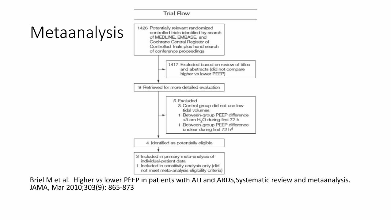
Metaanalysis
Briel M et al. Higher vs lower PEEP in patients with ALI and ARDS,Systematic review and metaanalysis. JAMA, Mar 2010;303(9): 865-873

Trial selection
• Randomized trials eligible for this review compared higher with lower levels of PEEP (mean difference of at least 3 cm H2O between groups during first 3 days following randomization)in critically ill adults (>16 years) with a diagnosis of acute lung injury or ARDS as defined by the American-European Consensus Conference.
• 12 Eligible trials incorporated a target tidal volume of less than 8mL/kg of predicted body weight in both the experimental and the control ventilation strategies and provided patient follow-up to death or for at least 20 days.

• Primary outcome: Hospital mortality, measured to at least 60 days in all eligible trials
• Prespecified secondary outcomes:Death before discharge from the intensive care unit Pneumothorax with need for chest tube drainage in the first 28 daysDeath following pneumothorax with need for chest tube drainageTime-to-unassisted breathing within the first 28days Days with unassisted breathing between day 1 and day 28Use of rescue therapy Death following rescue therapyuse of neuromuscular blockers, vasopressors, and corticosteroids


Baseline charecteristics of included patients

ALVEOLI Study Group

Lung Open Ventilation group


Express study group

Respiratory variables during first week of treatment

Clinical outcomes in all patients and stratified by presence of ARDS at baseline

In-hospital time to death

In-hospital time to death

Time to unassisted breathing


Strengths of this meta-analysis
• An explicit study protocol and analysis plan
• Access to trial protocols, case report forms, and complete, unedited data sets
• Standardized outcome definitions across trials (except for rescue therapies)
• Analyses based on the intention-to-treat principle.

Limitations of this meta-analysis
• Limited statistical power: A post hoc calculation estimated that the primary analysis had a power of 72% to detect a 5% absolute risk reduction in hospital mortality(2-sided α=0.05)
• Caregivers were not blinded to allocated PEEP strategies. Differing thresholds for rescue therapy in the high and low PEEP groups could explain the lower use of rescue therapies and mortality following rescue therapy in the higher PEEP group
• Rescue therapies were not standardized across the trials
• Analyses involving lung compliance are limited by missing data

Clinical implications of the study
• The potentially lower hospital mortality and the absence of increased serious adverse events associated with higher PEEP levels in patients with moderate and severe ARDS support the safety of higher PEEP in these patients. For this purpose, clinicians could titrate PEEP as described in the 3 major trials in this review
• For patients with mild ARDS, the results lack statistical power; still, the 95% CI of 0.98-1.92 for hospital mortality in patients with mild ARDS indicates that a RR reduction of 2%(0.4% absolute reduction) associated with higher PEEP is plausible but that larger, important risk reductions are unlikely

Open lung strategy (LIP approach): Amato NEJM 1998

Stabilizing procedures and randomization
• All patients underwent a standardized regimen of ventilatory–hemodynamic procedures for at least 30 minutes (control period), during which time their initial clinical condition was evaluated and stabilized
• Subsequently, a bedside procedure was performed to calculate the inspiratory and static pressure–volume curve without disconnecting the ventilator
• A well-defined P FLEX (corresponding to an upward shift in the slope of the curve and signaling an increment in lung compliance) could be determined for 49 patients, but the corresponding value was used to adjust PEEP only in the group assigned to protective mechanical ventilation

Inspiratory static P-V curve of the respiratory system obtained from a patient
Amato et al.Beneficial Effects of the "Open Lung Approach" with Low Distending Pressures in Acute Respiratory Distress Syndrome. AM J RESPIR CRIT CARE MED 1995;152:1835-46

• Conventional ventilation: Strategy of maintaining the lowest PEEP for acceptable oxygenation, with a tidal volume of 12 ml per kilogram of body weight and normal arterial carbon dioxide levels (35 to 38 mm Hg)
• Protective ventilation: End-expiratory pressures above the lower inflection point on the static pressure–volume curve, a tidal volume of less than 6 ml per kilogram, driving pressures of less than 20 cm of water above the PEEP value, permissive hypercapnia, and preferential use of pressure-limited ventilatory modes.
Effect of a Protective-Ventilation Strategy on Mortality in the ARDS. Amato NEJM 1998

• PEEP was preset at 2 cm of water above PFLEX. When auto-PEEP was present, the total PEEP (external PEEP plus auto-PEEP) was considered and adjusted to equal PFLEX plus 2 cm of water.
• If a sharp PFLEX could not be determined on the pressure–volume curve, an empirical total PEEP value of 16 cm of water was used.
• Recruiting maneuvers were frequently used, especially after inadvertent disconnections from the ventilator. Continuous positive airway pressures of 35 to 40 cm of water were applied for 40 seconds, followed by a careful return to previous PEEP levels

Study outcomes according to intention to treat analysis

Esophageal pressure measurements
• Physiologic background:
• At the end of a relaxed exhalation (to functional residual capacity) and with the mouth open,the alveolar pressure(Palv),the pressure at the airway opening (PAO), and the atmospheric pressure (Patm) are equal

How to measure pleural pressure in clinical practice?
• Because the body of the esophagus is essentially a passive structure (except during a swallow), able to transmit pressure from the adjacent pleural space (Ppl) to the measurement catheter in the esophagus, Pes in lower 1/3rd of esophagus is a reasonably close surrogate for Ppl in a human being in the upright posture

Equipments required for recording pressure from esophageal balloon catheter

• The device consists of a thin polyethylene catheter with multiple small holes in the distal 5–7 cm of its length
• The distal end of the catheter is placed in a 10-cm latex balloon that prevents the holes in the catheter from being occluded by esophageal tissue and maintains a column of air within and around the catheter, in order to measure pressure in the surrounding structures
• The proximal end of the catheter is attached to the pressure transducers and recording equipment
Benditt JO.Esophageal and gastric pressure measurements.Respir Care Jan 2005;50(1):70-71

Hypothesis of using esophageal pressure to set PEEP
• Calculated transpulmonary pressure is often a negative value
at end-expiration. This is presumed to reflect closed airways.
• In the presence of closed airways and flooded or atelectatic lung, the Paw measured proximally(the set PEEP) may underestimate alveolar pressure, resulting in a negative calculated transpulmonary pressure.
• Raising PEEP until transpulmonary pressure becomes positive at end expiration could assure that airways remain open

• Wide variation in pleural pressure in patients of ARDS due to factors like ascites, intra-abdominal hypertension, resuscitation with large fluid volumes, obesity
• Estimating pleural pressure to calculate transpulmonary pressure may allow better control of both end inspiratory and end expiratory lung volume, and thereby reduce VILI caused by overdistension or atelectrauma
• Optimizing inflating pressures to the mid lung may prevent over distension of the upper, nondependent portions of the aerated lung while preventing collapse of the lower, dependent portions


• RCT involving patients of ARDS comparing mechanical ventilation directed by esophageal pressure measurements with mechanical ventilation managed according to the ARDSNet recommendations
• Methods: trial performed in the medical and surgical ICUs of Beth Israel Deaconess Medical Center in Boston
• Inclusion criteria: Patients with acute lung injury or ARDS according to the American–European Consensus Conference definitions
• Exclusion criteria: Recent injury or other pathologic condition of the esophagus, major bronchopleural fistula, and solid organ transplantation

Experimental protocol
• Subjects were supine, with head of bed elevated to 30 degrees
• An esophageal balloon catheter was passed to a depth of 60 cm from the incisors for measurement of gastric pressure and then withdrawn to a depth of 40 cm to record esophageal pressure during mechanical ventilation
• Mixed expired partial pressure of carbon dioxide was measured to allow calculation of physiological dead space
• Patients were randomly assigned with the use of a block-randomization scheme to the control or esophageal pressure–guided group

• Each patient underwent a recruitment maneuver under heavy sedation or paralysis, in which airway pressure was increased to 40 cm of water for 30 seconds
• Esophageal pressure guided group:
Tidal volume= 6 ml/kg of predicted body weight
PEEP set to achieve a transpulmonary pressure of 0 to 10 cm of water at end expiration according to a sliding scale based on PaO2 and FiO2
Tidal volume limited to keep end inspiratory transpulmonary pressure
at less than 25cm of water

• Control group:
Treated according to the low tidal volume strategy reported by the ARDSNet study
Tidal volume= 6 ml/kg of predicted body weight
PEEP based on patient’s PaO2 and FiO2
• Goals of mechanical ventilation( Both groups):
PaO2: 55-120 mmHg or pulse oximetry reading of 88-98%
Arterial pH: 7.30- 7.45
PaCO2: 40-60 mmHg

Ventilator settings according to the protocol
• Esophageal pressure guided group
• Control group
FiO2 0.4 0.5 0.5 0.6 0.6 0.7 0.7 0.8 0.8 0.9 0.9 1.0
P Lep 0 0 2 2 4 4 6 6 8 8 10 10
FiO2 0.3 0.4 0.4 0.5 0.5 0.6 0.7 0.7 0.7 0.8 0.9 0.9 0.9 1.0
PEEP 5 5 8 8 10 10 10 12 14 14 14 16 18 20-24

• All measurements were repeated 5 minutes after initiation of mechanical ventilation, at 24, 48 and 72 hours and whenever changes were made in ventilator settings
• Primary end point: Arterial oxygenation as measured by PaO2: FiO2 ratio, 72 hours after randomization
• Secondary end points:
Respiratory system compliance and the ratio of physiological dead space to tidal volume
the number of ventilator-free days at 28 days
length of stay in the ICU
death within 28 days and 180 days after treatment


Criticism of study
• Mean PaO2 was 122±44 mmHg in the esophageal pressure guided group at 72 hours.
• More than half of patients in the esophageal pressure guided group had a PaO2 above the range stipulated by the protocol.
• Poor protocol compliance
• Profound artifact on PaO2: FiO2 at the critical time of assessment for the primary outcome

• Effects on important clinical outcomes such as long term mortality, ventilator free days, and length of stay in ICU remain unanswered
• Improved oxygenation need not result in clinical improvement.
• Only large trial to show a lowering of mortality rates in association with a particular method of ventilation also found that group with improved oxygenation had higher mortality rates
The Acute Respiratory Distress Syndrome Network. Ventilation with lower tidal volumes as compared with traditional tidal volumes for acute lung injury and the acute respiratory distress syndrome. N Engl J Med 2000;342:1301-8.

Other potential sources of error
• Balloon placement was inadequate in 1/3rd of patients
• A correction factor of 5 cm of water was subtracted from the esophageal pressure in an attempt to compensate for the known artifacts of mediastinal weight and balloon air volume on the observed pressures

? Too many assumptions
• the balloon pressure reflects the esophageal pressure
• the transmural pressure in the esophagus is 0 cm of water
• the esophagus is not compressed by intrathoracic structures such as the heart
• the pressures in the periesophageal area are the same as the pleural pressure
• pleural pressure is relatively uniform throughout the thorax

Comparison of changes in PEEP and PaO2:FiO2 From baseline to day 3 in the study by Talmor et al. and in the ARDSNet ALVEOLI Trial
• Improvements in oxygenation in the study by Talmor et al. were reflective of generally higher level of PEEP, rather than a unique response to PEEP titration method