severe hypercholesterolemia md, mmsc, connor a. emdin...
TRANSCRIPT

Accepted Manuscript
Diagnostic Yield of Sequencing Familial Hypercholesterolemia Genes in Patients withSevere Hypercholesterolemia
Amit V. Khera, MD, Hong-Hee Won, PhD, Gina M. Peloso, PhD, Kim S. Lawson, MS,Traci M. Bartz, MS, Xuan Deng, MSc, Elisabeth M. van Leeuwen, Pradeep Natarajan,MD, MMSc, Connor A. Emdin, HBSc, Alexander G. Bick, BS, Alanna C. Morrison,PhD, Jennifer A. Brody, BA, Namrata Gupta, PhD, Akihiro Nomura, MD, ThorstenKessler, MD, Stefano Duga, PhD, Joshua C. Bis, PhD, Cornelia M. van Duijn, PhD, L.Adrienne Cupples, PhD, Bruce Psaty, MD, PhD, Daniel J. Rader, MD, John Danesh,FMedSci, Heribert Schunkert, MD, Ruth McPherson, MD, Martin Farrall, FRCPath,Hugh Watkins, MD, PhD, Eric Lander, PhD, James G. Wilson, MD, Adolfo Correa,MD, PhD, Eric Boerwinkle, PhD, Piera Angelica Merlini, MD, Diego Ardissino, MD,Danish Saleheen, MB, BS, PhD, Stacey Gabriel, PhD, Sekar Kathiresan, MD
PII: S0735-1097(16)32399-3
DOI: 10.1016/j.jacc.2016.03.520
Reference: JAC 22438
To appear in: Journal of the American College of Cardiology
Received Date: 2 March 2016
Revised Date: 22 March 2016
Accepted Date: 22 March 2016
Please cite this article as: Khera AV, Won H-H, Peloso GM, Lawson KS, Bartz TM, Deng X, vanLeeuwen EM, Natarajan P, Emdin CA, Bick AG, Morrison AC, Brody JA, Gupta N, Nomura A, Kessler T,Duga S, Bis JC, van Duijn CM, Cupples LA, Psaty B, Rader DJ, Danesh J, Schunkert H, McPherson R,Farrall M, Watkins H, Lander E, Wilson JG, Correa A, Boerwinkle E, Merlini PA, Ardissino D, SaleheenD, Gabriel S, Kathiresan S, Diagnostic Yield of Sequencing Familial Hypercholesterolemia Genes inPatients with Severe Hypercholesterolemia, Journal of the American College of Cardiology (2016), doi:10.1016/j.jacc.2016.03.520.
This is a PDF file of an unedited manuscript that has been accepted for publication. As a service toour customers we are providing this early version of the manuscript. The manuscript will undergocopyediting, typesetting, and review of the resulting proof before it is published in its final form. Please

note that during the production process errors may be discovered which could affect the content, and alllegal disclaimers that apply to the journal pertain.

MANUSCRIP
T
ACCEPTED
ACCEPTED MANUSCRIPT
1
Diagnostic Yield of Sequencing Familial Hypercholesterolemia Genes in Patients with Severe Hypercholesterolemia
Amit V. Khera, MD*,a,b Hong-Hee Won, PhD*,c Gina M. Peloso, PhD*,b,d Kim S. Lawson, MS,e Traci M. Bartz, MS,f Xuan Deng, MSc,d Elisabeth M. van Leeuwen,g Pradeep Natarajan, MD, MMSc,a,b Connor A. Emdin, HBSc,b Alexander G. Bick, BS,b Alanna C. Morrison, PhD,e Jennifer A. Brody,h BA, Namrata Gupta, PhD,b Akihiro Nomura, MD,b, i Thorsten Kessler, MD,j Stefano Duga, PhD,k Joshua C. Bis, PhD, h Cornelia M. van Duijn, PhD,g L. Adrienne Cupples, PhD,d Bruce Psaty, MD, PhD,h,l Daniel J. Rader, MD,m John Danesh, FMedSci,n Heribert Schunkert,j MD, Ruth McPherson, MD,o Martin Farrall, FRCPath,p Hugh Watkins, MD,p PhD, Eric Lander, PhD,b James G. Wilson, MD,q Adolfo Correa, MD, PhD,r Eric Boerwinkle, PhD,e Piera Angelica Merlini, MD,s Diego Ardissino, MD,t Danish Saleheen, MB,BS,PhD,u Stacey Gabriel, PhD,b Sekar Kathiresan, MDa,b * Drs. Amit V. Khera, Hong-Hee Won, and Gina M. Peloso contributed equally to this work a Center for Human Genetic Research, Cardiovascular Research Center and Cardiology Division (Khera, Natarajan, Kathiresan), Massachusetts General Hospital, Harvard Medical School, Boston MA b Program in Medical and Population Genetics, Broad Institute, Cambridge, MA c Samsung Advanced Institute for Health Sciences and Technology (SAIHST), Sungkyunkwan University, Samsung Medical Center, Seoul, Korea d Department of Biostatistics, Boston University School of Public Health e Human Genetics Center and Institute of Molecular Medicine, University of Texas-Houston Health Science Center, Houston, TX f Department of Biostatistics, University of Washington, Seattle, Washington; g Department of Epidemiology, Erasmus Medical Center, Rotterdam, The Netherlands h Cardiovascular Health Research Unit, University of Washington i Division of Cardiovascular Medicine, Kanazawa University Graduate School of Medical Science, Kanazawa, Japan j Deutsches Herzzentrum München, Technische Universität München, Deutsches Zentrum für Herz-Kreislauf-Forschung (DZHK); Munich Heart Alliance, München, Germany (Kessler, Schunkert); k Department of Biomedical Sciences, Humanitas University, Via Manzoni 113, 20089 Rozzano, Milan, Italy; Humanitas Clinical and Research Center, Via Manzoni 56, 20089 Rozzano, Milan, Italy l Departments of Medicine, Epidemiology, and Health Services, University of Washington m Departments of Genetics, University of Pennsylvania, Philadelphia n Public Health and Primary Care, University of Cambridge, Cambridge, Wellcome Trust Sanger Institute, Wellcome Trust Genome Campus, Hinxton, Cambridge, UK, and NIHR Blood and Transplant Research Unit in Donor Health and Genomics, Department of Public Health and Primary Care, University of Cambridge, Cambridge, United Kingdom o University of Ottawa Heart Institute, Ottawa, Canada p Division of Cardiovascular Medicine, Radcliffe Department of Medicine and the Wellcome Trust Centre for Human Genetics, University of Oxford, Oxford, United Kingdom

MANUSCRIP
T
ACCEPTED
ACCEPTED MANUSCRIPT
2
q Department of Physiology and Biophysics, University of Mississippi Medical Center, Jackson, Mississippi r Jackson Heart Study, Department of Medicine, University of Mississippi Medical Center s Ospedale Niguarda, Milano Italy
t Division of Cardiology, Azienda Ospedaliero-Universitaria di Parma, University of Parma Parma, Italy; ASTC - Associazione Per Lo Studio Della Trombosi In Cardiologia, Pavia Italy (Ardissino) u Biostatistics and Epidemiology, Perelman School of Medicine, University of Pennsylvania Funding/Support: Field-work, genotyping, and standard clinical chemistry assays in PROMIS were principally supported by grants awarded to the University of Cambridge from the British Heart Foundation, UK Medical Research Council, Wellcome Trust, EU Framework 6–funded Bloodomics Integrated Project, Pfizer, Novartis, and Merck. Additional support for PROMIS was provided by by the UK Medical Research Council (MR/L003120/1), British Heart Foundation (RG/13/13/30194), UK National Institute for Health Research Cambridge Biomedical Research Centre, European Research Council (268834), and European Commission Framework Programme 7 (HEALTH-F2-2012-279233). The Jackson Heart Study is supported by contracts HHSN268201300046C, HHSN268201300047C, HHSN268201300048C, HHSN268201300049C, HHSN268201300050C from the National Heart, Lung, and Blood Institute and the National Institute on Minority Health and Health Disparities. The Munich Study is supported by the German Federal Ministry of Education and Research (BMBF) in the context of the e:Med program (e:AtheroSysMed) and the FP7 European Union project CVgenes@target (261123). Further grants were received by the Fondation Leducq (CADgenomics: Understanding Coronary Artery Disease Genes, 12CVD02). This study was also supported through the Deutsche Forschungsgemeinschaft (DFG) cluster of excellence ‘Inflammation at Interfaces and SFB 1123. Funding support for "Building on GWAS for NHLBI-diseases: the U.S. CHARGE Consortium" was provided by the NIH through the American Recovery and Reinvestment Act of 2009 (ARRA) (5RC2HL102419). Data for "Building on GWAS for NHLBI-diseases: the U.S. CHARGE Consortium" was provided by Eric Boerwinkle on behalf of the Atherosclerosis Risk in Communities Study, L. Adrienne Cupples, principal investigator for the Framingham Heart Study, and Bruce Psaty, principal investigator for the Cardiovascular Health Study (CHS). Sequencing was carried out at the Baylor Genome Center (U54 HG003273). The ARIC Study is carried out as a collaborative study supported by National Heart, Lung, and Blood Institute (NHLBI) contracts (HHSN268201100005C, HHSN268201100006C, HHSN268201100007C, HHSN268201100008C, HHSN268201100009C, HHSN268201100010C, HHSN268201100011C, and HHSN268201100012C). The Framingham Heart Study is conducted and supported by the NHLBI in collaboration with Boston University (Contract No. N01-HC-25195), and its contract with Affymetrix, Inc., for genome-wide genotyping services (Contract No. N02-HL-6-4278), for quality control by Framingham Heart Study investigators using genotypes in the SNP Health Association Resource (SHARe) project. A portion of this research was conducted using the Linux Cluster for Genetic Analysis (LinGA-II) funded by the Robert Dawson Evans Endowment of the Department of Medicine at Boston University School of Medicine and Boston Medical Center. This CHS research was supported by NHLBI contracts HHSN268201200036C, HHSN268200800007C, N01HC55222, N01HC85079, N01HC85080, N01HC85081,

MANUSCRIP
T
ACCEPTED
ACCEPTED MANUSCRIPT
3
N01HC85082, N01HC85083, N01HC85086; and NHLBI grants U01HL080295, R01HL087652, R01HL105756, R01HL103612, and R01HL120393 with additional contribution from the National Institute of Neurological Disorders and Stroke (NINDS). Additional support was provided through R01AG023629 from the National Institute on Aging (NIA). A full list of principal CHS investigators and institutions can be found at CHS-NHLBI.org. The Italian ATVB Study was supported by a grant from RFPS-2007-3-644382 and Programma di ricerca Regione-Università 2010-2012 Area 1 - Strategic Programmes – Regione Emilia-Romagna. Funding for the exome sequencing project (ESP) was provided by RC2 HL103010 (HeartGO), RC2 HL102923 (LungGO) and RC2 HL102924 (WHISP). Exome sequencing was performed through RC2 HL102925 (BroadGO) and RC2 HL102926 (SeattleGO). Exome sequencing in ATVB, PROCARDIS, Ottawa, PROMIS, Munich Study, and Jackson Heart Study was supported by 5U54HG003067 (to E.S.L. and S.G.). Dr. Kathiresan is supported by a Research Scholar award from the Massachusetts General Hospital (MGH), the Donovan Family Foundation, and R01 HL127564. The views expressed in this manuscript are those of the authors and do not necessarily represent the views of the National Heart, Lung, and Blood Institute; the National Institutes of Health; or the U.S. Department of Health and Human Services. Role of the Funder/Sponsor: The sponsors had no role in the design and conduct of the study; collection, management, analysis, and interpretation of the data; preparation, review, or approval of the manuscript; and decision to submit the manuscript for publication. Conflict of Interest Disclosures: Dr Khera is supported by an ACC/Merck Fellowship award and reported consulting fees from Merck and Amarin. Dr Peloso is supported by the National Heart, Lung, and Blood Institute of the National Institutes of Health under Award Number K01HL125751. Dr Kessler is supported by a DZHK Rotation Grant. Dr Rader reported consulting fees from Aegerion, Alnylam, Eli Lilly, Pfizer, and Novartis, is an inventor on a patent related to lomitapide that is owned by the University of Pennsylvania and licensed to Aegerion, and is a co-founder of VascularStrategies and Staten Biotechnology. Dr Kathiresan has received grants from Bayer Healthcare, Aegerion, and Regeneron and reported consulting fees from Merck, Quest Diagnostics, Genomics PLC, and Eli Lilly. Corresponding Author: Sekar Kathiresan, MD Cardiovascular Research Center & Center for Human Genetics Massachusetts General Hospital 185 Cambridge Street, CPZN 5.252 Boston, MA 02114 Telephone: 617 643 6120 Fax: 8779915996 Email: [email protected]

MANUSCRIP
T
ACCEPTED
ACCEPTED MANUSCRIPT
4
Abstract: Background: About 7% of US adults have severe hypercholesterolemia (untreated LDL cholesterol ≥190 mg/dl). Such high LDL levels may be due to familial hypercholesterolemia (FH), a condition caused by a single mutation in any of three genes. Lifelong elevations in LDL cholesterol in FH mutation carriers may confer CAD risk beyond that captured by a single LDL cholesterol measurement. Objectives: Assess the prevalence of a FH mutation among those with severe hypercholesterolemia and determine whether CAD risk varies according to mutation status beyond the observed LDL cholesterol. Methods: Three genes causative for FH (LDLR, APOB, PCSK9) were sequenced in 26,025 participants from 7 case-control studies (5,540 CAD cases, 8,577 CAD-free controls) and 5 prospective cohort studies (11,908 participants). FH mutations included loss-of-function variants in LDLR, missense mutations in LDLR predicted to be damaging, and variants linked to FH in ClinVar, a clinical genetics database. Results: Among 8,577 CAD-free control participants, 430 had LDL cholesterol ≥190 mg/dl; of these, only eight (1.9%) carried a FH mutation. Similarly, among 11,908 participants from 5 prospective cohorts, 956 had LDL cholesterol ≥190 mg/dl and of these, only 16 (1.7%) carried a FH mutation. Within any stratum of observed LDL cholesterol, risk of CAD was higher among FH mutation carriers when compared with non-carriers. When compared to a reference group with LDL cholesterol <130 mg/dl and no mutation, participants with LDL cholesterol ≥190 mg/dl and no FH mutation had six-fold higher risk for CAD (OR 6.0; 95%CI 5.2–6.9) whereas those with LDL cholesterol ≥190 mg/dl as well as a FH mutation demonstrated twenty-two fold increased risk (OR 22.3; 95%CI 10.7–53.2). Conclusions: Among individuals with LDL cholesterol ≥190 mg/dl, gene sequencing identified a FH mutation in <2%. However, for any given observed LDL cholesterol, FH mutation carriers are at substantially increased risk for CAD. Clinical trial: ??? Please query authors. Keywords: familial hypercholesterolemia, low-density lipoprotein cholesterol, gene sequencing, coronary artery disease, genetics Abbreviations: APOB = apolipoprotein B CAD = coronary artery disease FH = familial hypercholesterolemia HDL = high-density lipoprotein LDL = low-density lipoprotein LDLR = low-density lipoprotein receptor PCSK9 = proprotein convertase subtilisin/kexin type 9

MANUSCRIP
T
ACCEPTED
ACCEPTED MANUSCRIPT
5
Introduction
Primary, severe hypercholesterolemia, defined as having a low-density lipoprotein (LDL)
cholesterol ≥ 190 mg/dl, is a treatable risk factor for coronary artery disease (CAD) (1,2); current
treatment guidelines place particular emphasis on intensive lifestyle and pharmacologic therapy
in this population (3). One etiology of severely elevated LDL cholesterol is familial
hypercholesterolemia (FH), an autosomal dominant monogenic disorder linked to impaired
hepatic clearance of LDL cholesterol particles (4). It is often assumed that individuals with LDL
cholesterol ≥ 190 mg/dl have FH but this may not be the case. Large-scale gene sequencing
provides an opportunity to clarify the diagnostic yield and clinical impact of identifying a FH
mutation in severely hypercholesterolemic patients.
Previous studies of the diagnostic yield of genetic testing in severe hypercholesterolemia
have focused on individuals with clinically-suspected FH and in these samples, a FH mutation
prevalence ranging from 20 to 80% has been reported (5-16). This variability is likely due to
differing ascertainment schemes based on family history, physical exam features, elevated LDL
cholesterol at a young age, or referral to specialized clinics, each of which may enrich for
monogenic etiologies. In contrast, if ascertainment from the general population is based solely on
elevated LDL cholesterol, the extent to which FH mutations contribute to severe
hypercholesterolemia is unknown. Such knowledge may inform design and effectiveness of
universal FH screening proposals (17,18).
Knowledge of FH mutation status may also provide CAD risk information beyond that of
a single LDL cholesterol measurement (4,18). A FH mutation leads to cumulative exposure to
higher LDL cholesterol levels over a lifetime and as such, within any stratum of LDL
cholesterol, the risk of CAD may be greater if the LDL elevation is due to a monogenic mutation

MANUSCRIP
T
ACCEPTED
ACCEPTED MANUSCRIPT
6
versus other causes. The extent to which CAD risk is modulated by the presence of a causal FH
mutation is uncertain.
We analyzed gene sequences of three FH genes, low-density lipoprotein receptor
(LDLR), apolipoprotein B (APOB), and proprotein convertase subtilisin/kexin type 9 (PCSK9), in
twelve distinct cohorts including >26,000 participants to determine: 1) the diagnostic yield of
gene sequencing to identify a FH mutation in severely hypercholesterolemic individuals; and 2)
the clinical impact of a FH mutation with regard to CAD risk within any given stratum of LDL
cholesterol levels.
Methods
Study Populations
Whole exome sequencing was performed in seven previously described CAD case-
control cohorts of the Myocardial Infarction Genetics Consortium (Online Table 1). Studies
included the Italian Atherosclerosis Thrombosis and Vascular Biology study (19), the Exome
Sequencing Project Early-Onset Myocardial Infarction (ESP-EOMI) study (20), a nested case-
control of the Jackson Heart Study (JHS) (15), the Munich Myocardial Infarction study (22), the
Ottawa Heart Study (23), the Precocious Coronary Artery Disease (PROCARDIS) study (24),
and the Pakistan Risk of Myocardial Infarction Study (PROMIS) (25). The effect of lipid-
lowering therapy in those reporting use at the time of lipid measurement was taken into account
by dividing the measured total cholesterol and LDL cholesterol by 0.8 and 0.7 respectively as
has been implemented previously (26-28). Primary, severe LDL cholesterol elevation was
defined as ≥ 190 mg/dl in accordance with current cholesterol treatment guidelines (3).
The prevalence of a FH mutation in individuals with a LDL cholesterol > 190 mg/dl was
additionally determined in 11,908 participants from five prospective cohort studies derived from

MANUSCRIP
T
ACCEPTED
ACCEPTED MANUSCRIPT
7
the Cohorts for Heart and Aging Research in Genomic Epidemiology (CHARGE) Consortium
(29). Atherosclerosis Risk in Communities Study (ARIC), Cardiovascular Health Study,
Framingham Heart Study (FHS), Rotterdam Baseline Study, and Erasmus Rucphen Family
Study (Online Table 2).
In order to determine the cumulative exposure to LDL cholesterol according to FH
mutation status, publically available data from the National Center for Biotechnology
Information dbGAP database was analyzed. These data included 5,727 ARIC cohort participants
and 2,714 FHS Offspring Study participants.
Gene Sequencing
The CAD case-control whole exome sequencing was performed as previously described
at the Broad Institute (Cambridge, MA) (20). The population-based cohort sequencing was
performed at the Baylor College of Medicine (Houston, Texas) for the ARIC, CHS, and FHS
cohorts and at Erasmus Medical Center (Rotterdam, Netherlands) for the RS and ERF cohorts
respectively. Additional sequencing methodology details available in Supplementary Methods.
Genetic Variant Annotation
Three classes of genetic variants were aggregated with respect to association with FH: 1)
loss of function variants in LDLR: single base changes that introduce a stop codon leading to
premature truncation of a protein (nonsense), insertions or deletions (indels) of DNA that
scramble the protein translation beyond the variant site (frameshift), or point mutations at sites of
pre-mRNA splicing that alter the splicing process (splice-site); 2) missense variants in LDLR
predicted to be deleterious by each of five in silico prediction algorithms (LRT score,
MutationTaster, PolyPhen-2 HumDiv, PolyPhen-2 HumVar and Sorting Intolerant From
Tolerant (SIFT)) as described previously (20,30); and 3) Variants in LDLR, APOB, or PCSK9,

MANUSCRIP
T
ACCEPTED
ACCEPTED MANUSCRIPT
8
annotated as “pathogenic” or “likely pathogenic” in ClinVar, a publically available archive of
genetic variations associated with clinical phenotypes (31). Additional sensitivity analyses
aggregated all rare (allele frequency < 0.01) missense mutations in LDLR, exon 26 of APOB
which encodes key components of apolipoprotein B binding to the LDL receptor and harbor the
majority of APOB variants linked to FH (32), and those that produce a change in PCSK9 at an
amino acid associated with FH in ClinVar. Rare synonymous variants at these same locations
were included as a negative control. Software used to annotate observed variants included
Variant Effect Predictor (version 77) (33) and associated LOFTEE plugin (34), and the dbNSFP
database (version 3.0b1) (35).
Longitudinal Analysis of LDL Cholesterol Exposure
Individuals with a FH mutation and LDL cholesterol ≥ 130 mg/dl were identified in
ARIC and FHS Offspring Study cohorts. LDL cholesterol values were adjusted in those
reporting lipid-lowering therapy by dividing measured value by 0.7. Mean LDL cholesterol
exposure was calculated as cumulative exposure, determined via an area under the curve
analysis, divided by length of follow-up. 27 FH mutation carriers met the above inclusion criteria
and were matched 1:1 to a mutation negative control according to age (within 10 years), gender,
statin use at time of last visit, and similar LDL cholesterol at last visit (within 10 mg/dl). A
match was available in 25 of 27 (93%) individuals. Mean LDL cholesterol exposure was
compared among those with and without FH mutation using a paired t-test.
Statistical Analysis
The impact of aggregations of genetic variants on levels of LDL cholesterol was assessed
using linear regression with adjustment for age, age2, gender, cohort, and the first five principal
components of ancestry. Odds ratios for CAD were calculated using logistic regression with

MANUSCRIP
T
ACCEPTED
ACCEPTED MANUSCRIPT
9
adjustment for gender, cohort, and the first five principal components of ancestry. In analyses
conducted on ordinal strata of LDL cholesterol, individuals with LDL cholesterol <130 mg/dl
and no mutation linked to FH served as the reference group.
Analyses were performed using R version 3.2.2 software (The R Project for Statistical
Computing, Vienna, Austria). Figures were creating using the “ggplot2” package within R (36).
Results
Within the Myocardial Infarction Genetics Consortium CAD case-control cohorts, a total
of 14,117 participants with both LDL cholesterol level and sequence data for FH genes were
available for analysis – 8,577 CAD-free controls and 5,540 CAD cases (Online Table 3). The
study population included 10,421 (74%) males with mean age 53 years (SD 14). Proportions of
self-identified race were 47%, 46%, and 7% for white, South Asian, and black, respectively. 47%
of study participants had a history of hypertension, 27% had a history of diabetes, 34% were
current smokers, and 14% were on lipid-lowering medications.
Sequencing identified 86 variants linked to FH on the basis of leading to loss of function
in LDLR, missense mutations in LDLR predicted to be damaging by each of five computer
prediction algorithms, or a variant in LDLR, APOB, or PCSK9 previously linked to FH in the
ClinVar genetics database. These included 13 premature stop (“nonsense”) codons, 6 splice
acceptor/donor variants, 4 frameshift mutations, and 63 missense mutations (Online Table 4).
164 individuals harbored a mutation linked to FH, including 48 CAD-free controls (0.6%;
95%CI 0.4 – 0.7%) and 116 CAD cases (2.1%; 95%CI 1.7 – 2.5%) (Online Table 5). The
mutation was located in LDLR for 141 participants (86%), in APOB for 22 (13%), and in PCSK9
for 1 (0.6%) (Online Table 4). Only one homozygote (or compound heterozygote) participant

MANUSCRIP
T
ACCEPTED
ACCEPTED MANUSCRIPT
10
was identified; a 33-year old patient with LDL cholesterol of 539 mg/dl and CAD was
homozygous for a p.Q33* premature stop codon in LDLR.
Diagnostic Yield of Gene Sequencing in Severe Hypercholesterolemia
Among 8,577 CAD-free control participants from the Myocardial Infarction Genetics
Consortium cohorts, LDL cholesterol approximated a normal distribution (Online Figure 1).
The prevalence of a FH mutation increased across categories of LDL cholesterol levels (P <
0.001) (Online Figure 2). Of 8,577 control participants, 430 participants (5% of control sample)
had LDL cholesterol ≥ 190 mg/dl and of these 430, only 8 carried a FH mutation (1.9%; 95%CI
0.9 – 3.8%) (Table 1 & Central Illustration ).
This prevalence estimate was replicated in 11,908 participants from five prospective
cohort studies of the CHARGE consortium; 956 (8%) had a LDL cholesterol >190 mg/dl and of
these, 16 (1.7%; 95%CI 1.0 – 2.8%) harbored a FH mutation. Across the twelve studies
combined (n=20,485), 1386 (7%) displayed LDL cholesterol ≥ 190 mg/dl and of these, 24
(1.7%) carried a FH mutation (Table 1).
Clinical Impact of FH Mutation Identification on CAD Risk
In the Myocardial Infarction Genetics Consortium case-control studies, the presence of a
FH mutation was associated with a 50 mg/dl (95%CI 44– 57) increase in LDL cholesterol and a
3.8 fold (95%CI 2.6 – 5.4) increase in odds of CAD. These effects were most pronounced in
those with loss of function mutations in LDLR (Figure 1). Average LDL cholesterol was 190
mg/dl in those with a FH mutation and 73/164 (45%) had a LDL cholesterol ≥ 190 mg/dl.
However, despite the observed large effect on average levels, a wide spectrum of circulating
LDL cholesterol concentrations was noted in those who were mutation positive (Figure 2). 44 of
164 (27%) mutation carriers had an observed LDL cholesterol less than 130 mg/dl; reflecting

MANUSCRIP
T
ACCEPTED
ACCEPTED MANUSCRIPT
11
incomplete penetrance. An aggregation of all rare missense mutations had a modest impact on
both LDL cholesterol and CAD risk. As expected, synonymous mutations, which do not lead to
a change in amino acid sequence, had no effect on either parameter (Figure 1). Beyond LDL
cholesterol levels, a FH mutation was associated with a nominally significant reduction in high-
density lipoprotein cholesterol (-1.9 mg/dl; 95%CI -3.7 – -0.1; p = 0.04) but no association with
circulating triglycerides (p = 0.36).
Within the Myocardial Infarction Genetics Consortium case-control cohort populations,
those with a FH mutation were at substantially higher risk compared to those without a mutation
(Table 2, p-value for difference = 0.001). For participants with both LDL cholesterol ≥ 190
mg/dl and a FH mutation, the odds of coronary artery disease were increased twenty-two fold
(OR 22.3; CI 10.7 – 53.2) when compared to those with LDL cholesterol < 130 mg/dl and no
mutation. For participants with LDL cholesterol ≥ 190 mg/dl and no FH mutation, odds of
coronary artery disease were increased six-fold (OR 6.0; CI 5.2 – 6.9) compared to the same
reference group. This difference persisted after additional adjustment for measured LDL
cholesterol level (p = 0.02).
Separation of the population into clinically relevant categories of LDL cholesterol levels
demonstrated trends towards higher risk in those with a FH mutation within each stratum
(Central Illustration; Supplementary Table 6). The impact of a FH mutation was similar
across strata of LDL cholesterol levels (p-interaction = 0.51). Within the subgroup of participants
with a LDL cholesterol in the ≥ 190 to 220 mg/dl range, those with a mutation had 17-fold
increased CAD risk versus 5-fold for those without a mutation. This difference was noted despite
similar observed LDL cholesterol levels in this stratum (mean LDL cholesterol in those with

MANUSCRIP
T
ACCEPTED
ACCEPTED MANUSCRIPT
12
mutation=205 mg/dl versus mean LDL cholesterol in those without a FH mutation = 203 mg/dl;
p-value for difference = 0.22).
Cumulative LDL Cholesterol Exposure According to FH Mutation Status
For any given observed LDL cholesterol, those harboring a mutation might have a higher
average LDL cholesterol exposure over their lifetime compared to those who do not harbor a
mutation and this could explain a higher CAD risk among mutation carriers. We tested this
hypothesis using two prospective cohort studies – ARIC and the FHS Offspring Study – where
sequencing data and serial measurements of LDL cholesterol were available. We identified 25
individuals with a FH mutation and LDL cholesterol ≥ 130 mg/dl. Mean LDL cholesterol at time
of last study visit was 185 mg/dl. As compared to matched non-carriers with similar LDL
cholesterol at the last visit, individuals with a FH mutation had a 17 mg/dl (95%CI 5 – 29; p =
0.007) higher average LDL cholesterol exposure in the years preceding the last visit (Figure 3;
Online Table 7).
Discussion
Among 20,485 multiethnic participants from 12 studies, we found that 1,386 (7%) had
severe hypercholesterolemia (LDL cholesterol ≥ 190 mg/dl) and of those with severe
hypercholesterolemia, only a small fraction (<2%) also carried a FH mutation. However, within
any stratum of LDL cholesterol, those who carried a FH mutation were at substantially higher
risk for CAD compared to those who did not. This increased CAD risk among mutation carriers
was at least partially explained by a greater cumulative exposure to LDL cholesterol over a
lifetime.
These results permit several conclusions. First, FH mutations explain only a small
fraction of severe hypercholesterolemia in the population. Previous reports have noted a

MANUSCRIP
T
ACCEPTED
ACCEPTED MANUSCRIPT
13
substantially higher rate of mutation detection in those with clinically-suspected FH, as
ascertained on the basis of features (e.g. family history, physical exam, or severe
hypercholesterolemia at a young age) that enrich for a monogenic etiology (5-16). Here, we
address a scientific question – what fraction of severely hypercholesterolemic individuals carry a
mutation in any of three high LDL genes – that is distinct from these earlier, seminal reports.
When participants are ascertained solely on the basis of a single elevated LDL cholesterol level,
we identify a FH mutation in fewer than 2% of severely hypercholesterolemic individuals. These
sequencing results are broadly consistent with those of a recent study of 98,098 individuals from
the Copenhagen General Population Study in which genotyping of the four most common FH
mutations was used to extrapolate overall FH mutation prevalence. In this Danish study, of 5,332
individuals with LDL cholesterol ≥ 5 mmol/l (193 mg/dl), fewer than 5% were predicted to
harbor a FH mutation (28).
If not a monogenic mutation in the three FH genes, what might be the cause of elevated
LDL cholesterol in the remaining >95% of participants with severe hypercholesterolemia?
Possibilities include polygenic hypercholesterolemia, lifestyle factors, and/or a combination of
these. For example, individuals in the top quartile of a polygenic LDL cholesterol gene score
comprised of 95 common variants were 13 fold more likely to have high LDL cholesterol (37).
Similarly, individuals in the top decile of a LDL cholesterol gene score comprised of 12 common
variants were 4.2 fold more likely to have a LDL ≥ 190 mg/dl in the UK Whitehall II study (38).
Future genetics studies may identify additional causal variants, genes beyond those considered in
this study, or large-effect regulatory variants that underlie severe hypercholesterolemia. Other
non-genetic explanations for severe elevations in LDL cholesterol include secondary causes (e.g.

MANUSCRIP
T
ACCEPTED
ACCEPTED MANUSCRIPT
14
hypothyroidism or nephrotic syndrome), lifestyle factors such as dietary fat, or some
combination of these.
Second, within any stratum of a single observed LDL cholesterol, CAD risk was higher
in those with a FH mutation when compared to those without, reinforcing the potential utility of
genetic testing. We analyzed 25 matched pairs of individuals with similarly elevated LDL
cholesterol levels at time of ascertainment and found a higher cumulative exposure to LDL
cholesterol in those with a FH mutation. These data support the hypothesis that a FH mutation,
present since birth, increases CAD risk via lifelong exposure to high LDL cholesterol (39). By
contrast, an isolated elevation in LDL cholesterol in those without a genetic predisposition may
reflect a time-limited exposure related to a current environmental perturbation or a value that is
more likely to regress towards the mean in the future. Future studies may identify additional
metabolic parameters, such as increased lipoprotein(a) levels (40), that also contribute to the
excess CAD risk noted in those with a FH mutation.
Finally, these data contribute to ongoing discussion regarding how to define FH.
Classically, FH refers to elevated LDL cholesterol caused by a single mutation in any of several
genes segregating in an autosomal dominant manner. Alternate approaches to two features –
LDL cholesterol threshold and mutation definition – impact FH prevalence estimates (Table 3).
An approach that includes all individuals with untreated LDL cholesterol ≥ 190 mg/dl (i.e.,
without a FH mutation requirement) would combine non-genetic and genetic causes and classify
about 7% of the US adult population as having FH. An alternative possibility is to withhold an
LDL cholesterol threshold and require only a stringent mutation definition; in such an analysis of
20,485 participants, we identified a FH mutation in 97 individuals (1 in 211). This estimate is
nearly identical to a population-based analysis in the Copenhagen General Population Study (1 in

MANUSCRIP
T
ACCEPTED
ACCEPTED MANUSCRIPT
15
217).28 However, if one additionally requires that a FH mutation is accompanied by an elevated
LDL cholesterol, FH prevalence in our study declines (1 in 301 with LDL threshold ≥130 mg/dl
and 1 in 853 with LDL threshold ≥ 190 mg/dl).
With regard to defining a FH mutation, all schema agree on the inclusion of loss of
function alleles in LDLR but differ on how to handle missense mutations. For missense
mutations, we applied a rigorous threshold – requiring that the mutation be designated as
damaging by each of five computer prediction algorithms or be previously annotated as
pathogenic in the ClinVar clinical genetics database. A key advantage of this approach is that it
ensures that classification is both fully reproducible and generalizable to genes beyond those
related to FH.
When routine genetic testing is not available, clinical scoring systems such as the Dutch
Lipid Clinical Network, Simon Broome, and MEDPED criteria have been developed to
approximate FH status.4 Ongoing collaborative efforts on how to optimally incorporate
population-based genetic sequencing data into existing frameworks for the clinical diagnosis of
FH will be critically important.
Study Limitations
Our data did not permit stratifying individuals by family history or physical exam
features, as suggested by the Dutch Lipid Clinic Network and Simon Broome criteria (41,42).
Secondly, we accounted for an estimated 30% reduction in LDL cholesterol in those on lipid-
lowering therapy as has been previously implemented (26-28). This approach may imperfectly
estimate untreated LDL cholesterol given heterogeneity in drug selection, dosing, response, and
variability across baseline LDL cholesterol levels or mutation status. However, a sensitivity
analysis limited to Myocardial Infarction Genetics Consortium cohort participants not on lipid-

MANUSCRIP
T
ACCEPTED
ACCEPTED MANUSCRIPT
16
lowering therapy similarly noted a pronounced difference in risk among severely
hypercholesterolemic individuals when stratified by mutation status (Online Table 8). Third,
structural and copy number genetic variation are inadequately captured by current exome
sequencing techniques and as such, some FH mutations may have been missed. Fourth, our
approach to annotating missense variants using prediction algorithms and the ClinVar database
may have led to misclassification in some cases. Additional studies that implement large-scale
functional screens of identified variants or pool phenotypes across additional studies may
provide additional refinement of pathogenicity annotations. Lastly, FH mutation prevalence was
determined in CAD-free controls and population-based cohorts. These individuals survived to
middle-age and few had clinically manifest CAD, raising the possibility of survivorship or
selection bias. Our case-control population was enriched for individuals with premature CAD;
effect estimates of mutations on coronary risk may be different in patients with later disease
onset.
Conclusions
Genetic sequencing identified a FH mutation in only a small proportion of severely
hypercholesterolemic individuals. For any given observed LDL cholesterol level, risk for CAD is
substantially higher in carriers of a FH mutation versus non-carriers, likely related in large part to
higher lifelong exposure to atherogenic LDL particles. A primary goal of precision medicine is
to use molecular diagnostics to identify a small subset of the population at increased disease risk
in which to deliver an intervention. Systematic efforts to identify and treat severely
hypercholesterolemic individuals who carry a FH mutation may represent one such opportunity.

MANUSCRIP
T
ACCEPTED
ACCEPTED MANUSCRIPT
17
CLINICAL PERSPECTIVES
Competency in Medical Knowledge: Sequencing of three genes causing familial
hypercholesterolemia identifies a mutation in only a small fraction of severely
hypercholesterolemic individuals.
Translational Outlook: Additional research is needed to determine the relative contributions of
other genetic variants and lifestyle factors and evaluate the clinical utility of genetic testing in
patients with severe hypercholesterolemia.

MANUSCRIP
T
ACCEPTED
ACCEPTED MANUSCRIPT
18
References
1. Emerging Risk Factors Collaboration. Major lipids, apolipoproteins, and risk of vascular
disease. JAMA. 2009;302(18):1993-2000.
2. Baigent C, Keech A, Kearney PM, et al. Efficacy and safety of cholesterol-lowering
treatment: prospective meta-analysis of data from 90,056 participants in 14 randomised
trials of statins. Lancet. 2005;366(9493):1267-1278.
3. Stone NJ, Robinson JG, Lichtenstein AH, et al. 2013 ACC/AHA guideline on the
treatment of blood cholesterol to reduce atherosclerotic cardiovascular risk in adults: a
report of the American College of Cardiology/American Heart Association Task Force on
Practice Guidelines. J Am Coll Cardiol. 2014;63(25_PA):2889-2934
4. Gidding SS, Ann Champagne M, de Ferranti SD, et al. The Agenda for Familial
Hypercholesterolemia: A Scientific Statement From the American Heart Association.
Circulation. 2015 Dec 1;132(22):2167-92.
5. Fouchier SW, Defesche JC, Umans-Eckenhausen MW, Kastelein JP. The molecular basis
of familial hypercholesterolemia in The Netherlands. Hum Genet. 2001;109(6):602-15.
6. Graham CA, McIlhatton BP, Kirk CW, et al. Genetic screening protocol for familial
hypercholesterolemia which includes splicing defects gives an improved mutation
detection rate. Atherosclerosis. 2005 Oct;182(2):331-40.
7. Humphries SE, Whittall RA, Hubbart CS, et al. Genetic causes of familial
hypercholesterolaemia in patients in the UK: relation to plasma lipid levels and coronary
heart disease risk. J Med Genet. 2006; 43: 943–49.
MANUSCRIP
T
ACCEPTED
ACCEPTED MANUSCRIPT
19
8. Lombardi MP, Redeker EJ, van Gent DH, et al. Molecular genetic testing for familial
hypercholesterolemia in the Netherlands: a stepwise screening strategy enhances the
mutation detection rate. Genet Test. 2006 Summer;10(2):77-84.
9. Civeira F, Ros E, Jarauta E, et al. Comparison of genetic versus clinical diagnosis in
familial hypercholesterolemia. Am J Cardiol. 2008;102(9):1187-93
10. Taylor A, Wang D, Patel K, et al. Mutation detection rate and spectrum in familial
hypercholesterolaemia patients in the UK pilot cascade project. Clin Genet.
2010;77(6):572-80.
11. Medeiros AM, Alves AC, Francisco V, Bourbon M; investigators of the Portuguese FH
Study. Update of the Portuguese Familial Hypercholesterolaemia Study. Atherosclerosis.
2010;212(2):553-8.
12. Chmara M, Wasag B, Zuk M, et al. Molecular characterization of Polish patients with
familial hypercholesterolemia: novel and recurrent LDLR mutations. J Appl Genet.
2010;51(1):95-106.
13. Marduel M, Carrié A, Sassolas A, et al. Molecular spectrum of autosomal dominant
hypercholesterolemia in France. Hum Mutat. 2010;31(11):E1811-24.
14. van der Graaf A, Avis HJ, et al. Molecular basis of autosomal dominant
hypercholesterolemia: assessment in a large cohort of hypercholesterolemic children.
Circulation. 2011;123(11):1167-73.
15. Ahmad Z, Adams-Huet B, Chen C, Garg A. Low prevalence of mutations in known loci
for autosomal dominant hypercholesterolemia in a multiethnic patient cohort. Circ
Cardiovasc Genet. 2012;5(6):666-75.

MANUSCRIP
T
ACCEPTED
ACCEPTED MANUSCRIPT
20
16. Klančar G, Grošelj U, Kovač J, et al. Universal Screening for Familial
Hypercholesterolemia in Children. J Am Coll Cardiol. 2015;66(11):1250-7.
17. Goldberg AC, Hopkins PN, Toth PP, et al. Familial hypercholesterolemia: screening,
diagnosis and management of pediatric and adult patients: clinical guidance from the
National Lipid Association Expert Panel on Familial Hypercholesterolemia. J Clin
Lipidol. 2011;5:1–8.
18. Nordestgaard BG, Chapman MJ, Humphries SE, et al. Familial hypercholesterolaemia is
underdiagnosed and undertreated in the general population: guidance for clinicians to
prevent coronary heart disease: consensus statement of the European Atherosclerosis
Society. Eur Heart J. 2013;34:3478-90a.
19. Atherosclerosis, Thrombosis, and Vascular Biology Italian Study Group. No evidence of
association between prothrombotic gene polymorphisms and the development of acute
myocardial infarction at a young age. Circulation. 2003;107:1117-22.
20. Do R, Stitziel NO, Won H-H, et al. Exome sequencing identifies multiple rare alleles at
LDLR and APOA5 that confer risk for myocardial infarction. Nature. 2015;519:102-106.
21. Taylor HA, Jr. The Jackson Heart Study: an overview. Ethnicity & disease. 2005;15:S6-
1-3.
22. Crosby J, Peloso GM, Auer PL, et al. Loss-of-function mutations in APOC3,
triglycerides, and coronary disease. N Engl J Med 2014;371:22-31.
23. McPherson R, Pertsemlidis A, Kavaslar N, et al. A common allele on chromosome 9
associated with coronary heart disease. Science. 2007;316:1488- 91.
24. Clarke R, Peden JF, Hopewell JC, et al. Genetic variants associated with Lp(a)
lipoprotein level and coronary disease. N Engl J Med. 2009;361:2518-28.

MANUSCRIP
T
ACCEPTED
ACCEPTED MANUSCRIPT
21
25. Saleheen D, Zaidi M, Rasheed A, et al. The Pakistan Risk of Myocardial Infarction
Study: a resource for the study of genetic, lifestyle and other determinants of myocardial
infarction in South Asia. European journal of epidemiology. 2009;24:329-38.
26. Baigent C, Keech A, Kearney PM, et al. Efficacy and safety of cholesterol-lowering
treatment: prospective meta-analysis of data from 90,056 participants in 14 randomised
trials of statins. Lancet. 2005;366(9493):1267-1278.
27. Myocardial Infarction Genetics Consortium Investigators. Inactivating mutations in
NPC1L1 and protection from coronary heart disease. N Engl J Med. 2014;371(22):2072-
82.
28. Benn M, Watts GF, Tybjærg-Hansen A, Nordestgaard BG. Mutations causative of
familial hypercholesterolaemia: screening of 98 098 individuals from the Copenhagen
General Population Study estimated a prevalence of 1 in 217. Eur Heart J. 2016 Feb 22.
[Epub ahead of print]
29. Psaty BM, O'Donnell CJ, Gudnason V, et al. Cohorts for Heart and Aging Research in
Genomic Epidemiology (CHARGE) Consortium: Design of prospective meta-analyses of
genome-wide association studies from five cohorts. Circ Cardiovasc Genet 2:73-80,
2009.
30. Purcell SM, Moran JL, Fromer M, et al. A polygenic burden of rare disruptive mutations
in schizophrenia. Nature. 2014;506:185-90.
31. Landrum MJ, Lee JM, Riley GR, Jang W, Rubinstein WS, Church DM, Maglott DR.
ClinVar: public archive of relationships among sequence variation and human phenotype.
Nucleic Acids Res. 2014;42:D980-5.

MANUSCRIP
T
ACCEPTED
ACCEPTED MANUSCRIPT
22
32. Borén J, Ekström U, Agren B, Nilsson-Ehle P, Innerarity TL. The molecular mechanism
for the genetic disorder familial defective apolipoprotein B100. J Biol Chem.
2001;276:9214–9218.
33. McLaren W, Pritchard B, Rios D, Chen Y, Flicek P, Cunningham F. Deriving the
consequences of genomic variants with the Ensembl API and SNP Effect Predictor.
Bioinformatics. 2010;26:2069-70.
34. Karczewski, K. J. LOFTEE (Loss-Of-Function Transcript Effect Estimator),
<https://github.com/konradjk/loftee> (2015).
35. Liu X, Jian X, Boerwinkle E. dbNSFP v2.0: a database of human non-synonymous SNVs
and their functional predictions and annotations. Hum Mutat. 2013 Sep;34(9):E2393-402.
36. Wikham, H. ggplot2: elegant graphics for data analysis. Springer New York, 2009.
37. Teslovich TM, Musunuru K, Smith AV, et al. Biological, clinical and population
relevance of 95 loci for blood lipids. Nature. 2010;466(7307):707-13.
38. Talmud PJ, Shah S, Whittall R, et al. Use of low-density lipoprotein cholesterol gene
score to distinguish patients with polygenic and monogenic familial
hypercholesterolaemia: a case-control study. Lancet. 2013;381:1293-301.
39. Brown MS, Goldstein JL. Biomedicine: lowering LDL — not only how low, but how
long? Science. 2006;311:1721-3.
40. Alonso R, Andres E, Mata N, et al. Lipoprotein(a) levels in familial
hypercholesterolemia: an important predictor of cardiovascular disease independent of
the type of LDL receptor mutation. J Am Coll Cardiol;63(19):1982-9.
41. World Health Organization 1999 Familial hypercholesterolaemia (FH). Report of a
second WHO consultation. Geneva: World Health Organization

MANUSCRIP
T
ACCEPTED
ACCEPTED MANUSCRIPT
23
42. National Collaborating Centre for Primary Care 2008 Identification and management of
familial hypercholesterolaemia. NICE clinical guideline 71. London: National Institute
for Health and Clinical Excellence.

MANUSCRIP
T
ACCEPTED
ACCEPTED MANUSCRIPT
24
Figure Legends
Central Illustration: Sequencing Familial Hypercholesterolemia Genes in Severe
Hypercholesterolemia: Prevalence and Impact
A. Prevalence of a FH mutation amongst severely hypercholesterolemic individuals. B. Risk of
coronary artery disease across LDL cholesterol and familial hypercholesterolemia mutation
status categories. Odds ratios for CAD were calculated via logistic regression with adjustment
for gender, cohort, and principal components of ancestry relative to a reference category of LDL
cholesterol <130 mg/dl without a familial hypercholesterolemia (FH) mutation. Counts of CAD-
free controls vs. CAD cases in each category are provided in Supplementary Table 6. P-value
for mutation carriers vs. noncarriers across strata of LDL cholesterol < 0.0001. P-interaction
between LDL cholesterol category and mutation status = 0.51
Figure 1. Impact of Familial Hypercholesterolemia, Rare Missense, and Rare Synonymous
Mutations on LDL Cholesterol and Coronary Artery Di sease.
For each class of variants, the number of individuals within the 14,117 participants of the
Myocardial Infarction Genetics Consortium case-control studies and % of CAD cases and CAD-
free controls is provided. Number of individuals within each mutation category sum to more
than the overall familial hypercholesterolemia mutation numbers due to overlap across variant
classification. Increase in LDL cholesterol values determined via linear regression with
adjustment for age, age2, gender, cohort, and principal components of ancestry. Odds ratios for
CAD were calculated via logistic regression with adjustment for gender, cohort, and principal
components of ancestry.
Figure 2. LDL Cholesterol Values According to Familial Hypercholesterolemia Mutation
Status.

MANUSCRIP
T
ACCEPTED
ACCEPTED MANUSCRIPT
25
The distribution of low-density lipoprotein (LDL) cholesterol values according to familial
hypercholesterolemia (FH mutation status) among the Myocardial Infarction Genetics
Consortium studies is displayed. LDL cholesterol values were higher in FH mutation carriers (N
= 164) as compared to noncarriers (N=13,954), p < 0.001.
Figure 3. Cumulative LDL cholesterol Exposure in Familial Hypercholesterolemia
Mutation Carriers Compared on Non-carriers Matched on LDL cholesterol at
Ascertainment
Hypercholesterolemic [low-density lipoprotein (LDL) cholesterol ≥ 130 mg/dl] carriers of a
familial hypercholesterolemia (FH) mutation were identified in the Atherosclerosis Risk in
Communities (ARIC) and Framingham Heart Study (FHS) Offspring cohorts and matched 1:1 to
a FH mutation non-carriers according to age, gender, statin use, and LDL cholesterol at time of
ascertainment. Mean ± standard error (SE) LDL cholesterol values at each study visit are
displayed in each cohort according to mutation status. A matched pairs t-test demonstrated
higher cumulative exposure to LDL cholesterol in FH mutation carriers versus non-carriers.

MANUSCRIP
T
ACCEPTED
ACCEPTED MANUSCRIPT
26
Table 1. Prevalence of a Familial Hypercholesterolemia Mutation Among Participants with
Severe Hypercholesterolemia (LDL Cholesterol ≥ 190 mg/dl)
N with LDL
Cholesterol ≥ 190 mg/dl (% of
Cohort)
N with FH Mutation (% of Individuals
with LDL Cholesterol ≥
190) Controls of the Myocardial Infarction Genetics (MIGen) Consortium
Atherosclerosis, Thrombosis and Vascular Biology Italian Study (N = 1,050)
44 (4%) 1 (2.3%)
Exome Sequencing Project; Early-Onset Myocardial Infarction (N = 1,213)
160 (13%) 3 (1.9%)
Jackson Heart Study (N = 599) 26 (4%) 1 (3.8%) Munich Myocardial Infarction Study (N = 272)
15 (6%) 0 (0%)
Ottowa Heart Study (N = 889) 59 (7%) 0 (0%) Precocious Coronary Artery Disease (N = 870)
36 (4%) 1 (2.8%)
Pakistani Risk of Myocardial Infarction Study (N = 3,684)
90 (2%) 2 (2.2%)
Total (N = 8,577) 430 (5%) 8 (1.9%) Cohorts for Heart and Aging Research in Genomic Epidemiology (CHARGE) Consortium
Atherosclerosis Risk in Communities Study (N = 7,959)
657 (8%) 12 (1.8%)
Cardiovascular Health Study (n = 732) 47 (4%) 1 (2.1%) Framingham Heart Study (N = 1,175) 38 (5%) 2 (5.3%) Rotterdam Baseline Study (N = 794) 99 (12%) 0 (0%) Erasmus Rucphen Family Study (N = 1,248) 115 (9%) 1 (0.9%)
Total (N = 11,908) 956 (8%) 16 (1.7%) Combined MIGen Controls + CHARGE (N = 20,485)
1386 (7%) 24 (1.7%)

MANUSCRIP
T
ACCEPTED
ACCEPTED MANUSCRIPT
27
Table 2. Risk of Coronary Artery Disease in those with Elevated LDL cholesterol (≥190 mg/dl)
According to Familial Hypercholesterolemia Mutation Status.
N (N CAD-
free Controls /
N CAD Case)
OR for CAD
(95%CI) P-value*
P-value (FH
Mutation + vs. –)y
LDL Cholesterol-
Adjusted OR
for CAD (95%CI) P-value*
P-value (FH
Mutation + vs. –) y
LDL Cholesterol ≥ 190 mg/dl
Familial Hypercholesterolemia Mutation –
1,264 (422 / 842)
6.0 5.2 – 6.9) P < 0.001
0.001 1.6 (1.3 – 2.1) P < 0.001
0.02
Familial Hypercholesterolemia Mutation +
73 (8 / 65)
22.3 (10.7 – 53.2)
P < 0.001
4.2 (1.9 – 10.4) P < 0.001
LDL Cholesterol < 130 mg/dl and Familial Hypercholesterolemia Mutation –
7,485 (5,175 / 2,310)
Reference Reference
Odds ratios (OR) for coronary artery disease (CAD) calculated via logistic regression with adjustment for gender, cohort, and principal components of ancestry relative to a reference category of LDL cholesterol <130 mg/dl without a familial hypercholesterolemia (FH) mutation. Odds ratio values with and without additional adjustment for observed LDL cholesterol, expressed as a continuous variable, are provided. * P-value for difference in OR compared to reference category. y P-value for difference in OR between FH Mutation + vs. FH Mutation – among participants with LDL cholesterol (≥190 mg/dl)

MANUSCRIP
T
ACCEPTED
ACCEPTED MANUSCRIPT
28
Table 3. Prevalence of Familial Hypercholesterolemia According to Different LDL Cholesterol Thresholds and Mutation Classification Schemes.
LDL Cholesterol Criteria
Mutation Criterion Prevalence of Familial Hypercholesterolemia
LDL Cholesterol ≥ 190 mg/dl
No mutation required 1,386 / 20,485 (1 in 14)
No threshold requirement
*LDLR loss of function variant; or *LDLR predicted damaging rare missense variant; or *LDLR, APOB, PCSK9 variant pathogenic in ClinVar
97 / 20,485 (1 in 211)
LDL Cholesterol ≥ 190 mg/dl
*LDLR loss of function variant; or *Any rare LDLR missense variant
80 / 20,485 (1 in 256)
LDL Cholesterol ≥ 130 mg/dl
*LDLR loss of function variant: or *LDLR predicted damaging rare, missense variant; or *LDLR, APOB, PCSK9 variant pathogenic in ClinVar
68 / 20,485 (1 in 301)
No threshold requirement
*LDLR loss of function variant; or *LDLR predicted damaging rare missense variant
60 / 20,485 (1 in 341)
LDL Cholesterol ≥ 190 mg/dl
*LDLR loss of function variant; or *LDLR predicted damaging rare missense variant; or *LDLR, APOB, PCSK9 variant pathogenic in ClinVar
24 / 20,485 (1 in 853)
For each classification scheme, we provide the number who meet the criteria out of a total 20,485 participants (CAD-free controls of the Myocardial Infarction Genetics Consortium combined with CHARGE Consortium participants). Loss of function variants defined as single base changes that introduce a stop codon leading to premature truncation of a protein (nonsense), insertions or deletions (indels) of DNA that scramble the protein translation beyond the variant site (frameshift), or point mutations at sites of pre-mRNA splicing that alter the splicing process (splice-site). Predicted damaging variants refer to those LDLR predicted to be deleterious by each of five in silico prediction algorithms (LRT score, MutationTaster, PolyPhen-2 HumDiv, PolyPhen-2 HumVar and Sorting Intolerant From Tolerant (SIFT)). Rare variants refers to those with minor allele frequency < 1% in the sequenced population.

MANUSCRIP
T
ACCEPTED
ACCEPTED MANUSCRIPT

MANUSCRIP
T
ACCEPTED
ACCEPTED MANUSCRIPT

MANUSCRIP
T
ACCEPTED
ACCEPTED MANUSCRIPT

MANUSCRIP
T
ACCEPTED
ACCEPTED MANUSCRIPT
1
Appendix:
Diagnostic Yield of Sequencing Familial Hypercholesterolemia Genes in Severe Hypercholesterolemia Amit V. Khera, MD*,a,b Hong-Hee Won, PhD*,c Gina M. Peloso, PhD*,b,d Kim S. Lawson, MS,e Traci M. Bartz, MS,f Xuan Deng, MSc,d Elisabeth M. van Leeuwen,g Pradeep Natarajan, MD, MMSc,a,b Connor A. Emdin, HBSc,b Alexander G. Bick, BS,b Alanna C. Morrison, PhD,e Jennifer A. Brody,h BA, Namrata Gupta, PhD,b Akihiro Nomura, MD,b, i Thorsten Kessler, MD,j Stefano Duga, PhD,k Joshua C. Bis, PhD, h Cornelia M. van Duijn, PhD,g L. Adrienne Cupples, PhD,d Bruce Psaty, MD, PhD,h,l Daniel J. Rader, MD,m John Danesh, FMedSci,n Heribert Schunkert,j MD, Ruth McPherson, MD,o Martin Farrall, FRCPath,p Hugh Watkins, MD,p PhD, Eric Lander, PhD,b James G. Wilson, MD,q Adolfo Correa, MD, PhD,r Eric Boerwinkle, PhD,e Piera Angelica Merlini, MD,s Diego Ardissino, MD,t Danish Saleheen, MB,BS,PhD,u Stacey Gabriel, PhD,b Sekar Kathiresan, MDa,b
Supplementary Methods.
Coronary artery disease case-control cohort
The coronary disease case-control exome sequencing was performed as previously
described (1). Sequence data of all participants were aligned to a human reference genome
(hg19) using the Burrows–Wheeler Aligner algorithm. Aligned non-duplicate reads were locally
realigned and base qualities were recalibrated using the Genome Analysis ToolKit (GATK)
software version 3.4 (2-4). Variants were jointly called using the GATK HaploTypeCaller and
filtered using the Variant Quality Score Recalibration (VQSR), quality over depth metrics, and
strand bias. The sensitivity of the selected VQSR threshold was 99.6% for single nucleotide
polymorphisms and 95% for insertion/deletion variants as empirically assessed using hapmap
controls with known genotypes included in the genotyping call set. Previous studies using similar
approaches have estimated a false-positive genotyping error rate of 0.001% (5). We also
excluded outlier samples with respect to relatedness with any other samples and number of
variants, increased heterozygous to non-reference homozygous genotypes ratio, high missing
genotypes, discordance between reported and genotypic gender, or a high discordant rate with

MANUSCRIP
T
ACCEPTED
ACCEPTED MANUSCRIPT
2
genotype array data when available. Population genetic substructure was assessed by calculation
of principal components of ancestry using Eigenstrat 4.2 as previously described (6,7).
Population-based cohort studies
The prevalence of a familial hypercholesterolemia mutation in individuals with a LDL
cholesterol > 190 mg/dl was additionally determined in 11,908 participants from five prospective
cohort studie: Atherosclerosis Risk in Communities Study (ARIC), Cardiovascular Health Study
(CHS), Framingham Heart Study (FHS), Rotterdam Baseline Study (RS), and Erasmus Rucphen
Family Study (ERF) (eTable 2). Whole exome sequencing for 9,866 individuals from ARIC,
CHS, and FHS was performed using Illumina HiSeq instruments (San Diego, CA) after exome
capture with VCRome 2.1 (NimbleGen Inc., Madison, WI) using chemistry recommended by the
manufacturer at Baylor College of Medicine. Sequence alignment and variant calling were
carried out via the Mercury pipeline in the DNAnexus platform. Whole exome sequencing for
2,042 individuals from RS and ERF was performed at Erasmus Medical Center, Rotterdam, The
Netherlands using Illumina HiSeq instruments (San Diego, CA). Fasting LDL cholesterol in
mg/dL was used from the earliest available exam in each contributing study. For participants
known to be on lipid-lowering therapy, we estimated the untreated LDL cholesterol value. This
approach has been demonstrated to perform well in accounting for treatment effects in studies of
quantitative traits. Statins are the most widely used treatment to lower plasma lipids and a statin
at average dose reduces total cholesterol by 20% and LDL cholesterol by 30%. Statins became
routinely used after the publication of the seminal 4S randomized control trial in 1994. If the
sample for LDL cholesterol was collected after 1994, we accounted for lipid-lowering
medication in the following manner: the treated total cholesterol value was divided by 0.8. No
adjustment was done on data collected before 1994 unless specific information on statin use was
MANUSCRIP
T
ACCEPTED
ACCEPTED MANUSCRIPT
3
available. LDL cholesterol was calculated using the Friedewald equation (LDL cholesterol =
total cholesterol – high-density lipoprotein cholesterol – (triglycerides/5)) for those with
triglycerides <400 mg/dl. If triglycerides were ≥400 mg/dl, calculated LDL cholesterol was set to
missing.
Longitudinal Analysis of LDL Cholesterol Exposure
In order to determine whether the cumulative exposure to LDL cholesterol differed
according to familial hypercholesterolemia mutation status, individuals with a familial
hypercholesterolemia mutation and LDL cholesterol ≥ 130 mg/dl were identified in ARIC and
FHS Offspring Study cohorts. ARIC is a prospective, community-based sample of 15,792 adults
ages 45–64 years recruited from four US communities between 1987 and 1989 (8). Participants
attended a baseline examination (visit 1) and follow-up examinations in 1990–1992 (visit 2),
1993–1995 (visit 3), and 1995–1998 (visit 4). Lipid levels from visits 1-4 were available for
analysis. All ARIC phenotypic and sequence data was retrieved from NCBI dbGaP (accession:
phs000090.v3.p1 and phs000572.v6.p4). The FHS Offspring Cohort consisted of 5,124 children
of the original cohort and their spouses and has been examined every three to eight years. Lipid
levels from exam 1 (1971-1975) to exam 7 (1998-2001) were available for analysis. All FHS
phenotypic and sequence data used in the longitudinal analysis were retrieved from NCBI dbGaP
(Accession: phs000007.v26.p10 and phs000572.v6.p4).
Exome sequencing data from 1091 FHS Offspring individuals and 5727 ARIC
participants were downloaded from NCBI dbGaP. The sequences were generated from three
independent sequencing efforts, the NHLBI Exome Sequencing Project, the Alzheimer’s Disease
Sequencing Project and the CHARGE consortium, as previously described (9,10). Sequences
were mapped to the human genome assembly hg19 human reference with BWA and single-

MANUSCRIP
T
ACCEPTED
ACCEPTED MANUSCRIPT
4
nucleotide variants and small indel variants were jointly called in each cohort with GATK
version 3.4 using the haplotype caller tool and subsequently filtered using GATK best practices.
Additionally, in the FHS Offspring Study, NCBI dbGaP data were downloaded for a set
of 1,623 unrelated FHS Offspring Cohort individuals resequenced for 200 cardiovascular disease
genes including APOB, LDLR and PCSK9, as previously described (11). Sequence reads were
first aligned to human genome assembly hg19 with BWA, recalibrated with GATK and used for
variant calling by the UnifiedGenotyper module. Samples from analysis with below 95%
concordance with prior SNP array data were removed.
We excluded outlier samples with respect to relatedness with other samples, number of
variants, increased heterozygous/non-reference homozygous ratio, high missing genotypes,
discordance between reported and genotype-derived gender, or a high discordant rate with
genotype array data when available. Individuals were included in the longitudinal analysis if
missing a LDL cholesterol value at no more than one study visit. In those missing a single visit
value, the last measured value was carried forward.

MANUSCRIP
T
ACCEPTED
ACCEPTED MANUSCRIPT
5
Online References
1. Do R, Stitziel NO, Won HH, et al. Exome sequencing identifies rare LDLR and APOA5 alleles
conferring risk for myocardial infarction. Nature. 2015;518:102-6.
2. McKenna A, Hanna M, Banks E, et al. The Genome Analysis Toolkit: a MapReduce framework
for analyzing next-generation DNA sequencing data. Genome Res. 2010;20(9):1297-303.
3. DePristo MA, Banks E, Poplin R, et al. A framework for variation discovery and genotyping
using next-generation DNA sequencing data. Nat Genet. 2011;43:491-8.
4. Van der Auwera GA, Carneiro MO, et al. From FastQ data to high confidence variant calls: the
Genome Analysis Toolkit best practices pipeline. Curr Protoc Bioinformatics.
2013;11(1110):11.10.1-11.10.33.
5. Heinrich V, Kamphans T, Stange J, et al. Estimating exome genotyping accuracy by comparing
to data from large scale sequencing projects. Genome Med. 2013;5(7):69.
6. Price, AL et al. Principal components analysis corrects for stratification in genome-wide
association studies. Nature Genet. 2006:38, 904–09.
7. TG and HDL Working Group of the Exome Sequencing Project, National Heart, Lung, and
Blood Institute. Loss-of-function mutations in APOC3, triglycerides, and coronary disease. N
Engl J Med. 2014;371:22-31.
8. The ARIC Investigators: The Atherosclerosis Risk in Communities (ARIC) Study: Design and
objectives. The ARIC investigators. Am J Epidemiol. 1989:129:687–702.
9. Fu W, O'Connor TD, Jun G, et al. Analysis of 6,515 exomes reveals the recent origin of most
human protein-coding variants. Nature. 2013 Jan 10;493(7431):216-20.

MANUSCRIP
T
ACCEPTED
ACCEPTED MANUSCRIPT
6
10. Li AH, Morrison AC, Kovar C, et al. Analysis of loss-of-function variants and 20 risk factor
phenotypes in 8,554 individuals identifies loci influencing chronic disease. Nat Genet.
2015;47(6):640-2.
11. Bick AG, Flannick J, Ito K, et al. Burden of rare sarcomere gene variants in the Framingham and
Jackson Heart Study cohorts. Am J Hum Genet. 2012;91(3):513-9.
12. Atherosclerosis, Thrombosis, and Vascular Biology Italian Study Group. No evidence of
association between prothrombotic gene polymorphisms and the development of acute
myocardial infarction at a young age. Circulation 2003;107:1117-22.
13. Do R, Stitziel NO, Won H-H, et al. Exome sequencing identifies multiple rare alleles at LDLR
and APOA5 that confer risk for myocardial infarction. Nature. 2015;519:102-106.
14. Taylor HA, Jr. The Jackson Heart Study: an overview. Ethnicity & disease. 2005;15:S6-1-3.
15. Crosby J, Peloso GM, Auer PL, et al. Loss-of-function mutations in APOC3, triglycerides, and
coronary disease. N Engl J Med. 2014;371:22-31.
16. McPherson R, Pertsemlidis A, Kavaslar N, et al. A common allele on chromosome 9 associated
with coronary heart disease. Science. 2007;316:1488- 91.
17. Clarke R, Peden JF, Hopewell JC, et al. Genetic variants associated with Lp(a) lipoprotein level
and coronary disease. N Engl J Med.2009;361:2518-28.
18. Saleheen D, Zaidi M, Rasheed A, et al. The Pakistan Risk of Myocardial Infarction Study: a
resource for the study of genetic, lifestyle and other determinants of myocardial infarction in
South Asia. European journal of epidemiology. 2009;24:329-38.
19. Fried, L.P., Borhani, N.O., Enright, P., Furberg, C.D., Gardin, J.M., Kronmal, R.A., Kuller, L.H.,
Manolio, T.A., Mittelmark, M.B., Newman, A., et al. (1991). The Cardiovascular Health Study:
design and rationale. Ann Epidemiol. 1, 263-276.

MANUSCRIP
T
ACCEPTED
ACCEPTED MANUSCRIPT
7
20. Kannel, W.B., Dawber, T.R., Kagan, A., Revotskie, N., and Stokes, J., 3rd. (1961). Factors of
risk in the development of coronary heart disease--six year follow-up experience. The
Framingham Study. Ann Intern Med. 55, 33-50.
21. Hofman, A., Grobbee, D.E., de Jong, P.T., and van den Ouweland, F.A. (1991). Determinants of
disease and disability in the elderly: the Rotterdam Elderly Study. Eur J Epidemiol. 7, 403-422.
22. Pardo, L.M., MacKay, I., Oostra, B., van Duijn, C.M., and Aulchenko, Y.S. (2005). The effect of
genetic drift in a young genetically isolated population. Annals of human genetics. 69, 288-295.

MANUSCRIP
T
ACCEPTED
ACCEPTED MANUSCRIPT
8
Online Table 1. Coronary Artery Disease Definitions Across Cohorts Cohort Controls Cases CAD Definition Control
Definition Reference
ATVB 1050 1248 MI in men or women ≤ 45y
No history of thromboembolic
disease
12
EOMI 1213 189 MI (men ≤ 50y or women ≤ 60y)
Hospital-based, no report of MI by
history
13
JHS 599 13 Combination of prevalent CHD (self-
reported or electrocardiographic evidence of MI) and incident CHD (MI or
coronary revascularization)
Free of CHD during follow-up
14
Munich-MI 272 341 MI in men ≤40y or women ≤55y
Controls without CAD, men ≥ 65y and women ≥ 75y
15
OHS 889 386 MI or CABG or angiographic disease (>50% stenosis) in
men ≤ 50 y or women ≤ 60 y)
Asymptomatic 16
PROCARDIS 870 560 MI (men ≤ 50 y or women ≤ 60 y)
No history of CAD
17
PROMIS 3684 2803 MI, age 30-80y Age and gender frequency-
matched. No history of MI/CVD
18
ATVB: Atherosclerosis, Thrombosis and Vascular Biology Italian Study; EOMI: NHLBI Exome Sequencing Project Early-Onset Myocardial Infarction; JHS: Jackson Heart Study; OHS: Ottawa Heart Study; PROCARDIS: Precocious coronary artery disease; PROMIS: Pakistan Risk of Myocardial Infarction Study MI: myocardial infarction; CAD: coronary artery disease; CABG: coronary artery bypass; y: years of age

MANUSCRIP
T
ACCEPTED
ACCEPTED MANUSCRIPT
9
Online Table 2. Cohort Descriptions for the Prospective Cohort Studies
Cohort
Descriptions N N (%) with LDL
Cholesterol > 190 mg/dl
Atherosclerosis Risk in Communities Study (ARIC)
The ARIC study has been described in detail previously.8 Men and women aged 45-64 years at baseline were recruited from four communities: Forsyth County, North Carolina; Jackson, Mississippi; Minneapolis, Minnesota; and Washington County, Maryland. A total of 15,792 individuals, predominantly White and African American, participated in the baseline examination in 1987-1989, with three triennial follow-up examinations. 2671 African-American and 5604 European-American individuals with LDL cholesterol were exome sequenced at Baylor University.
2486
(AA)
229 (9%)
5473
(EA)
428 (8%)

MANUSCRIP
T
ACCEPTED
ACCEPTED MANUSCRIPT
10
Cardiovascular Health Study (CHS)
The CHS has been described in detail previously.19 The CHS is a population-based cohort study of risk factors for coronary heart disease and stroke in adults ≥65 years conducted across four field centers. The original cohort of 5201 persons was recruited in 1989-1990 (84% Caucasian) from random samples of the Medicare eligibility lists. DNA was extracted from blood available blood samples drawn on participants at their baseline examination. 732 European-American individuals with LDL cholesterol were exome sequenced at Baylor University. CHS was approved by institutional review commitees at each field center and the coordinating center. Participants gave informed consent including consent to use of genetic information for the study of cardiovascular disease.
732 38 (5%)
Framingham Heart Study (FHS)
The FHS is a three generational prospective cohort that has been described in detail previously.20 Individuals were initially recruited in 1948 in Framingham, USA to evaluate cardiovascular disease risk factors. The second generation cohort (5124 offspring of the original cohort) was recruited between 1971 and 1975. The third generation cohort (4095 grandchildren of the original cohort) was collected between 2002 and 2005. Fasting lipid levels were measured at exam 1 of the Offspring (1971-1975) and third generation (2002-2005) cohorts, using standard LRC protocols.
1175 47 (4%)
Rotterdam Baseline Study (RS)
The Rotterdam Study is an ongoing prospective population-based cohort study, focused on chronic disabling conditions of the elderly. The study comprises an outbred ethnically homogenous population of Dutch Caucasian origin. The rationale of the study has been described in detail elsewhere.21 In summary, 7983 men and women aged 55 years or older, living in Ommoord, a suburb of Rotterdam, the Netherlands, were invited to participate. 794 European individuals with LDL cholesterol were exome
794 99 (12%)

MANUSCRIP
T
ACCEPTED
ACCEPTED MANUSCRIPT
11
sequenced.
Erasmus Rucphen Family (ERF) Study
The ERF study has been described in detail previously.22 A total of approximately 3000 participants descend from 22 couples who lived in the Rucphen region in The Netherlands in the 19th century. 1248 European individuals with LDL cholesterol were exome sequenced.
1248 115 (9%)
SD: standard deviation. SI conversion factor: To convert cholesterol to mmol/L, multiply values by 0.0259. To convert triglyceride levels to mmol/l, multiple values by 0.01129.

MANUSCRIP
T
ACCEPTED
ACCEPTED MANUSCRIPT
12
Online Table 3. Baseline Characteristics According to CAD Case-Control Status within the Myocardial Infarction Genetics Consortium Studies
CAD Control
(N = 8,577) CAD Case (N = 5,540)
Age 58 (14) 45 (8) Male Gender 5,871 (68%) 4,550 (82%) Race White 3,908 (46%) 2,672 (48%) Black 985 (11%) 65 (1%) South Asian 3,684 (43%) 2,803 (51%) Hypertension 2,940 (44%) 1,928 (52%) Diabetes Mellitus 1,715 (23%) 1,655 (32%) Current Smoking 1,961 (23%) 2,821 (52%) Total Cholesterol, mg/dl 198 (47) 221 (56) LDL-Cholesterol, mg/dl 121 (41) 140 (52) HDL-Cholesterol, mg/dl 44 (15) 39 (13) Triglycerides, mg/dl 138 (95 – 202) 165 (116 –
242) Lipid-lowering Medication
342 (4%) 1,502 (27%)
Values represent n (% of individuals with nonmissing data), mean (SD), or median (IQR). CAD – coronary artery disease. SI conversion factor: To convert cholesterol to mmol/L, multiply values by 0.0259. To convert triglyceride levels to mmol/l, multiple values by 0.01129.

MANUSCRIP
T
ACCEPTED
ACCEPTED MANUSCRIPT
13
Online Table 4. Variant Characteristics and Frequency in Coronary Artery Disease Controls vs. Cases
CHR:POS_REF/ALT GENE Consequence Amino Acid Change
Loss of Function
Predicted Damaging
ClinVar Pathogenic
N Controls
N Cases
1:55518037_G/A PCSK9 Missense Asp204Asn -- -- Yes 0 1 2:21229068_G/A APOB Missense Arg3558Cys -- -- Yes 8 8 2:21229160_C/T APOB Missense Arg3527Gln -- -- Yes 2 4 19:11210912_C/G LDLR Missense Cys27Trp -- Yes -- 0 1 19:11210970_G/A LDLR Missense Asp47Asn -- Yes -- 1 1 19:11210928_C/T LDLR Premature
Stop Gln33Ter Yes -- -- 0 1
19:11210974_G/A LDLR Missense Gly48Asp -- Yes -- 1 0 19:11211016_C/T LDLR Missense Thr62Met -- Yes -- 4 3 19:11213418_A/G LDLR Missense Asp90Gly -- Yes -- 0 1 19:11213450_G/A LDLR Missense Glu101Lys -- Yes Yes 0 1 19:11213453_C/T LDLR Premature
Stop Gln102Ter Yes -- -- 0 1
19:11213463_G/A LDLR Splice Donor Yes -- -- 0 4 19:11213463_G/T LDLR Splice Donor Yes -- -- 0 1 19:11215908_G/A LDLR Missense Cys109Tyr -- Yes -- 0 1 19:11215937_G/A LDLR Missense Gly119Arg -- Yes -- 0 1 19:11215974_A/G LDLR Missense Asp131Gly -- Yes -- 0 1 19:11215991_G/A LDLR Missense Gly137Ser -- Yes -- 1 0 19:11215992_G/T LDLR Missense Gly137Val -- Yes -- 0 1 19:11215995_C/G LDLR Premature
Stop Ser138Ter Yes -- -- 0 2
19:11216000_G/T LDLR Premature Stop
Glu140Ter Yes -- -- 0 2
19:11216011_C/A LDLR Premature Stop
Cys143Ter Yes -- -- 1 0
19:11216090_G/A LDLR Missense Asp170Asn -- Yes -- 1 0 19:11216102_G/T LDLR Premature
Stop Glu174Ter Yes -- -- 0 1

MANUSCRIP
T
ACCEPTED
ACCEPTED MANUSCRIPT
14
19:11216112_C/T LDLR Missense Ser177Leu -- Yes Yes 0 2 19:11216242_C/CTG LDLR Frameshift Asp221TrpfsTer45 Yes -- -- 1 0 19:11216244_A/G LDLR Missense Asp221Gly -- Yes -- 0 8 19:11216264_G/T LDLR Premature
Stop Glu228Ter Yes -- Yes 0 1
19:11217344_T/A LDLR Missense Asp266Glu -- Yes -- 1 1 19:11218077_G/C LDLR Missense Cys276Ser -- Yes -- 0 1 19:11218096_C/A LDLR Missense Phe282Leu -- Yes -- 0 1 19:11218136_T/TA LDLR Premature
Stop Cys296Ter Yes -- -- 0 1
19:11218148_A/G LDLR Missense Arg300Gly -- Yes -- 0 1 19:11221334_A/G LDLR Missense Asn316Ser -- Yes -- 1 1 19:11221354_G/A LDLR Missense Gly323Ser -- Yes -- 1 0 19:11221366_C/T LDLR Missense His327Tyr -- Yes -- 2 5 19:11221390_G/A LDLR Missense Gly335Ser -- Yes -- 1 1 19:11221391_G/A LDLR Missense Gly335Asp -- Yes -- 1 0 19:11221406_G/T LDLR Missense Cys340Phe -- Yes -- 0 2 19:11221435_C/T LDLR Premature
Stop Arg350Ter Yes -- -- 0 1
19:11221449_T/G LDLR Splice Donor Yes -- -- 0 1 19:11222190_A/G LDLR Missense Asp354Gly -- Yes -- 0 1 19:11222295_C/T LDLR Missense Thr389Met -- Yes -- 0 1 19:11222305_C/A LDLR Premature
Stop Cys392Ter Yes -- -- 0 1
19:11223962_G/A LDLR Missense Ala399Thr -- Yes -- 0 1 19:11223983_C/T LDLR Missense Arg406Trp -- Yes -- 1 0 19:11224005_C/T LDLR Missense Thr413Met -- Yes -- 0 1 19:11224013_C/T LDLR Missense Arg416Trp -- Yes -- 0 1 19:11224019_G/A LDLR Missense Glu418Lys -- Yes -- 0 1 19:11224024_C/G LDLR Premature
Stop Tyr419Ter Yes -- -- 0 1
19:11224052_G/A LDLR Missense Val429Met -- Yes Yes 0 1

MANUSCRIP
T
ACCEPTED
ACCEPTED MANUSCRIPT
15
19:11224061_C/G LDLR Missense Leu432Val -- Yes -- 0 3 19:11224066_C/G LDLR Missense Asp433Glu -- Yes -- 1 0 19:11224126_G/A LDLR Splice Donor Yes -- -- 0 1 19:11224296_G/A LDLR Missense Asp482Asn -- Yes -- 0 2 19:11224326_G/A LDLR Missense Asp492Asn -- Yes -- 0 1 19:11224354_C/T LDLR Missense Ala501Val -- Yes -- 0 1 19:11224419_G/A LDLR Missense Val523Met -- Yes Yes 0 1 19:11224422_G/A LDLR Missense Val524Met -- Yes -- 0 1 19:11224428_C/T LDLR Missense Pro526Ser -- Yes -- 1 0 19:11224437_G/C LDLR Missense Gly529Arg -- Yes -- 0 2 19:11226781_G/A LDLR Premature
Stop Trp533Ter Yes -- -- 0 1
19:11226801_G/A LDLR Missense Ala540Thr -- Yes -- 1 1 19:11226829_G/A LDLR Missense Gly549Asp -- -- Yes 0 4 19:11227549_C/T LDLR Missense Arg574Cys -- Yes -- 0 1 19:11227576_C/G LDLR Missense His583Asp -- Yes -- 0 1 19:11227576_C/T LDLR Missense His583Tyr -- Yes -- 1 1 19:11227590_C/G LDLR Missense Ser587Arg -- Yes -- 0 1 19:11227603_G/A LDLR Missense Gly592Arg -- Yes -- 1 0 19:11227604_G/A LDLR Missense Gly592Glu -- Yes Yes 0 2 19:11227613_G/A LDLR Missense Arg595Gln -- Yes -- 0 1 19:11227645_G/T LDLR Missense Ala606Ser -- -- Yes 4 2 19:11227676_T/C LDLR Splice Donor Yes -- -- 0 1 19:11230888_C/A LDLR Missense His656Asn -- Yes -- 1 0 19:11231057_T/C LDLR Missense Cys667Arg -- Yes -- 0 1 19:11231084_G/A LDLR Missense Gly676Ser -- Yes -- 1 0 19:11231087_T/C LDLR Missense Cys677Arg -- Yes -- 0 1 19:11231112_C/T LDLR Missense Pro685Leu -- Yes Yes 1 3 19:11231118_T/TC LDLR Frameshift Asn688GlnfsTer29 Yes -- -- 1 0 19:11231154_C/T LDLR Missense Pro699Leu -- Yes -- 0 1 19:11231159_G/A LDLR Missense Gly701Ser -- Yes -- 2 2 19:11231198_G/T LDLR Premature Glu714Ter Yes -- -- 0 1

MANUSCRIP
T
ACCEPTED
ACCEPTED MANUSCRIPT
16
CHR: Chromosome; POS: Chromosomal positions based on the hg19 build of the human reference genome; REF: Reference allele; ALT: Alternate allele.
Stop 19:11234017_C/T LDLR Premature
Stop Gln770Ter Yes -- -- 0 1
19:11238683_G/T LDLR Splice Acceptor
Yes -- -- 0 1
19:11238706_AG/A LDLR Frameshift Gly779GlufsTer9 Yes -- -- 0 1 19:11240210_T/TG LDLR Frameshift Val806GlyfsTer11 Yes -- -- 1 2 19:11240239_C/T LDLR Missense Arg814Trp -- Yes -- 0 1 19:11240278_G/A LDLR Missense Val827Ile -- Yes -- 4 1

MANUSCRIP
T
ACCEPTED
ACCEPTED MANUSCRIPT
17
Online Table 5. Baseline Characteristics According to Familial Hypercholesterolemia Mutation Status within the Myocardial Infarction Genetics Consortium Studies
Familial Hypercholesterolemia
Mutation Negative (N = 13,954)
Familial Hypercholesterolemia
Mutation Positive (N = 164)
Age 53 (13) 46 (12) Male Gender 10,291 (74%) 130 (79%) Race White 6,462 (46%) 118 (72%) Black 1,044 (7%) 6 (4%) South Asian 6,447 (46%) 40 (24%) Hypertension 4,832 (47%) 36 (46%) Diabetes Mellitus 3,351 (27%) 19 (13%) Current Smoking 4,722 (34%) 60 (37%) Total Cholesterol, mg/dl 206 (51) 263 (84) LDL-Cholesterol, mg/dl 130 (46) 190 (84) HDL-Cholesterol, mg/dl 42 (15) 41 (14) Triglycerides, mg/dl 150 (102 – 218) 137 (102 – 212) Lipid-lowering Medication
1,812 (14%) 37 (20%)
Values represent n (% of individuals with nonmissing data), mean (SD), or median (IQR). SI conversion factor: To convert cholesterol to mmol/L, multiply values by 0.0259. To convert triglyceride levels to mmol/l, multiple values by 0.01129.

MANUSCRIP
T
ACCEPTED
ACCEPTED MANUSCRIPT
18
Online Table 6. Coronary artery disease status by category of observed LDL cholesterol and FH mutation status within Myocardial Infarction Genetics Consortium Studies
LDL Cholesterol Category
FH Mutation Status
N N CAD-free
Controls
N CAD Cases
< 130 mg/dl Mutation + 7,485 5,175 2,310 < 130 mg/dl Mutation – 44 22 22 ≥ 130 – 160 mg/dl Mutation + 3,325 1,978 1,347 ≥ 130 – 160 mg/dl Mutation – 28 12 16 ≥ 160 – 190 mg/dl Mutation + 1,879 954 925 ≥ 160 – 190 mg/dl Mutation – 19 6 13 ≥ 190 – 220 mg/dl Mutation + 784 288 496 ≥ 190 – 220 mg/dl Mutation – 22 3 19 ≥ 220 mg/dl Mutation + 51 5 46 ≥ 220 mg/dl Mutation – 480 134 346

MANUSCRIP
T
ACCEPTED
ACCEPTED MANUSCRIPT
19
Online Table 7. Matching characteristics of average LDL cholesterol exposure analysis. Values, mean (SD) or N (%), refer to characteristics at time of most recent study visit.
Atherosclerosis Risk in Communities
Familial Hypercholesterolemia Mutation Carriers (n =
18)
Familial Hypercholesterolemia
Mutation Noncarriers (n = 18)
Age, years 63 (5) 64 (6) Male Gender 9 (50%) 9 (50%) Statin use 9 (50%) 9 (50%) LDL Cholesterol 195 (11) 196 (11)
Framingham Offspring Study
Familial Hypercholesterolemia
Mutation Carriers (n = 7)
Familial Hypercholesterolemia
Mutation Noncarriers (n = 7)
Age, years 66 (8) 67 (9) Male Gender 2 (29%) 2 (29%) Statin use 2 (29%) 2 (29%) LDL Cholesterol 161 (9) 158 (8)
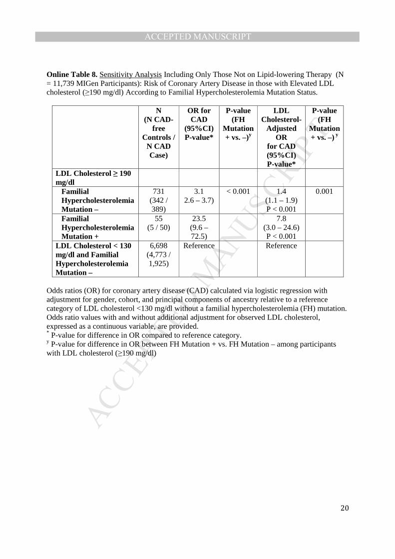
MANUSCRIP
T
ACCEPTED
ACCEPTED MANUSCRIPT
20
Online Table 8. Sensitivity Analysis Including Only Those Not on Lipid-lowering Therapy (N = 11,739 MIGen Participants): Risk of Coronary Artery Disease in those with Elevated LDL cholesterol (≥190 mg/dl) According to Familial Hypercholesterolemia Mutation Status.
N (N CAD-
free Controls /
N CAD Case)
OR for CAD
(95%CI) P-value*
P-value (FH
Mutation + vs. –)y
LDL Cholesterol-
Adjusted OR
for CAD (95%CI) P-value*
P-value (FH
Mutation + vs. –) y
LDL Cholesterol ≥ 190 mg/dl
Familial Hypercholesterolemia Mutation –
731 (342 / 389)
3.1 2.6 – 3.7)
< 0.001 1.4 (1.1 – 1.9) P < 0.001
0.001
Familial Hypercholesterolemia Mutation +
55 (5 / 50)
23.5 (9.6 – 72.5)
7.8 (3.0 – 24.6) P < 0.001
LDL Cholesterol < 130 mg/dl and Familial Hypercholesterolemia Mutation –
6,698 (4,773 / 1,925)
Reference Reference
Odds ratios (OR) for coronary artery disease (CAD) calculated via logistic regression with adjustment for gender, cohort, and principal components of ancestry relative to a reference category of LDL cholesterol <130 mg/dl without a familial hypercholesterolemia (FH) mutation. Odds ratio values with and without additional adjustment for observed LDL cholesterol, expressed as a continuous variable, are provided. * P-value for difference in OR compared to reference category. y P-value for difference in OR between FH Mutation + vs. FH Mutation – among participants with LDL cholesterol (≥190 mg/dl)

MANUSCRIP
T
ACCEPTED
ACCEPTED MANUSCRIPT
21
Online Figure Titles and Legends Online Figure 1. LDL Cholesterol Values in MIGen Control Participants (n=8,577) Online Figure 2. Frequency of Familial Hypercholesterolemia Mutations According to LDL Cholesterol Level and Coronary Artery Disease Status within MIGen Consortium Studies.

MANUSCRIP
T
ACCEPTED
ACCEPTED MANUSCRIPT
22
Online Figure 1.
0.000
0.002
0.004
0.006
0.008
0.010
10 70 130 190 250 310 370 430
LDL−Cholesterol (mg/dl)
Den
sity

MANUSCRIP
T
ACCEPTED
ACCEPTED MANUSCRIPT
23
Online Figure 2.