sex difference in the association between plasma selenium
TRANSCRIPT

RESEARCH Open Access
Sex difference in the association betweenplasma selenium and first stroke: acommunity-based nested case-controlstudyHuan Hu1,2, Chonglei Bi3, Tengfei Lin4, Lishun Liu4, Yun Song4,5, Binyan Wang5,6, Ping Wang7, Ziyi Zhou4,Chongqian Fang3, Hai Ma8, Xiao Huang1,2, Lihua Hu1,9, Xiping Xu4, Hao Zhang4, Yong Huo9, Xiaobin Wang10,Huihui Bao1,2, Xiaoshu Cheng1,2 and Ping Li1,2*
Abstract
Background: To date, there is no clearly defined association between plasma selenium levels and first stroke. Weaimed to investigate the association between baseline plasma selenium and first stroke risk in a community-basedChinese population.
Methods: Using a nested case-control study design, a total of 1255 first stroke cases and 1255 matched controlswere analyzed. Participant plasma selenium concentrations were measured by inductively coupled plasma massspectrometry (ICP-MS), and the association of plasma selenium with first stroke risk was estimated by conditionallogistic regression models.
Results: Overall, a non-linear negative association between plasma selenium and first total stroke and first ischemicstroke risks was found in males but not in females. Compared with participants with lower selenium levels (tertile1–2, < 94.1 ng/mL), participants with higher selenium levels (tertile 3, ≥ 94.1 ng/mL) had significantly lower risks offirst total stroke (OR 0.63; 95% CI 0.48, 0.83) and first ischemic stroke (OR 0.61; 95% CI 0.45, 0.83) in males but not infemales with first total stroke (OR 0.92; 95% CI 0.69, 1.22) and first ischemic stroke (OR 0.89; 95% CI 0.65, 1.22).Furthermore, a stronger association between plasma selenium and first total stroke was found in males with highervitamin E levels (≥ 13.5 μg/mL vs. < 13.5 μg/mL P-interaction = 0.007). No significant association was observedbetween plasma selenium and first hemorrhagic stroke risk in either males or females.
Conclusion: Our study indicated a significant, non-linear, negative association between plasma selenium and firststroke in males but not in females.
Trial registration: ChiCTR1800017274.
Keywords: Selenium, First stroke, First ischemic stroke, First hemorrhagic stroke, Vitamin E
© The Author(s). 2021 Open Access This article is licensed under a Creative Commons Attribution 4.0 International License,which permits use, sharing, adaptation, distribution and reproduction in any medium or format, as long as you giveappropriate credit to the original author(s) and the source, provide a link to the Creative Commons licence, and indicate ifchanges were made. The images or other third party material in this article are included in the article's Creative Commonslicence, unless indicated otherwise in a credit line to the material. If material is not included in the article's Creative Commonslicence and your intended use is not permitted by statutory regulation or exceeds the permitted use, you will need to obtainpermission directly from the copyright holder. To view a copy of this licence, visit http://creativecommons.org/licenses/by/4.0/.The Creative Commons Public Domain Dedication waiver (http://creativecommons.org/publicdomain/zero/1.0/) applies to thedata made available in this article, unless otherwise stated in a credit line to the data.
* Correspondence: [email protected] of Cardiovascular Medicine, The Second Affiliated Hospital ofNanchang University, No. 1 Minde Road, Nanchang, Jiangxi Province, China2Center for Prevention and Treatment of Cardiovascular Diseases, TheSecond Affiliated Hospital of Nanchang University, Nanchang, No. 1 MindeRoad, Nanchang, Jiangxi Province, ChinaFull list of author information is available at the end of the article
Hu et al. Biology of Sex Differences (2021) 12:39 https://doi.org/10.1186/s13293-021-00383-2

HighlightsThis study is the first work to find a significant non-linear, inverse association between baseline plasma sel-enium and first total stroke and first ischemic strokerisks in males but not in females. The present study isalso the first to indicate a stronger non-linear negativerelationship between plasma selenium and first stroke inmale participants with higher plasma vitamin E levels (≥13.5 μg/mL) than in those with lower plasma vitamin Elevels (< 13.5 μg/mL). This study found no significantassociation between plasma selenium and firsthemorrhagic stroke risk in either males or females.
IntroductionStroke is a leading cause of mortality and disabilityworldwide. Accounting for almost one-third of strokemortality worldwide, China bears the heaviest strokeburden in the world [1, 2]. Since control of risk factorsfor stroke helps decrease stroke burden [3, 4], the identi-fication of novel risk factors is urgent to further lowerstroke risk. The potential effects of nutritional determi-nants on stroke have attracted increasing attention forcontrolling the occurrence of stroke. Appropriate intakeof electrolyte minerals, one type of micronutrient, playsa vital role in maintaining brain health. There was an in-verse association between potassium intake and strokerisk, whereas higher sodium intake was associated with ahigher risk of stroke [5, 6]. We previously found thatthere was a significant inverse association betweenplasma zinc and first hemorrhagic stroke [7], whereasexcess zinc also led to neurotoxicity [8]. Thus, appropri-ate intake of nutritional determinants and maintainingtheir reasonable levels play important roles in control-ling stroke risk. Although accumulating evidence has in-dicated that trace elements might exert effects on stroke[9, 10], there is still limited evidence of dietary supple-mentation for stroke prevention [11], suggesting thatmuch more evidence is needed to clarify this issue.Selenium (Se), an essential trace element, acts as the
active center of selenoproteins or selenoenzymes (e.g.,glutathione peroxidases), which have many importantbiological functions, including antioxidant, anti-inflammatory, and immunoregulatory roles [12–14]. In-sufficient or excessive Se intake may be associated withmany adverse health outcomes [15–17]. Se overexposuremight have toxic effects, which should not be ignored, ashigh Se levels have been reported to be positively associ-ated with type 2 diabetes, and this association might ex-ists only in females [18–21], whereas Se deficiency ismainly positively associated with Keshan disease andKashin-Beck disease [22, 23]. Low Se status was also re-ported to be related to an increased risk of thyroid dis-ease, including autoimmune thyroiditis, subclinicalhypothyroidism, and hypothyroidism, and Se deficiency
might constitute a risk factor for hyperthyroidism, espe-cially in males [24, 25]. Low serum Se was an independ-ent predictor of cancer mortality only in maleparticipants with a median plasma Se concentration of75.9 ng/mL, while another nested case-control studywith a mean plasma Se concentration of 84.0 ng/mLfrom the European Prospective Investigation into Cancerand Nutrition indicated that Se status is suboptimal inmany Europeans (with low Se status) and suggested aninverse association between colorectal cancer risk andhigher serum Se status, which was more evident in fe-males [26, 27]. Previous studies from two prominent Seintervention trials in North America (with currently highselenium status) observed conflicting results with regardto Se intake and cancer risk, which indicated that aninteraction of Se status and cancer risk could be foundin the Nutritional Prevention of Cancer Trial (NPC) [28]with a median plasma Se concentration of 115 ng/mL atbaseline but not in Selenium and Vitamin E Cancer Pre-vention Trial (SELECT) [29] with a median plasma Seconcentration of 136 ng/mL at baseline. High Se statuswas also positively associated with cognitive performancein males only among the population of the NationHealth and Nutrition Survey (NHANES, 2011–2014),with a median whole blood Se concentration of 196.7ng/mL [30]. Thus, there was an interaction of Se withdiabetes, thyroid disease, cancers, and cognitive perform-ance, which seemed to be associated with sex differencesand needs to be further understood. Different baselineSe levels and any other poorly defined confoundersmight affect these complex associations. In particular,health problems caused by Se deficiency need to begiven great attention in China, a Se-deficient countrywhere it is estimated that over 105 million people poten-tially face adverse health impacts due to Se deficiency[31, 32]. Based on the aforementioned conclusions, morestudies are still needed to identify the appropriate levelof Se that should be considered safe to address the un-certainties regarding the health risks of Se exposure. Al-though cross-sectional epidemiologic studies haveindicated inverse associations between Se levels andstroke risk [33–35], previous prospective epidemiologicstudies have reported inconsistent findings on the asso-ciations between Se concentrations and stroke risk [36–38]. Moreover, few studies have thoroughly analyzed thepotential modifiers affecting this association; in particu-lar, whether this relationship was associated with sex dif-ferences remains unclear. Therefore, the prospectiverelationship between plasma Se and the risk of firststroke remains inconclusive and deserves furtherinvestigation.To narrow the knowledge gap mentioned above, we
performed a nested case-control study to investigate theassociation between baseline plasma Se levels and the
Hu et al. Biology of Sex Differences (2021) 12:39 Page 2 of 15

risk of first total stroke and stroke subtypes (ischemicstroke and hemorrhagic strokes) and examined any pos-sible effect modifiers using data from a community-based population in China.
MethodsStudy population and designOur present study is a subset of the China H-typeHypertension Registry Study (CHHRS; URL: http://www.chictr.org.cn; Unique identifier: ChiCTR1800017274),which is an ongoing community-based non-intervention,prospective, observational, multicenter, real-world regis-try study and was mainly conducted in RongchengCounty, Shandong Province, and Lianyungang, JiangsuProvince, China. It was designed to establish a nationalregistry of patients with hypertension, to investigate theprevalence and treatment of H-type hypertension inChina and the related factors affecting its prognosis, andfinally to construct a risk prediction model of cardio-cerebral and renal vascular diseases. Eligible participantswere men and women aged ≥ 18 years with essentialhypertension, defined as seated systolic blood pressure(SBP) ≥ 140 mmHg and/or seated diastolic blood pres-sure (DBP) ≥ 90 mmHg at the screening visit. Theboundaries for elevated blood pressure (140 and 90mmHg, respectively) were applied irrespective of age.Participants were excluded if they had psychological ornervous system impairment resulting in an inability todemonstrate informed consent or were unable to befollowed-up according to the study protocol. The trialconsisted of two stages: screening and recruitment and a3-year observation follow-up period. Participants werescheduled for follow-up every 3 months. At each visit,blood pressure, heart rate, the usage of medications, ad-verse events, and study outcome events were measuredand recorded. The primary outcome was the first com-posite of cardiovascular events consisting of non-fatalstroke, myocardial infarction, vascular death, and all-cause death.The current nested case-control study utilized data
from the CHHRS, which was conducted in Rongcheng, acoastal area of Shandong Province, China. This studymatched stroke cases with an equal number of controls(patients without stroke) by age ± 1 year, sex, and vil-lage. Patients with stroke data from the Chinese Centersfor Disease Control and Prevention (CDC, 2016-2018)who had complete records (physical exam, question-naire, and biological samples) were selected as cases.The initial sample consisted of 1401 incident cases and1401 matched controls, both of which were recruitedfrom the same trial population. Next, we excluded par-ticipants with missing serum selenium values (n = 287)and unpaired individuals (n = 5). Based on the inclusionand exclusion criteria, 1255 stroke cases and 1255
matched controls with complete Se measurements wereselected for final data analysis (Supplementary Fig. 1).
EthicsThe present study was approved by the Ethics Commit-tee of the Institute of Biomedicine, Anhui Medical Uni-versity, Hefei, China. All participants signed an approvedwritten consent form after the study protocol was thor-oughly explained to them.
OutcomesThe primary outcome of the present study was a firstnon-fatal or fatal stroke. Secondary outcomes includedfirst ischemic stroke (fatal and non-fatal) and firsthemorrhagic stroke (fatal and non-fatal).Information on the incidence of first stroke for all par-
ticipants was obtained via the Centers for Disease Con-trol and Prevention of Rongcheng County and checkedagainst the national health insurance system with elec-tronic linkage to all hospitalizations or ascertainedthrough active follow-up. Diseases were coded accordingto the International Classification of Diseases, 10th Revi-sion (ICD-10). Secondary outcomes included first ische-mic stroke (I63) and first hemorrhagic stroke (I60-I61).The primary outcome (first non-fatal or fatal stroke) in-cluded first ischemic stroke (I63), first hemorrhagicstroke (I60-I61), and no type stroke (I64).According to government regulations, local authorities
from medical institutions are required to report all newcases of stroke to the local Centers for Disease Controland Prevention. A report card that includes informationon demographics, diagnostic basis and date of stroke isrequired to be submitted on the 28th of each month.Quality control, including finding and deleting repeatedcases, error checking, and determining any missed cases,is completed by trained officials. Furthermore, the stafffrom the local Centers for Disease Control and Preven-tion double checked these information and were respon-sible for deleting repeated cases and finding logisticalerrors and missed cases. In addition, 5% of all uploadedcases were randomly chosen for further confirmation byphone or door-to-door interviews.
Laboratory assaysBaseline serum total homocysteine (tHcy), fasting glu-cose levels, and lipids were measured using automaticclinical analyzers (Beckman Coulter, AU680) at theShenzhen Tailored Medical Laboratory in Shenzhen,China. Estimated glomerular filtration rates (eGFRs)were estimated by the Chronic Kidney Disease Epidemi-ology Collaboration equation. Baseline plasma vitamin Econcentrations were measured using liquid chromatog-raphy–tandem quadrupole mass spectrometry (LC-MS/MS), and plasma selenium (Se) concentrations were
Hu et al. Biology of Sex Differences (2021) 12:39 Page 3 of 15

measured by inductively coupled plasma mass spectrom-etry (ICP-MS) using a Thermo Fisher iCAP Q ICP-MSin a commercial laboratory (Beijing DIAN Medical La-boratory, China). In the present study, the intra-assayCV for Se ranged from 1.02 to 7.93%, while the inter-assay CV for Se ranged from 2.79 to 3.51%. Accordingto a previous study [39], the reference value (50–120 ng/mL) for plasma Se levels was used in this study.
Statistical analysisBaseline characteristics are presented as the means ±SDs for continuous variables and as frequencies (%) forcategorical variables. Differences in baseline characteris-tics between males and females and cases and controlswere compared using chi-square tests for categoricalvariables and t tests for continuous variables. Differencesin population characteristics according to Se tertileswere compared using ANOVA tests, or chi-square tests.Variables that are known as traditional or suspected
risk factors for stroke [40] and matched variables or vari-ables that showed significant differences between casesand controls were adjusted for in the models. Odds ra-tios (ORs) and 95% confidence intervals (95% CIs) forfirst stroke, first ischemic stroke, and first hemorrhagicstroke were calculated by modeling plasma Se as tertilesusing conditional logistic regression, without and withadjustment for matched variables (sex and age), bodymass index (BMI), baseline systolic blood pressure(SBP), baseline diastolic blood pressure (DBP), smokingstatus, alcohol consumption, labor intensity, baselinetotal homocysteine (tHcy), plasma vitamin E, fasting glu-cose, estimated glomerular filtration rate (eGFR), anti-platelet drugs, lipoprotein-lowering drugs, glucose-lowering drugs, anti-hypertensive drugs, self-reportedhypertension, self-reported diabetes, self-reported atrialfibrillation, and self-reported hyperlipidemia. A general-ized additive model (GAM) and smooth curve fitting(penalized spline method) were evaluated to furthercharacterize the shape of the association between serumSe and first stroke and its subtypes. As additional ex-ploratory analyses, possible modifications of the associ-ation between plasma Se (tertile 3, ≥ 94.1 vs. tertile 1–2,< 94.1 ng/mL) and first total stroke in male and femaleparticipants were also assessed for variables includingage (< 70 vs. ≥ 70 years), BMI (< 24 vs. ≥ 24 kg/m2),current smoking (yes vs. no), current alcohol consump-tion (yes vs. no), baseline SBP (< 140 vs. ≥ 140 mmHg),fasting glucose (< 6.1 vs. ≥ 6.1 mmol/L or diabetes), totalcholesterol (< 5.8 [median] vs. ≥ 5.8 mmol/L), triglycer-ides (< 1.2 [median] vs. ≥ 1.2 mmol/L), estimated glom-erular filtration rate (< 90 vs. ≥ 90 mL/min/1.73 m2),total homocysteine (< 12.5 [median] vs. ≥ 12.5 μmol/L),and vitamin E (< 13.5 [median] vs. ≥ 13.5 μmol/L) usingmultivariate logistic regression models. Diabetes was
defined as fasting serum glucose ≥ 7.0 mmol/L, self-reported use of anti-diabetic medications, or physician-diagnosed diabetes. Potential interactions were examinedby including the interaction terms in those logistic re-gression models with the greatest number of confound-ing variables.A 2-tailed P < 0.05 was considered to be statistically
significant in all analyses. R software version 3.4.3 (www.R-project.org) and Empower version 2.17.8 (www.empowerstats.com, X&Y Solutions, Inc.) were used forall statistical analyses.
ResultsStudy participants and baseline characteristicsA total of 1255 first stroke cases (1079 cases of first is-chemic stroke, 171 cases of first hemorrhagic stroke, and5 cases of first uncertain type of stroke) and 1255matched controls were included in this analysis. Themean age of all participants at baseline was 70.8 years(SD, 8.1), 49.5% of the participants were male, and themean Se level was 87.2 ng/mL (SD, 18.6). The baselinecharacteristics of the male and female participants areshown in Table 1. The reference value of 50–120 ng/mLfor serum Se concentration was used in this study. Thedetailed plasma Se concentration distribution of subjectsis listed in Supplemental Table 1, and 95.1% of the par-ticipants were within the normal range of Se levels withrespect to the reference value (50–120 ng/mL) forplasma Se concentrations. As shown in Table 1, maleparticipants had non-significantly higher Se levels (87.7± 18.6 vs. 86.8 ± 18.6 ng/mL; P = 0.230) and significantlylower vitamin E (12.9 ± 3.3 vs. 15.2 ± 4.3 ng/mL; P <0.001) than females. The distribution graph of serum Seand vitamin E levels in males versus females showedsimilar results (Supplemental Fig. 2A-B). Males alsotended to be older, were more likely to be currentsmokers and current drinkers, had higher DBP and tHcylevels, as well as lower BMI, SBP, TC, TG, and glucoselevels at baseline and a lower frequency of lipid-lowering, glucose-lowering, and anti-hypertensive druguse, and were less likely to be hypertensive patients orself-reported diabetic and self-reported hyperlipidemiapatients compared with female participants.The baseline characteristics of cases and control par-
ticipants by sex are shown in Table 2. For stroke cases,male stroke cases had non-significantly lower Se levelsthan females (86.2 ± 16.8 vs. 87.0 ± 18.4 ng/mL; P =0.436). Male stroke cases also tended to be older, weremore likely to be current smokers and current drinkers,had higher DBP and tHcy levels, as well as lower BMI,SBP, TC, TG, glucose, and vitamin E levels at baselineand a lower frequency of glucose-lowering and anti-hypertensive drug use, and were less likely to be hyper-tensive patients or self-reported diabetic and self-
Hu et al. Biology of Sex Differences (2021) 12:39 Page 4 of 15

reported hyperlipidemia patients compared with femalestroke cases. For non-stroke controls, male non-strokecontrols had significantly higher Se levels than females(89.1 ± 20.1 vs. 86.6 ± 18.7 ng/mL; P = 0.020). Malenon-stroke controls also tended to be older, were morelikely to be current smokers, current drinkers and self-reported atrial fibrillation patients, had higher DBP andtHcy levels, as well as lower BMI, SBP, TC, TG, and vita-min E levels at baseline and a lower frequency ofglucose-lowering and anti-hypertensive drug use, andwere less likely to be hypertensive patients or self-reported diabetic patients compared with female non-stroke controls.
In addition, plasma Se was positively associated withBMI, current smoking, current alcohol consumption,self-reported diabetes, a higher frequency of glucose-lowering drug use, TC, high-density lipoprotein choles-terol, fasting glucose, and vitamin E levels and was in-versely associated with labor intensity and tHcy levels atbaseline (Supplemental Table 2).
Association between plasma selenium concentration andfirst stroke in all participantsOverall, there was a non-linear negative association be-tween plasma Se levels and the risk of first total strokeand first ischemic stroke (Fig. 1A, B) but not with the
Table 1 Baseline characteristics of male and female participantsa
Characteristics Total Male Female P value
N n = 2510 n=1242 n = 1268
Age, years 70.8 ± 8.1 71.4 ± 8.1 70.1 ± 8.0 < 0.001
BMI, kg/m2 26.2 ± 4.1 25.3 ± 3.6 27.0 ± 4.4 < 0.001
Current smoking, n (%) 551 (22.0) 547 (44.0) 4 (0.3) < 0.001
Current alcohol drinking, n (%) 612 (24.4) 599 (48.2) 13 (1.0) < 0.001
Baseline SBP, mmHg 153.3 ± 23.1 150.8 ± 22.3 155.7 ± 23.6 < 0.001
Baseline DBP, mmHg 85.3 ± 12.3 86.2 ± 12.5 84.3 ± 12.0 < 0.001
Self-reported hypertension, n (%) 1312 (52.3) 553 (44.5) 759 (59.9) < 0.001
Self-reported diabetes, n (%) 424 (16.9) 161 (13.0) 263 (20.7) < 0.001
Self-reported hyperlipidemia, n (%) 264 (10.5) 108 (8.7) 156 (12.3) 0.003
Self-reported atrial fibrillation, n (%) 46 (1.8) 26 (2.1) 20 (1.6) 0.335
Hypertension, n (%)b 2016 (80.3) 940 (75.7) 1076 (84.9) < 0.001
Labor intensity, n (%) < 0.001
Mild 1885 (75.1) 887 (71.4) 998 (78.7)
Moderate 488 (19.4) 280 (22.5) 208 (16.4)
Severe 137 (5.5) 75 (6.0) 62 (4.9)
Medication use, n (%)
Anti-platelet drugs 83 (3.3) 47 (3.8) 36 (2.8) 0.186
Lipid-lowering drugs 44 (1.8) 15 (1.2) 29 (2.3) 0.039
Glucose-lowering drugs 301 (12.0) 112 (9.0) 189 (14.9) < 0.001
Anti-hypertensive drugs 1152 (45.9) 476 (38.3) 676 (53.3) < 0.001
Laboratory results
TC, mmol/L 5.9 ± 1.2 5.6 ± 1.1 6.1 ± 1.3 < 0.001
TG, mmol/L 1.4 ± 0.9 1.2 ± 0.7 1.6 ± 0.9 < 0.001
HDL-C, mmol/L 1.6 ± 0.4 1.6 ± 0.4 1.6 ± 0.4 0.558
Glucose, mmol/L 6.3 ± 2.3 6.1 ± 2.1 6.4 ± 2.5 < 0.001
tHcy, μmol/L 13.9 ± 7.2 15.3 ± 8.6 12.5 ± 5.0 < 0.001
eGFR, mL/min/1.73 m2 92.5 ± 14.4 92.3 ± 15.1 92.7 ± 13.8 0.428
Vitamin E, μg/mL 14.0 ± 4.0 12.9 ± 3.3 15.2 ± 4.3 < 0.001
Selenium, ng/mL 87.2 ± 18.6 87.7 ± 18.6 86.8 ± 18.6 0.230
Abbreviations: BMI body mass index, SBP systolic blood pressure, DBP diastolic blood pressure, TC total cholesterol, TG triglycerides, HDL-C high-density lipoprotein-cholesterol, tHcy total homocysteine, eGFR estimated glomerular filtration rateaVariables are presented as the mean ± SD or n (%)bHypertension was defined as a self-reported history of hypertension, use of anti-hypertensive drugs, SBP ≥ 140 mmHg, or DBP ≥ 90 mmHg
Hu et al. Biology of Sex Differences (2021) 12:39 Page 5 of 15

risk of first hemorrhagic stroke (Supplemental Fig. 3A) inall participants. Consistently, when plasma Se was assessedas tertiles that were calculated among the whole popula-tion, significantly lower risks of first total stroke (Model 2,OR 0.77; 95% CI 0.61, 0.97) and first ischemic stroke(model 2, OR 0.78; 95% CI 0.60, 0.99) were found in par-ticipants in tertile 3 (≥ 94.1 ng/mL) than in those in tertile1 (< 79.1 ng/mL) (Table 3). It is worth noting that non-significantly higher risks of first total stroke (model 2, OR1.02; 95% CI 0.82, 1.26) and first ischemic stroke (model2, OR 1.05; 95% CI 0.83, 1.33) were found in participantsin tertile 2 (79.1 to < 94.1 ng/mL) than in those in tertile 1
(< 79.1 ng/mL) (Table 3). Due to the similar first totalstroke and first ischemic stroke prevalence in participantswith Se levels in tertile 1 and tertile 2 (Table 3), we com-bined these two groups into one group called tertile 1–2.Compared with participants with lower Se levels in tertile1–2 (< 94.1 ng/mL), significantly lower risks of first totalstroke (model 2, OR 0.76; 95% CI 0.63, 0.93) and first is-chemic stroke (model 2, OR 0.75; 95% CI 0.61, 0.93) werefound in those with higher Se levels in tertile 3 (≥ 94.1 ng/mL) (Table 3). However, no significant association wasfound between plasma Se concentrations and firsthemorrhagic stroke (Table 3).
Table 2 Baseline characteristics of cases and control participants by sexa
Characteristics First stroke cases Pvalue
Non-stroke controls PvalueMales Females Males Females
N n = 621 n = 634 n = 621 n = 634
Age, years 71.4 ± 8.1 70.1± 8.0 0.004 71.4 ± 8.1 70.1 ± 8.0 0.003
BMI, kg/m2 25.7 ± 3.6 27.3 ± 4.9 < 0.001 25.0 ± 3.5 26.7 ± 3.8 < 0.001
Current smoking, n (%) 293 (47.2) 3 (0.5) < 0.001 254 (40.9) 1 (0.2) < 0.001
Current alcohol drinking, n (%) 291 (46.9) 4 (0.6) < 0.001 308 (49.6) 9 (1.4) < 0.001
Baseline SBP, mmHg 154.8 ± 23.1 159.5 ± 24.2 < 0.001 146.8 ± 20.8 151.8 ± 22.2 < 0.001
Baseline DBP, mmHg 88.0 ± 13.4 86.4 ± 12.2 0.030 84.4 ± 11.3 82.2 ± 11.4 < 0.001
Self-reported hypertension, n (%) 319 (51.4) 427 (67.4) < 0.001 234 (37.7) 332 (52.4) < 0.001
Self-reported diabetes, n (%) 97 (15.6) 164 (25.9) < 0.001 64 (10.3) 99 (15.6) 0.005
Self-reported hyperlipidemia, n (%) 46 (7.4) 83 (13.1) < 0.001 62 (10.0) 73 (11.5) 0.382
Self-reported atrial fibrillation, n (%) 13 (2.1) 16 (2.5) 0.612 13 (2.1) 4 (0.6) 0.025
Hypertension, n (%)b 511 (82.3) 564 (89.0) < 0.001 429 (69.1) 512 (80.8) < 0.001
Labor intensity, n (%) < 0.001 0.006
Mild 457 (73.6) 521 (82.2) 430 (69.2) 477 (75.2)
Moderate 125 (20.1) 96 (15.1) 155 (25.0) 112 (17.7)
Severe 39 (6.3) 17 (2.7) 36 (5.8) 45 (7.1)
Medication use, n (%)
Anti-platelet drugs 37 (6.0) 23 (3.6) 0.053 10 (1.6) 13 (2.1) 0.561
Lipid-lowering drugs 8 (1.3) 14 (2.2) 0.214 7 (1.1) 15 (2.4) 0.095
Glucose-lowering drugs 72 (11.6) 121 (19.1) < 0.001 40 (6.4) 68 (10.7) 0.007
Anti-hypertensive drugs 281 (45.2) 383 (60.4) < 0.001 195 (31.4) 293 (46.2) < 0.001
Laboratory results
TC, mmol/L 5.6 ± 1.1 6.1 ± 1.3 < 0.001 5.7 ± 1.1 6.1 ± 1.3 < 0.001
TG, mmol/L 1.3 ± 0.8 1.7 ± 1.0 < 0.001 1.1 ± 0.7 1.5 ± 0.8 < 0.001
HDL-C, mmol/L 1.6 ± 0.4 1.6 ± 0.4 0.935 1.7 ± 0.4 1.7 ± 0.4 0.376
Glucose, mmol/L 6.3 ± 2.2 6.8 ± 2.8 0.001 5.9 ± 1.9 6.1 ± 2.2 0.051
tHcy, μmol/L 15.8 ±9.5 12.8 ± 5.7 < 0.001 14.7 ± 7.5 12.2 ± 4.3 < 0.001
eGFR, mL/min/1.73 m2 91.4 ± 15.9 92.0 ± 14.7 0.485 93.1 ± 14.1 93.4 ± 12.7 0.685
Vitamin E, μg/mL 13.0 ± 3.4 15.2 ± 4.3 < 0.001 12.8 ± 3.1 15.1 ± 4.3 < 0.001
Selenium, ng/mL 86.2 ± 16.8 87.0 ± 18.4 0.436 89.1 ± 20.1 86.6 ± 18.7 0.020
Abbreviations: BMI body mass index, SBP systolic blood pressure, DBP diastolic blood pressure, TC total cholesterol, TG triglycerides, HDL-C high-density lipoprotein-cholesterol, tHcy total homocysteine, eGFR estimated glomerular filtration rateaVariables are presented as the mean ± SD or n (%)bHypertension was defined as a self-reported history of hypertension, use of anti-hypertensive drugs, SBP ≥ 140 mmHg, or DBP ≥ 90 mmHg
Hu et al. Biology of Sex Differences (2021) 12:39 Page 6 of 15

Association between plasma selenium concentrations andfirst stroke by sexGiven the differences in plasma Se levels between maleand female participants (87.7 ± 18.6 vs. 86.8 ± 18.6 ng/mL), we further investigated the possible effect of sex onthe Se-first stroke association. Overall, there was a non-linear negative association between plasma Se levels andthe risks of first total stroke and first ischemic stroke inmales (Fig. 1C, D) but not in females (Fig. 1E, F).
Furthermore, there was no significant association be-tween plasma Se and first hemorrhagic stroke in eithersex (Supplemental Fig. 3B-C). Consistently, when theplasma Se of males and females was assessed as tertilesaccording to the tertile cut-off value of the whole popu-lation, the highest tertile (T3, ≥ 94.1 ng/mL) of plasmaSe was associated with a lower first total stroke risk inmales (model 2, OR 0.67; 95% CI 0.48, 0.93, P = 0.017)but not in females (model 2, OR 0.85; 95% CI 0.61, 1.19,
Fig. 1 The association between baseline plasma selenium and first total stroke and first ischemic stroke risks. Odds ratios for first total stroke inthe (A) total population, (C) males, and (E) females, and for first ischemic stroke in the (B) total population, (D) males, and (F) females by plasmaselenium levels. In addition to the matching factors (age and sex), the splines also adjusted for BMI, baseline SBP, baseline DBP, smoking status,alcohol consumption, labor intensity, baseline total homocysteine, vitamin E, fasting glucose, estimated glomerular filtration rate (eGFR), anti-platelet drugs, lipoprotein-lowering drugs, glucose-lowering drugs, anti-hypertensive drugs, self-reported hypertension, self-reported diabetes, self-reported atrial fibrillation, and self-reported hyperlipidemia
Hu et al. Biology of Sex Differences (2021) 12:39 Page 7 of 15

P = 0.353) compared with the lowest tertile (T1, < 79.1ng/mL) of plasma Se (Table 4). It is also worth notingthat non-significantly higher risks of first total stroke(model 2, OR 1.11; 95% CI 0.80, 1.54) and first ischemicstroke (model 2, OR 1.17; 95% CI 0.82, 1.66) were foundin male participants in tertile 2 (79.1 to < 94.1 ng/mL)than in those in tertile 1 (< 79.1 ng/mL) (Table 3). Ac-cordingly, higher Se levels in tertile 3 (≥ 94.1 ng/mL)were associated with a lower first total stroke risk inmales (model 2 OR 0.63; 95% CI 0.48, 0.83, P = 0.001),but not in females (model 2, OR 0.92; 95% CI 0.69, 1.22,P = 0.563) compared with lower Se levels in tertile 1–2(< 94.1 ng/mL) (Table 4).Similar effects of sex on the Se-first ischemic stroke
association were also observed and are displayed inTable 4. However, no significant association was foundbetween plasma Se and first hemorrhagic stroke riskamong either males or females (Table 4).
Stratified analysis by potential effect modifiers in maleand female participantsStratified analyses were conducted to explore potentialmodifiers affecting the association between plasma Se(tertile 3, ≥ 94.1 vs. tertile 1–2, < 94.1 ng/mL) and firsttotal stroke risk among male participants (Table 5). Astronger non-linear negative association between base-line plasma Se and first total stroke was found amongmales with higher vitamin E levels than among thosewith lower vitamin E levels (≥ 13.5 μg/mL; OR 0.39; 95%CI 0.25, 0.60; vs. < 13.5 μg/mL; OR 0.85; 95% CI 0.62,1.17; P for interaction = 0.007). None of the other vari-ables, including age (< 70 vs. ≥ 70 years), BMI (< 24 vs. ≥24 kg/m2), current smoking (yes vs. no), current alcoholconsumption (yes vs. no), baseline SBP (< 140 vs. ≥ 140mmHg), fasting glucose (< 6.1 vs. ≥ 6.1 mmol/L or dia-betes), TC (< 5.8 [median] vs. ≥ 5.8 mmol/L), TG (< 1.2[median] vs. ≥ 1.2 mmol/L), eGFR (< 90 vs. ≥ 90 mL/
Table 3 Risk of first stroke (total and subtypes) associated with plasma selenium concentrations in all participantsa
Selenium, ng/mL Cases/controls
Model 1 Model 2
OR (95% CI) P value OR (95% CI) P value
First total stroke
Tertiles
T1 (< 79.1) 420/417 Ref. Ref.
T2 (79.1 to < 94.1) 434/402 1.07 (0.88, 1.30) 0.516 1.02 (0.82, 1.26) 0.891
T3 (≥ 94.1) 401/436 0.90 (0.73, 1.11) 0.317 0.77 (0.61, 0.97) 0.027
Categories
T1–T2 (< 94.1) 854/819 Ref. Ref.
T3 (≥ 94.1) 401/436 0.87 (0.73, 1.04) 0.119 0.76 (0.63, 0.93) 0.007
Ischemic stroke
Tertiles
T1 (< 79.1) 360/363 Ref. Ref.
T2 (79.1 to < 94.1) 372/340 1.10 (0.89, 1.35) 0.376 1.05 (0.83, 1.33) 0.672
T3 (≥ 94.1) 347/376 0.92 (0.74, 1.14) 0.444 0.78 (0.60, 0.99) 0.045
Categories
T1–T2 (< 94.1) 732/703 Ref. Ref.
T3 (≥ 94.1) 347/376 0.87 (0.72, 1.06) 0.163 0.75 (0.61, 0.93) 0.009
Hemorrhagic stroke
Tertiles
T1 (< 79.1) 58/54 Ref. Ref.
T2 (79.1 to < 94.1) 60/61 0.88 (0.50, 1.57) 0.673 0.76 (0.38, 1.55) 0.455
T3 (≥ 94.1) 53/56 0.85 (0.47, 1.54) 0.587 0.72 (0.35, 1.48) 0.371
Categories
T1–T2 (< 94.1) 118/115 Ref. Ref.
T3 (≥ 94.1) 53/56 0.92 (0.57, 1.47) 0.718 0.85 (0.48, 1.50) 0.577a ORs of first stroke (total), first ischemic, and hemorrhagic stroke in relation to plasma selenium (tertiles) were analyzed using conditional logistic regressionmodels. Model 1 is conditioned on the matching factors of age and sex; model 2 is conditioned on the matching factors of age and sex, as well as adjusted forBMI, baseline SBP, baseline DBP, smoking status, alcohol consumption, labour intensity, baseline total homocysteine, vitamin E, fasting glucose, estimatedglomerular filtration rate (eGFR), anti-platelet drugs, lipoprotein-lowering drugs, glucose-lowering drugs, anti-hypertensive drugs, self-reported hypertension, self-reported atrial fibrillation, self-reported diabetes, and self-reported hyperlipidemia.
Hu et al. Biology of Sex Differences (2021) 12:39 Page 8 of 15
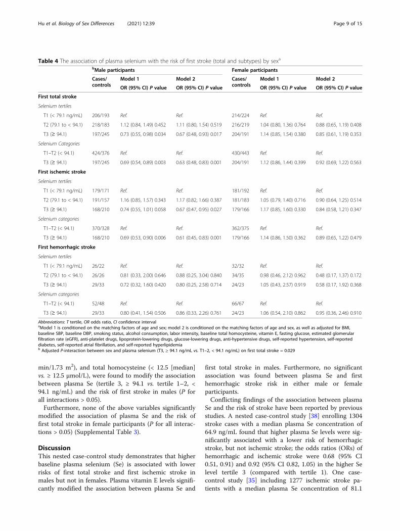
min/1.73 m2), and total homocysteine (< 12.5 [median]vs. ≥ 12.5 μmol/L), were found to modify the associationbetween plasma Se (tertile 3, ≥ 94.1 vs. tertile 1–2, <94.1 ng/mL) and the risk of first stroke in males (P forall interactions > 0.05).Furthermore, none of the above variables significantly
modified the association of plasma Se and the risk offirst total stroke in female participants (P for all interac-tions > 0.05) (Supplemental Table 3).
DiscussionThis nested case-control study demonstrates that higherbaseline plasma selenium (Se) is associated with lowerrisks of first total stroke and first ischemic stroke inmales but not in females. Plasma vitamin E levels signifi-cantly modified the association between plasma Se and
first total stroke in males. Furthermore, no significantassociation was found between plasma Se and firsthemorrhagic stroke risk in either male or femaleparticipants.Conflicting findings of the association between plasma
Se and the risk of stroke have been reported by previousstudies. A nested case-control study [38] enrolling 1304stroke cases with a median plasma Se concentration of64.9 ng/mL found that higher plasma Se levels were sig-nificantly associated with a lower risk of hemorrhagicstroke, but not ischemic stroke; the odds ratios (ORs) ofhemorrhagic and ischemic stroke were 0.68 (95% CI0.51, 0.91) and 0.92 (95% CI 0.82, 1.05) in the higher Selevel tertile 3 (compared with tertile 1). One case-control study [35] including 1277 ischemic stroke pa-tients with a median plasma Se concentration of 81.1
Table 4 The association of plasma selenium with the risk of first stroke (total and subtypes) by sexa
bMale participants Female participants
Cases/controls
Model 1 Model 2 Cases/controls
Model 1 Model 2
OR (95% CI) P value OR (95% CI) P value OR (95% CI) P value OR (95% CI) P value
First total stroke
Selenium tertiles
T1 (< 79.1 ng/mL) 206/193 Ref. Ref. 214/224 Ref. Ref.
T2 (79.1 to < 94.1) 218/183 1.12 (0.84, 1.49) 0.452 1.11 (0.80, 1.54) 0.519 216/219 1.04 (0.80, 1.36) 0.764 0.88 (0.65, 1.19) 0.408
T3 (≥ 94.1) 197/245 0.73 (0.55, 0.98) 0.034 0.67 (0.48, 0.93) 0.017 204/191 1.14 (0.85, 1.54) 0.380 0.85 (0.61, 1.19) 0.353
Selenium Categories
T1–T2 (< 94.1) 424/376 Ref. Ref. 430/443 Ref. Ref.
T3 (≥ 94.1) 197/245 0.69 (0.54, 0.89) 0.003 0.63 (0.48, 0.83) 0.001 204/191 1.12 (0.86, 1.44) 0.399 0.92 (0.69, 1.22) 0.563
First ischemic stroke
Selenium tertiles
T1 (< 79.1 ng/mL) 179/171 Ref. Ref. 181/192 Ref. Ref.
T2 (79.1 to < 94.1) 191/157 1.16 (0.85, 1.57) 0.343 1.17 (0.82, 1.66) 0.387 181/183 1.05 (0.79, 1.40) 0.716 0.90 (0.64, 1.25) 0.514
T3 (≥ 94.1) 168/210 0.74 (0.55, 1.01) 0.058 0.67 (0.47, 0.95) 0.027 179/166 1.17 (0.85, 1.60) 0.330 0.84 (0.58, 1.21) 0.347
Selenium categories
T1–T2 (< 94.1) 370/328 Ref. Ref. 362/375 Ref. Ref.
T3 (≥ 94.1) 168/210 0.69 (0.53, 0.90) 0.006 0.61 (0.45, 0.83) 0.001 179/166 1.14 (0.86, 1.50) 0.362 0.89 (0.65, 1.22) 0.479
First hemorrhagic stroke
Selenium tertiles
T1 (< 79.1 ng/mL) 26/22 Ref. Ref. 32/32 Ref. Ref.
T2 (79.1 to < 94.1) 26/26 0.81 (0.33, 2.00) 0.646 0.88 (0.25, 3.04) 0.840 34/35 0.98 (0.46, 2.12) 0.962 0.48 (0.17, 1.37) 0.172
T3 (≥ 94.1) 29/33 0.72 (0.32, 1.60) 0.420 0.80 (0.25, 2.58) 0.714 24/23 1.05 (0.43, 2.57) 0.919 0.58 (0.17, 1.92) 0.368
Selenium categories
T1–T2 (< 94.1) 52/48 Ref. Ref. 66/67 Ref. Ref.
T3 (≥ 94.1) 29/33 0.80 (0.41, 1.54) 0.506 0.86 (0.33, 2.26) 0.761 24/23 1.06 (0.54, 2.10) 0.862 0.95 (0.36, 2.46) 0.910
Abbreviations: T tertile, OR odds ratio, CI confidence intervalaModel 1 is conditioned on the matching factors of age and sex; model 2 is conditioned on the matching factors of age and sex, as well as adjusted for BMI,baseline SBP, baseline DBP, smoking status, alcohol consumption, labor intensity, baseline total homocysteine, vitamin E, fasting glucose, estimated glomerularfiltration rate (eGFR), anti-platelet drugs, lipoprotein-lowering drugs, glucose-lowering drugs, anti-hypertensive drugs, self-reported hypertension, self-reporteddiabetes, self-reported atrial fibrillation, and self-reported hyperlipidemiab Adjusted P-interaction between sex and plasma selenium (T3, ≥ 94.1 ng/mL vs. T1–2, < 94.1 ng/mL) on first total stroke = 0.029
Hu et al. Biology of Sex Differences (2021) 12:39 Page 9 of 15

ng/mL indicated that higher plasma Se levels were asso-ciated with a decreased risk of ischemic stroke, wherethe OR for those with higher Se levels in quartile 4(compared with quartile 1) was 0.10 (95% CI 0.06, 0.17).Moreover, the Canadian Health Measures Survey(CHMS 2007–2011) enrolling 7065 adult subjects with a
median whole blood Se concentration of 184 ng/mL andthe National Health and Nutrition Examination Study(NHANES 2011–2012) enrolling 5030 adult subjectswith a median whole blood Se concentration of 181 ng/mL found inverse, cross-sectional associations betweenwhole blood Se and the prevalence of stroke, and the
Table 5 Stratified analysis of the association between plasma selenium concentrations (T3, ≥ 94.1 ng/mL vs. T1–2, < 94.1 ng/mL)and incident risk of first total stroke in males
Subgroups No. of cases/no. of controls aAdjusted model P forinteractionSelenium ≥ 94.1 ng/mL Selenium < 94.1 ng/mL OR (95% CI)
Age, years 0.288
< 70 98/110 173/161 0.73 (0.50, 1.08)
≥ 70 99/135 251/215 0.54 (0.38, 0.76)
Body mass index, kg/m2 0.067
< 24 44/89 151/152 0.39 (0.24, 0.63)
≥ 24 153/156 273/224 0.75 (0.55, 1.02)
Current smoking 0.739
No 95/136 233/231 0.72 (0.51, 1.01)
Yes 102/109 191/145 0.54 (0.36, 0.80)
Current alcohol drinking 0.230
No 98/106 232/207 0.74 (0.51, 1.06)
Yes 99/139 192/169 0.53 (0.37, 0.77)
SBP, mmHg 0.759
< 140 46/92 124/156 0.60 (0.38, 0.96)
≥ 140 151/153 300/220 0.64 (0.47, 0.88)
Glucose, mmol/L 0.898
< 6.1 108/168 274/284 0.66 (0.48, 0.90)
≥ 6.1 or diabetesb 89/77 150/92 0.66 (0.42, 1.02)
TC, mmol/L 0.402
< 5.8 95/107 262/236 0.77 (0.54, 1.10)
≥ 5.8 102/138 162/140 0.53 (0.36, 0.78)
TG, mmol/L 0.611
< 1.2 115/160 246/255 0.71 (0.52, 0.98)
≥ 1.2 82/85 178/121 0.55 (0.36, 0.84)
eGFR, mL/min/1.73 m2 0.753
< 90 64/68 173/141 0.71 (0.45, 1.10)
≥ 90 133/177 251/235 0.61 (0.45, 0.83)
tHcy, μmol/L 0.117
< 12.5 89/105 129/142 0.81 (0.54, 1.22)
≥ 12.5 108/140 293/234 0.55 (0.39, 0.76)
Vitamin E, μg/mL 0.007
< 13.5 117/132 270/277 0.85 (0.62, 1.17)
≥ 13.5 80/113 154/99 0.39 (0.25, 0.60)
Abbreviations: TC, total cholesterol, T tertile, OR odds ratio, CI confidence intervalaORs of first total stroke in relation to serum selenium levels were calculated using multivariate logistic regression models. Each subgroup analysis was adjusted, ifnot stratified, for age, BMI, baseline SBP, baseline DBP, smoking status, alcohol consumption, labor intensity, baseline total homocysteine, vitamin E, fastingglucose, estimated glomerular filtration rate (eGFR), anti-platelet drugs, lipoprotein-lowering drugs, glucose-lowering drugs, anti-hypertensive drugs, self-reportedhypertension, self-reported diabetes, self-reported atrial fibrillation, and self-reported hyperlipidemiabDiabetes was defined as a self-reported history of diabetes mellitus, use of anti-diabetic medications, or fasting glucose ≥ 7.0 mmol/L
Hu et al. Biology of Sex Differences (2021) 12:39 Page 10 of 15

Inuit Health Survey (IHS) enrolling 49 stroke cases witha median whole blood Se concentration of 260 ng/mLindicated a reverse relation of whole blood and dietarySe levels with stroke but revealed an L-shaped relation-ship [33, 34]. However, the Reasons for Geographic andRacial Differences in Stroke Study (REGARDS) [36] re-vealed that higher environmental Se levels were associ-ated with increased stroke risk; the hazard ratio (HR) forthose in quartile 4 (0.45–2.20 ppm) of environmental Seexposure (compared with quartile 1, 0.10–0.30 ppm) was1.33 (95% CI 1.09, 1.62). It is noteworthy that all of thesestudies used different sources (plasma, whole blood, diet,and environment) of Se levels to assess the associationbetween Se levels and stroke, which might be one reasonfor the discrepancy in these findings. Different Se sta-tuses at baseline might be another important reason forthe discrepancy in these findings.Several studies have also explored the association be-
tween Se and stroke mortality specifically. A cohortstudy [37] enrolling 23 stroke death cases among 1100Finnish males with a mean plasma Se concentration of55.4 ng/mL found that low Se selenium (< 45 μg/L) wasassociated with a higher risk of stroke mortality, report-ing an adjusted relative risk of 3.7 (95% CI 1.0, 13.1).The NHANES III cohort study [41] including 13,887participants with a mean plasma Se concentration of125.6 ng/mL found that the association curve for Se andstroke mortality had a reversed U-shape. However, an-other cohort study including 1103 Chinese participantswith a mean plasma Se concentration of 73 ng/mLfound no significant association of plasma Se levels andstroke mortality [42]. Notably, these studies focused onstroke mortality, and these findings might not representthe association of plasma Se levels and first stroke risk.Similarly, prospective associations between Se status/in-take and cardiovascular outcomes remain inconclusive[43–45]. The Selenium and Vitamin E Cancer Preven-tion Trial (SELECT) with a median plasma Se concen-tration of 136 ng/mL at baseline and the NutritionalPrevention of Cancer Trial (NPC) with a median plasmaSe concentration of 115 ng/mL at baseline found nobeneficial effects of Se supplementation on cardiovascu-lar outcomes in relatively similar North American popu-lations. However, an interaction of Se status and cancerrisk could be found in NPC but not in SELECT. A previ-ous meta-analysis enrolling 16 prospective studies indi-cated that the Se level was negatively related tocardiovascular risk within a narrow Se range of 55–145ng/mL [46]. Therefore, the findings of the studies men-tioned above suggested that baseline Se status was animportant factor affecting the association between Seand human health. The median serum Se level in thepresent study was 86.7 ng/mL, which was slightly higherthan those in European populations (German, mean Se
level of 73.9 ng/mL; Sweden, mean serum Se level of67.1 ng/mL) but significantly lower than those in theAmerican population (mean serum Se level of 137.1 ng/mL). Several studies found an inverse relationship be-tween the Se level and cardiovascular disease amongsome Chinese and European populations with low Selevels, which was not observed in other studies per-formed among some American populations with high Selevels [35, 37, 47]. Thus, the present study found a nega-tive association between serum Se and stroke risk at arelatively low level of Se at baseline, which further sug-gested that baseline Se status should be taken into ac-count when evaluating the association between Se andstroke risk. None of the previous studies reported a sexdifference in the association between plasma Se andstroke risk, and the results of these studies remain in-conclusive. The present study provides an opportunityto explore the possible relationship between plasma Seand first stroke and to examine the potential effect mod-ifiers in a community-based Chinese population.Our current study provides three new insights into the
field. First, to the best of our knowledge, this is the firststudy to find a significant non-linear, inverse associationbetween plasma Se and first total stroke and first ische-mic stroke risks in males but not in females. The differ-ences in the primary outcome (first stroke) between thesexes in our study may be explained by the differencesin the way Se is metabolized between the male and fe-male reproductive systems. The retention rate for Se ishighly efficient in the testes, while it appears that the fe-male reproductive system does not retain significantlevels of Se as efficiently [48–50]. The interaction of Sewith the thyroid axis may be another reason for the dif-ferences. Wang et al. [25] proved strong sex-specific dif-ferences in the risk and development of hyperthyroidismin relation to baseline Se intake. Se deficiency mightconstitute a risk factor for hyperthyroidism in males, butno substantial association was found between hyperthy-roidism prevalence and Se status in females. Hyperthy-roidism has been reported to be associated with a 2- to3-fold increased risk of ischemic and non-ischemicstroke [51]. Therefore, we speculate that high Se levelsmay reduce the adverse effects in males due to Se defi-ciency, which may explain why the non-linear inverse as-sociation between serum Se and first stroke was mainlyfound among males. Further prospective studies areneeded to verify this differential association by sex.Second, we observed a sharp decline in the risk of first
stroke when plasma Se was over 94.1 ng/mL, suggestingthat this value might serve as a high plasma Se cut-offpoint marking a decreased risk of first stroke or a lowplasma Se cut-off point marking an increased risk of firststroke. This cut-off value agrees with a previous studythat reported that plasma Se > 90 μg/L was sufficient to
Hu et al. Biology of Sex Differences (2021) 12:39 Page 11 of 15

optimize the functions of selenoproteins [52], which arebelieved to carry out the functions of Se in its role of Secompounds. Schomburg Lutz et al. [53] also reportedthat deficiency of selenoprotein P, the main carrier of Seto target organs and reduces tissue oxidative stress bothdirectly and by delivering Se to protective selenopro-teins, was associated with an increased risk of stroke in aNorth European population without a history of cardio-vascular disease. Se concentrations approximately 110–125 ng/mL and 90–100 ng/mL are needed to maximizethe expression of selenoprotein P and GPX3, respect-ively, both of which are biomarkers for a replete Se sta-tus [54, 55]. From this perspective, it is reasonable thatno significant inverse relationship between serum selen-ium level and stroke risk was observed in the Americanpopulations with baseline selenium above 130 ng/mL be-cause the Se level is replete for the populations, whereasa significant negative association between Se level andstroke risk was found in the present study with baselineselenium of approximately 86.66 ng/mL because the Selevel is inadequate for this population. The referencevalue of serum Se concentration used in the presentstudy was 50–120 ng/mL, which was based on the evalu-ation of data from the literature summarized by the Hu-man Biomonitoring Commission, 2002 [56]. However, itshould also be noted that the cut-off value of Se in thepresent study is still within the normal range for humanplasma Se (50–120 ng/mL) [39], and our findings werefound mainly among a population with normal Se levels,with a prevalence of plasma Se < 50, 50 to 120, and >120 ng/mL of 1.2%, 95.1%, and 3.7% in this study (Sup-plemental Table 1). Therefore, the use of the cut-offvalue and normal range for human plasma Se (50–120ng/mL) among stroke patients needs careful consider-ation. Our results, if further confirmed, might have vitalclinical and public health implications for communityresidents in China.Third, our study is the first to indicate a stronger non-
linear negative relationship between plasma Se and firststroke in male participants with higher plasma vitamin Elevels (≥ 13.5 μg/mL) than in those with lower plasmavitamin E levels (<13.5 μg/mL). This finding suggeststhat higher plasma Se and higher plasma vitamin E levelsmay jointly decrease the first stroke risk. A previousmeta-analysis demonstrated a significant inverse associ-ation between dietary vitamin E intake and stroke risk,where a higher dietary vitamin E intake was associatedwith a lower risk of stroke [57]. The exact mechanismsunderlying a high Se × high vitamin E interaction remainunclear. One plausible biological explanation for theinteraction may be that both Se and vitamin E belong tothe vital antioxidants and participate in protectingagainst brain oxidative stress [58], one of the hallmarksof stroke. Accordingly, high plasma vitamin E and Se
levels may share some cellular and molecular mecha-nisms with the pathogenesis of stroke, which couldcause the interaction in the non-linear negative relation-ship between plasma Se and first stroke in males. Fur-ther studies are warranted to verify this hypothesis.While the mechanisms underlying the effect of Se on
first stroke remain inconclusive, an association seemsreasonable due to several vital biological functions of Se.Hosnedlova et al. [59] demonstrated that Se mainly ex-erts a protective effect against oxidative lipid damage inthe brain and modulates neurotoxicity and oxidativestress in nervous tissue. Furthermore, modulation of in-flammatory and metabolic signaling, as well as preserva-tion of mitochondrial function, may also be involved inthe protective role of Se in stroke [60, 61]. Se deficiencyin heart failure patients was independently associatedwith impaired exercise tolerance and a 50% higher mor-tality rate and impaired mitochondrial function in vitroin human cardiomyocytes [62]. Ishrap et al. [63] re-ported that pharmacological Se supplementation mighthave an unexpected ability to drive adaptive transcrip-tion to counter ferroptosis and protect neurons afterstroke both in vitro and in vivo in animal models. Fur-ther studies are needed to illuminate the mechanismsunderlying the association between plasma Se andstroke.Several possible limitations in this study should be
mentioned. First, the plasma Se concentrations only rep-resent the baseline Se levels of all participants; more fre-quent measurements during the follow-up would havestrengthened the accuracy of our results. Second, onlyplasma Se concentrations were used as the biomarker ofSe levels in our study; other biomarkers, including wholeblood and urinary Se concentrations, should also be con-sidered when performing a sensitivity analysis to confirmour findings. Third, this was a nested, case-controlstudy, not a cohort study, with a relatively small sam-ple size from a community-based population, and allstratified analyses were not prespecified; thus, thiswork was a product of hypothesis generation. Al-though the use of first stroke in this study may bereasonable to avoid reverse causation, it cannot en-tirely clarify the prospective relationship between Seand first stroke, and further larger-scale cohort stud-ies are needed to verify this issue. Fourth, the findingswere observed among subjects with H-type hyperten-sion, which might not be entirely representative ofthe general population; thus, the findings of this studymight not be applied to other populations. Finally,since Se is renally eliminated under the influence ofdiuretics, we adjusted for all anti-hypertensive drugstogether and did not analyze the effects of diureticsseparately on the association; thus, further analysis isneeded to clarify this issue.
Hu et al. Biology of Sex Differences (2021) 12:39 Page 12 of 15
In summary, we found a significant non-linear, inverse asso-ciation between baseline plasma Se and the risks of first strokeand first ischemic stroke in males but not in females. Plasmavitamin E level was a potential modifier affecting this associ-ation. In addition, no significant association between plasmaSe and first hemorrhagic stroke was found among either sex.
Perspectives and significanceData from the present study show that there was a sexdifference in the association between plasma Se concen-tration and first stroke risk. This relationship existedamong males but not in females. Thus, our findings sug-gest that we should take sex differences into accountand stratify the data by sex when evaluating the associ-ation between plasma Se and first stroke risk in the fu-ture. If further confirmed, our findings may provideimportant data for clinical and nutritional guidelines onthe primary prevention of first stroke among males bytaking plasma Se into account to serve as a potentiallymodifiable risk factor and a possible biomarker for thepurposes of monitoring and intervention. Furthermore,one future direction of this work is to clarify the poten-tial mechanisms of sex disparities in plasma Se and firststroke risk.
AbbreviationsCHHRS: China H-type Hypertension Registry Study; CDC: Chinese Centers forDisease Control and Prevention; OR: Odds ratio; CI: Confidence intervals;BMI: Body mass index; SBP: Systolic blood pressure; DBP: Diastolic bloodpressure; tHcy: Total homocysteine; eGFR: Estimated glomerular filtration rate;TC: Total cholesterol; TG: Triglycerides; HDL-C: High-density lipoprotein-cholesterol
Supplementary InformationThe online version contains supplementary material available at https://doi.org/10.1186/s13293-021-00383-2.
Additional file 1: Supplemental Table 1. Distributions of plasmaselenium concentrations. Supplemental Table 2. Characteristics ofstudy participants by tertiles of baseline plasma selenium concentrationsa.Supplemental Table 3. Stratified analysis of the association betweenplasma selenium concentrations (T3, ≥94.1 ng/mL vs. T1-2, <94.1 ng/mL)and incident risk of first total stroke in females. Supplemental Figure 1.Flow chart of the study participants using a nested case-control design.*1401 controls were individually matched with 1401 cases by age (within1 year), sex and village at a 1:1 ratio. Abbreviations: CHHRS: China H-typeHypertension Registry Study. Supplemental Figure 2. Distributions ofplasma selenium (A) and vitamin E (B) levels by sex. Supplemental Fig-ure 3. The association between baseline plasma selenium and the risk offirst hemorrhagic stroke. Odds ratios for first hemorrhagic stroke in the(A) total population, (B) males, and (C) females by plasma selenium levels.In addition to the matching factors (age and sex), the splines also ad-justed for BMI, baseline SBP, baseline DBP, smoking status, alcohol con-sumption, labor intensity, baseline total homocysteine, vitamin E, fastingglucose, estimated glomerular filtration rate (eGFR), anti-platelet drugs,lipoprotein-lowering drugs, glucose-lowering drugs, anti-hypertensivedrugs, self-reported hypertension, self-reported diabetes, self-reportedatrial fibrillation, and self-reported hyperlipidemia.
AcknowledgementsWe acknowledge the contribution of all staff who participated in the presentstudy as well as the study participants who shared their time with us.
Authors’ contributionsConcept and design of this study: Dr. Ping Li and Xiping Xu. Manuscriptcomposition: Dr. Huan Hu and Ping Li. Data acquisition and collation: LishunLiu. Statistical analysis: Huan Hu, Ping Wang, and Ziyi Zhou. Reviewed andrevised the manuscript: Ping Li, Xiao Huang, Huihui, Bao, and Xiping Xu. Theother authors coordinated this analysis. All authors read and approved thefinal manuscript.
FundingThe study was supported by funding from the following: the National KeyResearch and Development Program [2016YFE0205400, 2018ZX09739010,2018ZX09301034003], the Science and Technology Planning Project ofGuangzhou, China [201707020010]; the Science, Technology and InnovationCommittee of Shenzhen [GJHS20170314114526143,JSGG20180703155802047]; the Economic, Trade and InformationCommission of Shenzhen Municipality [20170505161556110,20170505160926390]; the National Natural Science Foundation of China[81960074, 81860058, 81500233, 81560079]; the Jiangxi Outstanding PersonFoundation [20192BCBL23024], the Major Projects of the Science andTechnology Department, Jiangxi [20171BAB205008, 20152ACB20022,20202ACBL206004], the Funding Scheme for Academic and TechnicalLeaders of Major Disciplines, Jiangxi [20172BCB22027], and Special Funds forGuiding Local Scientific and Technological Development by the CentralGovernment of China (S2019CXSFG0016).
Availability of data and materialsAll data are available from the corresponding author upon request.
Declarations
Ethics approval and consent to participateThe protocol of the present study was approved by the Ethics Committee ofthe Institute of Biomedicine, Anhui Medical University, Hefei, China. Allparticipants signed an approved written consent form.
Competing interestsDr. Xiping Xu reports grants from the National Key Research andDevelopment Program [2016YFE0205400, 2018ZX09739010,2018ZX09301034003), the Science and Technology Planning Project ofGuangzhou, China (201707020010), the Science, Technology and InnovationCommittee of Shenzhen [GJHS20170314114526143,JSGG20180703155802047), and the Economic, Trade and InformationCommission of Shenzhen Municipality [20170505161556110,20170505160926390]. Dr. Xiao Huang reports grants from the NationalNatural Science Foundation of China [81960074, 81500233], the JiangxiOutstanding Person Foundation [20192BCBL23024], and Major projects ofthe Science and Technology Department, Jiangxi [20171BAB205008]. Dr. PingLi reports grants from the National Natural Science Foundation of China[81560079, 81860058], Major Projects of the Science and TechnologyDepartment, Jiangxi, [20152ACB20022, 20202ACBL206004], Funding Schemefor Academic and Technical Leaders of Major Disciplines, Jiangxi[20172BCB22027], and Special Funds for Guiding Local Scientific andTechnological Development by the Central Government of China(S2019CXSFG0016). No other disclosures were reported. The authors declarethat they have no competing interests.
Author details1Department of Cardiovascular Medicine, The Second Affiliated Hospital ofNanchang University, No. 1 Minde Road, Nanchang, Jiangxi Province, China.2Center for Prevention and Treatment of Cardiovascular Diseases, TheSecond Affiliated Hospital of Nanchang University, Nanchang, No. 1 MindeRoad, Nanchang, Jiangxi Province, China. 3People’s Hospital of Rongcheng,No. 298 Chengshan Avenue, Rongcheng, Shandong Province, China. 4BeijingAdvanced Innovation Center for Food Nutrition and Human Health, Collegeof Food Science and Nutritional Engineering, China Agricultural University,No. 17 Tsinghua East Road, Beijing, China. 5Institute of Biomedicine, AnhuiMedical University, No. 81 Meishan Road, Hefei, Anhui Province, China.6Shenzhen Evergreen Medical Institute, No. 16 Gaoxin Middle 1 Road,Shenzhen, China. 7School of Public Health (Shenzhen), Sun Yat-SenUniversity, No. 135 Xingang West Road, Guangzhou, Guangdong Province,China. 8Health and Family Planning Commission, No. 688 Qingshan East
Hu et al. Biology of Sex Differences (2021) 12:39 Page 13 of 15

Road, Rongcheng, Shandong Province, China. 9Department of Cardiology,Peking University First Hospital, No. 8 Xishiku Street, Beijing, China.10Department of Population, Family and Reproductive Health, Johns HopkinsUniversity Bloomberg School of Public Health, 3400 N. Charles Street,Baltimore, MD 21205, USA.
Received: 2 January 2021 Accepted: 17 May 2021
References1. Zhou M, Wang H, Zeng X, Yin P, Zhu J, Chen W, et al. Mortality, morbidity,
and risk factors in China and its provinces, 1990-2017: a systematic analysisfor the Global Burden of Disease Study 2017. Lancet. 2019;394(10204):1145–58. https://doi.org/10.1016/S0140-6736(19)30427-1.
2. Wang W, Jiang B, Sun H, Ru X, Sun D, Wang L, et al. Prevalence, incidence,and mortality of stroke in China: results from a nationwide population-based survey of 480 687 adults. Circulation. 2017;135(8):759–71. https://doi.org/10.1161/CIRCULATIONAHA.116.025250.
3. Wu S, Wu B, Liu M, Chen Z, Wang W, Anderson CS, et al. Stroke in China:advances and challenges in epidemiology, prevention, and management.Lancet Neurol. 2019;18(4):394–405. https://doi.org/10.1016/S1474-4422(18)30500-3.
4. Lackland DT, Roccella EJ, Deutsch AF, Fornage M, George MG, Howard G,et al. Factors influencing the decline in stroke mortality: a statement fromthe American Heart Association/ American Stroke Association. Stroke. 2014;45:315–53.
5. Vinceti M, Filippini T, Crippa A, de Sesmaisons A, Wise LA, Orsini N. Meta-analysis of potassium intake and the risk of stroke. J Am Heart Assoc. 2016;5:e004210.
6. Jayedi A, Ghomashi F, Zargar MS, Shab-Bidar S. Dietary sodium, sodium-to-potassium ratio, and risk of stroke: A systematic review and nonlinear dose-response meta-analysis. Clin Nutr. 2019;38(3):1092–100. https://doi.org/10.1016/j.clnu.2018.05.017.
7. Zhang J, Cao J, Zhang Y, Li H, Zhang H, Huo Y, et al. Baseline plasma zincand risk of first stroke in hypertensive patients: a nested case-control study.Stroke. 2019;50(11):3255–8. https://doi.org/10.1161/STROKEAHA.119.027003.
8. Morris DR, Levenson CW. Neurotoxicity of Zinc. Adv Neurobiol. 2017;18:303–12. https://doi.org/10.1007/978-3-319-60189-2_15.
9. Uesugi S, Ishihara J, Iso H, Sawada N, Takachi R, Inoue M, et al. Dietaryintake of antioxidant vitamins and risk of stroke: the Japan Public HealthCenter-based Prospective Study. Eur J Clin Nutr. 2017;71(10):1179–85.https://doi.org/10.1038/ejcn.2017.71.
10. Mohammadifard N, Humphries KH, Gotay C, Mena-Sánchez G, Salas-SalvadóJ. Esmaillzadeh. A, et al. Trace minerals intake: Risks and benefits forcardiovascular health. Crit Rev Food Sci Nutr. 2019;59(8):1334–46. https://doi.org/10.1080/10408398.2017.1406332.
11. Jenkins DJA, Spence JD, Giovannucci EL, Kim YI, Josse R, Vieth R, et al.Supplemental vitamins and minerals for CVD prevention and treatment. JAm Coll Cardiol. 2018;71(22):2570–84. https://doi.org/10.1016/j.jacc.2018.04.020.
12. Rayman MP. Selenium and human health. Lancet. 2012;379(9822):1256–68.https://doi.org/10.1016/S0140-6736(11)61452-9.
13. Roman M, Jitaru P, Barbante C. Selenium biochemistry and its role forhuman health. Metallomics. 2014;6(1):25–54. https://doi.org/10.1039/C3MT00185G.
14. Zoidis E, Seremelis I, Kontopoulos N, Danezis GP. Selenium-dependentantioxidant enzymes: actions and properties of selenoproteins. Antioxidants(Basel). 2018;7:66.
15. Loscalzo J. Keshan disease, selenium deficiency, and the selenoproteome. NEngl J Med. 2014;370(18):1756–60. https://doi.org/10.1056/NEJMcibr1402199.
16. Fang LQ, Goeijenbier M, Zuo SQ, Wang LP, Liang S, Klein SL, et al. Theassociation between hantavirus infection and selenium deficiency inmainland China. Viruses. 2015;7(1):333–51. https://doi.org/10.3390/v7010333.
17. Wang XL, Yang TB, Wei J, Lei GH, Zeng C. Association between serumselenium level and type 2 diabetes mellitus: a non-linear dose-responsemeta-analysis of observational studies. Nutr J. 2016;15:48–56.
18. Zhang Y, Li H, Lin T, Guo H, Jiang C, Xie L, et al. Plasma selenium levels andrisk of new-onset diabetes in hypertensive adults. J Trace Elem Med Biol.2019;56:6–12. https://doi.org/10.1016/j.jtemb.2019.07.003.
19. Liao XL, Wang ZH, Liang XN, Liang J, Wei XB, Wang SH, et al. Theassociation of circulating selenium concentrations with diabetes mellitus.
Diabetes Metab Syndr Obes. 2020;13:4755–61. https://doi.org/10.2147/DMSO.S284120.
20. Vinceti M, Filippini T, Wise LA. Environmental Selenium and Human Health:an Update. Curr Environ Health Rep. 2018;5(4):464–85. https://doi.org/10.1007/s40572-018-0213-0.
21. Vinceti M, Filippini T, Rothman KJ. Selenium exposure and the risk of type 2diabetes: a systematic review and meta-analysis. Eur J Epidemiol. 2018;33(9):789–810. https://doi.org/10.1007/s10654-018-0422-8.
22. Liu X, Wang Y, Han S, Zhang Y, Zou Y, Su S, et al. A spatial ecological studyon serum selenium and Keshan disease in Heilongjiang Province. China. BiolTrace Elem Res. 2020;7. https://doi.org/10.1007/s12011-020-02478-0.
23. Ning Y, Wang X, Zhang P, Anatoly SV, Prakash NT, Li C, et al. Imbalance ofdietary nutrients and the associated differentially expressed genes andpathways may play important roles in juvenile Kashin-Beck disease. J TraceElem Med Biol. 2018;50:441–10. https://doi.org/10.1016/j.jtemb.2018.01.012.
24. Wu Q, Rayman MP, Lv H, Schomburg L, Cui B, Gao C, et al. Low populationselenium status is associated with increased prevalence of thyroid disease. JClin Endocrinol Metab. 2015;100(11):4037–47. https://doi.org/10.1210/jc.2015-2222.
25. Wang Y, Zhao F, Rijntjes E, Wu L, Wu Q, Sui J, et al. Role of selenium intakefor risk and development of Hyperthyroidism. J Clin Endocrinol Metab. 2019;104(2):568–80. https://doi.org/10.1210/jc.2018-01713.
26. Kornitzer M, Valente F, De Bacquer D, Neve J, De Backer G. Serum seleniumand cancer mortality: a nested case-control study within an age- and sex-stratified sample of the Belgian adult population. Eur J Clin Nutr. 2004;58(1):98–104. https://doi.org/10.1038/sj.ejcn.1601754.
27. Hughes DJ, Fedirko V, Jenab M, Schomburg L, Méplan C, Freisling H, et al.Selenium status is associated with colorectal cancer risk in the Europeanprospective investigation of cancer and nutrition cohort. Int J Cancer. 2015;136(5):1149–61. https://doi.org/10.1002/ijc.29071.
28. Clark LC, Combs GF, Turnbull BW, Slate EH, Chalker DK, Chow J, et al. Effectsof selenium supplementation for cancer prevention in patients withcarcinoma of the skin. A randomized controlled trial. Nutritional Preventionof Cancer Study Group. JAMA. 1996;276:1957–63.
29. Lippman SM, Klein EA, Goodman PJ, Lucia MS, Thompson IM, Ford LG, et al.Effect of selenium and vitamin E on risk of prostate cancer and othercancers: the Selenium and Vitamin E Cancer Prevention Trial (SELECT).JAMA. 2009;301(1):39–51. https://doi.org/10.1001/jama.2008.864.
30. Cardoso RB, Hare DJ, Macpherson H. Sex-dependent association betweenselenium status and cognitive performance in older adults. Eur J Nutr. 2021;60(2):1153–9. https://doi.org/10.1007/s00394-020-02384-0.
31. Xu ZC, Shao HF, Li S, Zheng C. Relationships between the selenium contentin flue-cured tobacco leaves and the selenium content in soil in ENSHI,China tobacco-growing area. Pak J Bot. 2012;44:1563–8.
32. Dinh QT, Cui Z, Huang J, Tran TAT, Wang D, Yang W, et al. Selenium distribution inthe Chinese environment and its relationship with human health: A review. EnvironInt. 2018;112:294–309. https://doi.org/10.1016/j.envint.2017.12.035.
33. Hu XF, Stranges S, Chan LHM. Circulating selenium concentration isinversely associated with the prevalence of stroke: results from theCanadian Health Measures Survey and the National Health and NutritionExamination Survey. J Am Heart Assoc. 2019;8:e012290.
34. Hu XF, Sharin T, Chan HM. Dietary and blood selenium are inverselyassociated with the prevalence of stroke among Inuit in Canada. J. Trace.Elem. Med. Biol. 2017;44:322–30. https://doi.org/10.1016/j.jtemb.2017.09.007.
35. Wen Y, Huang S, Zhang Y, Zhang H, Zhou L, Li D, et al. Associations of multipleplasma metals with the risk of ischemic stroke: A case-control study. Environ.Int. 2019;125:125–34. https://doi.org/10.1016/j.envint.2018.12.037.
36. Merrill PD, Ampah SB, He K, Rembert NJ, Brockman J, Kleindorfer D, et al.Association between trace elements in the environment and stroke risk: Thereasons for geographic and racial differences in stroke (REGARDS) study. JTrace Elem Med Biol. 2017;42:45–9. https://doi.org/10.1016/j.jtemb.2017.04.003.
37. Virtamo J, Valkeila E, Alfthan G, Punsar S, Huttunen JK, Karvonen MJ. Serumselenium and the risk of coronary heart disease and stroke. Am J Epidemiol.1985;122(2):276–82. https://doi.org/10.1093/oxfordjournals.aje.a114099.
38. Xiao Y, Yuan Y, Liu Y, Yu Y, Jia N, Zhou L, et al. Circulating multiple metalsand incident stroke in Chinese adults. Stroke. 2019;50(7):1661–8. https://doi.org/10.1161/STROKEAHA.119.025060.
39. Wilhelm M, Ewers U, Schulz C. Revised and new reference values for sometrace elements in blood and urine for human biomonitoring inenvironmental medicine. Int J Hyg Environ Health. 2004;207(1):69–73.https://doi.org/10.1078/1438-4639-00260.
Hu et al. Biology of Sex Differences (2021) 12:39 Page 14 of 15

40. Meschia JF, Bushnell C, Boden-Albala B, Braun LT, Bravata DM, Chaturvedi S,et al. Guidelines for the primary prevention of stroke: a statement forhealthcare professionals from the American Heart Association/AmericanStroke Association. Stroke. 2014;45(12):3754–832. https://doi.org/10.1161/STR.0000000000000046.
41. Bleys J, Navas-Acien A, Guallar E. Serum selenium levels and all-cause,cancer, and cardiovascular mortality among US adults. Arch Intern Med.2008;168(4):404–10. https://doi.org/10.1001/archinternmed.2007.74.
42. Wei WQ, Abnet CC, Qiao YL, Dawsey SM, Dong ZW, Sun XD, et al.Prospective study of serum selenium concentrations and esophageal andgastric cardia cancer, heart disease, stroke, and total death. Am J Clin Nutr.2004;79(1):80–5. https://doi.org/10.1093/ajcn/79.1.80.
43. Stranges S, Navas-Acien A, Rayman MP, Guallar E. Selenium status andcardiometabolic health: state of the evidence. Nutr Metab Cardiovasc Dis.2010;20(10):754–60. https://doi.org/10.1016/j.numecd.2010.10.001.
44. Khan SU, Khan MU, Riaz H, Valavoor S, Zhao D, Vaughan L, et al. Effects ofnutritional supplements and dietary interventions on cardiovascularoutcomes: an umbrella review and evidence map. Ann Intern Med. 2019;171(3):190–8. https://doi.org/10.7326/M19-0341.
45. Rees K, Hartley L, Day C, Flowers N, Clarke A, Stranges S. Seleniumsupplementation for the primary prevention of cardiovascular disease.Cochrane. Database Syst Rev. 2013;31:CD009671.
46. Zhang X, Liu C, Guo J, Song Y. Selenium status and cardiovascular diseases:meta-analysis of prospective observational studies and randomizedcontrolled trials. Eur J Clin Nutr. 2016;70(2):162–9.
47. Salvini S, Hennekens CH, Morris JS, Willett WC, Stampfer MJ. Plasma levels ofthe antioxidant selenium and risk of myocardial infarction among U.S.physicians. Am J Cardiol. 1995;76(17):1218–21.
48. Brown DG, Burk RF. Selenium retention in tissues and sperm of rats fed aTorula yeast diet. J Nutr. 1973;103(1):102–8. https://doi.org/10.1093/jn/103.1.102.
49. Hardy G, Hardy I. Selenium: the Se-XY nutraceutical. Nutrition. 2004;20(6):590–3. https://doi.org/10.1016/j.nut.2004.03.014.
50. Hatfield DL, Schweizer U, Tsuji PA, Gladyshev VN, Schomburg L. Sex-specificdifferences in biological effects and metabolism of selenium. In: HatfifieldDL, et al., editors. Selenium - Its Molecular Biology and Role in HumanHealth. 4th ed. New York: Springer; 2016. p. 214–20.
51. Dekkers OM, Horváth-Puhó E, Cannegieter SC, Vandenbroucke JP, SørensenHT, Jørgensen JOL. Acute cardiovascular events and all-cause mortality inpatients with hyperthyroidism: a population-based cohort study. Eur JEndocrinol. 2017;176(1):1–9. https://doi.org/10.1530/EJE-16-0576.
52. Xia Y, Hill KE, Li P, Xu J, Zhou D, Motley AK, et al. Optimization ofselenoprotein P and other plasma selenium biomarkers for the assessmentof the selenium nutritional requirement: a placebo-controlled, double-blindstudy of selenomethionine supplementation in selenium-deficient Chinesesubjects. Am J Clin Nutr. 2010;92(3):525–31. https://doi.org/10.3945/ajcn.2010.29642.
53. Schomburg L, Orho-Melander M, Struck J, Bergmann A, Melander O, et al.Selenoprotein-P deficiency predicts cardiovascular disease and death.Nutrients. 2019;11:1852.
54. Duffield AJ, Thomson CD, Hill KE, Williams S. An estimation of seleniumrequirements for New Zealanders. Am J Clin Nutr. 1999;70(5):896–903.https://doi.org/10.1093/ajcn/70.5.896.
55. Hurst R, Armah CN, Dainty JR, Hart DJ, Teucher B, Goldson AJ. Establishingoptimal selenium status: results of a randomized, double-blind, placebo-controlled trial. Am J Clin Nutr. 2010;91(4):923–31. https://doi.org/10.3945/ajcn.2009.28169.
56. HBC–Human Biomonitoring Commission (Kommission Human-Biomonitoring des Umweltbundesamtes), Stellungnahme der Kommission.Bekanntmachung des UmweltbundesamtesSelen und Human-Biomonitoring. Bundesgesundheitsbl Gesundheitsforsch Gesundheitsschutz.2002;45:190–5. https://doi.org/10.1007/s00103-001-0357-0.
57. Cheng P, Wang L, Ning S, Liu Z, Lin H, Chen S, et al. Vitamin E intake andrisk of stroke: a meta-analysis. Br J Nutr. 2018;120(10):1181–8. https://doi.org/10.1017/S0007114518002647.
58. Beytut E, Yilmaz S, Aksakal M, Polat S, et al. The possible protective effects ofvitamin E and selenium administration in oxidative stress caused by highdoses of glucocorticoid administration in the brain of rats. J Trace ElemMed Biol. 2018;45:131–5. https://doi.org/10.1016/j.jtemb.2017.10.005.
59. Hosnedlova B, Kepinska M, Skalickova S, Fernandez C, Ruttkay-Nedecky B,Malevu TD, et al. A summary of new findings on the biological effects of
selenium in selected animal species-a critical review. Int J Mol Sci. 2017;18(10):2209. https://doi.org/10.3390/ijms18102209.
60. Amani H, Habibey R, Shokri F, Hajmiresmail SJ, Akhavan O, Mashaghi A,et al. Selenium nanoparticles for targeted stroke therapy throughmodulation of inflammatory and metabolic signaling. Sci Rep. 2019;9(1):6044. https://doi.org/10.1038/s41598-019-42633-9.
61. Mehta SL, Kumari S, Mendelev N, Li PA. Selenium preserves mitochondrialfunction, stimulates mitochondrial biogenesis, and reduces infarct volumeafter focal cerebral ischemia. BMC Neurosci. 2012;13(1):79. https://doi.org/10.1186/1471-2202-13-79.
62. Bomer N, Beverborg NG, FHoes M, WStreng K, Vermeer M, Dokter MM, et al.Selenium and outcome in heart failure. Eur J Heart Fail. 2020;22(8):1415–23.https://doi.org/10.1002/ejhf.1644.
63. Alim I, Caulfield JT, Chen Y, Swarup V, Geschwind DH, Ivanova E, et al.Selenium Drives a Transcriptional Adaptive Program to Block Ferroptosisand Treat Stroke. Cell. 2019;177(5):1262–79. https://doi.org/10.1016/j.cell.2019.03.032.
Publisher’s NoteSpringer Nature remains neutral with regard to jurisdictional claims inpublished maps and institutional affiliations.
Hu et al. Biology of Sex Differences (2021) 12:39 Page 15 of 15