sex differences in frontal lobe connectivity in adults ...centaur.reading.ac.uk/70133/1/sex...
TRANSCRIPT

Sex differences in frontal lobe connectivity in adults with autism spectrum conditions Article
Published Version
Creative Commons: Attribution 4.0 (CCBY)
Open access
Zeestraten, E. A., Gudbrandsen, M. C., Daly, E., de Schotten, M. T., Catani, M., Dell'Acqua, F., Lai, M.C., Ruigrok, A. N. V., Lombardo, M. V., Chakrabarti, B., BaronCohen, S., Ecker, C., Murphy, D. G. M. and Craig, M. C. (2017) Sex differences in frontal lobe connectivity in adults with autism spectrum conditions. Translational Psychiatry, 7. e1090. ISSN 21583188 doi: https://doi.org/10.1038/tp.2017.9 Available at http://centaur.reading.ac.uk/70133/
It is advisable to refer to the publisher’s version if you intend to cite from the work. See Guidance on citing .
To link to this article DOI: http://dx.doi.org/10.1038/tp.2017.9
Publisher: Nature
All outputs in CentAUR are protected by Intellectual Property Rights law, including copyright law. Copyright and IPR is retained by the creators or other copyright holders. Terms and conditions for use of this material are defined in the End User Agreement .
www.reading.ac.uk/centaur

CentAUR
Central Archive at the University of Reading
Reading’s research outputs online
OPEN
ORIGINAL ARTICLE
Sex differences in frontal lobe connectivity in adults withautism spectrum conditionsEA Zeestraten1, MC Gudbrandsen1, E Daly1, MT de Schotten1, M Catani1, F Dell'Acqua1, M-C Lai2,3,4, ANV Ruigrok2, MV Lombardo2,5,B Chakrabarti2,6, S Baron-Cohen2,7, C Ecker1, MRC AIMS Consortium11, DGM Murphy1,8,10 and MC Craig1,9,10
Autism spectrum conditions (ASC) are more prevalent in males than females. The biological basis of this difference remains unclear.It has been postulated that one of the primary causes of ASC is a partial disconnection of the frontal lobe from higher-orderassociation areas during development (that is, a frontal ‘disconnection syndrome’). Therefore, in the current study we investigatedwhether frontal connectivity differs between males and females with ASC. We recruited 98 adults with a confirmed high-functioning ASC diagnosis (61 males: aged 18–41 years; 37 females: aged 18–37 years) and 115 neurotypical controls (61 males:aged 18–45 years; 54 females: aged 18–52 years). Current ASC symptoms were evaluated using the Autism Diagnostic ObservationSchedule (ADOS). Diffusion tensor imaging was performed and fractional anisotropy (FA) maps were created. Mean FA values weredetermined for five frontal fiber bundles and two non-frontal fiber tracts. Between-group differences in mean tract FA, as well assex-by-diagnosis interactions were assessed. Additional analyses including ADOS scores informed us on the influence of currentASC symptom severity on frontal connectivity. We found that males with ASC had higher scores of current symptom severity thanfemales, and had significantly lower mean FA values for all but one tract compared to controls. No differences were found betweenfemales with or without ASC. Significant sex-by-diagnosis effects were limited to the frontal tracts. Taking current ASC symptomseverity scores into account did not alter the findings, although the observed power for these analyses varied. We suggest thesefindings of frontal connectivity abnormalities in males with ASC, but not in females with ASC, have the potential to inform us onsome of the sex differences reported in the behavioral phenotype of ASC.
Translational Psychiatry (2017) 7, e1090; doi:10.1038/tp.2017.9; published online 11 April 2017
INTRODUCTIONAutism spectrum conditions (ASC) affect ~1% of the UK population,1
with a male:female prevalence ratio estimated at 2–5:1.2 The cause(s)of this sex difference remains unclear.2 One putative explanation isthat only the most ‘severe’ or evident cases of females with ASC arediagnosed, as it is thought females may be more able to compensatefor, or mask, their disabilities related to autism.3–7 Others haveargued that ASC in females is not more severe, but represents apartially different behavioral phenotype,7 which may be under-detected by current diagnostic criteria.8 Demand avoidance andextreme determination are, for example, more commonly associatedwith the behavioral phenotype in females with ASC.3,4 The limitedneuroimaging studies to date, have further shown that in differentage ranges, neuroanatomical features of ASC in females seem toinvolve different structures or growth trajectories than males withASC.9–15 However, to date there have been insufficient well-poweredstudies into the neurological basis of sex differences in ASC. This hascontributed to the current difficulties in our understanding for theroots of the skewed male:female prevalence ratio.
Previous structural neuroimaging studies in females withASC10,16,17 reported little overlap of atypical brain areas found inmeta-analyses of predominantly male samples.18,19 Further, werecently reported significant differences in the regional gray andwhite matter neuroanatomy of ASC when directly studyingdifferences between adult males and females with ASC.10
However, advances in neuroimaging technology have enabledresearch to focus on the brain as a network of connections. Also, ithas been postulated that one of the primary causes of ASC isunderpinned by a partial disconnection of the frontal lobe fromhigher-order association areas during development.20–22 Studiesof connectivity in ASC, using for example diffusion tensor imaging(DTI) tractography to visualize connectivity fiber tracts are of greatresearch interest.The hypothesis that ASC is associated with a frontal disconnec-
tion syndrome has been supported by DTI tractography and tract-based spatial statistics (TBSS) studies. These studies have reporteddifferences in the microstructure of tracts such as the inferiorfronto-occipital fasciculus (IFOF) and uncinate fasciculus (UF) inASC.22–24 White matter (WM) tracts central to language, the
1Department of Forensic and Neurodevelopmental Sciences, Institute of Psychiatry, Psychology and Neuroscience, King’s College London, London, UK; 2Autism Research Centre,Department of Psychiatry, University of Cambridge, Cambridge, UK; 3Child and Youth Mental Health Collaborative at the Centre for Addiction and Mental Health and The Hospitalfor Sick Children, Department of Psychiatry, University of Toronto, Toronto, ON, Canada; 4Department of Psychiatry, National Taiwan University Hospital and College of Medicine,Taipei, Taiwan; 5Department of Psychology and Center for Applied Neuroscience, University of Cyprus, Nicosia, Cyprus; 6School of Psychology and Clinical Language Sciences,Centre for Integrative Neuroscience and Neurodynamics, University of Reading, Reading, UK; 7CLASS Clinic, Cambridgeshire and Peterborough NHS Foundation Trust, Cambridge,UK; 8Sackler Institute for Translational Neurodevelopment, Institute of Psychiatry, King's College London, London, UK and 9National Autism Unit, Bethlem Royal Hospital, SLAMNHS Foundation Trust, London, UK. Correspondence: Dr MC Craig, National Autism Unit, Bethlem Royal Hospital, SLAM NHS Foundation Trust, Department of Forensic andDevelopmental Sciences, King’s College London, PO 50 Institute of Psychiatry, Psychology and Neuroscience, 16 De Crespigny Park, London SE5 8AF, UK.E-mail: [email protected] authors contributed equally to this work.11The Members of The Medical Research Council Autism Imaging Multicentre Study Consortium listed above References.Received 27 April 2016; revised 28 November 2016; accepted 30 November 2016
Citation: Transl Psychiatry (2017) 7, e1090; doi:10.1038/tp.2017.9
www.nature.com/tp
arcuate fasciculus (AF),22 and socioemotional processing, such asthe inferior longitudinal fasciculus (ILF), have also been shown tohave reduced FA in male-only or male-dominated studies ofASC.23,25–27
However, previous studies often focused on males with ASC,and it remains unclear whether these differences also exist infemales with ASC. In the light of our previous findings wehypothesized there would be minimal overlap in these tractswhen analyzing how males and females with ASC, respectively,differ from typically developing males and females. If correct, thiswould lend support to the hypothesis that sex differences inbehavioral phenotype in ASC are, in part, underpinned bydifferences in brain connectivity.
MATERIALS AND METHODSParticipants and assessmentSixty-one right-handed male adults with a diagnosis of ASC (mean age:26.0 ± 7.0 years; range: 18–41), 61 neurotypical male controls (mean age:28.5 ± 6.8 years; range: 18–45), 37 adult ASC females (mean age: 25.4 ± 6.1years; range: 18–37) and 54 neurotypical female controls (mean age:27.9 ± 7.3 years; range: 18–52) were included and underwent MRI with DTIand neurobehavioural assessment at the Institute of Psychiatry, Psychologyand Neuroscience, King’s College London (males with ASC: 35; malecontrols: 33; females with ASC: 10; female controls: 21) or the AutismResearch Centre, University of Cambridge (males with ASC: 26; malecontrols: 28; females with ASC: 27; female controls: 33) as part of the UKMedical Research Council Autism Imaging Multicentre Study (MRC AIMS).Inclusion criteria for the ASC group included a diagnosis of autism
according to the International Statistical Classification of Diseases, 10thRevision (ICD-10) research criteria. A childhood diagnosis was confirmedusing the Autism Diagnostic Interview-Revised (ADI-R).28 These interviewson retrospective childhood behaviors with parents or carers confirmed allindividuals with ASC exceeded cutoff scores within the domains of socialinteraction, communication, and repetitive and stereotypical behaviors.However, failure to reach cutoff was permitted by one point in any one ofthe domains. Current symptoms within the domains of impairedcommunication and reciprocal social interaction were measured usingthe Autism Diagnostic Observation Schedule (ADOS), module 4.29 TheADOS is an observational assessment of standardized activities, whichallows an examiner to observe behaviors of interest in an ASC diagnosis.The occurrence of behaviors and interactions during the activities is rated,with higher scores representing behavior more typically associated withASC. As all study participants were adults, these observations representcurrent ASC severity. The Wechsler Abbreviated Scale of Intelligence30 wasused to assess overall intellectual ability. All individuals reached full-scaleintelligence quotient (IQ) values 470 (details in Table 1). Adults with ahistory of head injury, genetic disorder associated with autism (forexample, fragile X syndrome or tuberous sclerosis) or other neurologicalconditions that may affect brain function (for example, epilepsy) wereexcluded from the study. Further, exclusion criteria included drug abuse(for example, alcohol) and regular use of mood stabilizers, benzodiazepinesor current antipsychotic medications.In accordance with ethics approval by the National Research Ethics
Committee, Suffolk, England, written informed consent was obtained fromall participants.
DTI acquisition protocol and analysesMRI scans were performed using a 3-tesla GE magnet and an 8-channelreceive-only radio frequency head coil (GE Medical Systems HDx, King’sCollege London, UK and University of Cambridge, UK). Diffusion weightedimages were acquired with a spin-echo pulse sequence together withecho-planar readout providing 2.4 mm3 isotropic resolution and wholehead coverage. A double refocusing pulse was used to reduce eddycurrent induced artefacts. A set of 60 slices without slice gap was obtainedwith a field of view of 30.7 × 30.7cm2 and an acquisition matrix of128× 128. At each slice location 6 non-diffusion-weighted and 32diffusion-weighted volumes with different non-collinear diffusion direc-tions with a b-value of 1300 s mm− 2 were acquired. Using a peripheralgating device placed on the participants’ forefinger, the acquisition wascardiac gated with a repetition time (TR) equivalent to 20R-R intervals and
an echo time (TE) of 104.5 ms. More details on the acquisition sequenceare provided by Jones et al.31
Pre-processing and generation of fiber tract data were performed usingExploreDTI.32 This consisted of correction for head motion and eddycurrent induced geometric distortions of raw diffusion-weighted data;33
further details can be found in Catani et al.22 Subsequently, the diffusiontensor was estimated in each voxel using a nonlinear least squaremethod34 and fractional anisotropy (FA), a measure giving information onthe degree of directionality of the diffusion tensor, was determined ineach voxel.As the number of streamlines and the tract volume may vary
substantially between participants, we used a region of interest approachwithin a recent DTI atlas35–37 (http://www.natbrainlab.com). We coregis-tered individual whole-brain FA volumes to the FMRIB58 template usingnonlinear registration as implemented in the FSL software package38
(http://www.fmrib.ox.ac.uk/fsl). Bilaterally, we defined five specific brainregions in each hemisphere in the FMRIB58 space containing fiber tractsoriginating in the frontal lobe: the cingulum (the fiber bundle that runsaround the corpus callosum with the cingulated gyrus), UF (the bundle offibers connecting the medial and lateral orbitofrontal cortex with theanterior temporal lobe), IFOF (the long ventral bundle running from theorbitofrontal cortex to the ventral occipital lobe) and anterior and longsegments of the AF (anterior: connecting the precentral, inferior frontaland middle frontal gyri, known as Broca’s territory, to Geschwind’s territoryin the supramarginal gyrus; long: the fiber bundle between Broca’sterritory and Wernicke’s territory in the superior and middle temporallobe). We also identified two non-frontal fiber bundles, the inferiorlongitudinal fasciculus (ILF; connecting the anterior temporal lobe to thecentral occipital lobe) and posterior segments of the AF (linking Gesch-wind’s and Wernicke’s territories), in order to identify between-groupdifferences in FA.39 The tracts analyzed were based on recent findings offrontospecific abnormalities in adult males with ASC, which were absent inthe ILF and posterior segments of the AF.22
Statistical analysesStatistical testing was undertaken using SPSS 20.0 (IBM, Armonk, NY, USA)in which statistical significance was defined as Po0.05 (two-tailed) for allanalyses.Independent sample t-tests were used to calculate demographic
differences between sexes. To compare tract-specific FA values betweengroups, multivariate analysis of covariance (MANCOVA) models were used.In these models tract mean FA values served as dependent variables,diagnostic group and sex as fixed factors, and scanning centre, age andFSIQ were added as covariates. We also tested whether there was aninteraction effect over-and-above the main effects of sex and diagnosisseparately (that is, the effect of an ASC diagnosis differs in strength and/ordirection between sexes). Holm–Bonferroni correction was applied toaccount for multiple comparisons.To exclude current symptom severity (that is, determined by the ADOS)
as the driving factor for significant interactions, we compared FA betweenASC individuals who did and did not reach ADOS cutoff for ‘autismspectrum’ (that is, ADOS Total score of 7) scores using a MANCOVA foreach sex. In addition, we calculated Bayes factors post hoc. These factorsrepresent a weighted measure of the plausibility of the prior hypothesisthat there was no difference between groups, versus the presence of asignificant difference.40 They are particularly useful in the interpretation ofnull results, as they can distinguish between the two underlying causes ofa null result (that is, a real absence of differences, versus insensitivity of theinvestigated data to provide a significant result). For computing Bayesfactors, a freely available calculator was used (http://www.lifesci.sussex.ac.uk/home/Zoltan_Dienes/inference/Bayes.htm) which required the datasummary (that is, mean difference between FA of those who did anddid not reach ADOS cutoff scores, per sex and the standard error of thisdifference) and specification of the theory tested against the nullhypothesis. For the latter, a uniform distribution of plausibilities ofpopulation effects was assumed, with a lower limit of 0 and upper limitdefined as the maximum observed difference. Bayes factor thresholds of0.33 and 3 were applied, where values below 0.33 suggest the datasupport the prior hypothesis of no difference between groups, valuesabove 3 support the alternative hypothesis, and values in between suggestthe data are insensitive to draw conclusions from Dienes et al.40 Inaddition, we determined ASC-specific sex differences with furtheradjustment for ADOS Total scores (that is, ADOS Total is the sum of theSocial Interaction and Communication scores); together, this informed us
Sex differences in frontal connectivity in autismEA Zeestraten et al
2
Translational Psychiatry (2017), 1 – 8
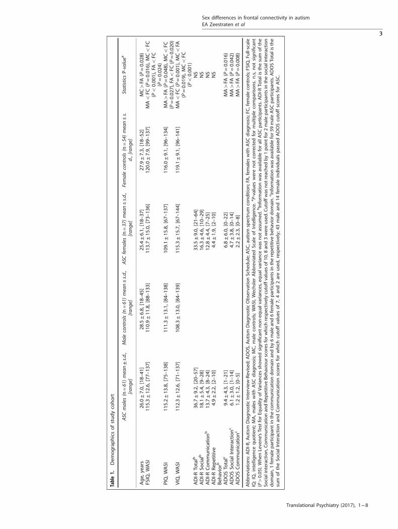
Table1.
Dem
ographicsofstudyco
hort
ASC
males
(n=61)mean±s.d.,
[ran
ge]
Malecontrols(n
=61)mean±s.d.,
[ran
ge]
ASCfemales
(n=37)mean±s.d.,
[ran
ge]
Femalecontrols(n
=54)mean±s.
d.,[rang
e]StatisticsP-valuea
Age,
years
26.0±7.0,
[18–
41]
28.5±6.8,
[18–
45]
25.4±6.1,
[18–
37]
27.9±7.3,
[18–
52]
MC4
FA(P=0.02
8)FS
IQ,W
ASI
115.3±12
.6,[77
–13
7]11
0.9±11
.8,[88
–13
3]11
3.7±15
.0,[73
–13
6]12
0.0±7.9,
[99–
137]
MAo
FC(P=0.01
6),M
Co
FC(Po
0.00
1),FAo
FC(P=0.02
4)PIQ,W
ASI
115.2±13
.8,[75
–13
8]11
1.3±13
.1,[84
–13
8]10
9.1±15
.8,[67
–13
7]11
6.0±9.1,
[96–
134]
MA4
FA(P=0.04
8),M
Co
FC(P=0.02
7),FAo
FC(P=0.02
0)VIQ,W
ASI
112.3±12
.6,[71
–13
7]10
8.3±13
.0,[84
–13
9]11
5.3±15
.7,[67
–14
4]11
9.1±9.1,
[96–
141]
MAo
FC(P=0.00
1),M
Co
FA(P=0.01
9),M
Co
FC(Po
0.00
1)ADI-R
Totalb
36.7±9.2,
[20–
57]
33.5±9.0,
[21–
64]
NS
ADI-R
Socialb
18.1±5.4,
[9–28
]16
.3±4.6,
[10–
29]
NS
ADI-R
Communicationb
13.7±4.3,
[8–24
]12
.8±4.4,
[7–25
]NS
ADI-R
Rep
etitive
Beh
aviorb
4.9±2.2,
[2–10
]4.4±1.9,
[2–10
]NS
ADOSTo
talc
9.4±4.3,
[1–21
]6.8±6.0,
[0–22
]MA4FA
(P=0.01
6)ADOSSo
cial
Interactionc
6.1±3.0,
[1–14
]4.7±3.8,
[0–14
]MA4FA
(P=0.04
2)ADOSCommunicationc
1.2±1.2,
[0–5]
2.2±2.3,
[0–8]
MA4FA
(P=0.00
8)
Abbreviations:ADI-R
,Autism
DiagnosticInterview-Rev
ised
;ADOS,Autism
DiagnosticObservationSched
ule;A
SC,autism
spectrum
condition;FA,fem
ales
withASC
diagnosis;FC
,fem
aleco
ntrols;FSIQ,Full-scale
IQ;IQ,intelligen
cequotien
t;MA,males
withASC
diagnosis;MC,maleco
ntrols;WASI,Wechsler
Abbreviated
ScaleofIntelligen
ce.a P-values
werenotco
rrectedformultiple
comparisons.n.s,notsignificant
(P4
0.05
).When
Leve
ne’sTestforEq
ualityofVa
rian
cesshowed
significantnon-equal
varian
ces,eq
ual
varian
cewas
notassumed
.bInform
ationwas
available
forallA
SCparticipan
ts.A
DI-R
Totalisthesum
ofthe
Socialinteraction,C
ommunicationan
dRep
etitiveBeh
aviourscoresforwhichrespective
lycu
toffvalues
of1
0,8an
d3wereused.C
utoffwas
notreached
by1pointfor2maleparticipan
tsin
thesocialinteraction
domain,1
femaleparticipan
tin
theco
mmunicationdomainan
dby6malean
d6femaleparticipan
tsin
therepetitivebeh
aviordomain.cInform
ationwas
availablefor59
maleASC
participan
ts.A
DOSTo
talisthe
sum
oftheSo
cial
Interactionan
dCommunicationscoresforwhichcu
toffvalues
of7,
4an
d2areused,respective
ly;4
3malean
d14
femaleindividualspassedADOScu
toffscoresforASC
.
Sex differences in frontal connectivity in autismEA Zeestraten et al
3
Translational Psychiatry (2017), 1 – 8
the effect current ASC severity had on tract-specific mean FA values. Toensure our study had sufficient power to detect significant sex differencesafter ADOS adjustment, post hoc power analyses were performed using theG*Power software package.41
RESULTSParticipant demographicsASC groups were matched for age and severity of childhoodautistic symptoms (Table 1). ADOS scores were significantly higherin ASC males (ADOS Total score males: 9.4 ± 4.3; females: 6.8 ± 6.0,P= 0.016). Full scale IQ (FSIQ) did not differ between sexes in theASC groups, but FSIQ scores of female controls were higher thanthose of females with an ASC diagnosis and both male diagnosticgroups. Comparisons between verbal and performance IQ scoresshowed similar results. To adjust for the IQ differences, FSIQ wasincluded as a covariate for all following analyses.
Sex-specific effects and sex-by-diagnosis interaction effectsComparison of tract mean FA values of male and female controlsrevealed comparable microstructural integrity levels in all frontaltracts. However, of the non-frontal tracts, the right ILF was shownto have significantly higher mean FA in females, FA = 0.44004,than males, FA = 0.43191 (F(1,113) = 6.82, P= 0.010).In males we found significant diagnostic effects of lower tract
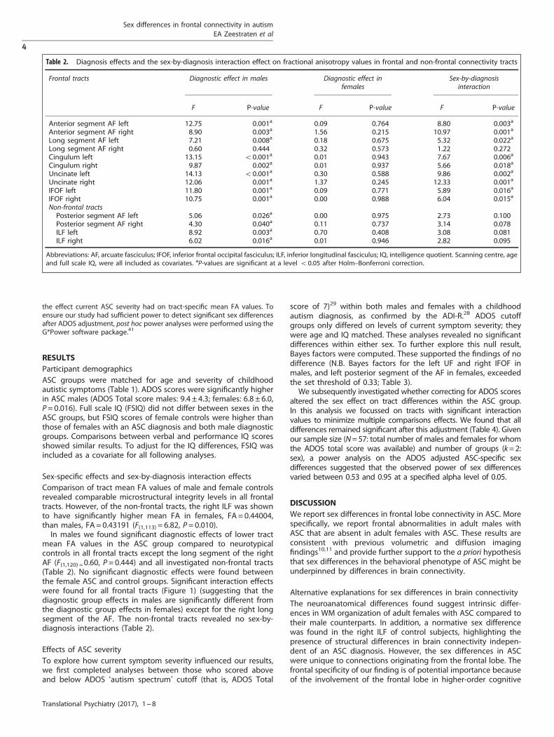
mean FA values in the ASC group compared to neurotypicalcontrols in all frontal tracts except the long segment of the rightAF (F(1,120) =0.60, P= 0.444) and all investigated non-frontal tracts(Table 2). No significant diagnostic effects were found betweenthe female ASC and control groups. Significant interaction effectswere found for all frontal tracts (Figure 1) (suggesting that thediagnostic group effects in males are significantly different fromthe diagnostic group effects in females) except for the right longsegment of the AF. The non-frontal tracts revealed no sex-by-diagnosis interactions (Table 2).
Effects of ASC severityTo explore how current symptom severity influenced our results,we first completed analyses between those who scored aboveand below ADOS ‘autism spectrum’ cutoff (that is, ADOS Total
score of 7)29 within both males and females with a childhoodautism diagnosis, as confirmed by the ADI-R.28 ADOS cutoffgroups only differed on levels of current symptom severity; theywere age and IQ matched. These analyses revealed no significantdifferences within either sex. To further explore this null result,Bayes factors were computed. These supported the findings of nodifference (N.B. Bayes factors for the left UF and right IFOF inmales, and left posterior segment of the AF in females, exceededthe set threshold of 0.33; Table 3).We subsequently investigated whether correcting for ADOS scores
altered the sex effect on tract differences within the ASC group.In this analysis we focussed on tracts with significant interactionvalues to minimize multiple comparisons effects. We found that alldifferences remained significant after this adjustment (Table 4). Givenour sample size (N=57: total number of males and females for whomthe ADOS total score was available) and number of groups (k=2:sex), a power analysis on the ADOS adjusted ASC-specific sexdifferences suggested that the observed power of sex differencesvaried between 0.53 and 0.95 at a specified alpha level of 0.05.
DISCUSSIONWe report sex differences in frontal lobe connectivity in ASC. Morespecifically, we report frontal abnormalities in adult males withASC that are absent in adult females with ASC. These results areconsistent with previous volumetric and diffusion imagingfindings10,11 and provide further support to the a priori hypothesisthat sex differences in the behavioral phenotype of ASC might beunderpinned by differences in brain connectivity.
Alternative explanations for sex differences in brain connectivityThe neuroanatomical differences found suggest intrinsic differ-ences in WM organization of adult females with ASC compared totheir male counterparts. In addition, a normative sex differencewas found in the right ILF of control subjects, highlighting thepresence of structural differences in brain connectivity indepen-dent of an ASC diagnosis. However, the sex differences in ASCwere unique to connections originating from the frontal lobe. Thefrontal specificity of our finding is of potential importance becauseof the involvement of the frontal lobe in higher-order cognitive
Table 2. Diagnosis effects and the sex-by-diagnosis interaction effect on fractional anisotropy values in frontal and non-frontal connectivity tracts
Frontal tracts Diagnostic effect in males Diagnostic effect infemales
Sex-by-diagnosisinteraction
F P-value F P-value F P-value
Anterior segment AF left 12.75 0.001a 0.09 0.764 8.80 0.003a
Anterior segment AF right 8.90 0.003a 1.56 0.215 10.97 0.001a
Long segment AF left 7.21 0.008a 0.18 0.675 5.32 0.022a
Long segment AF right 0.60 0.444 0.32 0.573 1.22 0.272Cingulum left 13.15 o0.001a 0.01 0.943 7.67 0.006a
Cingulum right 9.87 0.002a 0.01 0.937 5.66 0.018a
Uncinate left 14.13 o0.001a 0.30 0.588 9.86 0.002a
Uncinate right 12.06 0.001a 1.37 0.245 12.33 0.001a
IFOF left 11.80 0.001a 0.09 0.771 5.89 0.016a
IFOF right 10.75 0.001a 0.00 0.988 6.04 0.015a
Non-frontal tractsPosterior segment AF left 5.06 0.026a 0.00 0.975 2.73 0.100Posterior segment AF right 4.30 0.040a 0.11 0.737 3.14 0.078ILF left 8.92 0.003a 0.70 0.408 3.08 0.081ILF right 6.02 0.016a 0.01 0.946 2.82 0.095
Abbreviations: AF, arcuate fasciculus; IFOF, inferior frontal occipital fasciculus; ILF, inferior longitudinal fasciculus; IQ, intelligence quotient. Scanning centre, ageand full scale IQ, were all included as covariates. aP-values are significant at a level o0.05 after Holm–Bonferroni correction.
Sex differences in frontal connectivity in autismEA Zeestraten et al
4
Translational Psychiatry (2017), 1 – 8

functioning affected in ASC, and the postulated ‘disconnectionsyndrome’ underlying ASC during development.20–22 The neuroa-natomical sex differences observed in the current study maypartially account for the different behavioral phenotype of ASCfemales.7
It could also be argued that our findings are due to a skewedpattern of ASC symptom severity. It has been proposed, forexample, that in order for women to reach the threshold for aclinical ASC diagnosis, they require the presence of more severebrain abnormalities as they are better able to compensate for, or
Figure 1. Visualizations of investigated tracts and mean fractional anisotropy graphs showing significant sex-by-diagnosis interaction effects.(a). Left anterior segment of the AF; (b) right segment of the AF; (c) left UF; (d) right UF. Bars indicate s.e. AF, arcuate fasciculus; ASC, autismspectrum condition; UF, uncinate fasciculus.
Sex differences in frontal connectivity in autismEA Zeestraten et al
5
Translational Psychiatry (2017), 1 – 8

mask their autistic disabilities than men.3–6 This hypothesis issupported by some findings of greater structural brainabnormalities42,43 and a greater genetic mutation load44 infemales with ASC. To minimize this potential effect, we matchedthe male and female groups on the severity of their childhood ASCsymptoms (that is, ADI-R scores28) as opposed to their currentsymptom severity (that is, ADOS scores29). A consequence of thisapproach was a sex bias with fewer women scoring above ADOScutoff than men. To determine whether this difference accountedfor our findings, we first carried out within-sex analyses based onscoring above or below ADOS cutoff. These analyses revealed theabsence of mean FA differences in any of the tracts based onADOS status in either sex. This suggests that current symptomseverity does not modulate the FA values of frontal tracts. Furtherpost hoc analyses also found that, after correction for the ADOSscores, the sex-by-diagnosis interactions remained significant.
However, these analyses were underpowered for some tracts (forexample, the left anterior AF segment, right cingulum and left UF)and larger studies are still needed to verify these findings.Another issue to consider is the developmental nature of ASC.
Although the observed variance in WM organization in our adultsample might represent an innate sex difference, it is alsoplausible that it is secondary to other experiential factors. Forexample, due to culturally defined sex differences, girls with ASCmay receive more social interaction, and subsequently adoptmore intrapersonal skills than boys.45 This may exert a protectiveeffect on ASC etiology and/or a modulating effect on neurode-velopment in females.46 Equally, early diagnosis of ASC in malesand under-detection of the condition in females may lead todifferences in the pharmacological management of common co-morbidities (for example, depression, anxiety and attention deficit/hyperactivity disorder) during development. Differential exposureto medications could in turn influence critical periods of braindevelopment, such as myelination and pruning.47 Finally, sex-specific physiological features, such as sex hormones (see below),may also affect sexual differentiation of the brain.48 Longitudinalstudies of ASC are required to elucidate the sex-specific effects ofthese factors on lifespan development in individuals with ASC.
Possible biological explanations: biological differencesASC is a complex condition that involves multiple geneticvariations. The biological basis of sex differences in frontal brainconnectivity in ASC may additionally involve an interactionbetween sex hormones and sex chromosomes. It has beenhypothesized, for example, that genes on the paternal Xchromosome protect against social and communication impair-ments. This protective effect is absent in males due to theirinheriting a single maternal X chromosome.49 It has also beenpostulated that differential peaks of testosterone during prenatalneurodevelopment may predispose to sex differences in vulner-ability to autism.50 Fetal testosterone concentration has beenreported to be positively associated with a number of autistictraits in neurotypical males and females.51,52 Fetal testosteronealso influences brain structures associated with language andcommunication in boys with ASC.53 Our findings therefore raise
Table 3. Sex-specific differences between fractional anisotropy values in adults with an ASC diagnosis with and without severe current symptoms asmeasured using the ADOS total score
Frontal tracts Males (N = 59) 16 ADOS− and 43 ADOS+ Females (N= 36) 22 ADOS− and 14 ADOS+
F P-value Bayes factor F P-value Bayes factor
Anterior segment AF left 0.37 0.546 0.23 1.90 0.178 0.02Anterior segment AF right 1.13 0.293 0.19 0.92 0.346 0.05Long segment AF left 0.59 0.444 0.25 0.71 0.407 0.15Long segment AF right 0.11 0.742 0.08 0.21 0.648 o0.01Cingulum left 0.27 0.607 0.13 0.48 0.492 0.04Cingulum right 0.03 0.870 0.01 0.12 0.734 0.02Uncinate left 0.01 0.939 0.41a 3.48 0.072 0.01Uncinate right 0.29 0.593 0.09 0.80 0.377 0.02IFOF left 0.36 0.550 0.12 0.02 0.899 0.15IFOF right 0.32 0.577 0.45a 0.09 0.761 0.09Non-frontal tractsPosterior segment AF left 0.35 0.557 0.13 0.05 0.826 0.54a
Posterior segment AF right 0.06 0.802 0.19 0.43 0.515 0.03ILF left 0.13 0.718 0.05 0.23 0.634 0.32ILF right 0.18 0.671 0.21 0.93 0.342 0.05
Abbreviations: ADOS, Autism Diagnostic Observation Schedule; ADOS− , ASC participants not reaching ADOS cutoff score of 7; ADOS+, ASC participantsreaching ADOS Total cutoff score of 7; AF, arcuate fasciculus; ASC, autism spectrum conditions; IFOF, inferior frontal occipital fasciculus; ILF, inferior longitudinalfasciculus; IQ, intelligence quotient. Scanning centre, age, and full scale IQ, were all included as covariates. aBayes factors (40.33) indicate data sensitivity wasinsufficient to draw conclusions from.
Table 4. ASC-specific sex differences in fractional anisotropy values offrontal tracts corrected for current symptom severity as measuredusing the ADOS total score
Frontal tracts Sex differenceADOS corrected
Observed power
F P-value
Anterior segment AF left 4.02 0.048a 0.53Anterior segment AF right 8.83 0.004a 0.86Long segment AF left 8.80 0.004a 0.86Cingulum left 6.80 0.011a 0.76Cingulum right 4.64 0.034a 0.60Uncinate left 4.70 0.033a 0.60Uncinate right 7.01 0.010a 0.77IFOF left 8.11 0.005a 0.83IFOF right 12.29 0.001a 0.95
Abbreviations: ADOS, Autism Diagnostic Observation Schedule; AF, arcuatefasciculus; ASC, autism spectrum conditions; IFOF, Inferior Frontal OccipitalFasciculus; IQ, intelligence quotient. Scanning centre, age and full scale IQ,were all included as covariates. aP-values are significant at a level o0.05.
Sex differences in frontal connectivity in autismEA Zeestraten et al
6
Translational Psychiatry (2017), 1 – 8
the question of whether (fetal) testosterone modulates theneurodevelopment of frontal connectivity in ASC. Modulation offrontotemporal functional connectivity by testosterone levels hasalready been reported in neurotypical individuals,54 but to datewe are unaware of any studies on the putative effects of fetaltestosterone on WM organization. In brief, the contribution ofsexual differentiation mechanisms to sex-specific risks of devel-oping ASC should be a key area for future studies.2
Future investigations should also include other regions ofinterest and WM connections beyond those analyzed in thepresent study. These could, for example, include thecerebellum9,16 and temporoparietal junction10,17 as both regionshave previously been reported to exhibit sex differences in whiteand/or gray matter volume. Such studies may also benefit fromthe application of a 2 × 2 factorial design and TBSS. The mainadvantage of TBSS is that it is a fully automated, operator-independent approach that allows a ‘whole brain’ analysis ofglobal patterns of white matter integrity. It therefore has thepotential to identify WM differences in brain regions notpreviously considered to be of importance and is resistant tooperator-bias.
CONCLUSIONWe report sex differences in brain connectivity in ASC, with frontalabnormalities in adult males with ASC that are absent in adultfemales with ASC. These differences may explain some of the sexdifferences reported in the behavioral phenotype of ASC. Largerand longitudinal studies are required to replicate these findingsand to explore differences in brain connectivity between otherbrain regions that could contribute to the sex differences seen inbehavioral phenotypes.
CONFLICT OF INTERESTThe authors declare no conflict of interest.
ACKNOWLEDGMENTSWe are grateful to all participants and their parents for participating in this study. Wethank the National Autistic Society for their help in recruiting participants. We alsothank all of the radiographers and physicists of the Centre for NeuroimagingSciences, Institute of Psychiatry, Psychology & Neuroscience (IoPPN) King’s CollegeLondon and University of Cambridge. This work was supported by grant GO 400061(to DGMM) from the UK Medical Research Council (http://www.mrc.ac.uk/index.htm).The research leading to these results has received support from the InnovativeMedicines Initiative Joint Undertaking under grant agreement no. 115300, resourcesof which are composed of financial contribution from the European Union’s SeventhFramework Programme (FP7/2007–2013) and EFPIA companies’ in kind contribution.The National Institute for Health Research (NIHR) Collaboration for Leadership inApplied Health Research and Care—East of England (CLAHRC-EoE) also supportedthe project. This paper represents independent research (part) funded by theNational Institute for Health Research (NIHR) Biomedical Research Centre at SouthLondon and Maudsley NHS Foundation Trust and King’s College London. The viewsexpressed are those of the author(s) and not necessarily those of the NHS, the NIHRor the Department of Health. During the period of the study M-CL and AR weresupported by the William Binks Autism Neuroscience Fellowship and AutismResearch Trust, and M-CL was also supported by the O’Brien Scholars Programwithin the Child and Youth Mental Health Collaborative at the Centre for Addictionand Mental Health and The Hospital for Sick Children, Toronto. The Autism ResearchTrust supported SB-C, BC and ML.
MEMBERS OF THE MEDICAL RESEARCH COUNCIL AUTISMIMAGING MULTICENTRE STUDY CONSORTIUMThe Medical Research Council Autism Imaging Multicentre StudyConsortium (MRC AIMS Consortium) is a UK collaboration betweenthe Institute of Psychiatry (IoP) at King’s College London, theAutism Research Centre, University of Cambridge, and the AutismResearch Group, University of Oxford. The Consortium members
are in alphabetical order: Anthony J. Bailey (Oxford), Simon Baron-Cohen (Cambridge), Patrick F. Bolton (IoP), Edward T. Bullmore(Cambridge), Sarah Carrington (Oxford), Marco Catani (IoP),Bhismadev Chakrabarti (Cambridge), Michael C. Craig (IoP), EileenM. Daly (IoP), Sean C. L. Deoni (IoP), Christine Ecker (IoP), FrancescaHappé (IoP), Julian Henty (Cambridge), Peter Jezzard (Oxford),Patrick Johnston (IoP), Derek K. Jones (IoP), Meng-Chuan Lai(Cambridge), Michael V. Lombardo (Cambridge), Anya Madden(IoP), Diane Mullins (IoP), Clodagh M. Murphy (IoP), Declan G. M.Murphy (IoP), Greg Pasco (Cambridge), Amber N. V. Ruigrok(Cambridge), Susan A. Sadek (Cambridge), Debbie Spain (IoP),Rose Stewart (Oxford), John Suckling (Cambridge), Sally J. Wheel-wright (Cambridge), Steven C. Williams (IoP) and C. EllieWilson (IoP).
REFERENCES1 Baird G, Simonoff E, Pickles A, Chandler S, Loucas T, Meldrum D et al. Prevalence
of disorders of the autism spectrum in a population cohort of children in SouthThames: The Special Needs and Autism Project (SNAP). Lancet 2006; 368:210–215.
2 Lai M-C, Lombardo MV, Auyeung B, Chakrabarti B, Baron-Cohen S. Sex/genderdifferences and autism: setting the scene for future research. J Am Acad ChildAdolesc Psychiatry 2015; 54: 11–24.
3 Baron-Cohen S, Lombardo MV, Auyeung B, Ashwin E, Chakrabarti B, Knickmeyer R.Why are autism spectrum conditions more prevalent in males? PLoS Biol 2011; 9:e1001081.
4 Kopp S, Gillberg C. The Autism Spectrum Screening Questionnaire (ASSQ)-RevisedExtended Version (ASSQ-REV): an instrument for better capturing the autismphenotype in girls? A preliminary study involving 191 clinical cases and com-munity controls. Res Dev Disabil 2011; 32: 2875–2888.
5 Dworzynski K, Ronald A, Bolton P, Happé F. How different are girls and boysabove and below the diagnostic threshold for autism spectrum disorders? J AmAcad Child Adolesc Psychiatry 2012; 51: 788–797.
6 Fombonne E. The epidemiology of autism: a review. Psychol Med 1999; 29:769–786.
7 Lai M-C, Lombardo MV, Pasco G, Ruigrok ANV, Wheelwright SJ, Sadek SA et al. Abehavioral comparison of male and female adults with high functioning autismspectrum conditions. PLoS One 2011; 6: e20835.
8 Andersson GW, Gillberg C, Miniscalco C. Pre-school children with suspectedautism spectrum disorders: do girls and boys have the same profiles? Res DevDisabil 2013; 34: 413–422.
9 Bloss CS, Courchesne E. MRI neuroanatomy in young girls with autism: apreliminary study. J Am Acad Child Adolesc Psychiatry 2007; 46: 515–523.
10 Lai M-C, Lombardo MV, Suckling J, Ruigrok ANV, Chakrabarti B, Ecker C et al.Biological sex affects the neurobiology of autism. Brain 2013; 136: 2799–2815.
11 Beacher FD, Minati L, Baron-Cohen S, Lombardo MV, Lai M-C, Gray MA et al.Autism attenuates sex differences in brain structure: a Combined Voxel-BasedMorphometry and Diffusion Tensor Imaging Study. Am J Neuroradiol 2012; 33:83–89.
12 Nordahl CW, Lange N, Li DD, Barnett LA, Lee A, Buonocore MH et al. Brainenlargement is associated with regression in preschool-age boys with autismspectrum disorders. Proc Natl Acad Sci USA 2011; 108: 20195–20200.
13 Nordahl CW, Iosif A-M, Young GS, Perry LM, Dougherty R, Lee A et al. Sex dif-ferences in the corpus callosum in preschool-aged children with autism spectrumdisorder. Mol Autism 2015; 6: 1–11.
14 Schaer M, Kochalka J, Padmanabhan A, Supekar K, Menon V. Sex differences incortical volume and gyrification in autism. Mol Autism. Molecular Autism 2015; 6:42.
15 Lai M-C, Lerch JP, Floris DL, Ruigrok ANV, Pohl A, Lombardo MV et al. Imaging sex/gender and autism in the brain: Etiological implications. J Neurosci Res 2017; 95:380–397.
16 Craig MC, Zaman SH, Daly EM, Cutter WJ, Robertson DMW, Hallahan B et al.Women with autistic-spectrum disorder: magnetic resonance imaging study ofbrain anatomy. Br J Psychiatry 2007; 191: 224–228.
17 Calderoni S, Retico A, Biagi L, Tancredi R, Muratori F, Tosetti M. Female childrenwith autism spectrum disorder: An insight from mass-univariate and patternclassification analyses. Neuroimage 2012; 59: 1013–1022.
18 Radua J, Via E, Catani M, Mataix-Cols D. Voxel-based meta-analysis of regionalwhite-matter volume differences in autism spectrum disorder versus healthycontrols. Psychol Med 2011; 41: 1539–1550.
19 Via E, Radua J, Cardoner N, Happé F, Mataix-Cols D. Meta-analysis of gray matterabnormalities in autism spectrum disorder. Arch Gen Psychiatry 2011; 68: 409.
Sex differences in frontal connectivity in autismEA Zeestraten et al
7
Translational Psychiatry (2017), 1 – 8

20 Geschwind DH, Levitt P. Autism spectrum disorders: developmental disconnec-tion syndromes. Curr Opin Neurobiol 2007; 17: 103–111.
21 Belmonte MK, Allen G, Beckel-Mitchener A, Boulanger LM, Carper RA, Webb SJ.Autism and abnormal development of brain connectivity. J Neurosci 2004; 24:9228–9231.
22 Catani M, Dell’Acqua F, Budisavljevic S, Howells H, Thiebaut de Schotten M,Froudist-Walsh S et al. Frontal networks in adults with autism spectrum disorder.Brain 2016; 139: 616–630.
23 Kumar A, Sundaram SK, Sivaswamy L, Behen ME, Makki MI, Ager J et al. Alterationsin frontal lobe tracts and corpus callosum in young children with autism spectrumdisorder. Cereb Cortex 2010; 20: 2103–2113.
24 Shukla DK, Keehn B, Müller R-A. Tract-specific analyses of diffusion tensor imagingshow widespread white matter compromise in autism spectrum disorder. J ChildPsychol Psychiatry 2011; 52: 286–295.
25 Billeci L, Calderoni S, Tosetti M, Catani M, Muratori F. White matter connectivity inchildren with autism spectrum disorders: a tract-based spatial statistics study.BMC Neurol 2012; 12: 148–163.
26 Jou RJ, Jackowski AP, Papademetris X, Rajeevan N, Staib LH, Volkmar FR. Diffusiontensor imaging in autism spectrum disorders: preliminary evidence of abnormalneural connectivity. Aust N Z J Psychiatry 2011; 45: 153–162.
27 Ameis SH, Catani M. Altered white matter connectivity as a neural substrate forsocial impairment in Autism Spectrum Disorder. Cortex 2015; 62: 158–181.
28 Lord C, Rutter M, Le Couteur A. Autism diagnostic interview-revised: a revisedversion of a diagnostic interview for caregivers of individuals with possible per-vasive developmental disorders. J Autism Dev Disord 1994; 24: 659–685.
29 Lord C, Heemsbergen J, Jordan H, Mawhood L, Schopler E. Autism diagnosticobservation schedule : a standardized observation of communicative and socialbehavior. J Autism Dev Disord 1989; 19: 185–212.
30 Wechsler D. Wechsler Abbreviated Scale of Intelligence (WASI). Harcourt Assess-ment: San Antonio TX, 1999.
31 Jones DK, Williams SCR, Gasston D, Horsfield MA, Simmons A, Howard R. Isotropicresolution diffusion tensor imaging with whole brain acquisition in a clinicallyacceptable time. Hum Brain Mapp 2002; 230: 216–230.
32 Leemans A, Jeurissen B, Sijbers J. ExploreDTI: a graphical toolbox for processing,analyzing, and visualizing diffusion MR data. 17th Sci Meet Int Soc Magn Reson MedHonolulu 2009; 245: 3537.
33 Leemans A, Jones DK. The B-matrix must be rotated when correcting for subjectmotion in DTI data. Magn Reson Med 2009; 61: 1336–1349.
34 Jones DK, Basser PJ. “Squashing peanuts and smashing pumpkins”: how noisedistorts diffusion-weighted MR data. Magn Reson Med 2004; 52: 979–993.
35 Thiebaut de Schotten M, Ffytche DH, Bizzi A, Dell’Acqua F, Allin M,Walshe M et al. Atlasing location, asymmetry and inter-subject variability of whitematter tracts in the human brain with MR diffusion tractography. Neuroimage2011; 54: 49–59.
36 Catani M, Dell’acqua F, Vergani F, Malik F, Hodge H, Roy P et al. Short frontal lobeconnections of the human brain. Cortex 2012; 48: 273–291.
37 Catani M, Thiebaut de Schotten M. Atlas of Human Brain Connections. OxfordUniversity Press: New York, NY, USA, 2012.
38 Smith SM, Jenkinson M, Woolrich MW, Beckmann CF, Behrens TEJ, Johansen-BergH et al. Advances in functional and structural MR image analysis andimplementation as FSL. Neuroimage 2004; 23(Suppl 1): S208–S219.
39 Thiebaut de Schotten M, Cohen L, Amemiya E, Braga LW, Dehaene S. Learning toread improves the structure of the arcuate fasciculus. Cereb Cortex 2014; 24:989–995.
40 Dienes Z. Using Bayes to get the most out of non-significant results. Front Psychol2014; 5: 1–17.
41 Faul F, Erdfelder E, Lang AG, Buchner A. G*Power 3: a flexible statistical poweranalysis program for the social, behavioral, and biomedical sciences. Behav ResMethods 2007; 39: 175–191.
42 Murphy DGM, Beecham J, Craig MC, Ecker C. Autism in adults. New biologicialfindings and their translational implications to the cost of clinical services. BrainRes 2011; 1380: 22–33.
43 Schumann CM, Bloss CS, Carter Barnes C, Wideman GM, Carper RA, Akshoomoff Net al. Longitudinal magnetic resonance imaging study of cortical developmentthrough early childhood in autism. J Neurosci 2010; 30: 4419–4427.
44 Jacquemont S, Coe BP, Hersch M, Duyzend MH, Krumm N, Bergmann S et al. Ahigher mutational burden in females supports a “female protective model” inneurodevelopmental disorders. Am J Hum Genet 2014; 94: 415–425.
45 Kreiser NL, White SW. ASD in females: are we overstating the gender difference indiagnosis? Clin Child Fam Psychol Rev 2014; 17: 67–84.
46 Cheslack-Postava K, Jordan-Young RM. Autism spectrum disorders: toward agendered embodiment model. Soc Sci Med 2012; 74: 1667–1674.
47 Chugani DC. Pharmacological intervention in autism: targeting critical periods ofbrain development. Clin Neuropsychiatry 2005; 2: 346–353.
48 Li AA, Baum MJ, McIntosh LJ, Day M, Liu F, Earl Gray L. Building a scientificframework for studying hormonal effects on behavior and on the development ofthe sexually dimorphic nervous system. Neurotoxicology 2008; 29: 504–519.
49 Skuse DH. Imprinting, the X-chromosome, and the male brain: explaining sexdifferences in the liability to autism. Pediatr Res 2000; 47: 9–16.
50 Baron-Cohen S, Lutchmaya S, Knickmeyer R. Prenatal Testosterone in Mind. MITPress: Cambridge, MA, USA, 2004.
51 Auyeung B, Baron-Cohen S, Ashwin E, Knickmeyer R, Taylor K, Hackett G. Fetaltestosterone and autistic traits. Br J Psychol 2009; 100: 1–22.
52 Auyeung B, Taylor K, Hackett G, Baron-Cohen S. Foetal testosterone and autistictraits in 18 to 24-month-old children. Mol Autism 2010; 1: 11–18.
53 Lombardo MV, Ashwin E, Auyeung B, Chakrabarti B, Taylor K, Hackett G et al. Fetaltestosterone influences sexually dimorphic gray matter in the human brain. JNeurosci 2012; 32: 674–680.
54 Volman I, Toni I, Verhagen L, Roelofs K. Endogenous testosterone modulatesprefrontal-amygdala connectivity during social emotional behavior. Cereb Cortex2011; 21: 2282–2290.
This work is licensed under a Creative Commons Attribution 4.0International License. The images or other third party material in this
article are included in the article’s Creative Commons license, unless indicatedotherwise in the credit line; if the material is not included under the Creative Commonslicense, users will need to obtain permission from the license holder to reproduce thematerial. To view a copy of this license, visit http://creativecommons.org/licenses/by/4.0/
© The Author(s) 2017
Sex differences in frontal connectivity in autismEA Zeestraten et al
8
Translational Psychiatry (2017), 1 – 8