shaping of t cell functions by trogocytosis
TRANSCRIPT

cells
Review
Shaping of T Cell Functions by Trogocytosis
Masafumi Nakayama *, Arisa Hori, Saori Toyoura and Shin-Ichiro Yamaguchi
�����������������
Citation: Nakayama, M.; Hori, A.;
Toyoura, S.; Yamaguchi, S.-I. Shaping
of T Cell Functions by Trogocytosis.
Cells 2021, 10, 1155. https://doi.org/
10.3390/cells10051155
Academic Editors: Ryuta Koyama
and Kumiko Nakada-Tsukui
Received: 3 April 2021
Accepted: 6 May 2021
Published: 10 May 2021
Publisher’s Note: MDPI stays neutral
with regard to jurisdictional claims in
published maps and institutional affil-
iations.
Copyright: © 2021 by the authors.
Licensee MDPI, Basel, Switzerland.
This article is an open access article
distributed under the terms and
conditions of the Creative Commons
Attribution (CC BY) license (https://
creativecommons.org/licenses/by/
4.0/).
Laboratory of Immunology and Microbiology, College of Pharmaceutical Sciences, Ritsumeikan University,Shiga 525-8577, Japan; [email protected] (A.H.); [email protected] (S.T.);[email protected] (S.-I.Y.)* Correspondence: [email protected]; Tel.: +81-77-599-3264
Abstract: Trogocytosis is an active process whereby plasma membrane proteins are transferredfrom one cell to the other cell in a cell-cell contact-dependent manner. Since the discovery of theintercellular transfer of major histocompatibility complex (MHC) molecules in the 1970s, trogocytosisof MHC molecules between various immune cells has been frequently observed. For instance,antigen-presenting cells (APCs) acquire MHC class I (MHCI) from allografts, tumors, and virallyinfected cells, and these APCs are subsequently able to prime CD8+ T cells without antigen processingvia the preformed antigen-MHCI complexes, in a process called cross-dressing. T cells also acquireMHC molecules from APCs or other target cells via the immunological synapse formed at the cell-cellcontact area, and this phenomenon impacts T cell activation. Compared with naïve and effectorT cells, T regulatory cells have increased trogocytosis activity in order to remove MHC class II andcostimulatory molecules from APCs, resulting in the induction of tolerance. Accumulating evidencesuggests that trogocytosis shapes T cell functions in cancer, transplantation, and during microbialinfections. In this review, we focus on T cell trogocytosis and the related inflammatory diseases.
Keywords: acquisition; nibbling; stripping; cross-dressed; cross-presentation; dendritic cell (DC);TCR; Treg; chimeric antigen receptor (CAR); fratricide; escape variant
1. Introduction
In order to communicate with each other, immune cells express a wide variety of cellsurface molecules such as receptors, ligands, and adhesion molecules. Cell-cell communica-tion is required for the generation of appropriate immune responses to various pathogens.It has been established that during cell-cell interactions, membrane-associated proteinsare transferred between immune cells [1–4]. In the past this biological phenomenon hasbeen called acquisition, nibbling, or stripping, and is currently referred to as trogocytosis,derived from the ancient Greek word Trogo, meaning ‘gnaw’ [5]. In contrast to phagocytosis(Phago is the Greek meaning ‘to eat’), which is executed by phagocytes such as macrophagesand dendritic cells (DCs), trogocytosis is considered to be executed by any type of cells, asdescribed below. By acquiring membrane-associated proteins, so-called recipient cells gainalternative cellular functions. In contrast, donor cells may lose these proteins and cellularfunctions (Figure 1). In addition, under certain conditions, bi-directional trogocytosis isobserved [6] (see Section 2). In some cases, trogocytosis mediates the intercellular transfernot only of plasma membranes but also of intracellular contents [7,8], which may also altercellular functions. However, this possibility has not been extensively investigated.
The best characterized trogocytosis involves the transfer of major histocompatibil-ity complex (MHC) molecules from antigen-presenting cells (APCs) to T cells duringtheir interactions [1,3]. Trogocytosis of MHC molecules shapes T cell functions and isinvolved in various T cell-mediated diseases. Trogocytosis has been observed not only inimmune cell interactions, but also during epithelial cell communication [9] and in neu-ronal synapses [10,11]. Further, trogocytosis is used by amoebae to kill host cells [12,13],indicating that this biological phenomenon is widely conserved throughout eukaryotes.
Cells 2021, 10, 1155. https://doi.org/10.3390/cells10051155 https://www.mdpi.com/journal/cells

Cells 2021, 10, 1155 2 of 16
Recent findings of trogocytosis in microbes [14], mammalian neuronal networks [15,16],and non-T cell immune cell interactions [2,17,18] have been well summarized by otherspreviously and also in this issue. Thus, here we mainly focus on T cell trogocytosis andrelated diseases.
Cells 2021, 10, 1155 2 of 16
that this biological phenomenon is widely conserved throughout eukaryotes. Recent find-ings of trogocytosis in microbes [14], mammalian neuronal networks [15,16], and non-T cell immune cell interactions [2,17,18] have been well summarized by others previously and also in this issue. Thus, here we mainly focus on T cell trogocytosis and related dis-eases.
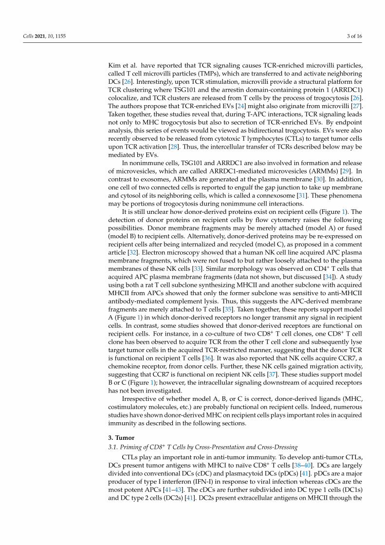
Figure 1. Principal models of trogocytosis. When two types of cells make physical contact via receptor-ligand interactions, the driving force for internalization of receptors expressed on one cell (here called recipient) co-opts ligand-containing plasma membrane fragments from the other cell (here called donor). Not only are receptors and ligands involved in this process, but adhesion molecules also play a role. The donor cells lose membrane molecules and their cellular functions, whereas the recipient cells gain donor-derived membrane molecules and functions. It is unclear how donor-derived mol-ecules exist on recipient cells. The three hypothesized models are shown as A, B, and C.
2. Possible Mechanisms Underlying Trogocytosis The molecular mechanisms underlying trogocytosis are not fully understood. How-
ever, trogocytosis of the T cell receptor (TCR) and MHC receptor ligand pair has been extensively characterized. Of note, Martinez-Martin et al. have shown that T cells acquire MHC class I (MHCI) from APCs through the action of small GTPases such as RhoG and TC21 [19]. RhoG is known to be involved in phagocytosis [20], and thus trogocytosis is characterized as incomplete phagocytosis. Indeed, PI3K inhibitors effectively inhibit TCR trogocytosis [19,21]. Likewise, natural killer (NK) cells expressing NKG2D, an NK activat-ing receptor, acquire ligands, including RaeI, MICA, and MICB, from tumor cells via a PI3K-dependent pathway [22]. Taken together, trogocytosis appears to be accompanied by recipient cell receptor internalization (Figure 1).
Donor cells may actively release their membrane fragments to recipient cells. These membrane fragments may include extracellular vesicles (EVs) [23]. Choudhuri et al. have shown that TCR signaling leads to secretion of TCR-enriched microvesicles via the central supramolecular activation cluster (c-SMAC) [24]. Sorting of TCR to the c-SMAC and the production of EVs is dependent on tumor susceptibility gene 101 (TSG101), an essential
Figure 1. Principal models of trogocytosis. When two types of cells make physical contact via receptor-ligand interactions,the driving force for internalization of receptors expressed on one cell (here called recipient) co-opts ligand-containingplasma membrane fragments from the other cell (here called donor). Not only are receptors and ligands involved in thisprocess, but adhesion molecules also play a role. The donor cells lose membrane molecules and their cellular functions,whereas the recipient cells gain donor-derived membrane molecules and functions. It is unclear how donor-derivedmolecules exist on recipient cells. The three hypothesized models are shown as A, B, and C.
2. Possible Mechanisms Underlying Trogocytosis
The molecular mechanisms underlying trogocytosis are not fully understood. How-ever, trogocytosis of the T cell receptor (TCR) and MHC receptor ligand pair has beenextensively characterized. Of note, Martinez-Martin et al. have shown that T cells acquireMHC class I (MHCI) from APCs through the action of small GTPases such as RhoG andTC21 [19]. RhoG is known to be involved in phagocytosis [20], and thus trogocytosisis characterized as incomplete phagocytosis. Indeed, PI3K inhibitors effectively inhibitTCR trogocytosis [19,21]. Likewise, natural killer (NK) cells expressing NKG2D, an NKactivating receptor, acquire ligands, including RaeI, MICA, and MICB, from tumor cells viaa PI3K-dependent pathway [22]. Taken together, trogocytosis appears to be accompaniedby recipient cell receptor internalization (Figure 1).
Donor cells may actively release their membrane fragments to recipient cells. Thesemembrane fragments may include extracellular vesicles (EVs) [23]. Choudhuri et al. haveshown that TCR signaling leads to secretion of TCR-enriched microvesicles via the centralsupramolecular activation cluster (c-SMAC) [24]. Sorting of TCR to the c-SMAC and theproduction of EVs is dependent on tumor susceptibility gene 101 (TSG101), an essen-tial component of the endosomal sorting complex required for transport (ESCRT)-I [25].These EVs are transferred to and activate neighboring B cells [24]. On the other hand,
Cells 2021, 10, 1155 3 of 16
Kim et al. have reported that TCR signaling causes TCR-enriched microvilli particles,called T cell microvilli particles (TMPs), which are transferred to and activate neighboringDCs [26]. Interestingly, upon TCR stimulation, microvilli provide a structural platform forTCR clustering where TSG101 and the arrestin domain-containing protein 1 (ARRDC1)colocalize, and TCR clusters are released from T cells by the process of trogocytosis [26].The authors propose that TCR-enriched EVs [24] might also originate from microvilli [27].Taken together, these studies reveal that, during T-APC interactions, TCR signaling leadsnot only to MHC trogocytosis but also to secretion of TCR-enriched EVs. By endpointanalysis, this series of events would be viewed as bidirectional trogocytosis. EVs were alsorecently observed to be released from cytotoxic T lymphocytes (CTLs) to target tumor cellsupon TCR activation [28]. Thus, the intercellular transfer of TCRs described below may bemediated by EVs.
In nonimmune cells, TSG101 and ARRDC1 are also involved in formation and releaseof microvesicles, which are called ARRDC1-mediated microvesicles (ARMMs) [29]. Incontrast to exosomes, ARMMs are generated at the plasma membrane [30]. In addition,one cell of two connected cells is reported to engulf the gap junction to take up membraneand cytosol of its neighboring cells, which is called a connexosome [31]. These phenomenamay be portions of trogocytosis during nonimmune cell interactions.
It is still unclear how donor-derived proteins exist on recipient cells (Figure 1). Thedetection of donor proteins on recipient cells by flow cytometry raises the followingpossibilities. Donor membrane fragments may be merely attached (model A) or fused(model B) to recipient cells. Alternatively, donor-derived proteins may be re-expressed onrecipient cells after being internalized and recycled (model C), as proposed in a commentarticle [32]. Electron microscopy showed that a human NK cell line acquired APC plasmamembrane fragments, which were not fused to but rather loosely attached to the plasmamembranes of these NK cells [33]. Similar morphology was observed on CD4+ T cells thatacquired APC plasma membrane fragments (data not shown, but discussed [34]). A studyusing both a rat T cell subclone synthesizing MHCII and another subclone with acquiredMHCII from APCs showed that only the former subclone was sensitive to anti-MHCIIantibody-mediated complement lysis. Thus, this suggests the APC-derived membranefragments are merely attached to T cells [35]. Taken together, these reports support modelA (Figure 1) in which donor-derived receptors no longer transmit any signal in recipientcells. In contrast, some studies showed that donor-derived receptors are functional onrecipient cells. For instance, in a co-culture of two CD8+ T cell clones, one CD8+ T cellclone has been observed to acquire TCR from the other T cell clone and subsequently lysetarget tumor cells in the acquired TCR-restricted manner, suggesting that the donor TCRis functional on recipient T cells [36]. It was also reported that NK cells acquire CCR7, achemokine receptor, from donor cells. Further, these NK cells gained migration activity,suggesting that CCR7 is functional on recipient NK cells [37]. These studies support modelB or C (Figure 1); however, the intracellular signaling downstream of acquired receptorshas not been investigated.
Irrespective of whether model A, B, or C is correct, donor-derived ligands (MHC,costimulatory molecules, etc.) are probably functional on recipient cells. Indeed, numerousstudies have shown donor-derived MHC on recipient cells plays important roles in acquiredimmunity as described in the following sections.
3. Tumor3.1. Priming of CD8+ T Cells by Cross-Presentation and Cross-Dressing
CTLs play an important role in anti-tumor immunity. To develop anti-tumor CTLs,DCs present tumor antigens with MHCI to naïve CD8+ T cells [38–40]. DCs are largelydivided into conventional DCs (cDC) and plasmacytoid DCs (pDCs) [41]. pDCs are a majorproducer of type I interferon (IFN-I) in response to viral infection whereas cDCs are themost potent APCs [41–43]. The cDCs are further subdivided into DC type 1 cells (DC1s)and DC type 2 cells (DC2s) [41]. DC2s present extracellular antigens on MHCII through the

Cells 2021, 10, 1155 4 of 16
conventional antigen presentation pathway whereas cDC1s are able to present extracellularantigens not only on MHCII, but also on MHCI, called cross-presentation (Figure 2) [39–41].In general, as observed in DC2s, extracellular antigens are processed and loaded on MHCIIin phagosomes. In DC1s, however, extracellular antigens are released from phagosomesto the cytosol and then translocated via TAP (transfer associated with antigen processing)molecules to the endoplasmic reticulum (ER) where extracellular antigen peptides as wellas intracellular antigen peptides are associated with MHCI [44,45]. Regarding the unusualpathway of extracellular antigens from phagosomes to cytosol, it has been recently reportedthat DNGR-1 (also known as CLEC9A) senses necrotic cell-derived F-actin [46,47] and itshemITAM-Syk signaling induces phagosomal membrane rupture to allow endocytosedantigens to enter the cytosol in DC1s [48].
Cells 2021, 10, 1155 5 of 16
Figure 2. Trogocytosis in T cell priming and effector phases. During the priming phase, dendritic cell (DC) type 2 cells (DC2s) present extracellular tumor antigens on MHCII to activate CD4+ T cells whereas DC type 1 cells (DC1s) are able to present them on MHCI, called cross-presentation, to activate CD8+ T cells. In addition, DC1s and/or DC2s acquire pre-formed antigen-MHCI complexes for antigen presentation to CD8+ T cells, which is called cross-dressing. In the cytotoxic T lymphocyte (CTL) effector phase, CTLs strip off target antigens from tumor cells. These CTLs with acquired tumor antigen-MHCI are then lysed by tumor-unexperienced CTLs through a process called fratricide cell death. On the other hand, tumor cells lose antigens, resulting in generation of CTL escape variants.
3.2. MHC Trogocytosis in the CTL Effector Phase Trogocytosis is also frequently observed in the CTL effector phase. When CTLs attack
tumor cells, they acquire MHCI from tumor cells (Figure 2) [7,65]. However, it is still un-der debate whether trogocytosis enhances or suppresses CTL activity. Given the positive correlation between cytotoxic activity and trogocytosis ability [66,67], CTLs with high avidity (high recognition efficiency) may exert both high cytotoxicity and trogocytosis ac-tivity. Alternatively, given that the acquired antigen-MHC complexes have been proposed to transmit sustained TCR signals in CD4+ T cells [67–70], trogocytosis may prolong CTL activation. In contrast, a regulatory function of the MHC on CTLs has been also reported. For instance, CTLs that have acquired the tumor antigen-MHC complex are recognized and lysed by tumor-unexperienced CTLs, which is called fratricide cell death (Figure 2) [7,65,71]. Likewise, it was recently reported that trogocytosis-mediated fratricide of chi-meric antigen receptor (CAR) T cells causes tumor escape [72] (see Section 3.3).
It is noteworthy that TCR-mediated trogocytosis strips tumor antigens from target tumor cells, causing antigen loss and tumor escape (Figure 2) [2,72,73]. For example, low-avidity CTLs remove tumor antigen-MHCI complexes from target tumor cells without killing, interfering with tumor killing by high-avidity CTLs [73]. Likewise, CAR and mon-oclonal antibodies (mAbs) also mediate tumor antigen loss via trogocytosis [2,72] (see Sec-tion 3.3).
Figure 2. Trogocytosis in T cell priming and effector phases. During the priming phase, dendritic cell (DC) type 2 cells(DC2s) present extracellular tumor antigens on MHCII to activate CD4+ T cells whereas DC type 1 cells (DC1s) are ableto present them on MHCI, called cross-presentation, to activate CD8+ T cells. In addition, DC1s and/or DC2s acquirepreformed antigen-MHCI complexes for antigen presentation to CD8+ T cells, which is called cross-dressing. In the cytotoxicT lymphocyte (CTL) effector phase, CTLs strip off target antigens from tumor cells. These CTLs with acquired tumorantigen-MHCI are then lysed by tumor-unexperienced CTLs through a process called fratricide cell death. On the otherhand, tumor cells lose antigens, resulting in generation of CTL escape variants.
In addition to the cross-presentation pathway, several recent studies have reportedthe cross-dressing pathway, in which DCs acquire MHCI molecules from neighboringDCs or tumor cells (Figure 2). These MHCI-dressed (cross-dressed) DCs activate CD8+ Tcells via the preformed antigen peptide-MHCI complexes without the above-mentionedantigen processing [3,49,50]. Prior to the first demonstration of the cross-dressing pathwayby Dolan et al. [51], the tumor-derived exosomes containing MHCI were previously con-sidered to provoke anti-tumor immunity [52,53]. In this study, when FVB mouse (MHChaplotype: H-2q) bone marrow-derived DCs (BMDCs) were co-cultured with dying H-2b
tumor cells expressing ovalbumin (OVA), the H-2q BMDCs acquired the OVA peptide-H-2Kb complexes from tumor cells and subsequently activated CD8+ T cells from OT-I mice

Cells 2021, 10, 1155 5 of 16
specific for OVA residues 257–264 on H-2Kb. Thus, this indicates that BMDMs do not useself-MHC, but instead use non-self MHC molecules to activate T cells. It should be men-tioned here that BMDCs are CD11c+ MHCII+ cells generated with GM-CSF; however, thesein vitro-cultured DCs are not equivalent to in vivo DCs and are neither DC1s nor DC2s [54].To address the role of cross-dressed DCs in vivo, the authors used CD11c-diphtheria toxinreceptor (DTR) transgenic BALB/c (H-2d) mice in which DCs are removable by diphtheriatoxin (DT) treatment [55]. In these mice inoculated subcutaneously with H-2b tumor cellsexpressing OVA, OT-I CD8+ T cells vigorously proliferated, an effect abolished by DTtreatment, indicating that DCs are essential for OT-I CD8+ T cell proliferation in response tothe tumor cell-derived OVA peptide-H-2Kb in vivo [51]. Subsequently, cross-dressing hasbeen demonstrated to be involved not only in cancer [56–58], but also in transplantationand during microbial infections (see Sections 4.1 and 5).
Depending on experimental conditions, cross-dressing has been shown to be con-ducted by both DC1s and DC2s. Further, DC1s are reportedly essential for cross-dressingof DNA vaccine antigens [59,60] whereas DC2s show higher cross-dressing of neighboringDC-derived MHCI [61–63]. This apparent discrepancy may be ascribed to the differencein type of donor cells that DCs acquire MHCI from. In addition to cDCs, pDCs alsoacquire antigen-MHC complexes from tumor cells and stimulate MHC-restricted T cellproliferation [64]. Interestingly, a recent study has shown that pDCs give the antigen-MHCIcomplexes to DC1s, which contribute to cross-dressing [60].
3.2. MHC Trogocytosis in the CTL Effector Phase
Trogocytosis is also frequently observed in the CTL effector phase. When CTLs attacktumor cells, they acquire MHCI from tumor cells (Figure 2) [7,65]. However, it is stillunder debate whether trogocytosis enhances or suppresses CTL activity. Given the positivecorrelation between cytotoxic activity and trogocytosis ability [66,67], CTLs with highavidity (high recognition efficiency) may exert both high cytotoxicity and trogocytosisactivity. Alternatively, given that the acquired antigen-MHC complexes have been proposedto transmit sustained TCR signals in CD4+ T cells [67–70], trogocytosis may prolong CTLactivation. In contrast, a regulatory function of the MHC on CTLs has been also reported.For instance, CTLs that have acquired the tumor antigen-MHC complex are recognized andlysed by tumor-unexperienced CTLs, which is called fratricide cell death (Figure 2) [7,65,71].Likewise, it was recently reported that trogocytosis-mediated fratricide of chimeric antigenreceptor (CAR) T cells causes tumor escape [72] (see Section 3.3).
It is noteworthy that TCR-mediated trogocytosis strips tumor antigens from targettumor cells, causing antigen loss and tumor escape (Figure 2) [2,72,73]. For example, low-avidity CTLs remove tumor antigen-MHCI complexes from target tumor cells withoutkilling, interfering with tumor killing by high-avidity CTLs [73]. Likewise, CAR andmonoclonal antibodies (mAbs) also mediate tumor antigen loss via trogocytosis [2,72] (seeSection 3.3).
3.3. CAR-Mediated Trogocytosis
CARs combine antigen-binding domains, most commonly, a single-chain variablefragment (scFv) derived from the variable domains of antibodies with the signaling do-mains of the TCRζ chain and additional costimulatory domains from receptors such asCD28, OX40, and 4-1BB [74]. Autologous T cells engineered to express a CAR specific forCD19 (CD19 CAR T cells) are highly effective against several types of B-cell malignanciesand have recently received FDA approval for use in children and young adults with re-lapse of chemotherapy refractory acute lymphoblastic leukemia (ALL) and for adults withchemotherapy-refractory non-Hodgkin lymphoma (NHL) [75]. Despite the high initialresponse rate with CD19 CAR T cells in ALL, relapse occurs with some tumors beingantigen-negative and others antigen-low [75–78]. A recent study using a mouse model ofleukemia demonstrated that CD19 is transferred to CAR T cells via trogocytosis, resultingin removal of the tumor antigen [72]. Such a loss of tumor antigen was also observed during

Cells 2021, 10, 1155 6 of 16
cancer therapies using mAbs such as rituximab and epratuzumab [2,79,80]. This processcould cause tumor escape variants. Further, CD19-acquired CAR T cells were shown tobe killed by tumor-unexperienced neighbor CAR T cells [72], a process called fratricide(Figure 2) [7]. Therefore, the inhibition of trogocytosis may improve the efficacy of CAR Ttherapy. Since the specific molecular mechanisms of trogocytosis remain unknown, as aninitial strategy, combinatorial targeting could overcome this trogocytosis-based side effect.
4. Transplantation4.1. Allospecific T Cell Priming by Cross-Dressing
T cell-mediated recognition of allogeneic transplants has been considered to occurthrough two main pathways (Figure 3). In the direct pathway recipient T cells recognizeintact MHC alloantigens on donor DCs resulting in acute rejection [81]. With the indirectpathway allograft antigens are internalized and processed by recipient DCs and recipientT cells subsequently recognize these antigens, which promotes chronic rejection [82,83].In addition to these pathways, there is accumulating evidence of a third, semidirect path-way (cross-dressing pathway) where MHC alloantigens are acquired by recipient DCs(Figure 3) [84–86] as described below.
In both human and mouse allogeneic DC coculture assays, recipient DCs acquireantigen-MHC complexes from donor DCs, and these donor MHC-dressed recipient DCsprime cognate T cells in a donor MHC-restricted manner, suggesting the role of cross-dressing in T cell alloreactions in vitro [84,87]. In several mouse models of allograft(skin, heart, or kidney) transplantation, recipient DCs infiltrate allografts and acquiredonor MHC [88,89]. Finally, these DCs prime alloreactive T cells in a donor MHC-restricted manner, suggesting that cross-dressing indeed occurs in allograft transplantation(Figure 3) [88,89]. However, these studies did not address whether cross-dressed DCs areinvolved in allograft rejection and which DC subset contributes to the cross-dressing [88].
The relative contribution of cross-presentation and cross-dressing to CD8+ T cellactivation can be addressed using TAP−/− mice. TAP molecules are generally requiredfor cross-presentation, but not for cross-dressing (see Section 3.1). On the other hand, thecontribution of DC1s to CD8+ T cell activation can be addressed with Batf3−/− mice, asthis transcription factor is required for the development of DC1s, but not of DC2s [90].Recently, Li et al. used these knockout mice and showed that when H-2Kd skin grafts weretransplanted into WT or Batf3−/− recipient H-2Kb mice, Batf3−/− recipient mice showeddelayed rejection, suggesting that recipient DC1s contribute to allograft rejection [91].Although DC1s have cross-presenting activity, alloreactive CD8+ T cell proliferation wasobserved in TAP−/− mice as well as in WT mice, suggesting that DC1 cross-dressing, ratherthan cross-presentation, contributes to alloreactive T cell activation [91]. However, it wasnot directly demonstrated that cross-dressing is involved in allograft rejection. To thisend, Hughes et al. used B6 (H-2Kb/b) WT or H-2K−/− recipient mice transplanted withH-2Kb/d kidneys expressing the membrane-bound form of OVA; both recipient DCs werefound to acquire H-2Kd and H-2Kb-SIINFEKL (OVA-derived peptide) complexes. Twodays after transplantation, these mice were adoptively transferred OT-I CD8+ T cells. Inboth recipients, acute rejection was equally observed, indicating that recipient MHCI isnot required for rejection. To exclude the possibility of direct pathways (Figure 3), theauthors showed that graft survival is prolonged when recipient DCs were depleted usingthe CD11c-DTR system. Taken together, this study clearly demonstrates that cross-dressedDCs are involved in allograft rejection [92].
In these mouse experiments, recipient DCs acquire allo-MHC from the graft not onlyvia trogocytosis [87,93,94] but also via extracellular vesicles [88,95], although it remainsunknown which is the dominant pathway for cross-dressing in transplantation. It alsoremains unknown whether MHC donor cells in grafts are DCs or parenchymal cells.Furthermore, the most important question concerns whether cross-dressing is essential forallograft rejection because genetically engineered mice in which cross-dressing pathway isspecifically impaired have not been developed so far.

Cells 2021, 10, 1155 7 of 16
Cells 2021, 10, 1155 7 of 16
2Kb/d kidneys expressing the membrane-bound form of OVA; both recipient DCs were found to acquire H-2Kd and H-2Kb-SIINFEKL (OVA-derived peptide) complexes. Two days after transplantation, these mice were adoptively transferred OT-I CD8+ T cells. In both recipients, acute rejection was equally observed, indicating that recipient MHCI is not required for rejection. To exclude the possibility of direct pathways (Figure 3), the authors showed that graft survival is prolonged when recipient DCs were depleted using the CD11c-DTR system. Taken together, this study clearly demonstrates that cross-dressed DCs are involved in allograft rejection [92].
In these mouse experiments, recipient DCs acquire allo-MHC from the graft not only via trogocytosis [87,93,94] but also via extracellular vesicles [88,95], although it remains unknown which is the dominant pathway for cross-dressing in transplantation. It also remains unknown whether MHC donor cells in grafts are DCs or parenchymal cells. Fur-thermore, the most important question concerns whether cross-dressing is essential for allograft rejection because genetically engineered mice in which cross-dressing pathway is specifically impaired have not been developed so far.
Figure 3. Trogocytosis in allograft transplantation. Alloreactive T cell activation is induced by three pathways. The first is the direct pathway where intact MHC alloantigens on donor DCs are recognized by recipient T cells, promoting acute rejection. The second is the indirect pathway where allograft antigens are internalized and processed by recipient DCs, on which donor antigen-recipient MHC complexes are recognized by recipient T cells, promoting chronic rejection. The third pathway is a semi-direct pathway of so-called cross-dressing where recipient DCs acquire preformed donor antigen-MHC complexes and are recognized by recipient T cells.
Figure 3. Trogocytosis in allograft transplantation. Alloreactive T cell activation is induced by three pathways. The firstis the direct pathway where intact MHC alloantigens on donor DCs are recognized by recipient T cells, promoting acuterejection. The second is the indirect pathway where allograft antigens are internalized and processed by recipient DCs, onwhich donor antigen-recipient MHC complexes are recognized by recipient T cells, promoting chronic rejection. The thirdpathway is a semi-direct pathway of so-called cross-dressing where recipient DCs acquire preformed donor antigen-MHCcomplexes and are recognized by recipient T cells.
4.2. Induction of Allospecific T Cell Tolerance by Cross-Dressing
In contrast to skin grafts, allogeneic liver grafts are accepted in mice without anyimmunosuppressive treatment [96]. In humans, complete immunosuppression withdrawalhas proven to be feasible in approximately 20% of liver transplant recipients [97]. Theseobservations led to the hypothesis of spontaneous tolerance in liver transplantation, al-though the underlying mechanism is not well understood. Ono et al. recently reportedthat, in a mouse model of allogeneic liver transplantation, recipient DCs infiltrate into livergrafts, and acquire donor MHC. These cross-dressed DCs express high levels of PD-L1,which in vitro did not prime alloreactive CD8+ T cells, but rather induced tolerance [98].Taken together, these results suggest that cross-dressing plays a role in tolerance inductionalthough whether the depletion of PD-L1high cross-dressed DCs causes breakdown oftolerance has not been addressed.
4.3. Induction of Allospecific T Cell Tolerance by Double-Negative T (DNT) Cell Trogocytosis
TCRαβ+ CD3+ CD4- CD8- T cells, so called double-negative T (DNT) cells, comprise asmall subset of mature peripheral T cells, and the number of DNT cells are expanded invarious inflammatory conditions [99]. Indeed, DNT cells have been reported to be involvedin several autoimmune diseases such as systemic lupus erythematosus (SLE), Sjogren’s

Cells 2021, 10, 1155 8 of 16
syndrome, and psoriasis, although the precise origin and function of DNT cells is still underdebate [99]. In contrast to such pro-inflammatory activity, a reported regulatory functionof DNT cells is the enhancement of allograft survival [100–102], in which trogocytosisis involved [100,103]. For instance, in a mouse model of skin allograft transplantation,recipient DNT cells acquire donor MHCI and interact with alloreactive CD8+ T cells. Duringthese cell-cell interactions, DNT cells lyse CD8+ T cells through the Fas/FasL pathway,which prevents allograft rejection [100,103]. In addition, it has been recently reported thatDNT cell trogocytosis suppresses CD4+ T cell activation in a mouse model of allergy [104](see Section 6).
5. Infection
Cross-dressing (see Section 3.1) also contributes to antiviral T cell responses, which hasbeen clearly demonstrated by Wakim and Bevan using mouse models of viral infection [62].In this study, the authors utilized irradiated (H-2Kd x H-2Kb) F1 mice reconstituted withH-2Kd CD11c-DTR bone marrow cells, in which DCs have only H-2Kd and are removableby DT treatment [62]. Following adoptive transfer of OT-I CD8+ T cells and infection withvesicular stomatitis virus expressing OVA, DCs acquired the OVA peptide-H-2Kb complexfrom the virally infected cells. These cross-dressed DCs were essential for memory, but notnaïve OT-I CD8+ T cell activation, in vivo [62]. Smyth et al. used a mouse model of OVA-expressing adenoviral infection to show that cross-dressing activates not only memory,but also naïve OT-I CD8+ T cells [63]. Both studies demonstrated that DC2s have morepotent cross-dressing activity than DC1s for antiviral immunity, although they did not useBatf3−/− mice [62,63]. The discrepancy regarding cross-dressing of naïve T cells may beascribed to different amounts of MHCI and costimulatory molecules on cross-dressed DCs.In other words, naive T cells can be primed by DCs with acquired membrane fragmentsharboring larger amounts of MHCI and costimulatory molecules of virally infected DCs,whereas memory T cells can be activated by DCs dressed with membrane fragments ofvirally infected parenchymal cells.
In addition to cDCs, pDCs play an important role in immune responses by producinglarge amount of IFN-I during antiviral immunity (see Section 3.1) [41,43]. Although it isstill under debate whether pDCs have antigen processing machinery, pDCs have beenreported to have cross-dressing activity [64]. It was also recently reported that pDCs giveMHCI to DC1s, which contributes to their cross-dressing [60]. Since these studies measuredonly CD8+ T cell activation, it remains unknown whether direct or indirect cross-dressingby pDCs indeed contributes to antiviral immunity.
6. Th2 Diseases
When naive CD4+ TCRs recognize antigen-MHCII complexes on APCs, these CD4+ Tcells expand and differentiate into functionally distinct effector helper T (Th) cell subsets,such as Th1, Th2, and Th17 cells [105]. Among these Th subsets, Th2 cells produce IL-4,IL-5, and IL-13, which play a central role in humoral immunity and host defense againstparasite infection, but also have a detrimental role in allergic diseases such as asthmaand atopic dermatitis [105]. There are numerous studies showing that naïve CD4+ T cellsas well as CD8+ T cells acquire antigen-MHC complexes from DCs during these cell-cellinteractions [68,69,94,106–110]. Upon interaction with DCs, CD4+ T cells acquire not onlyMHCII, but also costimulatory molecules and adhesion molecules that are recruited ontothe immunological synapse formed at the cell-cell contact area. Therefore, these MHCII-acquired CD4+ T cells are considered to act as APCs [94,106–109,111,112]. In addition,MHCII acquisition induces prolonged TCR signaling even after dissociation from APCs,which impacts CD4+ T cell activation, survival, and cytokine production [70].
In addition to CD4+ T cells and DCs, various immune cells acquire MHCII and areinvolved in Th2 responses. For instance, basophils, the major producer of IL-4 [113],acquire MHCII and act as APCs for Th2 differentiation [114]. Group 2 innate lymphoid

Cells 2021, 10, 1155 9 of 16
cells (ILC2s), which also produce high amounts of Th2 cytokines [115], per se synthesizeMHCII but also acquire MHCII from DCs and act as APCs in anti-parasitic immunity [116].
DNT cells (see Section 4.3) are also involved in allergic asthma. For instance, in amouse model of OVA-induced allergic asthma, adoptive transfer of DNT cells ameliorateslung inflammation, mucus production, and OVA-specific IgG/IgE production [104]. Inthis mouse study, DNT cells acquired MHCII molecules from DCs via Lag3/CD223, a CD4homologue [117] that binds to MHCII. However, it remains unknown how this trogocytosisis involved in suppression of allergic inflammation. Like T regulatory cells (Tregs) [118](see Section 7), DNT cells may impair the antigen-presenting activity of DCs by strippingoff MHCII from their surface. Alternatively, MHCII-acquired DNT cells act as regulatoryAPCs, such as MHCII-acquired NK cells [119] or lymph node stroma cells [120], which donot express costimulatory molecules and thus induce CD4+ T cell tolerance [3].
7. Treg Trogocytosis
Tregs suppress conventional T cell activation via multiple mechanisms [121,122]. Forinstance, Tregs absorb IL-2 and produce immunosuppressive cytokines such as IL-10 andTGF-β to inhibit T cell proliferation and function [121,122]. In addition to these directeffects on T cells, Tregs constitutively express CTLA-4 to down-regulate the expressionof costimulatory ligands such as CD80 and CD86 on DCs [123]. This extrinsic functionof CTLA-4 on Tregs is different from that on effector T cells, in which CTLA-4 transmitsthe intrinsic inhibitory signal. Treg-specific CTLA-4 deletion indicates that Treg CTLA-4is crucial for immune suppression [123]. Interestingly, trogocytosis is involved in thisprocess. Specifically, Tregs have been reported to use CTLA-4 to acquire CD80 and CD86from DCs via trogocytosis (Figure 4) [124,125]. A recent study also reported that inducedTregs (iTregs) have high trogocytosis activity to remove the antigen-MHCII complex fromDCs [118]. This activity of iTregs is higher than that of naïve and effector T cells [118],which is probably due to the Tregs form having a more stable immunological synapse(IS) than conventional T cells by excluding protein kinase C-θ (PKC-θ) from the IS [126].PKC-θ has shown to destabilize the IS [127]. Taken together, trogocytosis may be involvedin induction of antigen-specific tolerance by iTregs (Figure 4).
Cells 2021, 10, 1155 10 of 16
Figure 4. Trogocytosis in Treg-mediated immune suppression. Treg cells strip off MHCII and costimula-tory molecules from DCs and as a result these DCs have an impaired antigen-presenting activity.
8. Application of Trogocytosis As described above, T cells mediate various diseases such as cancer, autoimmunity,
allergy, and infectious diseases. However, in many cases, pathogenic T cells and their TCR antigens remain to be identified, which hampers understanding of pathogenesis and de-velopment of therapeutic approaches. To overcome this problem, several approaches for identification of TCR antigens have been developed [128–130]. In this context, trogocytosis may also be applied for clinical diagnosis. Specifically, several studies have utilized the ability of CD8+ T cells to acquire antigen peptide-MHCI complexes in order to detect an-tigen-specific T cells in peripheral blood mononuclear cells (PBMCs) from patients in-fected with human T-cell lymphotropic virus type I (HTLV-1) or lymphocytic choriomen-ingitis virus (LCMV). Tomaru et al. first successfully identified T cell populations that specifically recognize the HTLV-I Tax (11–19) peptide presented on HLA-A*201 [131]. In this study, the authors established Hmy2.CIR cells, an HLA-A and HLA-B locus-defective immortalized B cell line, transduced with HLA-A*201 fused with GFP. When these cells were cocultured with patient PBMCs, HTLV-I-specific T cells acquired the peptide-HLA-GFP complex and became GFP-positive [131]. This is useful for detection of antigen-spe-cific T cells from bulk PBMCs; however, this approach is limited by cell type, color spec-trum of GFP and related proteins, and restriction of each construct to a single MHC. To overcome these limitations, Beadling and Slifka developed a simple and versatile method to detect pathogen-specific T cells called T-cell recognition of APCs by protein transfer (TRAP) assay [132]. Specifically, the authors biotinylated the surface of APCs, followed by labeling with streptavidin-fluorochrome. When cocultured with LCMV-infected APCs labeled with fluorochrome, virus-specific T cells acquired APC membrane fragments and became fluorochrome-positive. Likewise, Daubeuf et al. established a method to detect antigen-specific CD8+ T cells by using Dil-labeled APCs [66]. Importantly, this simple method is not limited by type of APC and MHC [132].
In the coculture of T cells and APCs, T cells initially acquire the antigen peptide-MHC complex from APCs and subsequent TCR signaling stimulates the secretion of extracellu-lar vesicles, which are acquired by APCs [26]. As a consequence, the cell-cell contact-de-pendent intercellular transfer of membrane fragments/vesicles is viewed as bidirectional trogocytosis (see Section 2). By focusing on APCs that acquire T cell membrane fragments containing TCR, Li et al. have recently developed the method to identify TCR ligand [6]. The authors first generated the HLA-A2-restricted single-chain trimer cDNA library con-taining melanoma neoepitopes and then transduced K562 cells. When cognate TCR rec-ognizes antigen peptides, K562 cells acquire the TCR, which is highly detectable by FACS. After sort-purification, reading of the library-derived antigen sequence enabled identifi-cation of neo-tumor antigens [6].
Figure 4. Trogocytosis in Treg-mediated immune suppression. Treg cells strip off MHCII and costim-ulatory molecules from DCs and as a result these DCs have an impaired antigen-presenting activity.
8. Application of Trogocytosis
As described above, T cells mediate various diseases such as cancer, autoimmunity,allergy, and infectious diseases. However, in many cases, pathogenic T cells and theirTCR antigens remain to be identified, which hampers understanding of pathogenesis anddevelopment of therapeutic approaches. To overcome this problem, several approachesfor identification of TCR antigens have been developed [128–130]. In this context, tro-gocytosis may also be applied for clinical diagnosis. Specifically, several studies have

Cells 2021, 10, 1155 10 of 16
utilized the ability of CD8+ T cells to acquire antigen peptide-MHCI complexes in order todetect antigen-specific T cells in peripheral blood mononuclear cells (PBMCs) from patientsinfected with human T-cell lymphotropic virus type I (HTLV-1) or lymphocytic chori-omeningitis virus (LCMV). Tomaru et al. first successfully identified T cell populations thatspecifically recognize the HTLV-I Tax (11–19) peptide presented on HLA-A*201 [131]. Inthis study, the authors established Hmy2.CIR cells, an HLA-A and HLA-B locus-defectiveimmortalized B cell line, transduced with HLA-A*201 fused with GFP. When these cellswere cocultured with patient PBMCs, HTLV-I-specific T cells acquired the peptide-HLA-GFP complex and became GFP-positive [131]. This is useful for detection of antigen-specificT cells from bulk PBMCs; however, this approach is limited by cell type, color spectrumof GFP and related proteins, and restriction of each construct to a single MHC. To over-come these limitations, Beadling and Slifka developed a simple and versatile methodto detect pathogen-specific T cells called T-cell recognition of APCs by protein transfer(TRAP) assay [132]. Specifically, the authors biotinylated the surface of APCs, followedby labeling with streptavidin-fluorochrome. When cocultured with LCMV-infected APCslabeled with fluorochrome, virus-specific T cells acquired APC membrane fragments andbecame fluorochrome-positive. Likewise, Daubeuf et al. established a method to detectantigen-specific CD8+ T cells by using Dil-labeled APCs [66]. Importantly, this simplemethod is not limited by type of APC and MHC [132].
In the coculture of T cells and APCs, T cells initially acquire the antigen peptide-MHC complex from APCs and subsequent TCR signaling stimulates the secretion ofextracellular vesicles, which are acquired by APCs [26]. As a consequence, the cell-cellcontact-dependent intercellular transfer of membrane fragments/vesicles is viewed asbidirectional trogocytosis (see Section 2). By focusing on APCs that acquire T cell membranefragments containing TCR, Li et al. have recently developed the method to identify TCRligand [6]. The authors first generated the HLA-A2-restricted single-chain trimer cDNAlibrary containing melanoma neoepitopes and then transduced K562 cells. When cognateTCR recognizes antigen peptides, K562 cells acquire the TCR, which is highly detectableby FACS. After sort-purification, reading of the library-derived antigen sequence enabledidentification of neo-tumor antigens [6].
9. Conclusions
Trogocytosis has been frequently observed during immune cell interactions and ap-pears to be involved in various diseases. Nevertheless, the molecular mechanisms under-lying trogocytosis are still poorly understood. For instance, cross-dressing is involved inCD8+ T cell activation in cancer, viral infection, and transplantation; however, it remainsunknown how DCs acquire MHCI from donor cells such as other DCs, tumor cells, orvirally infected cells. Moreover, how the donor cell-derived MHCI molecules are expressedon the recipient DCs has not been carefully addressed. As a detrimental effect of trogocyto-sis during the CTL effector phase, the TCR as well as CAR strip off target antigens fromtumor cells, resulting in the generation of escape variants. Thus, the inhibition of receptor-mediated trogocytosis may improve the efficacy of some cancer therapies; however, it iscurrently impossible to inhibit trogocytosis without impairment of receptor functions. It isalso unknown how donor cells give their membrane fragments to recipient cells. Under-standing of the molecular mechanisms underlying these processes will enable the specificperturbation of trogocytosis pathways, resulting in the development of new therapeuticstrategies for treatment of immune diseases.
Author Contributions: M.N. wrote the manuscript. A.H., S.T., and S.-I.Y. edited the manuscript. Allauthors have read and agreed to the published version of the manuscript.
Funding: Work in the Nakayama laboratory is supported by Japan Science and Technology Agency(JST) PRESTO [JPMJPR17H9], Japan Society for the Promotion of Science (JSPS) [19H03880], andUehara Memorial Foundations.

Cells 2021, 10, 1155 11 of 16
Data Availability Statement: Not applicable.
Conflicts of Interest: The authors declare no conflict of interest.
References1. Ahmed, K.A.; Munegowda, M.A.; Xie, Y.; Xiang, J. Intercellular trogocytosis plays an important role in modulation of immune
responses. Cell. Mol. Immunol. 2008, 5, 261–269. [CrossRef] [PubMed]2. Taylor, R.P.; Lindorfer, M.A. Fcgamma-receptor-mediated trogocytosis impacts mAb-based therapies: Historical precedence and
recent developments. Blood 2015, 125, 762–766. [CrossRef] [PubMed]3. Nakayama, M. Antigen presentation by MHC-dressed cells. Front. Immunol. 2015, 5, 672. [CrossRef] [PubMed]4. Dance, A. Core Concept: Cells nibble one another via the under-appreciated process of trogocytosis. Proc. Natl. Acad. Sci. USA
2019, 116, 17608–17610. [CrossRef] [PubMed]5. Joly, E.; Hudrisier, D. What is trogocytosis and what is its purpose? Nat. Immunol. 2003, 4, 815. [CrossRef] [PubMed]6. Li, G.; Bethune, M.T.; Wong, S.; Joglekar, A.V.; Leonard, M.T.; Wang, J.K.; Kim, J.T.; Cheng, D.; Peng, S.; Zaretsky, J.M.; et al. T cell
antigen discovery via trogocytosis. Nat. Methods 2019, 16, 183–190. [CrossRef]7. Trambas, C.M.; Griffiths, G.M. Delivering the kiss of death. Nat. Immunol. 2003, 4, 399–403. [CrossRef] [PubMed]8. Steele, S.; Radlinski, L.; Taft-Benz, S.; Brunton, J.; Kawula, T.H. Trogocytosis-associated cell to cell spread of intracellular bacterial
pathogens. eLife 2016, 5, e10625. [CrossRef] [PubMed]9. Valenzuela, J.I.; Perez, F. Localized intercellular transfer of ephrin-as by trans-endocytosis enables long-term signaling. Dev. Cell
2020, 52, 104–117 e5. [CrossRef] [PubMed]10. Weinhard, L.; di Bartolomei, G.; Bolasco, G.; Machado, P.; Schieber, N.L.; Neniskyte, U.; Exiga, M.; Vadisiute, A.; Raggioli, A.;
Schertel, A.; et al. Microglia remodel synapses by presynaptic trogocytosis and spine head filopodia induction. Nat. Commun.2018, 9, 1228. [CrossRef]
11. Andoh, M.; Shibata, K.; Okamoto, K.; Onodera, J.; Morishita, K.; Miura, Y.; Ikegaya, Y.; Koyama, R. Exercise reverses behavioraland synaptic abnormalities after maternal inflammation. Cell Rep. 2019, 27, 2817–2825 e5. [CrossRef] [PubMed]
12. Ralston, K.S.; Solga, M.D.; Mackey-Lawrence, N.M.; Somlata; Bhattacharya, A.; Petri, W.A., Jr. Trogocytosis by Entamoebahistolytica contributes to cell killing and tissue invasion. Nature 2014, 508, 526–530. [CrossRef]
13. Saito-Nakano, Y.; Wahyuni, R.; Nakada-Tsukui, K.; Tomii, K.; Nozaki, T. Rab7D small GTPase is involved in phago-, trogocytosisand cytoskeletal reorganization in the enteric protozoan Entamoeba histolytica. Cell. Microbiol. 2021, 23, e13267. [CrossRef][PubMed]
14. Bettadapur, A.; Miller, H.W.; Ralston, K.S. Biting off what can be chewed: Trogocytosis in health, infection, and disease. Infect.Immun. 2020, 88, e00930-19. [CrossRef] [PubMed]
15. Thion, M.S.; Ginhoux, F.; Garel, S. Microglia and early brain development: An intimate journey. Science 2018, 362, 185–189.[CrossRef]
16. Otto, G. Synaptic nibbling. Nat. Rev. Neurosci. 2018, 19, 322. [CrossRef] [PubMed]17. Li, K.J.; Wu, C.H.; Lu, C.H.; Shen, C.Y.; Kuo, Y.M.; Tsai, C.Y.; Hsieh, S.C.; Yu, C.L. Trogocytosis between non-immune cells for cell
clearance, and among Immune-related cells for modulating immune responses and autoimmunity. Int. J. Mol. Sci. 2021, 22, 2236.[CrossRef]
18. Karasuyama, H.; Miyake, K.; Yoshikawa, S.; Kawano, Y.; Yamanishi, Y. How do basophils contribute to Th2 cell differentiationand allergic responses? Int. Immunol. 2018, 30, 391–396. [CrossRef] [PubMed]
19. Martinez-Martin, N.; Fernandez-Arenas, E.; Cemerski, S.; Delgado, P.; Turner, M.; Heuser, J.; Irvine, D.J.; Huang, B.; Bustelo, X.R.;Shaw, A.; et al. T cell receptor internalization from the immunological synapse is mediated by TC21 and RhoG GTPase-dependentphagocytosis. Immunity 2011, 35, 208–222. [CrossRef] [PubMed]
20. Goodridge, H.S.; Underhill, D.M.; Touret, N. Mechanisms of Fc receptor and dectin-1 activation for phagocytosis. Traffic 2012,13, 1062–1071. [CrossRef]
21. Aucher, A.; Magdeleine, E.; Joly, E.; Hudrisier, D. Capture of plasma membrane fragments from target cells by trogocytosisrequires signaling in T cells but not in B cells. Blood 2008, 111, 5621–5628. [CrossRef] [PubMed]
22. Nakamura, K.; Nakayama, M.; Kawano, M.; Amagai, R.; Ishii, T.; Harigae, H.; Ogasawara, K. Fratricide of natural killer cellsdressed with tumor-derived NKG2D ligand. Proc. Natl. Acad. Sci. USA 2013, 110, 9421–9426. [CrossRef]
23. Mathieu, M.; Martin-Jaular, L.; Lavieu, G.; Thery, C. Specificities of secretion and uptake of exosomes and other extracellularvesicles for cell-to-cell communication. Nat. Cell Biol. 2019, 21, 9–17. [CrossRef] [PubMed]
24. Choudhuri, K.; Llodra, J.; Roth, E.W.; Tsai, J.; Gordo, S.; Wucherpfennig, K.W.; Kam, L.C.; Stokes, D.L.; Dustin, M.L. Polarizedrelease of T-cell-receptor-enriched microvesicles at the immunological synapse. Nature 2014, 507, 118–123. [CrossRef] [PubMed]
25. Vietri, M.; Radulovic, M.; Stenmark, H. The many functions of ESCRTs. Nat. Rev. Mol. Cell Biol. 2020, 21, 25–42. [CrossRef][PubMed]
26. Kim, H.R.; Mun, Y.; Lee, K.S.; Park, Y.J.; Park, J.S.; Park, J.H.; Jeon, B.N.; Kim, C.H.; Jun, Y.; Hyun, Y.M.; et al. T cell microvilliconstitute immunological synaptosomes that carry messages to antigen-presenting cells. Nat. Commun. 2018, 9, 3630. [CrossRef][PubMed]

Cells 2021, 10, 1155 12 of 16
27. Kim, H.R.; Jun, C.D. T cell microvilli: Sensors or senders? Front. Immunol. 2019, 10, 1753. [CrossRef]28. Balint, S.; Muller, S.; Fischer, R.; Kessler, B.M.; Harkiolaki, M.; Valitutti, S.; Dustin, M.L. Supramolecular attack particles are
autonomous killing entities released from cytotoxic T cells. Science 2020, 368, 897–901. [CrossRef] [PubMed]29. Wang, Q.; Yu, J.; Kadungure, T.; Beyene, J.; Zhang, H.; Lu, Q. ARMMs as a versatile platform for intracellular delivery of
macromolecules. Nat. Commun. 2018, 9, 960. [CrossRef] [PubMed]30. Nabhan, J.F.; Hu, R.; Oh, R.S.; Cohen, S.N.; Lu, Q. Formation and release of arrestin domain-containing protein 1-mediated
microvesicles (ARMMs) at plasma membrane by recruitment of TSG101 protein. Proc. Natl. Acad. Sci. USA 2012, 109, 4146–4151.[CrossRef] [PubMed]
31. Norris, R.P. Transfer of mitochondria and endosomes between cells by gap junction internalization. Traffic 2021. Online ahead ofprint. [CrossRef] [PubMed]
32. Dopfer, E.P.; Minguet, S.; Schamel, W.W. A new vampire saga: The molecular mechanism of T cell trogocytosis. Immunity 2011,35, 151–153. [CrossRef] [PubMed]
33. Williams, G.S.; Collinson, L.M.; Brzostek, J.; Eissmann, P.; Almeida, C.R.; McCann, F.E.; Burshtyn, D.; Davis, D.M. Membranousstructures transfer cell surface proteins across NK cell immune synapses. Traffic 2007, 8, 1190–1204. [CrossRef] [PubMed]
34. Hudrisier, D.; Clemenceau, B.; Balor, S.; Daubeuf, S.; Magdeleine, E.; Daeron, M.; Bruhns, P.; Vie, H. Ligand binding butundetected functional response of FcR after their capture by T cells via trogocytosis. J. Immunol. 2009, 183, 6102–6113. [CrossRef]
35. Patel, D.M.; Dudek, R.W.; Mannie, M.D. Intercellular exchange of class II MHC complexes: Ultrastructural localization andfunctional presentation of adsorbed I-A/peptide complexes. Cell. Immunol. 2001, 214, 21–34. [CrossRef] [PubMed]
36. Chaudhri, G.; Quah, B.J.; Wang, Y.; Tan, A.H.; Zhou, J.; Karupiah, G.; Parish, C.R. T cell receptor sharing by cytotoxic Tlymphocytes facilitates efficient virus control. Proc. Natl. Acad. Sci. USA 2009, 106, 14984–14989. [CrossRef]
37. Somanchi, S.S.; Somanchi, A.; Cooper, L.J.; Lee, D.A. Engineering lymph node homing of ex vivo-expanded human natural killercells via trogocytosis of the chemokine receptor CCR7. Blood 2012, 119, 5164–5172. [CrossRef]
38. Palucka, K.; Banchereau, J. Cancer immunotherapy via dendritic cells. Nat. Rev. Cancer 2012, 12, 265–277. [CrossRef] [PubMed]39. Cruz, F.M.; Colbert, J.D.; Merino, E.; Kriegsman, B.A.; Rock, K.L. The biology and underlying mechanisms of cross-presentation
of exogenous antigens on MHC-I molecules. Annu. Rev. Immunol. 2017, 35, 149–176. [CrossRef] [PubMed]40. Blander, J.M. Regulation of the cell biology of antigen cross-presentation. Annu. Rev. Immunol. 2018, 36, 717–753. [CrossRef]41. Cabeza-Cabrerizo, M.; Cardoso, A.; Minutti, C.M.; Pereira da Costa, M.; Reis, E.S.C. Dendritic cells revisited. Annu. Rev. Immunol.
2021, 39, 131–166. [CrossRef] [PubMed]42. Veglia, F.; Gabrilovich, D.I. Dendritic cells in cancer: The role revisited. Curr. Opin. Immunol. 2017, 45, 43–51. [CrossRef]43. Reizis, B. Plasmacytoid dendritic cells: Development, regulation, and function. Immunity 2019, 50, 37–50. [CrossRef] [PubMed]44. Joffre, O.P.; Segura, E.; Savina, A.; Amigorena, S. Cross-presentation by dendritic cells. Nat. Rev. Immunol. 2012, 12, 557–569.
[CrossRef] [PubMed]45. Colbert, J.D.; Cruz, F.M.; Rock, K.L. Cross-presentation of exogenous antigens on MHC I molecules. Curr. Opin. Immunol. 2020,
64, 1–8. [CrossRef]46. Sancho, D.; Joffre, O.P.; Keller, A.M.; Rogers, N.C.; Martinez, D.; Hernanz-Falcon, P.; Rosewell, I.; Reis e Sousa, C. Identification of
a dendritic cell receptor that couples sensing of necrosis to immunity. Nature 2009, 458, 899–903. [CrossRef] [PubMed]47. Ahrens, S.; Zelenay, S.; Sancho, D.; Hanc, P.; Kjaer, S.; Feest, C.; Fletcher, G.; Durkin, C.; Postigo, A.; Skehel, M.; et al. F-actin is an
evolutionarily conserved damage-associated molecular pattern recognized by DNGR-1, a receptor for dead cells. Immunity 2012,36, 635–645. [CrossRef] [PubMed]
48. Canton, J.; Blees, H.; Henry, C.M.; Buck, M.D.; Schulz, O.; Rogers, N.C.; Childs, E.; Zelenay, S.; Rhys, H.; Domart, M.C.; et al. Thereceptor DNGR-1 signals for phagosomal rupture to promote cross-presentation of dead-cell-associated antigens. Nat. Immunol.2021, 22, 140–153. [CrossRef] [PubMed]
49. Pitt, J.M.; Charrier, M.; Viaud, S.; Andre, F.; Besse, B.; Chaput, N.; Zitvogel, L. Dendritic cell-derived exosomes as immunotherapiesin the fight against cancer. J. Immunol. 2014, 193, 1006–1011. [CrossRef]
50. Campana, S.; De Pasquale, C.; Carrega, P.; Ferlazzo, G.; Bonaccorsi, I. Cross-dressing: An alternative mechanism for antigenpresentation. Immunol. Lett. 2015, 168, 349–354. [CrossRef] [PubMed]
51. Dolan, B.P.; Gibbs, K.D., Jr.; Ostrand-Rosenberg, S. Dendritic cells cross-dressed with peptide MHC class I complexes prime CD8+
T cells. J. Immunol. 2006, 177, 6018–6024. [CrossRef]52. Wolfers, J.; Lozier, A.; Raposo, G.; Regnault, A.; Thery, C.; Masurier, C.; Flament, C.; Pouzieux, S.; Faure, F.; Tursz, T.; et al.
Tumor-derived exosomes are a source of shared tumor rejection antigens for CTL cross-priming. Nat. Med. 2001, 7, 297–303.[CrossRef] [PubMed]
53. Andre, F.; Schartz, N.E.; Movassagh, M.; Flament, C.; Pautier, P.; Morice, P.; Pomel, C.; Lhomme, C.; Escudier, B.; Le Chevalier, T.;et al. Malignant effusions and immunogenic tumour-derived exosomes. Lancet 2002, 360, 295–305. [CrossRef]
54. Helft, J.; Bottcher, J.; Chakravarty, P.; Zelenay, S.; Huotari, J.; Schraml, B.U.; Goubau, D.; Reis e Sousa, C. GM-CSF mouse bonemarrow cultures comprise a heterogeneous population of CD11c+ MHCII+ macrophages and dendritic cells. Immunity 2015,42, 1197–1211. [CrossRef] [PubMed]
55. Jung, S.; Unutmaz, D.; Wong, P.; Sano, G.; De los Santos, K.; Sparwasser, T.; Wu, S.; Vuthoori, S.; Ko, K.; Zavala, F.; et al. In vivodepletion of CD11c+ dendritic cells abrogates priming of CD8+ T cells by exogenous cell-associated antigens. Immunity 2002,17, 211–220. [CrossRef]

Cells 2021, 10, 1155 13 of 16
56. Zhang, Q.J.; Li, X.L.; Wang, D.; Huang, X.C.; Mathis, J.M.; Duan, W.M.; Knight, D.; Shi, R.; Glass, J.; Zhang, D.Q.; et al.Trogocytosis of MHC-I/peptide complexes derived from tumors and infected cells enhances dendritic cell cross-priming andpromotes adaptive T cell responses. PLoS ONE 2008, 3, e3097. [CrossRef] [PubMed]
57. Ziegler, P.K.; Bollrath, J.; Pallangyo, C.K.; Matsutani, T.; Canli, O.; De Oliveira, T.; Diamanti, M.A.; Muller, N.; Gamrekelashvili,J.; Putoczki, T.; et al. Mitophagy in intestinal epithelial cells triggers adaptive immunity during tumorigenesis. Cell 2018,174, 88–101.e16. [CrossRef] [PubMed]
58. Das Mohapatra, A.; Tirrell, I.; Benechet, A.P.; Pattnayak, S.; Khanna, K.M.; Srivastava, P.K. Cross-dressing of CD8α+ dendriticcells with antigens from live mouse tumor cells is a major mechanism of cross-priming. Cancer Immunol. Res. 2020, 8, 1287–1299.[CrossRef] [PubMed]
59. Li, L.; Kim, S.; Herndon, J.M.; Goedegebuure, P.; Belt, B.A.; Satpathy, A.T.; Fleming, T.P.; Hansen, T.H.; Murphy, K.M.; Gillanders,W.E. Cross-dressed CD8α+/CD103+ dendritic cells prime CD8+ T cells following vaccination. Proc. Natl. Acad. Sci. USA 2012,109, 12716–12721. [CrossRef] [PubMed]
60. Fu, C.; Peng, P.; Loschko, J.; Feng, L.; Pham, P.; Cui, W.; Lee, K.P.; Krug, A.B.; Jiang, A. Plasmacytoid dendritic cells cross-primenaive CD8 T cells by transferring antigen to conventional dendritic cells through exosomes. Proc. Natl. Acad. Sci. USA 2020,117, 23730–23741. [CrossRef]
61. Smyth, L.A.; Harker, N.; Turnbull, W.; El-Doueik, H.; Klavinskis, L.; Kioussis, D.; Lombardi, G.; Lechler, R. The relative efficiencyof acquisition of MHC:peptide complexes and cross-presentation depends on dendritic cell type. J. Immunol. 2008, 181, 3212–3220.[CrossRef]
62. Wakim, L.M.; Bevan, M.J. Cross-dressed dendritic cells drive memory CD8+ T-cell activation after viral infection. Nature 2011,471, 629–632. [CrossRef] [PubMed]
63. Smyth, L.A.; Hervouet, C.; Hayday, T.; Becker, P.D.; Ellis, R.; Lechler, R.I.; Lombardi, G.; Klavinskis, L.S. Acquisition ofMHC:peptide complexes by dendritic cells contributes to the generation of antiviral CD8+ T cell immunity in vivo. J. Immunol.2012, 189, 2274–2282. [CrossRef]
64. Bonaccorsi, I.; Morandi, B.; Antsiferova, O.; Costa, G.; Oliveri, D.; Conte, R.; Pezzino, G.; Vermiglio, G.; Anastasi, G.P.; Navarra,G.; et al. Membrane transfer from tumor cells overcomes deficient phagocytic ability of plasmacytoid dendritic cells for theacquisition and presentation of tumor antigens. J. Immunol. 2014, 192, 824–832. [CrossRef]
65. Huang, J.F.; Yang, Y.; Sepulveda, H.; Shi, W.; Hwang, I.; Peterson, P.A.; Jackson, M.R.; Sprent, J.; Cai, Z. TCR-mediatedinternalization of peptide-MHC complexes acquired by T cells. Science 1999, 286, 952–954. [CrossRef] [PubMed]
66. Daubeuf, S.; Puaux, A.L.; Joly, E.; Hudrisier, D. A simple trogocytosis-based method to detect, quantify, characterize and purifyantigen-specific live lymphocytes by flow cytometry, via their capture of membrane fragments from antigen-presenting cells. Nat.Protoc. 2006, 1, 2536–2542. [CrossRef] [PubMed]
67. Machlenkin, A.; Uzana, R.; Frankenburg, S.; Eisenberg, G.; Eisenbach, L.; Pitcovski, J.; Gorodetsky, R.; Nissan, A.; Peretz, T.;Lotem, M. Capture of tumor cell membranes by trogocytosis facilitates detection and isolation of tumor-specific functional CTLs.Cancer Res. 2008, 68, 2006–2013. [CrossRef] [PubMed]
68. Wetzel, S.A.; McKeithan, T.W.; Parker, D.C. Peptide-specific intercellular transfer of MHC class II to CD4+ T cells directly fromthe immunological synapse upon cellular dissociation. J. Immunol. 2005, 174, 80–89. [CrossRef] [PubMed]
69. Osborne, D.G.; Wetzel, S.A. Trogocytosis results in sustained intracellular signaling in CD4+ T cells. J. Immunol. 2012,189, 4728–4739. [CrossRef]
70. Reed, J.; Wetzel, S.A. Trogocytosis-mediated intracellular signaling in CD4+ T cells drives TH2-associated effector cytokineproduction and differentiation. J. Immunol. 2019, 202, 2873–2887. [CrossRef] [PubMed]
71. Stinchcombe, J.C.; Bossi, G.; Booth, S.; Griffiths, G.M. The immunological synapse of CTL contains a secretory domain andmembrane bridges. Immunity 2001, 15, 751–761. [CrossRef]
72. Hamieh, M.; Dobrin, A.; Cabriolu, A.; van der Stegen, S.J.C.; Giavridis, T.; Mansilla-Soto, J.; Eyquem, J.; Zhao, Z.; Whitlock, B.M.;Miele, M.M.; et al. CAR T cell trogocytosis and cooperative killing regulate tumour antigen escape. Nature 2019, 568, 112–116.[CrossRef] [PubMed]
73. Chung, B.; Stuge, T.B.; Murad, J.P.; Beilhack, G.; Andersen, E.; Armstrong, B.D.; Weber, J.S.; Lee, P.P. Antigen-specific inhibition ofhigh-avidity T cell target lysis by low-avidity T cells via trogocytosis. Cell Rep. 2014, 8, 871–882. [CrossRef]
74. June, C.H.; O’Connor, R.S.; Kawalekar, O.U.; Ghassemi, S.; Milone, M.C. CAR T cell immunotherapy for human cancer. Science2018, 359, 1361–1365. [CrossRef] [PubMed]
75. Salter, A.I.; Pont, M.J.; Riddell, S.R. Chimeric antigen receptor-modified T cells: CD19 and the road beyond. Blood 2018,131, 2621–2629. [CrossRef]
76. Sadelain, M.; Riviere, I.; Riddell, S. Therapeutic T cell engineering. Nature 2017, 545, 423–431. [CrossRef] [PubMed]77. Schuster, S.J.; Svoboda, J.; Chong, E.A.; Nasta, S.D.; Mato, A.R.; Anak, O.; Brogdon, J.L.; Pruteanu-Malinici, I.; Bhoj, V.; Landsburg,
D.; et al. Chimeric antigen receptor T cells in refractory B-cell lymphomas. N. Engl. J. Med. 2017, 377, 2545–2554. [CrossRef]78. Majzner, R.G.; Mackall, C.L. Tumor antigen escape from CAR T-cell therapy. Cancer Discov. 2018, 8, 1219–1226. [CrossRef]79. Lee, D.S.W.; Rojas, O.L.; Gommerman, J.L. B cell depletion therapies in autoimmune disease: Advances and mechanistic insights.
Nat. Rev. Drug Discov. 2020, 20, 179–199. [CrossRef]

Cells 2021, 10, 1155 14 of 16
80. Salles, G.; Barrett, M.; Foa, R.; Maurer, J.; O’Brien, S.; Valente, N.; Wenger, M.; Maloney, D.G. Rituximab in B-cell hematologicmalignancies: A review of 20 years of clinical experience. Adv. Ther. 2017, 34, 2232–2273. [CrossRef] [PubMed]
81. Sherman, L.A.; Chattopadhyay, S. The molecular basis of allorecognition. Ann. Rev. Immunol. 1993, 11, 385–402. [CrossRef]82. Auchincloss, H., Jr.; Lee, R.; Shea, S.; Markowitz, J.S.; Grusby, M.J.; Glimcher, L.H. The role of “indirect” recognition in initiating
rejection of skin grafts from major histocompatibility complex class II-deficient mice. Proc. Natl. Acad. Sci. USA 1993, 90, 3373–3377.[CrossRef] [PubMed]
83. Shoskes, D.A.; Wood, K.J. Indirect presentation of MHC antigens in transplantation. Immunol. Today 1994, 15, 32–38. [CrossRef]84. Herrera, O.B.; Golshayan, D.; Tibbott, R.; Salcido Ochoa, F.; James, M.J.; Marelli-Berg, F.M.; Lechler, R.I. A novel pathway of
alloantigen presentation by dendritic cells. J. Immunol. 2004, 173, 4828–4837. [CrossRef] [PubMed]85. Smyth, L.A.; Afzali, B.; Tsang, J.; Lombardi, G.; Lechler, R.I. Intercellular transfer of MHC and immunological molecules:
Molecular mechanisms and biological significance. Am. J. Transplant. 2007, 7, 1442–1449. [CrossRef] [PubMed]86. Siu, J.H.Y.; Surendrakumar, V.; Richards, J.A.; Pettigrew, G.J. T cell allorecognition pathways in solid organ transplantation. Front.
Immunol. 2018, 9, 2548. [CrossRef]87. Russo, V.; Zhou, D.; Sartirana, C.; Rovere, P.; Villa, A.; Rossini, S.; Traversari, C.; Bordignon, C. Acquisition of intact allogeneic
human leukocyte antigen molecules by human dendritic cells. Blood 2000, 95, 3473–3477. [CrossRef]88. Marino, J.; Babiker-Mohamed, M.H.; Crosby-Bertorini, P.; Paster, J.T.; LeGuern, C.; Germana, S.; Abdi, R.; Uehara, M.; Kim, J.I.;
Markmann, J.F.; et al. Donor exosomes rather than passenger leukocytes initiate alloreactive T cell responses after transplantation.Sci. Immunol. 2016, 1, aaf8759. [CrossRef] [PubMed]
89. Zhuang, Q.; Liu, Q.; Divito, S.J.; Zeng, Q.; Yatim, K.M.; Hughes, A.D.; Rojas-Canales, D.M.; Nakao, A.; Shufesky, W.J.; Williams,A.L.; et al. Graft-infiltrating host dendritic cells play a key role in organ transplant rejection. Nat. Commun. 2016, 7, 12623.[CrossRef] [PubMed]
90. Hildner, K.; Edelson, B.T.; Purtha, W.E.; Diamond, M.; Matsushita, H.; Kohyama, M.; Calderon, B.; Schraml, B.U.; Unanue, E.R.;Diamond, M.S.; et al. Batf3 deficiency reveals a critical role for CD8α+ dendritic cells in cytotoxic T cell immunity. Science 2008,322, 1097–1100. [CrossRef]
91. Li, B.; Lu, C.; Oveissi, S.; Song, J.; Xiao, K.; Zanker, D.; Duan, M.; Chen, J.; Xu, H.; Zou, Q.; et al. Host CD8α+ and CD103+
dendritic cells prime transplant antigen-specific CD8+ T cells via cross-dressing. Immunol. Cell Biol. 2020, 98, 563–576. [CrossRef]92. Hughes, A.D.; Zhao, D.; Dai, H.; Abou-Daya, K.I.; Tieu, R.; Rammal, R.; Williams, A.L.; Landsittel, D.P.; Shlomchik, W.D.; Morelli,
A.E.; et al. Cross-dressed dendritic cells sustain effector T cell responses in islet and kidney allografts. J. Clin. Investig. 2020,130, 287–294. [CrossRef] [PubMed]
93. Harshyne, L.A.; Watkins, S.C.; Gambotto, A.; Barratt-Boyes, S.M. Dendritic cells acquire antigens from live cells for cross-presentation to CTL. J. Immunol. 2001, 166, 3717–3723. [CrossRef]
94. Game, D.S.; Rogers, N.J.; Lechler, R.I. Acquisition of HLA-DR and costimulatory molecules by T cells from allogeneic antigenpresenting cells. Am. J. Transplant. 2005, 5, 1614–1625. [CrossRef] [PubMed]
95. Liu, Q.; Rojas-Canales, D.M.; Divito, S.J.; Shufesky, W.J.; Stolz, D.B.; Erdos, G.; Sullivan, M.L.; Gibson, G.A.; Watkins, S.C.;Larregina, A.T.; et al. Donor dendritic cell-derived exosomes promote allograft-targeting immune response. J. Clin. Investig. 2016,126, 2805–2820. [CrossRef] [PubMed]
96. Qian, S.; Demetris, A.J.; Murase, N.; Rao, A.S.; Fung, J.J.; Starzl, T.E. Murine liver allograft transplantation: Tolerance and donorcell chimerism. Hepatology 1994, 19, 916–924. [CrossRef] [PubMed]
97. Londono, M.C.; Rimola, A.; O’Grady, J.; Sanchez-Fueyo, A. Immunosuppression minimization vs. complete drug withdrawal inliver transplantation. J. Hepatol. 2013, 59, 872–879. [CrossRef] [PubMed]
98. Ono, Y.; Perez-Gutierrez, A.; Nakao, T.; Dai, H.; Camirand, G.; Yoshida, O.; Yokota, S.; Stolz, D.B.; Ross, M.A.; Morelli, A.E.; et al.Graft-infiltrating PD-L1hi cross-dressed dendritic cells regulate antidonor T cell responses in mouse liver transplant tolerance.Hepatology 2018, 67, 1499–1515. [CrossRef] [PubMed]
99. Brandt, D.; Hedrich, C.M. TCRαβ+ CD3+ CD4- CD8- (double negative) T cells in autoimmunity. Autoimmun. Rev. 2018,17, 422–430. [CrossRef] [PubMed]
100. Zhang, Z.X.; Yang, L.; Young, K.J.; DuTemple, B.; Zhang, L. Identification of a previously unknown antigen-specific regulatory Tcell and its mechanism of suppression. Nat. Med. 2000, 6, 782–789. [CrossRef] [PubMed]
101. Zhang, D.; Yang, W.; Degauque, N.; Tian, Y.; Mikita, A.; Zheng, X.X. New differentiation pathway for double-negative regulatoryT cells that regulates the magnitude of immune responses. Blood 2007, 109, 4071–4079. [CrossRef] [PubMed]
102. Ford, M.S.; Young, K.J.; Zhang, Z.; Ohashi, P.S.; Zhang, L. The immune regulatory function of lymphoproliferative doublenegative T cells in vitro and in vivo. J. Exp. Med. 2002, 196, 261–267. [CrossRef] [PubMed]
103. Ford McIntyre, M.S.; Young, K.J.; Gao, J.; Joe, B.; Zhang, L. Cutting edge: In vivo trogocytosis as a mechanism of double negativeregulatory T cell-mediated antigen-specific suppression. J. Immunol. 2008, 181, 2271–2275. [CrossRef] [PubMed]
104. Tian, D.; Yang, L.; Wang, S.; Zhu, Y.; Shi, W.; Zhang, C.; Jin, H.; Tian, Y.; Xu, H.; Sun, G.; et al. Double negative T cells mediateLag3-dependent antigen-specific protection in allergic asthma. Nat. Commun. 2019, 10, 4246. [CrossRef] [PubMed]
105. Nakayama, T.; Hirahara, K.; Onodera, A.; Endo, Y.; Hosokawa, H.; Shinoda, K.; Tumes, D.J.; Okamoto, Y. Th2 cells in health anddisease. Annu. Rev. Immunol. 2017, 35, 53–84. [CrossRef] [PubMed]

Cells 2021, 10, 1155 15 of 16
106. Tsang, J.Y.; Chai, J.G.; Lechler, R. Antigen presentation by mouse CD4+ T cells involving acquired MHC class II:peptide complexes:Another mechanism to limit clonal expansion? Blood 2003, 101, 2704–2710. [CrossRef] [PubMed]
107. Helft, J.; Jacquet, A.; Joncker, N.T.; Grandjean, I.; Dorothee, G.; Kissenpfennig, A.; Malissen, B.; Matzinger, P.; Lantz, O.Antigen-specific T-T interactions regulate CD4 T-cell expansion. Blood 2008, 112, 1249–1258. [CrossRef] [PubMed]
108. Zhou, J.; Tagaya, Y.; Tolouei-Semnani, R.; Schlom, J.; Sabzevari, H. Physiological relevance of antigen presentasome (APS),an acquired MHC/costimulatory complex, in the sustained activation of CD4+ T cells in the absence of APCs. Blood 2005,105, 3238–3246. [CrossRef] [PubMed]
109. Patel, D.M.; Arnold, P.Y.; White, G.A.; Nardella, J.P.; Mannie, M.D. Class II MHC/peptide complexes are released from APC andare acquired by T cell responders during specific antigen recognition. J. Immunol. 1999, 163, 5201–5210. [PubMed]
110. Arnold, P.Y.; Mannie, M.D. Vesicles bearing MHC class II molecules mediate transfer of antigen from antigen-presenting cells toCD4+ T cells. Eur. J. Immunol. 1999, 29, 1363–1373. [CrossRef]
111. Tatari-Calderone, Z.; Semnani, R.T.; Nutman, T.B.; Schlom, J.; Sabzevari, H. Acquisition of CD80 by human T cells at earlystages of activation: Functional involvement of CD80 acquisition in T cell to T cell interaction. J. Immunol. 2002, 169, 6162–6169.[CrossRef] [PubMed]
112. Nolte-’t Hoen, E.N.; Buschow, S.I.; Anderton, S.M.; Stoorvogel, W.; Wauben, M.H. Activated T cells recruit exosomes secreted bydendritic cells via LFA-1. Blood 2009, 113, 1977–1981. [CrossRef] [PubMed]
113. Karasuyama, H.; Miyake, K.; Yoshikawa, S.; Yamanishi, Y. Multifaceted roles of basophils in health and disease. J. Allergy Clin.Immunol. 2018, 142, 370–380. [CrossRef] [PubMed]
114. Miyake, K.; Shiozawa, N.; Nagao, T.; Yoshikawa, S.; Yamanishi, Y.; Karasuyama, H. Trogocytosis of peptide-MHC class IIcomplexes from dendritic cells confers antigen-presenting ability on basophils. Proc. Natl. Acad. Sci. USA 2017, 114, 1111–1116.[CrossRef] [PubMed]
115. Vivier, E.; Artis, D.; Colonna, M.; Diefenbach, A.; Di Santo, J.P.; Eberl, G.; Koyasu, S.; Locksley, R.M.; McKenzie, A.N.J.; Mebius,R.E.; et al. Innate lymphoid cells: 10 years on. Cell 2018, 174, 1054–1066. [CrossRef]
116. Oliphant, C.J.; Hwang, Y.Y.; Walker, J.A.; Salimi, M.; Wong, S.H.; Brewer, J.M.; Englezakis, A.; Barlow, J.L.; Hams, E.; Scanlon,S.T.; et al. MHCII-mediated dialog between group 2 innate lymphoid cells and CD4+ T cells potentiates type 2 immunity andpromotes parasitic helminth expulsion. Immunity 2014, 41, 283–295. [CrossRef] [PubMed]
117. Wang, J.; Sanmamed, M.F.; Datar, I.; Su, T.T.; Ji, L.; Sun, J.; Chen, L.; Chen, Y.; Zhu, G.; Yin, W.; et al. Fibrinogen-like protein 1 is amajor immune inhibitory ligand of LAG-3. Cell 2019, 176, 334–347 e12. [CrossRef] [PubMed]
118. Akkaya, B.; Oya, Y.; Akkaya, M.; Al Souz, J.; Holstein, A.H.; Kamenyeva, O.; Kabat, J.; Matsumura, R.; Dorward, D.W.; Glass,D.D.; et al. Regulatory T cells mediate specific suppression by depleting peptide-MHC class II from dendritic cells. Nat. Immunol.2019, 20, 218–231. [CrossRef] [PubMed]
119. Nakayama, M.; Takeda, K.; Kawano, M.; Takai, T.; Ishii, N.; Ogasawara, K. Natural killer (NK)-dendritic cell interactions generateMHC class II-dressed NK cells that regulate CD4+ T cells. Proc. Natl. Acad. Sci. USA 2011, 108, 18360–18365. [CrossRef] [PubMed]
120. Dubrot, J.; Duraes, F.V.; Potin, L.; Capotosti, F.; Brighouse, D.; Suter, T.; LeibundGut-Landmann, S.; Garbi, N.; Reith, W.; Swartz,M.A.; et al. Lymph node stromal cells acquire peptide-MHCII complexes from dendritic cells and induce antigen-specific CD4+
T cell tolerance. J. Exp. Med. 2014, 211, 1153–1166. [CrossRef]121. Sakaguchi, S.; Mikami, N.; Wing, J.B.; Tanaka, A.; Ichiyama, K.; Ohkura, N. Regulatory T cells and human disease. Annu. Rev.
Immunol. 2020, 38, 541–566. [CrossRef] [PubMed]122. Raffin, C.; Vo, L.T.; Bluestone, J.A. Treg cell-based therapies: Challenges and perspectives. Nat. Rev. Immunol. 2020, 20, 158–172.
[CrossRef] [PubMed]123. Wing, K.; Onishi, Y.; Prieto-Martin, P.; Yamaguchi, T.; Miyara, M.; Fehervari, Z.; Nomura, T.; Sakaguchi, S. CTLA-4 control over
Foxp3+ regulatory T cell function. Science 2008, 322, 271–275. [CrossRef] [PubMed]124. Qureshi, O.S.; Zheng, Y.; Nakamura, K.; Attridge, K.; Manzotti, C.; Schmidt, E.M.; Baker, J.; Jeffery, L.E.; Kaur, S.; Briggs, Z.; et al.
Trans-endocytosis of CD80 and CD86: A molecular basis for the cell-extrinsic function of CTLA-4. Science 2011, 332, 600–603.[CrossRef] [PubMed]
125. Gu, P.; Gao, J.F.; D’Souza, C.A.; Kowalczyk, A.; Chou, K.Y.; Zhang, L. Trogocytosis of CD80 and CD86 by induced regulatoryT cells. Cell. Mol. Immunol. 2012, 9, 136–146. [CrossRef]
126. Zanin-Zhorov, A.; Ding, Y.; Kumari, S.; Attur, M.; Hippen, K.L.; Brown, M.; Blazar, B.R.; Abramson, S.B.; Lafaille, J.J.; Dustin, M.L.Protein kinase C-theta mediates negative feedback on regulatory T cell function. Science 2010, 328, 372–376. [CrossRef]
127. Sims, T.N.; Soos, T.J.; Xenias, H.S.; Dubin-Thaler, B.; Hofman, J.M.; Waite, J.C.; Cameron, T.O.; Thomas, V.K.; Varma, R.; Wiggins,C.H.; et al. Opposing effects of PKCtheta and WASp on symmetry breaking and relocation of the immunological synapse. Cell2007, 129, 773–785. [CrossRef]
128. Gros, A.; Parkhurst, M.R.; Tran, E.; Pasetto, A.; Robbins, P.F.; Ilyas, S.; Prickett, T.D.; Gartner, J.J.; Crystal, J.S.; Roberts, I.M.; et al.Prospective identification of neoantigen-specific lymphocytes in the peripheral blood of melanoma patients. Nat. Med. 2016,22, 433–438. [CrossRef] [PubMed]
129. Stronen, E.; Toebes, M.; Kelderman, S.; van Buuren, M.M.; Yang, W.; van Rooij, N.; Donia, M.; Boschen, M.L.; Lund-Johansen, F.;Olweus, J.; et al. Targeting of cancer neoantigens with donor-derived T cell receptor repertoires. Science 2016, 352, 1337–1341.[CrossRef]

Cells 2021, 10, 1155 16 of 16
130. Gee, M.H.; Han, A.; Lofgren, S.M.; Beausang, J.F.; Mendoza, J.L.; Birnbaum, M.E.; Bethune, M.T.; Fischer, S.; Yang, X.; Gomez-Eerland, R.; et al. Antigen identification for orphan T cell receptors expressed on tumor-infiltrating lymphocytes. Cell 2018,172, 549–563 e16. [CrossRef]
131. Tomaru, U.; Yamano, Y.; Nagai, M.; Maric, D.; Kaumaya, P.T.; Biddison, W.; Jacobson, S. Detection of virus-specific T cells andCD8+ T-cell epitopes by acquisition of peptide-HLA-GFP complexes: Analysis of T-cell phenotype and function in chronic viralinfections. Nat. Med. 2003, 9, 469–476. [CrossRef] [PubMed]
132. Beadling, C.; Slifka, M.K. Quantifying viable virus-specific T cells without a priori knowledge of fine epitope specificity. Nat. Med.2006, 12, 1208–1212. [CrossRef] [PubMed]