shear unzipping of double-stranded dna
TRANSCRIPT

PHYSICAL REVIEW E 84, 031905 (2011)
Shear unzipping of double-stranded DNA
Shikha Prakash and Yashwant SinghDepartment of Physics, Banaras Hindu University, Varanasi-221 005, India
(Received 27 April 2011; revised manuscript received 26 July 2011; published 6 September 2011)
We use a simple nonlinear scaler displacement model to calculate the distribution of effects created by a shearstress on a double-stranded DNA (dsDNA) molecule and the value of shear force Fc that is required to separatethe two strands of a molecule at a given temperature. It is shown that for molecules of base pairs fewer than than21, the entire single strand moves in the direction of applied force, whereas for molecules having base pairs morethan 21, part of the strand moves in the opposite direction under the influence of force acting on the other strand.This result as well as the calculated values of Fc as a function of length of dsDNA molecules are in very goodagreement with the experimental values of Hatch et al. [Phys. Rev. E 78, 011920 (2008)].
DOI: 10.1103/PhysRevE.84.031905 PACS number(s): 87.15.−v, 64.70.qd, 05.90.+m, 82.37.Rs
I. INTRODUCTION
A double-stranded DNA (dsDNA) molecule consists oftwo polynucleotide strands connected loosely by hydrogenbonds through the base pairs and base stacking betweennearest-neighbor pairs of base pairs and wound around eachother to make a helix. The constraints of this helical structurerequire that the two base sequences on opposite strands mustbe complementary, with adenine (A) always binding tothymine (T) and guanine (G) binding to cytosine (C) [1]. Theforce that holds the complementary strands of DNA togetheris an important regulator of life processes because the bindingof regulatory proteins to DNA often involves the procedureof mechanical separation of its strands. The intermolecularforces of DNA have been studied extensively using a varietyof techniques, which cover a broad range of forces from a fewpiconewtons (pNs) up to several hundred pNs.
These techniques use either atomic force microscopy(AFM) or laser optical traps and magnetic tweezers [2–7]. Theexperiments fall into two general categories: those conductedon short DNA and those on long DNA. Experiments onlong DNA focused on the overall properties of the molecule[3,8] and resulted in the discovery of S-DNA [5,9–11]; Theoccurrence of this so called B-S transition has also been foundin a short DNA chain of 30 base pairs [12]. Those on shortduplexes focused on the reaction pathway of melting andcontributed to the understanding of the local unbinding ofDNA [13–17]. In the single-molecule experiments, the resultmay depend on choice of which variables are fixed and whichcan fluctuate. In some of the experiments (like AFM) extensionis fixed and force is allowed to fluctuate (constant extensionensemble), while in others (like magnetic tweezers) the forceis fixed and extension is allowed to fluctuate (constant forceensemble).
When a force is applied to pull apart the two strands fromone end of a dsDNA molecule in a direction perpendicular tothe helical axis, bases are sequentially stretched as the duplexis unzipped. On the other hand, in shear unzipping in whichthe applied force pulls the two strands from opposite endsas shown in Fig. 1, the stretching is spread over many basepairs. The study of distribution of shearing force along thelength of a DNA molecule may lead to valuable informationabout the force distribution across the phosphate backbone ofa single strand of DNA versus the force distribution across the
paired bases of complementary DNA. Such information is ofrelevance not only to understanding the biological processesbut also in material science application such as determiningthe strength of DNA and gold nanoparticle assemblies [18].
Following the work of Lee et al. [13] many workers[15,16,19–26] have measured the value of shear force thatseparates the two strands of a dsDNA molecule. It was foundthat the force increases linearly with the length. Using a simpleladder model of DNA and expressing the backbone bondenergy as well as the interaction energy among complementarybases in the form of harmonic springs, de Gennes [27] in 2001predicted that the critical force for shear unzipping, whichfor short chains shows linear dependence, would saturate toa finite value in the limit where the number of base pairsapproaches infinity and that the shear stress relaxes over adistance (number of base pairs) χ−1 (= √
κ2R
, where κ is thespring constant characteristic of stretching the backbone, and Ris the spring constant characteristic of stretching the hydrogenbonds between base pairs) on either side of the chain. Thecritical force Fc for shear unzipping as a function of numberof bound base pairs was given [27] as
Fc = 2fc
[χ−1 tanh
(χ
N
2
)+ 1
], (1)
where fc is the rupture force of a single bond, and (N + 1) isthe number of base pairs in the molecule.
Chakrabarti and Nelson [18] have generalized the simpleharmonic model of de Gennes [27] by representing theinteraction among complimentary bases by the Lennard-Jonespotential model and found that the strain is indeed localizedover a narrow range of χ−1 on either side of the chain, andthe chain unzips when the force exceeds a critical value ofFc ∼ fcN for short chains and Fc ∼ 2fcχ
−1 for long ones aspredicted by de Gennes [27]. It may, however, be noted thatthe calculations of de Gennes [27] and of Chakrabarti andNelson [18] correspond to zero temperature. The Langevindynamics simulation has recently been used to study shearunzipping of dsDNA at finite temperature [28]. The resultsfound for Fc are in agreement with the results of de Gennes [27]and Chakrabarti and Nelson [18].
Hatch et al. [26] have measured the value of Fc for severaldsDNA molecules of length ranging from 12 to 50 base pairsat room temperature and found that Fc is linear function of
031905-11539-3755/2011/84(3)/031905(8) ©2011 American Physical Society
SHIKHA PRAKASH AND YASHWANT SINGH PHYSICAL REVIEW E 84, 031905 (2011)
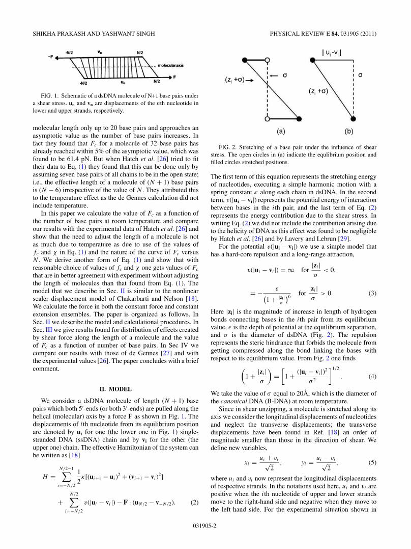
FIG. 1. Schematic of a dsDNA molecule of N+1 base pairs undera shear stress. un and vn are displacements of the nth nucleotide inlower and upper strands, respectively.
molecular length only up to 20 base pairs and approaches anasymptotic value as the number of base pairs increases. Infact they found that Fc for a molecule of 32 base pairs hasalready reached within 5% of the asymptotic value, which wasfound to be 61.4 pN. But when Hatch et al. [26] tried to fittheir data to Eq. (1) they found that this can be done only byassuming seven base pairs of all chains to be in the open state;i.e., the effective length of a molecule of (N + 1) base pairsis (N − 6) irrespective of the value of N . They attributed thisto the temperature effect as the de Gennes calculation did notinclude temperature.
In this paper we calculate the value of Fc as a function ofthe number of base pairs at room temperature and compareour results with the experimental data of Hatch et al. [26] andshow that the need to adjust the length of a molecule is notas much due to temperature as due to use of the values offc and χ in Eq. (1) and the nature of the curve of Fc versusN . We derive another form of Eq. (1) and show that withreasonable choice of values of fc and χ one gets values of Fc
that are in better agreement with experiment without adjustingthe length of molecules than that found from Eq. (1). Themodel that we describe in Sec. II is similar to the nonlinearscaler displacement model of Chakarbarti and Nelson [18].We calculate the force in both the constant force and constantextension ensembles. The paper is organized as follows. InSec. II we describe the model and calculational procedures. InSec. III we give results found for distribution of effects createdby shear force along the length of a molecule and the valueof Fc as a function of number of base pairs. In Sec IV wecompare our results with those of de Gennes [27] and withthe experimental values [26]. The paper concludes with a briefcomment.
II. MODEL
We consider a dsDNA molecule of length (N + 1) basepairs which both 5′-ends (or both 3′-ends) are pulled along thehelical (molecular) axis by a force F as shown in Fig. 1. Thedisplacements of ith nucleotide from its equilibrium positionare denoted by ui for one (the lower one in Fig. 1) single-stranded DNA (ssDNA) chain and by vi for the other (theupper one) chain. The effective Hamiltonian of the system canbe written as [18]
H =N/2−1∑i=−N/2
1
2κ[(ui+1 − ui)
2 + (vi+1 − vi)2]
+N/2∑
i=−N/2
v(|ui − vi |) − F · (uN/2 − v−N/2). (2)
FIG. 2. Stretching of a base pair under the influence of shearstress. The open circles in (a) indicate the equlibrium position andfilled circles stretched positions.
The first term of this equation represents the stretching energyof nucleotides, executing a simple harmonic motion with aspring constant κ along each chain in dsDNA. In the secondterm, v(|ui − vi|) represents the potential energy of interactionbetween bases in the ith pair, and the last term of Eq. (2)represents the energy contribution due to the shear stress. Inwriting Eq. (2) we did not include the contribution arising dueto the helicity of DNA as this effect was found to be negligibleby Hatch et al. [26] and by Lavery and Lebrun [29].
For the potential v(|ui − vi|) we use a simple model thathas a hard-core repulsion and a long-range attraction,
v(|ui − vi |) = ∞ for|zi |σ
< 0,
= − ε(1 + |zi|
σ
)6 for|zi |σ
> 0. (3)
Here |zi| is the magnitude of increase in length of hydrogenbonds connecting bases in the ith pair from its equilibriumvalue, ε is the depth of potential at the equilibrium separation,and σ is the diameter of dsDNA (Fig. 2). The repulsionrepresents the steric hindrance that forbids the molecule fromgetting compressed along the bond linking the bases withrespect to its equilibrium value. From Fig. 2 one finds
(1 + |zi |
σ
)=
[1 + (|ui − vi |)2
σ 2
]1/2
. (4)
We take the value of σ equal to 20A, which is the diameter ofthe canonical DNA (B-DNA) at room temperature.
Since in shear unzipping, a molecule is stretched along itsaxis we consider the longitudinal displacements of nucleotidesand neglect the transverse displacements; the transversedisplacements have been found in Ref. [18] an order ofmagnitude smaller than those in the direction of shear. Wedefine new variables,
xi = ui + vi√2
, yi = ui − vi√2
, (5)
where ui and vi now represent the longitudinal displacementsof respective strands. In the notations used here, ui and vi arepositive when the ith nucleotide of upper and lower strandsmove to the right-hand side and negative when they move tothe left-hand side. For the experimental situation shown in
031905-2

SHEAR UNZIPPING OF DOUBLE-STRANDED DNA PHYSICAL REVIEW E 84, 031905 (2011)
Fig. 1 it is clear that for i > 0, ui > 0 and magnitude of ui isgreater than that of vi and for i < 0, vi < 0, and the magnitudeof vi is greater than that of ui . This leads to following relationfor variables xi and yi :
x−i = −xi, x0 = 0, and y−i = yi. (6)
When we substitute these variables in Eq. (2) it decouples intotwo independent components:
H = Hx + Hy, (7)
where
Hx =N/2−1∑i=−N/2
1
2κ(xi+1 − xi)
2 − F√2
(xN/2 − x−N/2) (8)
and
Hy =N/2−1∑i=−N/2
1
2κ(yi+1 − yi)
2 +N/2∑
i=−N/2
v(yi)
− F√2
(yN/2 + y−N/2). (9)
Here the potential v(|ui − vi|) defined in Eq. (3) is expressedin terms of variable yi . Using Eqs. (4) and (5), one can rewriteEq. (3) as
v(yi) = − ε(1 + 2y2
i
σ 2
)3. (10)
Note that the expression of Hx does not contain the on-sitepotential v(yi) and simply corresponds to a harmonic chainthat is being pulled at the two ends by a force F/
√2, whereas
the expression of Hy contains the on-site potential v(yi) aswell as the force term.
In view of the relations given by Eq. (6) the average valueof displacement 〈xn〉 of the nth base pair can be calculatedfrom the relation
〈xn〉 =∫ ∏N/2
i=0 dxixn exp(−βHx)∫ ∏N/2i=0 dxi exp(−βHx)
, (11)
where β = (kBT )−1, kB being the Boltzmann constant, and T
is the temperature. Since for Hx given by Eq. (7) the integralsin Eq. (11) are Gaussians, one can solve them analytically togive
〈xn〉 = F√2κ
n. (12)
This result agrees with the one found by de Gennes [27].The average value of displacement yn of the nth base pair
can be found from the relation
〈yn〉 =∫ ∏N/2
i=−N/2 dyiyn exp(−βHy)∫ ∏N/2i=−N/2 dyi exp(−βHy)
. (13)
The integrals appearing in this expression cannot be evaluatedanalytically because of the form of on-site potential v(yi).However, for the expression of Hy given by Eq. (9) the integralappearing in Eq. (13) reduces to multiplication of (N + 1)matrices. The discretization of the coordinate variable andintroduction of a proper cutoff on the maximum values of y ′sdetermine the size of the matrices. We have taken −40 and40 A as the lower and upper limits of integration for eachcoordinate variable and discretized space using the Gaussian-Legendre method with the number of grid points equal to 900.By changing the limits of integration as well as the number ofgrid points we made sure that the values of 〈yn〉 are independentof the limit of integration and the number of grid points chosento discretize coordinate variables.
-8 -4 0 4 8n
0.4
0.8
1.2
1.6
2
2.4
<y n>
(Å)
-12 -8 -4 0 4 8 12n
0.4
0.8
1.2
1.6
2
2.4
<y n>
(Å)
-16 -12 -8 -4 0 4 8 12 16n
0.4
0.8
1.2
1.6
2
2.4
<y n>
(Å)
-24 -16 -8 0 8 16 24n
0.4
0.8
1.2
1.6
2
2.4
<y n>
(Å)
N+1=25N+1=17
N+1=33 N+1=49
FIG. 3. Displacement 〈yn〉 as function of n for four molecules of length 17, 25, 33, and 49 base pairs and for y−N/2 = yN/2 = 1.5 A (fullline), 2.0 A (dashed line), 2.38 A (dotted line), and 2.6 A (dot-dashed line).
031905-3
SHIKHA PRAKASH AND YASHWANT SINGH PHYSICAL REVIEW E 84, 031905 (2011)
III. RESULTS
When a shear force is applied on a dsDNA molecule, itstwo ssDNA strands get pulled in opposite directions, as shownin Fig. 1. The bonds in the backbone of DNA as well as bondsconnecting bases in a pair are stretched. In a case of κ beinginfinitely large the two strands will move like a rigid body,pulling all base pairs in the sequence in parallel. The effect ofthe shear force will then be uniformly distributed across all ofthe base pairs. However, if κ is finite, then both the backboneand the base pairs will stretch when a shear force is applied.The effect of the shear force may then be confined to limitedlengths on both ends of the molecule. To see how the effectcaused by shearing of a dsDNA molecule is distributed alongthe length of a molecule and how this depends on the energiesassociated with the stretching of backbone and base pairs, wecalculate the value of 〈yn〉 from Eq. (13) for different values ofn when the two end base pairs are stretched to a given lengthby the shear force.
In Fig. 3 we plot our results for four dsDNA molecules oflength 17, 25, 33, and 49 base pairs and for y−N/2 = yN/2 =1.50, 2.0, 2.38, and 2.60 A. The values shown in the figurecorrespond to ε = 0.04 eV,κ = 0.10eV/A
2, and T = 300 K.
When y−N/2 and yN/2 are allowed to be free (i.e., F = 0), then〈yn〉 is found to be zero for all n. From Fig. 3 it is clear thatfor short chains the effect created by shear force is distributedalong the entire length of a molecule affecting all base pairs,whereas for relatively larger molecules the base pairs in thecentral part of molecules are only marginally affected. Forexample, for a molecule of length 17 base pairs the value of 〈y0〉is 1.76 A when y−8 = y8 = 2.38 A, whereas, for the similarsituation (i.e., y−24 = y24 = 2.38 A), the value of 〈y0〉 for amolecule of length 49 base pairs is only 0.27 A. The qualitativenature of these results is in agreement with the results foundin Refs. [18] and [28]. The other point to be noted from thefigure is that the qualitative nature of the distribution of theeffect of shear stretching along the length of a molecule is thesame for all values of y−N/2 = yN/2 plotted in the figure.
In Fig. 4 we plot the value of 〈y0〉 when y−N/2 = yN/2 =2.38 A as a function of the length of dsDNA molecules.As discussed below, the value of y−N/2 = yN/2 = 2.38 A isassumed to be the critical value of stretching in the sense thatinitiation of separation of two strands starts at this value ofyN/2, and the shear force that creates this value of stretching of
10 20 30 40 50N+1
0
0.5
1
1.5
2
2.5
<y 0>
(Å)
yN/2
=y-N/2
=2.38Å
FIG. 4. Displacement 〈y0〉 of the central base pair as a functionof number of base pairs of dsDNA molecules.
-8 -4 0 4 8n
1
1.5
2
2.5
3
-10 -5 0 5 100
1
2
3
4
-24 -16 -8 0 8 16 24n
-2
0
2
4
6
<vn>(Å) <u
n>(Å)
<vn>(Å)
<vn>(Å)
<un>(Å)
<un>(Å)
N+1=17
N+1=21
N+1=49
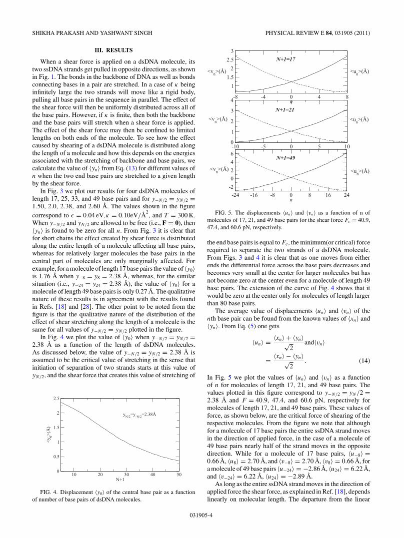
FIG. 5. The displacements 〈un〉 and 〈vn〉 as a function of n ofmolecules of 17, 21, and 49 base pairs for the shear force Fc = 40.9,47.4, and 60.6 pN, respectively.
the end base pairs is equal to Fc, the minimum(or critical) forcerequired to separate the two strands of a dsDNA molecule.From Figs. 3 and 4 it is clear that as one moves from eitherends the differential force across the base pairs decreases andbecomes very small at the center for larger molecules but hasnot become zero at the center even for a molecule of length 49base pairs. The extension of the curve of Fig. 4 shows that itwould be zero at the center only for molecules of length largerthan 80 base pairs.
The average value of displacements 〈un〉 and 〈vn〉 of thenth base pair can be found from the known values of 〈xn〉 and〈yn〉. From Eq. (5) one gets
〈un〉 = 〈xn〉 + 〈yn〉√2
and〈vn〉
= 〈xn〉 − 〈yn〉√2
. (14)
In Fig. 5 we plot the values of 〈un〉 and 〈vn〉 as a functionof n for molecules of length 17, 21, and 49 base pairs. Thevalues plotted in this figure correspond to y−N/2 = yN/2 =2.38 A and F = 40.9, 47.4, and 60.6 pN, respectively formolecules of length 17, 21, and 49 base pairs. These values offorce, as shown below, are the critical force of shearing of therespective molecules. From the figure we note that althoughfor a molecule of 17 base pairs the entire ssDNA strand movesin the direction of applied force, in the case of a molecule of49 base pairs nearly half of the strand moves in the oppositedirection. While for a molecule of 17 base pairs, 〈u−8〉 =0.66 A, 〈u8〉 = 2.70 A, and 〈v−8〉 = 2.70 A, 〈v8〉 = 0.66 A, fora molecule of 49 base pairs 〈u−24〉 = −2.86 A, 〈u24〉 = 6.22 A,and 〈v−24〉 = 6.22 A, 〈u24〉 = −2.89 A.
As long as the entire ssDNA strand moves in the direction ofapplied force the shear force, as explained in Ref. [18], dependslinearly on molecular length. The departure from the linear
031905-4

SHEAR UNZIPPING OF DOUBLE-STRANDED DNA PHYSICAL REVIEW E 84, 031905 (2011)
dependence of the shear force on molecular length is expectedto take place when 〈u−N/2〉 and 〈vN/2〉 become zero; i.e., thedisplacement of nucleotides on the opposite side of a ssDNAstrand remains unaffected by the applied force. Indeed, wefind that 〈u−10〉 and 〈v10〉 = 0 for a molecule of length 21 basepairs, which is in very good agreement with the experimentalvalue [26]. Since for molecules of base pairs larger than 21,part of a ssDNA moves in the opposite direction, a regiondevelops between each strand, which remains unaffected bythe force. This region moves toward the center while increasingthe molecular length; the force gets saturated as soon as theregion reaches the center of the chain and stays there whilefurther increasing the molecular length.
The value of shear force needed to separate the two strandsof a dsDNA molecule can be calculated in two different ways.In one, we follow a method proposed by de Gennes [27] andthat hereafter is referred to as a method of constant forceensemble. The other method is based on the constant extensionensemble.
In the method of constant force ensemble one first definesa critical distance y for the rupture of a base pair (i.e., whenyn of the nth base pair becomes larger than y, the bases ofthe pair become free) and calculate the value of force that canstretch a base pair to the critical distance y. This force can befound from the on-site potential v(y). Thus
fc = −∂v(y)
∂y
∣∣∣∣y=y
= 12εy
σ 2
(1 + 2y2
σ 2
)−4
. (15)
If we take the value of y = 2.38 A,we find fc = 4.1 pN for thevalue of ε and κ given above. This value of fc is close to thevalue used by Hatch et al. [26] to fit their experimental dataand the value estimated by Chakrabarti and Nelson [18]. Torupture the end base pairs of a given molecule they must be
0 10 20 30 40 50 60N+1
20
30
40
50
60
70
Fc(p
N)
FIG. 6. The value of critical force Fc in pN as a function ofnumber of base pairs of a dsDNA molecule. The curve shownby dotted line corresponds to constant force ensemble and dashedline to constant extension ensemble. The curve shown by full linecorresponds to values found by taking the effective length 10 and 15base pairs for molecules of length 12 and 16 base pairs, respectively.The experimental values of Hatch et al. [26] are shown by diamondwhen 5′-ends are pulled and by star when 3′-ends are pulled by shearforce.
stretched by the shear force to distance y. The balance of forceat one end of one of the ssDNA gives [27]
Fc = κ(〈uN/2〉 − 〈uN/2−1〉) + fc. (16)
Using the relations of Eq. (5) and Eq. (12) we get
Fc =√
2κ(〈yN/2〉 − 〈yN/2−1〉) + 2fc. (17)
Taking the value of 〈y−N/2〉 = 〈yN/2〉 = y = 2.38 A wecalculate the values of 〈yN/2−1〉 from Eq. (13) for severalmolecules of length 10–60 base pairs. The value of force Fc
found from Eq. (13) is shown by a dotted line in Fig. 6.In the constant extension ensemble one first calculates the
work done in stretching the two end base pairs of a givenmolecule to distance y. This work is found from the relation
W (y) = 1
β[ln ZN+1(y) − ln ZN+1], (18)
where
ZN+1(y) =∫ N/2∏
i=−N/2
dyi δ(y−N/2
−y) δ(yN/2 − y) exp(−βHy) (19)
is the constrained partition function and
ZN+1 =∫ N/2∏
i=−N/2
dyi exp(−βHy) (20)
is the partition function. Here δ is the Dirac function.We used the matrix multiplication method to evaluate W (y)
from the above equations for ε = 0.04 eV, κ = 0.1 eV/ A2,and T = 300 K. The derivative of W (y) with respect to y givesthe average force F (y) that is needed to keep the extension ofend base pairs of a given molecule equal to y. Thus
F (y) = ∂W (y)
∂y. (21)
To get the value of critical force we have to choose a value of y
that corresponds to rupturing of base pairs. The values shownin Fig. 6 by dashed line are found when y was taken equal to2.0 A; this value is slightly lower than the one taken for theconstant force ensemble. The difference in the value of criticalstretching in the two ensembles may be due to difference inthe path of unzipping.
We note that the values of Fc found by methods of theconstant force and the constant extension ensembles are closebut not identical. The difference between the two as expected[30] is large for molecules of smaller lengths but they becomesclose as molecular length increases. Both methods give thesame asymptotic value, equal to 61.2 pN, which is in verygood agreement with the experimental value 61.4 pN [26].The experimental values shown in the figure are of Hatchet al. [26]. In view of large spread in experimental data wefind good agreement between experimental values of Fc andthe theoretical values found using the constant force ensembleas well as the constant extension ensemble.
031905-5
SHIKHA PRAKASH AND YASHWANT SINGH PHYSICAL REVIEW E 84, 031905 (2011)
IV. DISCUSSIONS
A. Comparison with the experiment
Since the experimental values [26] of Fc given in Fig. 6are found using the fixed force ensemble, we concentrateour discussion with the theoretical values found using thesame ensemble and shown in Fig. 6 by a dotted line. Theagreement between theory and experiment is excellent exceptfor a molecule of length 12 base pairs. We know that at agiven temperature shorter dsDNA molecules are less stablecompared to longer molecules. It is quite possible that at roomtemperature due to surface effects a few of the base pairs at thetwo ends of a molecule of length 12 base pairs are nearly in anopen state. The curve shown in Fig. 6 by a solid line is foundwhen the effective length of a molecule of length 12 base pairsis taken to be equal to 10 base pairs and that of a moleculeof length 16 base pairs is equal to 15 base pairs. The valuesof force Fc shown by the solid line are in excellent agreementwith the experimental values for the entire range of molecularlength investigated by Hatch et al. [26]. The saturation valueof Fc found to be 61.2 pN is also in very good agreement withthe experimental value of 61.4 pN [26].
B. Comparison with the results of de Gennes [27]
In order to compare our results with those of de Gennes [27]
we first estimate the value of χ defined as χ =√
2Rκ
, where R
is the spring constant of a simple harmonic potential betweenthe bases of a pair. Expanding v(y) in ascending powers of y
one gets
v(yi) = −ε + 1
2
(12ε
σ 2
)y2
i , (22)
R = 12ε
σ 2and χ2 = 24ε
κσ 2. (23)
Substituting the values of ε, κ, and σ given above we findχ−1 = 6.44, which corresponds to κ/R = 83.3. This valueof κ/R is in good agreement with the predicted value of 77based on calculation of the spring constants for base pairs andbackbones [31].
The expression for the displacement 〈yn〉 found by deGennes [27] can be written as
〈yn〉 = 12 〈y0〉(eχn + e−χn). (24)
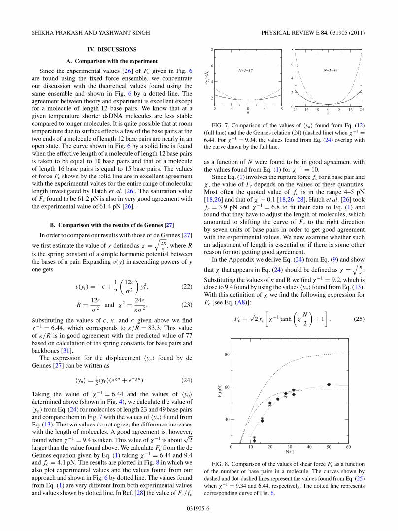
Taking the value of χ−1 = 6.44 and the values of 〈y0〉determined above (shown in Fig. 4), we calculate the value of〈yn〉 from Eq. (24) for molecules of length 23 and 49 base pairsand compare them in Fig. 7 with the values of 〈yn〉 found fromEq. (13). The two values do not agree; the difference increaseswith the length of molecules. A good agreement is, however,found when χ−1 = 9.4 is taken. This value of χ−1 is about
√2
larger than the value found above. We calculate Fc from the deGennes equation given by Eq. (1) taking χ−1 = 6.44 and 9.4and fc = 4.1 pN. The results are plotted in Fig. 8 in which wealso plot experimental values and the values found from ourapproach and shown in Fig. 6 by dotted line. The values foundfrom Eq. (1) are very different from both experimental valuesand values shown by dotted line. In Ref. [28] the value of Fc/fc
-8 -4 0 4 8n
2
4
6
8
<y n>
(Å)
-24 -16 -8 0 8 16 24n
0
2
4
6
8
N+1=17 N+1=49
FIG. 7. Comparison of the values of 〈yn〉 found from Eq. (12)(full line) and the de Gennes relation (24) (dashed line) when χ−1 =6.44. For χ−1 = 9.34, the values found from Eq. (24) overlap withthe curve drawn by the full line.
as a function of N were found to be in good agreement withthe values found from Eq. (1) for χ−1 = 10.
Since Eq. (1) involves the rupture force fc for a base pair andχ , the value of Fc depends on the values of these quantities.Most often the quoted value of fc is in the range 4–5 pN[18,26] and that of χ ∼ 0.1 [18,26–28]. Hatch et al. [26] tookfc = 3.9 pN and χ−1 = 6.8 to fit their data to Eq. (1) andfound that they have to adjust the length of molecules, whichamounted to shifting the curve of Fc to the right directionby seven units of base pairs in order to get good agreementwith the experimental values. We now examine whether suchan adjustment of length is essential or if there is some otherreason for not getting good agreement.
In the Appendix we derive Eq. (24) from Eq. (9) and show
that χ that appears in Eq. (24) should be defined as χ =√
Rκ
.
Substituting the values of κ and R we find χ−1 = 9.2, which isclose to 9.4 found by using the values 〈yn〉 found from Eq. (13).With this definition of χ we find the following expression forFc [see Eq. (A8)]:
Fc =√
2fc
[χ−1 tanh
(χ
N
2
)+ 1
]. (25)
0 10 20 30 40 50 60N+1
40
60
80
Fc(p
N)
FIG. 8. Comparison of the values of shear force Fc as a functionof the number of base pairs in a molecule. The curves shown bydashed and dot-dashed lines represent the values found from Eq. (25)when χ−1 = 9.34 and 6.44, respectively. The dotted line representscorresponding curve of Fig. 6.
031905-6
SHEAR UNZIPPING OF DOUBLE-STRANDED DNA PHYSICAL REVIEW E 84, 031905 (2011)
The appearance of√
2fc instead of 2fc [in Eq. (1)] is due to
difference in the definition of χ . The definition χ =√
Rκ
seemsmore appropriate in the sense that it is the ratio of the two
harmonic spring constants than using the definition χ =√
2Rκ
of Ref. [27], in which one spring constant is multiplied by two.The value of Fc found from Eq. (25) when χ−1 = 9.4 and
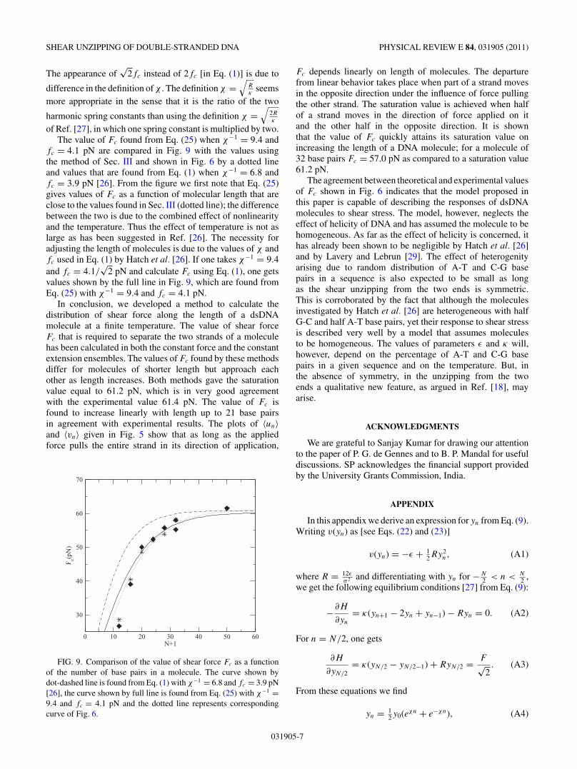
fc = 4.1 pN are compared in Fig. 9 with the values usingthe method of Sec. III and shown in Fig. 6 by a dotted lineand values that are found from Eq. (1) when χ−1 = 6.8 andfc = 3.9 pN [26]. From the figure we first note that Eq. (25)gives values of Fc as a function of molecular length that areclose to the values found in Sec. III (dotted line); the differencebetween the two is due to the combined effect of nonlinearityand the temperature. Thus the effect of temperature is not aslarge as has been suggested in Ref. [26]. The necessity foradjusting the length of molecules is due to the values of χ andfc used in Eq. (1) by Hatch et al. [26]. If one takes χ−1 = 9.4and fc = 4.1/
√2 pN and calculate Fc using Eq. (1), one gets
values shown by the full line in Fig. 9, which are found fromEq. (25) with χ−1 = 9.4 and fc = 4.1 pN.
In conclusion, we developed a method to calculate thedistribution of shear force along the length of a dsDNAmolecule at a finite temperature. The value of shear forceFc that is required to separate the two strands of a moleculehas been calculated in both the constant force and the constantextension ensembles. The values of Fc found by these methodsdiffer for molecules of shorter length but approach eachother as length increases. Both methods gave the saturationvalue equal to 61.2 pN, which is in very good agreementwith the experimental value 61.4 pN. The value of Fc isfound to increase linearly with length up to 21 base pairsin agreement with experimental results. The plots of 〈un〉and 〈vn〉 given in Fig. 5 show that as long as the appliedforce pulls the entire strand in its direction of application,
0 10 20 30 40 50 60N+1
30
40
50
60
70
Fc(p
N)
FIG. 9. Comparison of the value of shear force Fc as a functionof the number of base pairs in a molecule. The curve shown bydot-dashed line is found from Eq. (1) with χ−1 = 6.8 and fc = 3.9 pN[26], the curve shown by full line is found from Eq. (25) with χ−1 =9.4 and fc = 4.1 pN and the dotted line represents correspondingcurve of Fig. 6.
Fc depends linearly on length of molecules. The departurefrom linear behavior takes place when part of a strand movesin the opposite direction under the influence of force pullingthe other strand. The saturation value is achieved when halfof a strand moves in the direction of force applied on itand the other half in the opposite direction. It is shownthat the value of Fc quickly attains its saturation value onincreasing the length of a DNA molecule; for a molecule of32 base pairs Fc = 57.0 pN as compared to a saturation value61.2 pN.
The agreement between theoretical and experimental valuesof Fc shown in Fig. 6 indicates that the model proposed inthis paper is capable of describing the responses of dsDNAmolecules to shear stress. The model, however, neglects theeffect of helicity of DNA and has assumed the molecule to behomogeneous. As far as the effect of helicity is concerned, ithas already been shown to be negligible by Hatch et al. [26]and by Lavery and Lebrun [29]. The effect of heterogenityarising due to random distribution of A-T and C-G basepairs in a sequence is also expected to be small as longas the shear unzipping from the two ends is symmetric.This is corroborated by the fact that although the moleculesinvestigated by Hatch et al. [26] are heterogeneous with halfG-C and half A-T base pairs, yet their response to shear stressis described very well by a model that assumes moleculesto be homogeneous. The values of parameters ε and κ will,however, depend on the percentage of A-T and C-G basepairs in a given sequence and on the temperature. But, inthe absence of symmetry, in the unzipping from the twoends a qualitative new feature, as argued in Ref. [18], mayarise.
ACKNOWLEDGMENTS
We are grateful to Sanjay Kumar for drawing our attentionto the paper of P. G. de Gennes and to B. P. Mandal for usefuldiscussions. SP acknowledges the financial support providedby the University Grants Commission, India.
APPENDIX
In this appendix we derive an expression for yn from Eq. (9).Writing v(yn) as [see Eqs. (22) and (23)]
v(yn) = −ε + 12Ry2
n, (A1)
where R = 12εσ 2 and differentiating with yn for −N
2 < n < N2 ,
we get the following equilibrium conditions [27] from Eq. (9):
−∂H
∂yn
= κ(yn+1 − 2yn + yn−1) − Ryn = 0. (A2)
For n = N/2, one gets
∂H
∂yN/2= κ(yN/2 − yN/2−1) + RyN/2 = F√
2. (A3)
From these equations we find
yn = 12y0(eχn + e−χn), (A4)
031905-7

SHIKHA PRAKASH AND YASHWANT SINGH PHYSICAL REVIEW E 84, 031905 (2011)
where χ =√
Rκ
, and
y0 = F√2
1
Rcosh(
12χN
)[χ−1tanh
(12χN
) + 1] . (A5)
Let the force on the last hydrogen bonds (n = N/2) is RyN/2,when we reach the threshold fc (fc being the rupture force fora base pair). Thus
fc = Ry0cosh(
12χN
). (A6)
From Eq.(A3) we get
Fc =√
2κχyN/2tanh(
12χN
) + √2fc (A7)
=√
2fc
[χ−1tanh
(12χN
) + 1]. (A8)
This equation differs from Eq. (1) in the definition ofχ and the mutiplying factor, which is now
√2 instead
of 2.
[1] W. Saenger, Principle of Nucleic Acid Structure (Springer-Verlag, Berlin, 1984).
[2] T. Strick, J.-F. Allemand, V. Croquette, and D. Bensimon, Phys.Today 54, 46 (2001).
[3] U. Bockelmann, Curr. Opin. Struct. Biol. 14, 368 (2004);U. Bockelmann, B. Essevaz-Roulet, and F. Heslot, Phys. Rev.Lett. 79, 4489 (1997).
[4] S. B. Smith, Y. Cui, and C. Bustamante, Science 271, 795 (1996).[5] M. Reif, H. Clausen-Schaumann, and H. E. Gaub, Nat. Struct.
Biol. 6, 346 (1999).[6] R. S. Convory and C. Danilowicz, Contemp. Phys. 45, 277
(2004).[7] S. Kumar and M. S. Li, Phys. Rep. 486, 1 (2010).[8] Y. Zhang, H. Zhou, and Z. -C. Ou-Yang, Biophys. J. 81, 1133
(2001).[9] I. Rouzina and V. A. Bloomfield, Biophys. J. 80, 882 (2001).
[10] H. Clausen-Schaumann, M. Reif, C. Tolksdorf, and H. E. Gaub,Biophys. J. 78, 1997 (2000).
[11] C. Bustamante, S. B. Smith, J. Liphardt, and D. Smith, Curr.Opin. Struct. Biol. 10, 279 (2000).
[12] J. Morfill, F. Kuhner, K. Blank, R. A. Lugmaier, J. Sedlmair,and H. E. Gaub, Biophys. J. 93, 2400 (2007).
[13] G. U. Lee, L. A. Chrisey, and R. J. Colton, Science 266, 771(1994).
[14] A. D. Mc Kerall and G. U. Lee, Eur. Biophys. J. 28, 415 (1999).[15] L. H. Pope, M. L. Davies, C. A. Laughton, C. J. Roberts, S. J.
B. Tendler, and P. M. Williams, Eur. Biophys. J. 30, 53 (2000).[16] T. Strunz, K. Oroszlan, R. Schafer, and H.-J. Guntherodt, Proc.
Natl. Acad. Sci. USA 96, 11277 (1999).
[17] F. Kuhner, J. Morfill, R. A. Neher, K. Blank, and H. E. Gaub,Biophys. J. 92, 2491 (2007).
[18] B. Chakrabarti and D. R. Nelson, J. Phys. Chem. B 113, 3831(2009).
[19] B. D. Sattin, A. E. Pelling, and M. C. Goh, Nucleic Acids Res.32, 4876 (2004).
[20] W. Grange, T. Strunz, I. Schumakovitch, H.-J. Guntherodt, andM. Hegner, Single Mol. 2, 75 (2001).
[21] I. Schumakovitch, W. Grange, T. Strunz, P. Bertoncini, H.-J.Guntherodt, and M. Hegner, Biophys. J. 82, 517 (2002).
[22] G. Neuert, C. H. Albrecht, and H. E. Gaub, Biophys. J. 93, 1215(2007).
[23] Y. Zhang, V. T. Milam, D. J. Graves, and D. A. Hammer, Biophys.J. 90, 4128 (2006).
[24] M. V. Wal, S. Kamper, J. Headley, and K. Sinniah, Langmuir22, 882 (2006).
[25] A. Noy, D. V. Vezenov, J. F. Kayyem, T. J. Meade, and C. M.Leiber, Chem. Biol. 4, 519 (1997).
[26] K. Hatch, C. Danilowicz, V. Coljee, and M. Prentiss, Phys. Rev.E 78, 011920 (2008).
[27] P. G. de Gennes, Soryushiron Kenkyu 2, 1505 (2001).[28] R. K. Mishra, G. Mishra, M. S. Li, and S. Kumar, e-print
arXiv:1104.3059 (2011).[29] R. Lavery and A. Lebrun, Genetica 96, 75 (1999).[30] D. Keller, D. Swigon, and C. Bustamante, Biophys. J. 84, 733
(2003).[31] This value quoted in Ref. [26] was found from M. Fytas and
E. Kaxiras through private communication by the authors of thatreference.
031905-8