shiga toxin 1 induces apoptosis in the human myelogenous ... · the undifferentiated myelogenous...
TRANSCRIPT

INFECTION AND IMMUNITY, Aug. 2005, p. 5115–5126 Vol. 73, No. 80019-9567/05/$08.00�0 doi:10.1128/IAI.73.8.5115–5126.2005Copyright © 2005, American Society for Microbiology. All Rights Reserved.
Shiga Toxin 1 Induces Apoptosis in the Human MyelogenousLeukemia Cell Line THP-1 by a Caspase-8-Dependent, Tumor
Necrosis Factor Receptor-Independent MechanismSang-Yun Lee,* Rama P. Cherla, Isa Caliskan, and Vernon L. Tesh
Department of Medical Microbiology and Immunology, Texas A&M University System Health Science Center,College Station, Texas
Received 7 February 2005/Returned for modification 21 March 2005/Accepted 6 April 2005
Shiga toxins (Stxs) induce apoptosis in a variety of cell types. Here, we show that Stx1 induces apoptosis inthe undifferentiated myelogenous leukemia cell line THP-1 in the absence of tumor necrosis factor alpha(TNF-�) or death receptor (TNF receptor or Fas) expression. Caspase-8 and -3 inhibitors blocked, andcaspase-6 and -9 inhibitors partially blocked, Stx1-induced apoptosis. Stx1 induced the mitochondrial pathwayof apoptosis, as activation of caspase-8 triggered the (i) cleavage of Bid, (ii) disruption of mitochondrialmembrane potential, and (iii) release of cytochrome c into the cytoplasm. Caspase-8, -9, and -3 cleavage andfunctional activities began 4 h after toxin exposure and peaked after 8 h of treatment. Caspase-6 may alsocontribute to Stx1-induced apoptosis by directly acting on caspase-8. It appears that functional Stx1 holotoxinsmust be transported to the endoplasmic reticulum to initiate apoptotic signaling through the ribotoxic stressresponse. These data suggest that Stxs may activate monocyte apoptosis via a novel caspase-8-dependent, deathreceptor-independent mechanism.
Shiga toxins (Stxs) are a family of protein exotoxins ex-pressed by the enteric pathogens Shigella dysenteriae serotype 1and certain serotypes of Escherichia coli. Stxs share structuraland functional properties: Stxs are holotoxins composed of asingle A-subunit in noncovalent association with a pentamericring of identical B-subunits, and the toxins are potent proteinsynthesis inhibitors in susceptible mammalian cells (11, 31).The B-subunits mediate toxin binding through interaction withthe neutral membrane glycolipid globotriaosylceramide (Gb3)(24). Following internalization and retrograde transport of thetoxins through the Golgi apparatus to the endoplasmic reticu-lum (ER), A-subunits are proteolytically nicked and reduced,and the resultant A1-fragments are translocated into the targetcell cytoplasm (38). The A1-fragment acts as a highly specificN-glycosidase that cleaves a single adenine residue near the 3�end of the 28S rRNA component of eukaryotic ribosomes (8,39). Shigella dysenteriae serotype 1 produces Shiga toxin (Stx),while E. coli may express multiple toxins categorized as Shigatoxin type 1 (Stx1) or Shiga toxin type 2 (Stx2), based on theirantigenic similarity to Stx (27).
Shigella dysenteriae serotype 1 and Shiga toxin-producing E.coli cause the bloody diarrheal diseases bacillary dysentery andhemorrhagic colitis, respectively. Patients infected with Stx-producing bacteria are at increased risk for developing life-threatening complications, including acute renal failure (he-molytic uremic syndrome) and central nervous systemabnormalities such as disorientation, lethargy, seizures, paral-ysis, coma, and death (34, 40). Numerous studies in animalshave shown that the extra-intestinal complications seen in hu-
mans are reproduced by the intravenous administration ofpurified Stxs (29). Affected organs in humans and experimentalanimals show evidence of profound vascular damage. Theseobservations suggest that Stxs gain access to the bloodstreamand target vascular endothelial cells in the kidneys and centralnervous system for destruction. In addition to direct cytotoxiceffects on endothelial cells, Stxs have also been shown to stim-ulate macrophages to produce the proinflammatory cytokinestumor necrosis factor alpha (TNF-�) and interleukin-1� invitro (35, 48). These cytokines, in turn, up-regulate Gb3 bio-synthesis and expression on endothelial cells (47) and sensitizetarget cells to the cytotoxic action of the toxins (25). Thus, thehost innate immune response may exacerbate vascular damageinitiated by Stxs.
Stxs have been demonstrated to induce apoptosis in manyhuman cell types in vitro, including epithelial cell lines, primaryrenal epithelial cells, Burkitt’s lymphoma cells, microvascularendothelial cells, and myelogenous leukemia cell lines (re-viewed in reference 6). The mechanism(s) by which Stxs induceapoptosis in these cell types, and whether a common apoptosispathway is triggered by Stxs in all cell types, remains to be fullycharacterized. Kojio et al. (21) showed that Stx-induced apo-ptosis in the human monocytic cell line THP-1 required trans-port of toxins through functional Golgi complexes and theactivation of caspase-3. Fujii et al. (12) reported that Stx-induced HeLa cell apoptosis occurs via a pathway requiringcaspase-8, -6, and -3, but not caspase-9. In contrast to studiesusing holotoxin molecules, binding toxin receptors with puri-fied Stx B-subunits or anti-Gb3 antibodies has been reported tobe sufficient to induce caspase-8 activation and apoptosis inBurkitt’s lymphoma cells (20). Finally, it is unknown whetherStxs directly activate apoptosis in all cell types or if additionalhost factors are required in some instances. One potential hostfactor that may contribute to apoptosis induction is TNF-�
* Corresponding author. Mailing address: Department of MedicalMicrobiology and Immunology, Room 407, Reynolds Medical Build-ing, Texas A&M University System Health Science Center, CollegeStation, TX 77843-1114. Phone: (979) 845-1313. Fax: (979) 845-3479.E-mail: [email protected].
5115
on March 5, 2019 by guest
http://iai.asm.org/
Dow
nloaded from

(49). We previously showed that purified Stx1 induces TNF-�and interleukin-1� gene expression and apoptosis in the my-elomonocytic cell line THP-1 in a cell maturation-dependentmanner (14, 15). Differentiated (macrophage-like) THP-1 cellswere relatively resistant to killing by Stxs and expressed cyto-kines, whereas undifferentiated (monocytic) THP-1 cells failedto secrete TNF-� and were sensitive to Stx cytotoxicity. Plastic-adherent human peripheral blood monocytes, like differenti-ated THP-1 cells, were relatively insensitive to apoptosis in-duction by Stx2 but responded by producing TNF-� andgranulocyte-macrophage colony-stimulating factor (5). Thus,there appears to be a Stx-dependent, TNF-�-independent ap-optotic signaling mechanism operative in monocytic THP-1cells. Experiments reported here characterize Stx1-mediatedapoptotic signaling in monocytic THP-1 cells.
MATERIALS AND METHODS
Cells. The human myelogenous leukemia cell line THP-1 (46) was purchasedfrom the American Type Culture Collection, Manassas, VA, and cultured inRPMI 1640 medium (Gibco-BRL, Grand Island, NY) supplemented with pen-icillin (100 U/ml), streptomycin (100 �g/ml), and 10% fetal bovine serum (FBS;HyClone Laboratories, Logan, UT). Cells were maintained at 37°C in 5% CO2
in a humidified incubator.Toxins. Stx1 used in this study was prepared as previously described (44).
Briefly, Stx1 was purified from cell lysates prepared from E. coliDH5�(pCKS112) cells by sequential ion-exchange, chromatofocusing, and im-munoaffinity chromatography. Purity of toxin preparations was assessed by so-dium dodecyl sulfate-polyacrylamide gel electrophoresis (SDS-PAGE) with sil-ver staining and Western blot analysis using anti-Stx1 antibodies. Toxinpreparations contained �0.1 ng endotoxin per ml as determined by Limulusamoebocyte lysate assay (Associates of Cape Cod, Falmouth, ME). Purified Stx1pentameric B-subunits were the kind gift of Cheleste Thorpe, Tufts UniversitySchool of Medicine, Boston, MA. Purified Stx1A� (E167Q-R170L), an enzy-matic mutant in which glutamate at position 167 and arginine at position 170were replaced by glutamine and leucine, respectively, by oligonucleotide-directedsite-specific mutagenesis (32), was the kind gift of Yoshifumi Takeda, JissenWomen’s University, Tokyo, Japan.
Reagents. General caspase inhibitor N-benzyl-oxycarbonyl-Val-Ala-Asp-(OMe)-fluoromethyl-ketone (ZVAD-fmk), caspase-1 inhibitor Z-Tyr-Val-Ala-Asp-fmk (Z-YVAD-fmk), caspase-2 inhibitor Z-Val-Asp-Val-Ala-Asp-fmk (Z-VDVAD-fmk), caspase-3 inhibitor Z-Asp-Glu-Val-Asp-fmk (Z-DEVD-fmk),caspase-6 inhibitor Z-Val-Glu-Ile-Asp-fmk (Z-VEID-fmk), caspase-8 inhibitorZ-Ile-Glu-Thr-Asp-fmk (Z-IETD-fmk), and caspase-9 inhibitor Z-Leu-Glu-His-Asp-fmk (Z-LEHD-fmk) were purchased from Calbiochem, San Diego, CA. Allcaspase inhibitors were used at concentrations known to optimally inhibit specificcaspase activity. Rabbit anti-human cytochrome c antibody was purchased fromSan Cruz Biotechnology, Inc., Santa Cruz, CA. Antibodies directed againsthuman caspase-3, caspase-8, caspase-9, and Bid were obtained from Cell Signal-ing Technology, Beverly, MA. Cycloheximide and anisomycin were purchasedfrom Sigma, St. Louis, MO. Fluorescein-conjugated and nonfluoresceinatedmonoclonal antibodies directed against human membrane-bound TNF-�, TNFreceptor 1 (TNFR1), TNFR2, and Fas (APO/CD95) were purchased from R&DSystems, Inc., Minneapolis, MN. Human immunoglobulin G (IgG) and all otherreagents were obtained from Sigma Chemical Co., St. Louis, MO.
Analysis of cell surface receptor expression. THP-1 cells (2 � 106 cells/ml)were incubated in the presence or absence of Stx1 (400 ng/ml) for 0, 2, 4, and 6 h.We have previously shown that this toxin concentration produces optimal sig-naling for cytokine expression and apoptosis induction (14, 15). Cells werewashed twice with phosphate-buffered saline (PBS) containing 0.5% bovineserum albumin (BSA). Before staining for cell surface receptor expression usingantibodies, FcRs were blocked by treatment of the cells with 1.0 �g of humanIgG per 105 cells for 15 min at room temperature. FcR-blocked cells werewashed and suspended in PBS plus 0.5% BSA. Cells were then treated withfluorescein-conjugated monoclonal antibodies directed against human mem-brane-bound TNF-� (mTNF-�), TNFR1, TNFR2, or Fas for 30 to 45 min at 4°C.Following incubation, cells were washed and suspended in 0.5 ml PBS. Cellfluorescence was measured by flow cytometry using the FACSCalibur instrument(Becton Dickinson, Palo Alto, CA). Untreated cells and cells treated with fluo-rescein-labeled mouse IgG antibodies (Santa Cruz Biotechnology, Inc., Santa
Cruz, CA) served as control cells in the fluorescence-activated cell sorter (FACS)analysis.
Analysis of apoptosis by Annexin V and PI staining. THP-1 cells were treatedwith Stx1 (400 ng/ml) for 12 h in RPMI 1640 plus 0.5% FBS in the presence orabsence of anti-human TNF-� antibody (0.01 �g/ml), anti-human TNFR1 anti-body (5.0 �g/ml), or recombinant human TNF-� (rhTNF-�; 40 ng/ml). In aseparate set of experiments, cells were treated with Stx1 for 5 h in RPMI 1640plus 0.5% FBS in the presence or absence of caspase-1, -2, -3, -6, -8, and -9inhibitors or the general caspase inhibitor ZVAD-fmk (all at 40 �M). In someexperiments, THP-1 cells were treated with cycloheximide (10 to 200 �M) oranisomycin (0.1 to 10 �g/ml) for 5 h. Following treatment, cells were centrifugedat 200 � g for 5 min, washed in ice-cold sterile PBS, and stained using theAnnexin V-FLUOS staining kit (Roche Diagnostics Corp., Indianapolis, IN).Cells were incubated in the provided incubation buffer for 10 to 15 min at roomtemperature. Cells were then centrifuged, washed twice in incubation buffer, andsuspended in 0.5 ml of incubation buffer. Apoptosis was measured by flowcytometry (Becton Dickinson, Palo Alto, CA). Fluorescence parameters weregated using unstained and single-stained untreated cells. Total apoptosis wasexpressed as the percentages of Annexin V-positive plus Annexin V and pro-pidium iodide (PI) double-positive cells minus background fluorescence.
DNA fragmentation analysis. DNA fragmentation was assayed using the Ap-optotic DNA Ladder kit (Roche, Mannheim, Germany). THP-1 cells (2 � 106
cells/ml) were maintained in 12-well culture plates. Cells were treated with Stx1(400 ng/ml) in RPMI 1640 and 0.5% FBS in the presence or absence ofcaspase-1, -2, -3, -6, -8, and -9 inhibitors or the general caspase inhibitor ZVAD-fmk (all at 40 �M) for 5 h. Cells were washed with ice-cold PBS and lysed withlysis buffer (6.0 M guanidine-HCl, 10 mM urea, 10 mM Tris-HCl, 20% TritonX-100 [vol/vol], pH 4.4). After 10 min of incubation at room temperature, lysateswere centrifuged through DNA binding columns. DNA was eluted from thecolumns with 10 mM Tris (pH 8.5). Eluted DNA was treated with DNase-freeRNase for 30 min. DNA concentrations were measured, and equal amounts ofDNA (2.5 �g) were loaded on 1.2% agarose gels. Following electrophoresis, gelswere stained with ethidium bromide and photographed on a UV transilluminator(Gel Imager; Bio-Rad, Hercules, CA).
Preparation of cellular lysates. Prior to stimulation with Stx1, THP-1 cells (5� 106 cells/ml) were washed once in cold Dulbecco’s PBS and suspended inRPMI 1640 with 0.5% FBS for 2 h. Cells were then stimulated with Stx1 (400ng/ml) for the various time periods indicated in the figures. For Western blotanalyses of caspase and Bid activation, cells were harvested and lysed withmodified radioimmunoprecipitation assay buffer (1.0% Nonidet P-40, 1.0% Na-deoxycholate, 150 mM NaCl, 50 mM Tris-HCl [pH 7.5], 0.25 mM Na-pyrophos-phate, 2.0 mM Na-vanadate, 2.0 mM Na-fluoride, 10 �g/ml aprotinin, 1.0 �g/mlleupeptin, 1.0 �g/ml pepstatin, and 200 mM phenylmethylsulfonyl fluoride) at4°C. Extracts were collected and cleared by centrifugation at 15,000 � g for 10min. Cleared extracts were stored at �80°C until used in Western blot analyses.For measurement of cytochrome c release from mitochondria, cytosolic fractionswere prepared according to the method of Leist et al. (22). THP-1 cells (5 � 107
cells/ml) were stimulated with Stx1 as outlined above. Cells were harvested andsuspended for 20 min at 4°C in permeabilization buffer (pH 7.2) containing 210mM D-mannitol, 70 mM sucrose, 10 mM HEPES, 5.0 mM succinate, 0.2 mMEGTA, 0.15% BSA, and 80 �g/ml digitonin. Cells were centrifuged at 170 � g for10 min at 4°C. Supernatants were collected and centrifuged at 13,000 � g for 10min at 4°C. The resultant supernatants (cytosolic fraction) were used for cyto-chrome c analysis by Western blotting.
Western blot analysis of caspase activation, Bid cleavage, and cytochrome crelease. The protein content of cell extracts and cytosolic fractions was deter-mined using the Micro BCA protein assay kit (Pierce, Rockford, IL). Equalamounts of proteins (60 to 80 �g protein per gel lane) were separated by 12%Tris-glycine SDS-PAGE and transferred to nitrocellulose membranes. The mem-branes were blocked with 5% milk prepared in TBST (20 mM Tris [pH 7.6], 137mM NaCl, 0.1% Tween 20). Membranes were incubated with primary antibodiesspecific for human caspases, cytochrome c, or Bid in 5.0% BSA–TBST overnightat 4°C. The membranes were then incubated with the corresponding secondaryantibodies (rabbit or mouse anti-IgG coupled to horseradish peroxidase) for 2 hat room temperature. Bands were visualized using the Western Lightning chemi-luminescence system (NEN–Perkin-Elmer, Boston, MA). Data shown are fromat least two independent experiments.
Analysis of mitochondrial membrane potential. Alterations in mitochondrialmembrane potential were measured using the mitochondrial membrane poten-tial detection kit (Stratagene, La Jolla, CA). The mitochondrial membranepotential maintained in healthy cells allows the positively charged JC-1 reagent(5,5�,6,6�-tetraachloro-1,1�,3,3�-tetraethylbenz-imidazolocarbocyanine iodide) toaccumulate in mitochondrial membranes, forming aggregates with red fluores-
5116 LEE ET AL. INFECT. IMMUN.
on March 5, 2019 by guest
http://iai.asm.org/
Dow
nloaded from

cence. In apoptotic cells, as the mitochondrial membrane potential is disrupted,JC-1 does not accumulate in the membranes, and JC-1 in the cytoplasm has agreen fluorescence. THP-1 cells (2 � 106 cells/ml) in 12-well culture plates weretreated with Stx1 (400 ng/ml) in RPMI 1640 plus 0.5% FBS in the presence orabsence of 40 �M caspase-1, -2, -3, -6, -8, and -9 inhibitors or ZVAD-fmk for 0,2, 4, 6, and 8 h. Cells were harvested, washed in ice-cold PBS, and resuspendedin 0.5 ml JC-1 assay buffer containing the JC-1 reagent. Following incubation for15 min at 37°C in 5% CO2, the cells were centrifuged at 400 � g for 5 min andwashed twice in assay buffer. Washed cells were resuspended in 0.5 ml of assaybuffer, and maintenance of JC-1 in the mitochondrial membrane was assessed byflow cytometry.
Measurement of caspase activity. Caspase activity was determined usingcaspase assay kits purchased from Chemicon, Temecula, CA, with colorimetricsubstrates for caspase-3 (Ac-DEVD-pNA), caspase-8 (Ac-IETD-pNA), andcaspase-9 (Ac-LEHD-pNA). THP-1 cells (5 � 106 cells/ml) were treated with thecaspase inhibitors (40 �M) for 1 h prior to the addition of Stx1 (400 ng/ml). Atvarious time points after toxin stimulation, cells were washed in ice-cold PBS,treated with lysis buffer provided with the kits, and maintained on ice for 20 min.Cell lysates were centrifuged, and protein concentrations in the supernatantswere determined using the Bio-Rad Dc protein assay kit (Bio-Rad, Hercules,CA). Equal amounts of protein (60 �g per reaction mixture) were added to thecaspase assay buffer and incubated for 2 h at 37°C. Caspase activation resulted incleavage of the substrates, releasing free p-nitroaniline (pNA), which was de-tected spectrophotometrically (405 nm) in a microtiter plate reader. Caspaseactivity is expressed as pM pNA liberation per minute per �g protein. The datashown are the means standard errors of the means determined from threeindependent assays.
Statistics. Statistical analyses of experiments were performed with Excel (Mi-crosoft, Corp., Redmond, WA). All Annexin V/PI staining, Bid cleavage, andcaspase activity data were analyzed by using Student’s t test.
RESULTS
Roles of TNFR1, TNFR2, and Fas in Stx1-induced apopto-sis. Members of the TNFR family and Fas (APO/CD95) trans-duce apoptotic signals following engagement with their appro-priate ligands. We investigated, therefore, whether TNFR1,TNFR2, or Fas is involved in Stx1-induced apoptosis of mono-cytic THP-1 cells. Following exposure to Stx1 for 6 h, mem-brane expression of TNFR1, TNFR2, mTNF-� (reported tospecifically bind to TNFR2 [49]), and Fas was measured byflow cytometry (Fig. 1A). We failed to detect the expression ofmTNF-� or death receptors on Stx1-treated cells. Controlmonocytic THP-1 cells cultured without Stx1 also failed toexpress death receptors. As a positive control, treatment ofcells with phorbol-12-myristate-13-acetate induced the mem-brane expression of TNFR1, TNFR2, and mTNF-� (data notshown). Treatment of cells with Stx1 for 12 h resulted in 82.1 4.9% apoptosis as measured by Annexin V and PI staining(Fig. 1B). In keeping with the lack of death receptor expres-sion, purified rhTNF-� (40 ng/ml) failed to induce apoptosis inTHP-1 cells. Pretreatment of cells with anti-TNFR1 or anti-TNF-� neutralizing antibodies did not prevent apoptosis in-duced by Stx1. Treatment of cells with the antibodies alone didnot significantly affect cell viability. FasL has been reported tobe constitutively produced and stored within THP-1 cells (4),and Tsan et al. (45) demonstrated that binding of a FasLagonist did not affect THP-1 cell growth. Collectively, thesedata suggest that Stx1-mediated cell death of monocytic THP-1cells occurs through a TNFR/Fas-independent mechanism.
Requirement for caspase-8 and -3 activation for Stx1-in-duced apoptosis. The sequential activation of a cascade ofcysteine-dependent aspartate-specific proteases, called caspases,constitutes a major component of the programmed cell deathmachinery (43). In previous studies, we showed that Stx1-
induced apoptosis of THP-1 cells was blocked by the generalcaspase inhibitor ZVAD-fmk (14). To further clarify thecaspases required for Stx1-induced apoptosis, we measuredapoptosis in the presence of specific caspase inhibitors usingtwo different assays: DNA fragmentation and FACS analysiswith Annexin V and PI double staining. In addition to ZVAD-fmk, the caspase-8-specific inhibitor and, to a lesser extent, thecaspase-3-specific inhibitor were most effective in preventingStx1-induced DNA laddering (Fig. 2A). Furthermore, inhibi-tion of DNA fragmentation occurred in a caspase-8 inhibitordose-dependent manner (Fig. 2B). Figure 3A shows represen-tative scatter plots of FACS analyses of Annexin V/PI-stained
FIG. 1. (A) Effect of Stx1 on death receptor expression by THP-1cells. THP-1 cells were untreated (0 h) or treated with Stx1 (400 ng/ml)for 2, 4, and 6 h. After FcR blocking with human IgG (1.0 �g/105 cells),cells were incubated with anti-TNFR1, anti-TNFR2, anti-mTNF-�,anti-Fas, or control mouse IgG antibodies. Death receptor expressionwas assessed by flow cytometry. Representative results from threeindependent experiments are shown for untreated cells (shaded histo-gram) and 6-h Stx1 treatment (solid line). (B) Role of death receptorsin Stx1-mediated apoptosis. THP-1 cells were untreated or treatedwith rhTNF-� for 12 h or pretreated with neutralizing anti-TNFR1 oranti-TNF-� antibodies for 1 h followed by treatment with Stx1 (400ng/ml) for 12 h. Cells were stained with Annexin V and PI and ana-lyzed by flow cytometry. Percentages of apoptotic (Annexin V-positiveplus Annexin V/PI double-positive cells) were determined. Data shownare means standard errors of the means from three independentexperiments. *, a significant difference (P � 0.01) relative to control(untreated) cells.
VOL. 73, 2005 CASPASE-8-DEPENDENT Stx1-INDUCED APOPTOSIS 5117
on March 5, 2019 by guest
http://iai.asm.org/
Dow
nloaded from

cells treated with Stx1 with or without pretreatment with spe-cific caspase inhibitors. Percentages of total apoptosis ([An-nexin V-positive cells � Annexin V/PI double-positive cells] –background fluorescence) from three independent experi-ments are shown in Fig. 3B. After 5 h exposure to Stx1, 20.9 3.3% of THP-1 cells were Annexin V or Annexin V/PI positive.Caspase-3 and -8 inhibitors almost completely blocked apopto-
sis (4.3 2.5% and 3.55 2.7%, respectively; P � 0.01).Caspase-6 and -9 inhibitors were partially effective in blockingStx1-induced apoptosis, reducing Annexin V and AnnexinV/PI positivity by approximately 50% (10.5 2.1% and 10.1 0.9%, respectively; P � 0.01). Caspase-1 and -2 inhibitors didnot significantly decrease DNA laddering or Annexin V/PIstaining. These data show that Stx1 triggers apoptosis in mono-cytic THP-1 cells primarily through activation of caspase-3 and-8, although caspase-6 and -9 also contribute to apoptosis in-duction.
Activation of the mitochondrial pathway of apoptosisthrough Bid cleavage. Cleavage of caspase-8 may directly ac-
FIG. 2. (A) Effect of caspase inhibitors on Stx1-induced DNA frag-mentation in THP-1 cells. Cells were incubated with specific caspaseinhibitors (caspase-1 inhibitor [C1I], etc.) for 1 h and then treated withStx1 (400 ng/ml) for 5 h. All caspase inhibitors were used at 40 �M.DNA was isolated and analyzed by ethidium bromide-agarose gelelectrophoresis. ZVAD-fmk is a general caspase inhibitor. (B) Acaspase-8 inhibitor (Z-IETD-fmk) blocks Stx1-induced DNA fragmen-tation in a dose-dependent manner. THP-1 cells were pretreated withthe indicated concentrations of a caspase-8 inhibitor (C8I) for 1 h priorto the addition of Stx1 for 5 h. DNA was isolated and analyzed byethidium bromide-agarose gel electrophoresis. “DNA ladder” is anapoptosis DNA ladder marker.
FIG. 3. Effect of specific caspase inhibitors on Stx1-induced apo-ptosis of THP-1 cells. Cells (2 � 106 cells/ml) were incubated withvarious caspase inhibitors (all at 40 �M) for 1 h and then treated withStx1 (400 ng/ml) for 5 h. Cells were stained with Annexin V and PI andanalyzed by flow cytometry. (A) Representative scatter plots showingpercentages of cells staining Annexin V positive (lower right quad-rants) or Annexin V/PI double positive (upper right quadrants).(B) Percentages of total apoptosis of THP-1 cells treated with specificcaspase inhibitors and Stx1. Data shown are the means standarderrors of the means from three independent experiments. *, a signif-icant difference (P � 0.01) relative to Stx1 treatment.
5118 LEE ET AL. INFECT. IMMUN.
on March 5, 2019 by guest
http://iai.asm.org/
Dow
nloaded from
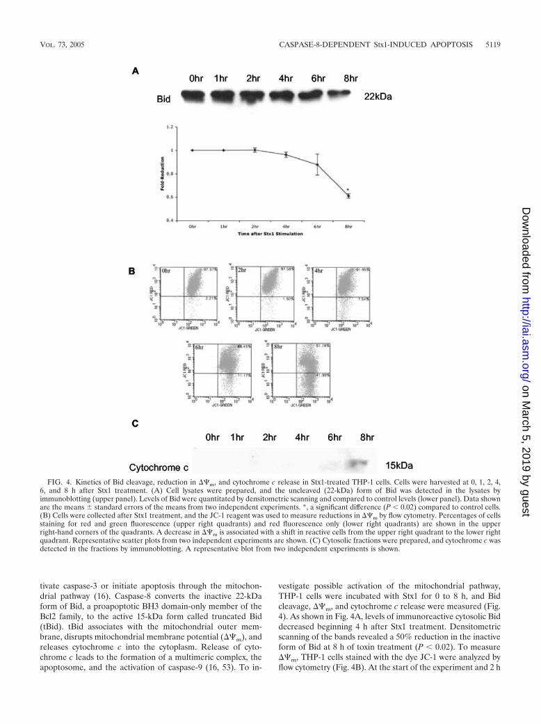
tivate caspase-3 or initiate apoptosis through the mitochon-drial pathway (16). Caspase-8 converts the inactive 22-kDaform of Bid, a proapoptotic BH3 domain-only member of theBcl2 family, to the active 15-kDa form called truncated Bid(tBid). tBid associates with the mitochondrial outer mem-brane, disrupts mitochondrial membrane potential (��m), andreleases cytochrome c into the cytoplasm. Release of cyto-chrome c leads to the formation of a multimeric complex, theapoptosome, and the activation of caspase-9 (16, 53). To in-
vestigate possible activation of the mitochondrial pathway,THP-1 cells were incubated with Stx1 for 0 to 8 h, and Bidcleavage, ��m, and cytochrome c release were measured (Fig.4). As shown in Fig. 4A, levels of immunoreactive cytosolic Biddecreased beginning 4 h after Stx1 treatment. Densitometricscanning of the bands revealed a 50% reduction in the inactiveform of Bid at 8 h of toxin treatment (P � 0.02). To measure��m, THP-1 cells stained with the dye JC-1 were analyzed byflow cytometry (Fig. 4B). At the start of the experiment and 2 h
FIG. 4. Kinetics of Bid cleavage, reduction in ��m, and cytochrome c release in Stx1-treated THP-1 cells. Cells were harvested at 0, 1, 2, 4,6, and 8 h after Stx1 treatment. (A) Cell lysates were prepared, and the uncleaved (22-kDa) form of Bid was detected in the lysates byimmunoblotting (upper panel). Levels of Bid were quantitated by densitometric scanning and compared to control levels (lower panel). Data shownare the means standard errors of the means from two independent experiments. *, a significant difference (P � 0.02) compared to control cells.(B) Cells were collected after Stx1 treatment, and the JC-1 reagent was used to measure reductions in ��m by flow cytometry. Percentages of cellsstaining for red and green fluorescence (upper right quadrants) and red fluorescence only (lower right quadrants) are shown in the upperright-hand corners of the quadrants. A decrease in ��m is associated with a shift in reactive cells from the upper right quadrant to the lower rightquadrant. Representative scatter plots from two independent experiments are shown. (C) Cytosolic fractions were prepared, and cytochrome c wasdetected in the fractions by immunoblotting. A representative blot from two independent experiments is shown.
VOL. 73, 2005 CASPASE-8-DEPENDENT Stx1-INDUCED APOPTOSIS 5119
on March 5, 2019 by guest
http://iai.asm.org/
Dow
nloaded from
after toxin stimulation, JC-1 accumulated in the mitochondrialmembrane as evidenced by 97% of the cells staining positivefor red and green fluorescence. Beginning 4 h after Stx1 treat-ment, red fluorescence was reduced, and at 6 and 8 h of toxintreatment, ��m was markedly reduced. Cytosolic fractionsprepared from THP-1 cells treated with Stx1 for 0 to 8 h wereanalyzed by immunoblotting for the presence of cytochrome c(Fig. 4C). No cytochrome c was detected in the cytoplasm until6 h after Stx1 treatment.
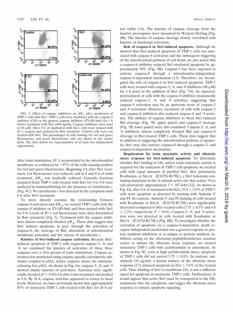
To more directly examine the relationship betweencaspase-8 activation and ��m, we treated THP-1 cells with thecaspase-8 inhibitor or ZVAD-fmk and then treated with Stx1for 8 h. Levels of JC-1 red fluorescence were then determinedby flow cytometry (Fig. 5). Treatment with the caspase inhib-itors almost completely blocked ��m. These data suggest thatStx1 induces apoptosis, in part, through the activation ofcaspase-8, the cleavage of Bid, alterations in mitochondrialmembrane potential, and the release of cytochrome c.
Kinetics of Stx1-induced caspase activation. Because Stx1-induced apoptosis of THP-1 cells required caspase-3, -8, and-9, we examined the kinetics of activation of these threecaspases over a 24-h period of toxin stimulation. Caspase ac-tivation was monitored using caspase-specific colorimetric sub-strates coupled to pNA. Active caspases cleave the substrate,releasing free pNA. As shown in Fig. 6A, caspase-3, -8, and -9showed similar kinetics of activation. Activities were signifi-cantly elevated (P � 0.05) 4 h after toxin treatment and peakedat 8 h. By 24 h, caspase activities appeared to return to basallevels. However, we have previously shown that approximately85% of monocytic THP-1 cells treated with Stx1 for 24 h are
not viable (14). The kinetics of caspase cleavage from theinactive procaspases were measured by Western blotting (Fig.6B). The kinetics of caspase cleavage closely correlated withkinetics of functional activation.
Role of caspase-6 in Stx1-induced apoptosis. Although weshowed that Stx1-induced apoptosis of THP-1 cells was asso-ciated with caspase-8 activation and the subsequent triggeringof the mitochondrial pathway of cell death, we also noted thata caspase-6 inhibitor reduced Stx1-mediated apoptosis by ap-proximately 50% (Fig. 3B). Caspase-3 has been reported toactivate caspase-8 through a mitochondria-independent,caspase-6-dependent mechanism (13). Therefore, we investi-gated the role of caspase-6 in Stx1-induced apoptosis. THP-1cells were treated with caspase-3, -6, and -8 inhibitors (40 �M)for 1 h prior to the addition of Stx1 (Fig. 7A). As expected,pretreatment of cells with the caspase-8 inhibitor dramaticallyreduced caspase-3, -8, and -9 activities, suggesting thatcaspase-8 activation may be an upstream event of caspase-3and -9 activation. However, treatment of cells with caspase-3and caspase-6 inhibitors also reduced caspase-8 and -9 activi-ties. The abilities of caspase inhibitors to block Stx1-inducedBid cleavage (Fig. 7B, upper panel) and caspase-8 cleavage(Fig. 7B, lower panel) were also examined. Caspase-3, -6, and-8 inhibitors almost completely blocked Bid and caspase-8cleavage in Stx1-treated THP-1 cells. These data suggest thatin addition to triggering the mitochondrial pathway of apopto-sis, Stx1 may also activate caspase-8 through a caspase-3- andcaspase-6-dependent mechanism.
Requirement for toxin enzymatic activity and ribotoxicstress response for Stx1-induced apoptosis. To determinewhether Stx1 binding to Gb3 and/or toxin enzymatic activity isrequired for the induction of THP-1 cell apoptosis, we treatedcells with equal amounts of purified Stx1, Stx1 pentamericB-subunits, or Stx1A� (E167Q-R170L), a Stx1 holotoxin con-taining mutations in the A-subunit active site that reduce Verocell cytotoxicity approximately 3 � 105-fold (32). As shown inFig. 8A, after 6 h of treatment with Stx1, 35.8 2.9% of THP-1cells were apoptotic as detected by staining with Annexin Vand PI. In contrast, Annexin V and PI staining of cells treatedwith B-subunits or Stx1A� (E167Q-R170L) were significantlydecreased compared to Stx1-treated cells (3.75 0.5% and 4.0 2.2%, respectively; P � 0.01). Caspase-3, -8, and -9 activa-tion were not detected in cells treated with B-subunits orStx1A� (E167Q-R170L) (Fig. 8B). To investigate whether theactivation of apoptosis via a caspase-8-dependent, death re-ceptor-independent mechanism was a general response to pro-tein synthesis inhibition or is unique to protein synthesis in-hibitors acting on the ribosomal peptidyltransferase reactioncenter to initiate the ribotoxic stress response, we treatedmonocytic THP-1 cells with cycloheximide or anisomycin. Asshown in Fig. 8C, even at high cycloheximide doses, apoptosisof THP-1 cells did not exceed 5.75 0.8%. In contrast, ani-somycin (10 �g/ml), a known inducer of the ribotoxic stressresponse (17), induced apoptosis in 28.6 3.6% of the treatedcells. Thus, binding of Stx1 to membrane Gb3 is not a sufficientsignal for apoptosis in monocytic THP-1 cells. Furthermore, itwould appear that active Stx1 must be transported to the ER,translocate into the cytoplasm, and trigger the ribotoxic stressresponse to initiate apoptotic signaling.
FIG. 5. Effect of caspase inhibitors on ��m after incubation ofTHP-1 cells with Stx1. THP-1 cells were incubated with the caspase-8inhibitor (C8I) or the general caspase inhibitor (ZVAD-fmk) for 1 hbefore treatment with Stx1 (400 ng/ml). Caspase inhibitors were usedat 40 �M. After 8 h of incubation with Stx1, cells were stained withJC-1 reagent and analyzed by flow cytometry. Control cells were nottreated with Stx1. The percentages of cells staining for red and greenfluorescence and green fluorescence only are shown in the scatterplots. The data shown are representative of at least two independentexperiments.
5120 LEE ET AL. INFECT. IMMUN.
on March 5, 2019 by guest
http://iai.asm.org/
Dow
nloaded from

FIG. 6. Kinetics of caspase activation and cleavage in Stx1-treated THP-1 cells. (A) Cells were incubated with or without Stx1 (400 ng/ml) for0 to 24 h. Caspase-3, -8, and -9 activities were then assayed as described in Materials and Methods. Control cells have not been treated with Stx1.Data shown are means standard errors of the means from three independent experiments (B) After Stx1 stimulation for 0, 2, 4, 6, and 8 h, celllysates were subjected to SDS-PAGE and caspase-3, -8, and -9 cleavage was detected by immunoblotting with antibodies directed against thecaspases. Data are representative of three independent experiments.
VOL. 73, 2005 CASPASE-8-DEPENDENT Stx1-INDUCED APOPTOSIS 5121
on March 5, 2019 by guest
http://iai.asm.org/
Dow
nloaded from

DISCUSSION
It is clear that Stxs induce apoptosis in a wide variety of celltypes, including cells of epithelial, endothelial, lymphocytic,and myelogenous origin (6). Apoptotic cell death is also evi-dent in tissues of humans suffering from the hemolytic uremicsyndrome (19). While cell death caused by Stxs may be criticalin the pathogenesis of hemorrhagic colitis and systemic vascu-lar complications, the mechanisms by which the toxins induceapoptosis remain to be fully characterized. We have shown thatmonocytic THP-1 cells are sensitive to the cytotoxic action ofStxs, with a 50% cytotoxic dose of approximately 14 pg/ml (35).Monocytic THP-1 cells undergo apoptosis via caspase-3 acti-vation (21), which is blocked by the general caspase inhibitorZVAD-fmk (14). The experiments performed in this studywere designed to further clarify the apoptotic signaling path-ways necessary for cell death in Stx1-stimulated monocyticTHP-1 cells.
The role of death receptors in signaling for apoptosis hasbeen well characterized. TNF-�/TNFR family or Fas/FasL li-gation initiates the formation of the death-inducing signaling
complex, an assembly of adaptor proteins, FADD andTRADD, and procaspase-8. Cleavage of procaspase-8 theninitiates caspase cascades that ultimately trigger apoptosis(28). Recently, several research groups have shown that in theabsence of death receptors of the TNFR family or Fas,caspase-8 may still be activated (12, 42). Our study and thestudies of Fujii et al. (12) demonstrate that similar apoptoticsignaling pathways may be triggered by Stxs in monocyticTHP-1 cells and HeLa cells. In contrast, Stx1-induced apopto-sis of Burkitt’s lymphoma cells appears to involve Fas-medi-ated signaling (20). Bremner et al. (4) reported that THP-1cells express FasL in the presence of lipopolysaccharide butthat cell viability was not affected by treatment with anti-Fasantibodies. Our data show that in the presence or absence ofStx1, monocytic THP-1 cells fail to express membrane TNFR1,TNFR2, TNF-�, or Fas. However, Stx1 treatment triggersDNA fragmentation and exposure of membrane phosphatidyl-serine in the absence of these membrane-expressed death re-ceptors. Thus, Stxs may induce apoptosis in different cell typesthrough different signaling mechanisms. Exogenously added
FIG. 7. Role of caspase-6 in Stx1-induced apoptosis. THP-1 cells were pretreated with caspase-3, -6, or -8 inhibitor (40 �M) for 1 h and thentreated with Stx1 (400 ng/ml) for 8 h. (A) Caspase activities were measured using specific colorimetric substrates as described in Materials andMethods. Control cells were not treated with Stx1. Data shown are means standard errors of the means from three independent experiments.*, **, and ***, significant differences (P � 0.01) within treatment groups relative to control (unstimulated) cells. (B) Cell lysates were separatedby SDS-PAGE, and Bid (upper panel) and caspase-8 (lower panel) cleavage was detected using anti-Bid and anti-caspase-8 antibodies. Controlcells were not treated with Stx1. A representative immunoblot from three independent experiments is shown.
5122 LEE ET AL. INFECT. IMMUN.
on March 5, 2019 by guest
http://iai.asm.org/
Dow
nloaded from

rhTNF-� did not induce THP-1 cell apoptosis, and anti-TNF-�or anti-TNFR1 neutralizing antibodies failed to inhibit Stx1-mediated apoptosis. As expected, treatment of cells withZVAD-fmk and a caspase-3 specific inhibitor reduced Stx1-
mediated DNA fragmentation and Annexin V staining, but wealso noted that a caspase-8 inhibitor blocked these indicatorsof apoptosis. While we cannot rule out the possibility of poorlydefined, novel death receptors participating in Stx1-mediated
FIG. 8. Requirement for Stx1 enzymatic activity and the ribotoxic stress response in Stx1-induced apoptosis of THP-1 cells. Cells wereincubated with Stx1 or Stx1A� (E167Q-R170L) or Stx1 B-subunits (400 ng/ml and 800 ng/ml, respectively) for 6 h. (A) Percent apoptosis wasanalyzed by flow cytometry following Annexin V and PI staining. Data shown are means standard errors of the means from at least twoindependent experiments. (B) Caspase-3, -8 and -9 activities were measured using specific colorimetric substrates as described in Materials andMethods. Control cells were not treated with Stx1, Stx1A�, or Stx1 B-subunits. Data shown are means standard errors of the means from threeindependent experiments. *, **, and ***, significant differences (P � 0.01) within treatment groups relative to Stx1-treated cells. (C) Cells weretreated with Stx1, cycloheximide (10 to 200 �M), or anisomycin (0.1 to 10 �g/ml) for 5 h, and apoptosis was measured by flow cytometry followingAnnexin V and PI staining. CHX, cycloheximide; Aniso, anisomycin.
VOL. 73, 2005 CASPASE-8-DEPENDENT Stx1-INDUCED APOPTOSIS 5123
on March 5, 2019 by guest
http://iai.asm.org/
Dow
nloaded from

apoptosis induction, our data suggest that Stx1 induces THP-1cell apoptosis in a TNFR/Fas-independent, caspase-8-depen-dent manner.
Activated caspase-8 may directly trigger the activation ofexecutioner caspases, such as caspase-3 (1), or may triggerapoptosis through the mitochondrial pathway by cleavage ofBid (9). Once cleaved, tBid translocates from the cytosol to themitochondrial outer membrane, mediating the homo-oli-gomerization of Bak and Bax proteins, the reduction in mito-chondrial membrane potential, and the release of cytochromec (51). Release of mitochondrial cytochrome c forms the ap-optosome, a complex containing procaspase-9, Apaf-1, ATP,and cytochrome c. Procaspase-9 is cleaved, which then cleavesprocaspase-3 (23, 53). We showed that Bid cleavage started 4 hafter exposure of THP-1 cells to Stx1, with a 50% reduction inthe uncleaved molecule by 8 h of treatment. Disruption of��m and cytochrome c release correlated with Bid cleavage.Furthermore, pretreatment of THP-1 cells with caspase-8 in-hibitor almost completely prevented mitochondrial membranedisruption. These data indicate that caspase-8 is involved inBid cleavage and cytochrome c release in Stx1-treated THP-1cells. In addition, caspase-8 activation occurs upstream ofcaspase-9 and -3 activation.
It should be noted that we have observed Stx1-inducedTHP-1 cell death beginning at 4 h (14), but cytochrome crelease was first evident 6 h after toxin treatment. These resultssuggest that caspase-8 may also directly activate caspase-3,while the mitochondrial pathway of apoptosis induction is trig-
gered with slightly slower kinetics. Our data suggest thatcaspase-6 may be involved in the direct activation of caspase-8.Caspase-6 is a lamin protease, and activated lamin A inducesnuclear disassembly and chromatin condensation (37). Fur-thermore, procaspase-6 is activated by caspase-3 and, in turn,activates procaspase-8 (7). Kojio et al. (21) showed a sixfoldincrease in caspase-6 activity in Stx-treated THP-1 cells, andwe show here that a caspase-6 inhibitor reduces apoptosis byapproximately 50%. To demonstrate a linkage amongcaspase-3, -6, and -8, we measured Stx1-induced caspase activ-ities and procaspase cleavage in the presence or absence ofspecific inhibitors for each of the caspases. Although the per-centages of inhibition differed among the different treatments,all the caspase-3, -6, and -8 inhibitors blocked caspase-3, -8,and -9 activities and procaspase-8 cleavage. Thus, blockingcaspase-3 or -6 prevents caspase-8 activation and blocks Bidcleavage. These data suggest that Stx1 may trigger separateapoptosis signaling pathways in THP-1 cells. These signalingpathways are depicted in Fig. 9.
Kojio et al. (21) reported caspase-3 activity peaking 5 h afterexposure of THP-1 cells to Stx1. We measured caspase-3, -8,and -9 activities from 0 to 24 h after toxin treatment and foundthat all caspase activities reached the highest levels after 8 h.For the remaining 16 h of the experiments, caspase activitiesappeared to return to basal levels, although we have shownsignificant cell death occurring within this time period (14).Caspase-2 activity was reported to be increased 12-fold inStx1-treated THP-1 cells (21), and we also found increased
FIG. 9. Proposed model of Stx1-induced apoptosis signaling pathways in monocytic THP-1 cells. See text for additional details.
5124 LEE ET AL. INFECT. IMMUN.
on March 5, 2019 by guest
http://iai.asm.org/
Dow
nloaded from

caspase-2 activity in Stx1-treated cells (data not shown). How-ever, we found that inhibition of caspase-2 activity failed todecrease Stx1-mediated DNA laddering or Annexin V stain-ing. The role of caspase-2 in apoptosis is controversial. Pro-caspase-2 is constitutively expressed in the nucleus and Golgiapparatus (26, 52). DNA damage by etoposide (an inhibitor ofnuclear topoisomerase 2), the binding of TNF-� to TNFR1,and the activation of caspase-3 have all been shown to activatecaspase-2 (33, 36). However, cleavage may not be required forcaspase-2 activation. Procaspase-2 may oligomerize and be-come partially activated, although the role of procaspase-2aggregates in apoptosis is unclear (2). Recently, Golgin-160, aGolgi apparatus-localized macromolecular complex, has beenshown to be a substrate for caspase-2 involved in the disassem-bly of the Golgi complex (26). Whether treatment of THP-1cells with Stx1 results in proteolysis of Golgin-160, and whethercleaved Golgin-160 is involved in apoptosis, will require addi-tional experiments.
The treatment of Ramos Burkitt’s lymphoma and astrocy-toma cells with Stx B-subunits or anti-Gb3 monoclonal anti-body has been reported to induce apoptosis and activate pro-caspase-8 (20). Thus, for these cells, binding of toxin receptorsat the cell surface may generate sufficient signals to activateprogrammed cell death cascades. We show here that purifiedStx1 B-subunits and an enzymatic (active site) mutant toxin areincapable of inducing apoptosis or caspase-3, -8, and -9 activ-ities in monocytic THP-1 cells. Inhibition of the formation offunctional Golgi complexes using brefeldin A has been shownto inhibit Stx1-induced caspase-3 activity and DNA fragmen-tation in THP-1 cells (21). Together, these data suggest thatStxs must be internalized and undergo retrograde transportthrough the Golgi apparatus to the ER, and the functionaltoxin A-subunit fragments must be translocated into the cyto-plasm in order to trigger the signals necessary for apoptosisinduction.
A key feature of the apoptotic pathways triggered by Stxsthat remains to be characterized is the linkage between proteinsynthesis inhibition and the activation of caspases. A family ofribosome inactivating proteins, including Stxs, has been shownto induce the ribotoxic stress response (5, 10, 17, 41). As aresult of the highly specific 28S rRNA depurination reactionmediated by Stxs, the stress-activated protein kinase cascades,JNK and p38, are activated. These mitogen-activated proteinkinases (MAPKs) phosphorylate a number of downstream sub-strates, leading to activation of transcriptional factors such asNF-�B and AP-1. In response to Stxs, therefore, the cellulartranscriptome may be rapidly altered by transcriptional andposttranscriptional mechanisms. The p38 MAPK cascade hasalso been reported to regulate the stability of some mRNAtranscripts (50). Our data suggest that the activation of apo-ptotic signaling is not a general response to protein synthesisinhibition but that the ribotoxic stress response may be selec-tively involved in triggering caspase-8- and mitochondrion-de-pendent programmed cell death. Ribosome inactivation maynot be the sole determinant in triggering apoptosis; rather,toxin effects directed to the ER membrane may contribute tosignaling. Jimbo et al. (18) showed that ER stress initiatedprocaspase-8 activation by direct interaction of caspase-8 withER membrane proteins. Bap31 is a polytopic integral mem-brane protein of the ER and a substrate for caspase-8 activity.
Bap31 may interact with Bcl-2 to inhibit the interaction be-tween procaspase-8 and Bap31. However, if Bap31 associateswith procaspase-8 in the absence of Bcl-2, then procaspase-8and Bap31 are cleaved and programmed cell death may beinitiated (3, 30). Even though the precise role of Bap31 inapoptosis remains to be elucidated, it may explain how pro-caspase-8 is activated in the absence of death receptor expres-sion by THP-1 cells.
ACKNOWLEDGMENTS
These studies were supported by U.S. Public Health Service grant2RO1-AI34530 from the National Institute of Allergy and InfectiousDiseases, National Institutes of Health.
We thank Yoshifumi Takeda and Cheleste Thorpe for their gifts oftoxins and Jane Miller for her excellent technical assistance with theFACS analyses. We thank David McMurray, Rajesh Miranda, andJane Welsh for their careful reading of the manuscript.
REFERENCES
1. Ashkenazi, A., and V. M. Dixit. 1998. Death receptors: signaling and mod-ulation. Science 281:1305–1308.
2. Baliga, B. C., S. H. Read, and S. Kumar. 2004. The biochemical mechanismof caspase-2 activation. Cell Death Diff. 11:1234–1241.
3. Breckenridge, D. G., M. Nguyen, S. Kuppig, M. Reth, and G. C. Shore. 2002.The procaspase-8 isoform, procaspase-8L, is recruited to the BAP31 com-plex at the endoplasmic reticulum. Proc. Natl. Acad. Sci. USA 99:4331–4336.
4. Bremner, T. A., D. Chatterjee, Z. Han, M.-F. Tsan, and J. H. Wyche. 1999.THP-1 monocytic leukemia cells express Fas ligand constitutively and killFas-positive Jurkat cells. Leukemia Res. 23:865–870.
5. Cameron, P., S. J. Smith, M. A. Giembycz, D. Rotondo, and R. Plevin. 2003.Verotoxin activates mitogen-activated protein kinase in human peripheralblood monocytes: role in apoptosis and proinflammatory cytokine release.Br. J. Pharmacol. 140:1320–1330.
6. Cherla, R. P., S.-Y. Lee, and V. L. Tesh. 2003. Shiga toxins and apoptosis.FEMS Microbiol. Lett. 228:159–166.
7. Cowling, V., and J. Downward. 2002. Caspase-6 is the direct activator ofcaspase-8 in the cytochrome c-induced apoptosis pathway: absolute require-ment for removal of caspase-6 prodomain. Cell Death Diff. 9:1046–1056.
8. Endo, Y., K. Tsurugi, T. Yutsudo, T., Y. Takeda, T. Ogasawara, and K.Igarashi. 1988. Site of action of a verotoxin (VT2) from Escherichia coliO157:H7 and of Shiga toxin on eukaryotic ribosomes. RNA N-glycosidaseactivity of the toxins. Eur. J. Biochem. 171:45–50.
9. Eskes, R., S. Desagher, B. Antonsson, and J.-C. Martinou. 2000. Bid inducesthe oligomerization and insertion of Bax into the outer mitochondrial mem-brane. Mol. Cell. Biol. 20:929–935.
10. Foster, G. H., and V. L. Tesh. 2002. Shiga toxin 1-induced activation of c-JunNH2-terminal kinase and p38 in the human monocytic cell line THP-1:possible involvement in the production of TNF-�. J. Leukoc. Biol. 71:107–114.
11. Fraser, M. E., M. M. Chernaia, Y. V. Kozlov, and M. N. G. James. 1994.Crystal structure of the holotoxin from Shigella dysenteriae at 2.5 Å resolu-tion. Nat. Struct. Biol. 1:59–64.
12. Fujii, J., T. Matsui, D. P. Heatherley, K. H. Schlegel, P. I. Lobo, T. Yutsudo,G. M. Ciraolo, R. E. Morris, and T. Obrig. 2003. Rapid apoptosis induced byShiga toxin in HeLa cells. Infect. Immun. 71:2724–2735.
13. Fumarola, C., and G. G. Guidotti. 2004. Stress-induced apoptosis: toward asymmetry with receptor-mediated cell death. Apoptosis 9:77–82.
14. Harrison, L. M., R. P. Cherla, C. van den Hoogen, W. C. E. van Haaften,S.-Y. Lee, and V. L. Tesh. 2005. Comparative evaluation of apoptosis inducedby Shiga toxin 1 and/or lipopolysaccharides in human monocytic and mac-rophage-like cells. Microb. Pathog. 38:63–76.
15. Harrison, L. M., W. C. E. van Haaften, and V. L. Tesh. 2004. Regulation ofproinflammatory cytokine expression by Shiga toxin 1 and/or lipopolysaccha-rides in the human monocytic cell line THP-1. Infect. Immun. 72:2618–2627.
16. Hengartner, M. O. 2000. The biochemistry of apoptosis. Nature 407:770–776.
17. Iordanov, M. S., D. Pribnow, J. L. Magun, T.-H. Dinh, J. A. Pearson, S. L.-Y.Chen, and B. E. Magun. 1997. Ribotoxic stress response: activation of thestress-activated protein kinase JNK1 by inhibitors of peptidyltransferasereaction and by sequence-specific RNA damage to the �-sarcin/ricin loop inthe 28S rRNA. Mol. Cell. Biol. 17:3373–3381.
18. Jimbo, A., E. Fujita, Y. Kouroku, J. Ohnishi, N. Inohara, K. Kuida, K.Sakamaki, S. Yonehara, and T. Momoi. 2003. ER stress induces caspase-8activation, stimulating cytochrome c release and caspase-9 activation. Exp.Cell Res. 83:156–166.
19. Karpman, D., A. Hakansson, M.-T. R. Perez, C. Isaksson, E. Carlemalm, A.
VOL. 73, 2005 CASPASE-8-DEPENDENT Stx1-INDUCED APOPTOSIS 5125
on March 5, 2019 by guest
http://iai.asm.org/
Dow
nloaded from

Caprioli, and C. Svanborg. 1998. Apoptosis of renal cortical cells in thehemolytic uremic syndrome: in vivo and in vitro studies. Infect. Immun.66:636–644.
20. Kiyokawa, N., T. Mori, T. Taguchi, M. Saito, K. Mimori, T. Suzuki, T.Sekino, N. Sato, H. Nakajima, Y. U. Katagiri, T. Takeda, and J. Fujimoto.2001. Activation of the caspase cascade during Stx1-induced apoptosis inBurkitt’s lymphoma cells. J. Cell. Biochem. 81:128–142.
21. Kojio, S., H.-M. Zhang, M. Ohmura, F. Gondaira, N. Kobayashi, and T.Yamamoto. 2000. Caspase-3 activation and apoptosis induction coupled withthe retrograde transport of Shiga toxin: inhibition by brefeldin A. FEMSImmunol. Med. Microbiol. 29:275–281.
22. Leist, M., C. Volbracht, E. Fava, and P. Nicotera. 1998. 1-Methyl-4-phenyl-pyridinium induces autocrine excitoxicity, protease activation, and neuronalapoptosis. Mol. Pharmacol. 54:789–801.
23. Li, P., D. Nijhawan, I. Budihardjo, S. M. Srinivasula, M. Ahmad, E. S.Alnemri, and X. Wang. 1997. Cytochrome c and dATP-dependent formationof Apaf-1/caspase-9 complex initiates an apoptotic protease cascade. Cell91:479–489.
24. Lingwood, C. A. 2003. Shiga toxin receptor glycolipid binding. Pathology andutility. Methods Mol. Med. 73:165–186.
25. Louise, C. B., and T. G. Obrig. 1991. Shiga toxin-associated hemolytic uremicsyndrome: combined cytotoxic effects of Shiga toxin, interleukin-1� andtumor necrosis factor alpha on human vascular endothelial cells in vitro.Infect. Immun. 59:4173–4179.
26. Mancini, M., C. E. Machamer, S. Roy, D. Nicholson, N. A. Thornberry, L.Casciola-Rosen, and A. Rosen. 2000. Caspase-2 is localized at the Golgicomplex and cleaves Golgin-160 during apoptosis. J. Cell Biol. 149:603–612.
27. Melton-Celsa, A. R., and A. D. O’Brien. 1998. Structure, biology and relativetoxicity of Shiga toxin family members for cells and animals, p. 121–128. InJ. B. Kaper and A. D. O’Brien (ed.), Escherichia coli O157:H7 and otherShiga toxin-producing E. coli strains. ASM Press, Washington, D.C.
28. Micheau, O., and J. Tschopp. 2003. Induction of TNF receptor I-mediatedapoptosis via two sequential signaling complexes. Cell 114:181–190.
29. Moxley, R. A., and D. H. Francis. 1998. Overview of animal models, p.249–260. In J. B. Kaper and A. D. O’Brien (ed.), Escherichia coli O157:H7and other Shiga toxin-producing E. coli strains. ASM Press, Washington,D.C.
30. Ng, F. W. H., M. Nguyen, T. Kawn, P. E. Branton, D. W. Nicholson, J. A.Cromlish, and G. C. Shore. 1997. p28 BAP31, a Bcl-2/Bcl-XL- and pro-caspase-8-associated protein in the endoplasmic reticulum. J. Cell Biol. 139:327–338.
31. O’Brien, A. D., V. L. Tesh, A. Donohue-Rolfe, M. P. Jackson, S. Olsnes, K.Sandvig, A. A. Lindberg, and G. T. Keusch. 1992. Shiga toxin: biochemistry,genetics, mode of action, and role in pathogenesis. Curr. Top. Microbiol.Immunol. 180:65–94.
32. Ohmura, M., S. Yamasaki, H. Kurazono, K. Kashiwagi, K. Igarashi, and Y.Takeda. 1993. Characterization of non-toxic mutants of verotoxin 1 that wereconstructed by replacing amino acids in the A subunit. Microb. Pathog.15:169–176.
33. Paroni, G., C. Henderson, C. Schneider, and C. Brancolini. 2001. Caspase-2-induced apoptosis is dependent on caspase-9, but its processing duringUV- or tumor necrosis factor-dependent cell death requires caspase-3.J. Cell Biol. 276:21907–21915.
34. Proulx, F., E. G. Seidman, and D. Karpman. 2001. Pathogenesis of Shigatoxin-associated hemolytic uremic syndrome. Pediatr. Res. 50:163–171.
35. Ramegowda, B., and V. L. Tesh. 1996. Differentiation-associated toxin re-ceptor modulation, cytokine production and sensitivity to Shiga-like toxins inhuman monocytes and monocytic cell lines. Infect. Immun. 64:1173–1180.
36. Robertson, J. D., V. Gogvadze, B. Zhitotovsky, and S. Orrenius. 2000. Dis-
tinct pathways for stimulation of cytochrome c release by etoposide. J. Biol.Chem. 275:32438–32443.
37. Ruchaud, S., N. Korfali, P. Villa, T. J. Kottke, C. Dingwall, S. H. Kaufmann,and W. C. Earnshaw. 2002. Caspase-6 gene disruption reveals a requirementfor lamin A cleavage in apoptotic chromatin condensation. EMBO J. 21:1967–1977.
38. Sandvig, K., S. Grimmer, S. U. Lauvrak, M. L. Torgersen, G. Skretting, B.van Deurs, and T. G. Iversen. 2002. Pathways followed by ricin and Shigatoxin into cells. Histochem. Cell. Biol. 117:131–141.
39. Saxena, S. K., A. D. O’Brien, and E. J. Ackerman. 1989. Shiga toxin, Shiga-like toxin II variant, and ricin are all single-site RNA N-glycosidases of 28SRNA when microinjected into Xenopus oocytes. J. Biol. Chem. 264:596–601.
40. Siegler, R. L. 1994. Spectrum of extrarenal involvement in postdiarrhealhemolytic uremic syndrome. J. Pediatr. 125:511–518.
41. Smith, W. E., A. V. Kane, S. T. Campbell, D. W. K. Acheson, B. H. Cochran,and C. M. Thorpe. 2003. Shiga toxin 1 triggers a ribotoxic stress responseleading to p38 and JNK activation and induction of apoptosis in intestinalepithelial cells. Infect. Immun. 71:1497–1504.
42. Souza-Fagundes, E. M., G. Brumatti, O. A. Martins-Filho, R. Correa-Ol-iveira, C. L. Zani, and G. P. Amarante-Mendes. 2003. Myriadenolide, alabdane diterpene isolated from Alomia myriadenia (asteraceae) inducesdepolarization of mitochondrial membranes and apoptosis associated withactivation of caspase-8, -9 and -3 in Jurkat and THP-1 cells. Exp. Cell Res.290:420–426.
43. Stennicke, H. R., and G. S. Salvesen. 2000. Caspases—controlling intracel-lular signals by protease zymogen activation. Biochim. Biophys. Acta 1477:299–306.
44. Tesh, V. L., J. A. Burris, J. W. Owens, V. M. Gordon, E. A. Wadolkowski,A. D. O’Brien, and J. E. Samuel. 1993. Comparison of the relative toxicitiesof Shiga-like toxins type I and type II for mice. Infect. Immun. 61:3392–3402.
45. Tsan, M.-F., J. E. White, J. G. Maheshware, T. A. Bremner, and J. Sacco.2000. Resveratrol induces Fas signaling-independent apoptosis in THP-1human monocytic leukemia cells. Br. J. Haematol. 109:405–412.
46. Tsuchiya, S., M. Yamabe, Y. Yamaguchi, Y. Kobayashi, T. Konno, and K.Tada.1980. Establishment and characterization of a human acute monocyticleukemia cell line (THP-1). Int. J. Cancer 26:171–176.
47. van de Kar, N. C. A. J., L. A. H. Monnens, M. A. Karmali, and V. W. M. vanHinsbergh. 1992. Tumor necrosis factor and interleukin 1 induce expressionof the verocytotoxin receptor globotriaosylceramide on human endothelialcells: implications for the pathogenesis of the hemolytic uremic syndrome.Blood 80:2755–2764.
48. van Setten, P. A., L. A. H. Monnens, R. G. G. Verstraten, L. P. W. J. van denHeuvel, and V. W. M. van Hinsbergh. 1996. Effects of verocytotoxin-1 onnonadherent monocytes: binding characteristics, protein synthesis inhibitionand induction of cytokine release. Blood 88:174–183.
49. Wajant, H., K. Pfizenmaier, and P. Scheurich. 2003. Tumor necrosis factorsignaling. Cell Death Diff. 10:45–65.
50. Wang, S. W., J. Pawlowski, S. T. Wathen, S. D. Kinney, H. S. Lichenstein,and C. L. Manthey. 1999. Cytokine mRNA decay is accelerated by aninhibitor of p38 mitogen-activated protein kinase. Inflamm. Res. 48:533–538.
51. Wei, M. C., T. Lindsten, V. K. Mootha, S. Weiler, A. Gross, M. Ashiya, C. B.Thompson, and S. J. Korsmeyer. 2000. tBID, a membrane-targeted deathligand, oligomerizes BAK to release cytochrome c. Genes Dev. 14:2060–2071.
52. Zhivotovsky, B., A. Samali, A. Gahm, and S. Orrenius. 1999. Caspases: theirintracellular localization and translocation during apoptosis. Cell Death Diff.6:644–651.
53. Zou, H., W. J. Henzel, X. Liu, A. Lutschg, and X. Wang. 1997. Apaf-1, humanprotein homologous to C. elegans CED-4, participates in cytochrome c-dependent activation of caspase-3. Cell 90:405–413.
Editor: A. D. O’Brien
5126 LEE ET AL. INFECT. IMMUN.
on March 5, 2019 by guest
http://iai.asm.org/
Dow
nloaded from