short and long term sequelae of radiation therapy to the ... · short and long term sequelae of...
TRANSCRIPT

Short and long term sequelae of radiation therapy to the oral cavity
Professor Alexander D Rapidis MD DDS PhD FACSProfessor Alexander D. Rapidis MD DDS PhD FACS
Chairman Dept. of Maxillofacial / Head and Neck Surgery
Greek Anticancer Institute Saint Savvas HospitalGreek Anticancer Institute, Saint Savvas Hospital
Athens Greece
Radiation TherapyRadiation TherapyGeneral Statements
Radiation alone or with other treatment d liti i d i i ifi t b fmodalities is used in a significant number of
patients with advanced stage oral cancer
A therapeutic dose of 5000-7000 cGy is externally delivered to the lesion
Increments of 200 cGy/day is delivered until the accumulated dose is achieved

Classification
Oral Complications f di thof radiotherapy
Acute Late
Mucositis Xerostomia
Skin Reactions Radiation caries
InfectionTrismus
Radiation induced
Infection
malignancies
Osteoradionecrosis
ChronicAcute
Acute
Mucositis

WHO Oral Mucositis ScaleWHO Oral Mucositis Scale
SevereOral Mucositis
Grade
43210Mucositis
to the extent Ulcers,
extensive Erythema,
ulcersSoreness
+/– erythemaNone
43210
that alimentation is not possible
erythema
Patients cannot swallow
Patients can swallow solid diet
No ulceration
cannot swallow solid diet
solid diet
Mucositis
Clinical Characteristics
Grade I White discoloration

Mucositis
Clinical Characteristics
Grade II Erythema
Mucositis
Clinical Characteristics
Grade III Pseudomembranous surface

Mucositis
Clinical Characteristics
Grade IV Ulcerations
Lessons Learned from Oral MucositisLessons Learned from Oral Mucositis
Mouth
» Extend to the entire GI tract Esophagus
Stomach
SmallIntestine
Colon
2004
Rectum
Anus
© M
AS
CC
2
Keefe D et al, Curr Opin Clin Nutr Metab Care. 2007

The Alimentary Canal
» The GI tract is all one tube from th t f d f
Mouth
mouth to anus—formed from primitive endoderm Esophagus
Pharyngeal gutPharyngeal gut
Stomach
SmallIntestine
Colon
Rectum
Anus
Midgut
2004
Hindgut
© M
AS
CC
2
Keefe D et al, Curr Opin Clin Nutr Metab Care. 2007
The Alimentary Canal
» Toxicities resulting from h th di th RadiationRadiationchemotherapy or radiotherapy
don’t occur in isolation
RadiationRadiationTherapyTherapy
ChemotherapyChemotherapy» There are common mechanisms &
systemic effects
» The GI system can provide a whole new paradigm for
h & i iresearch & new intervention strategies
2004
© M
AS
CC
2
Keefe D et al, Curr Opin Clin Nutr Metab Care. 2007

Mucositis
SymptomsSymptoms» Intense pain» Food and fluid intake decreases» Food and fluid intake decreases» Speech and swallowing difficult» May require ceasing therapyy q g py
Mucositis
Management» Mucosal coating agents» Mucosal coating agents
» Cleansing devices
» Chlorhexidine» Chlorhexidine
» Recombinant keratinocyte growth factor
» GMCSF (Combined Therapy)» GMCSF (Combined Therapy)
» Thalidomide?
Lo le el laser therap ?» Low-level laser therapy?
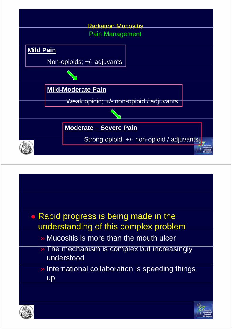
Radiation MucositisRadiation MucositisPain Management
Mild Pain
Non-opioids; +/- adjuvantsp ; j
Mild-Moderate Pain
Weak opioid; +/ non opioid / adjuvantsWeak opioid; +/- non-opioid / adjuvants
Moderate – Severe Pain
Strong opioid; +/- non-opioid / adjuvants
Rapid progress is being made in the understanding of this complex problemunderstanding of this complex problem» Mucositis is more than the mouth ulcer
Th h i i l b t i i l» The mechanism is complex but increasingly understood
» International collaboration is speeding things up

Acute
Ski tiSkin reactions


Late
X t iXerostomia
Xerostomia
Pathogenesis» Irreversible acinar cell damage
Clinical Characteristics» 50% decreased salivation after 1 week of radiation
» 75% decrease after 6 weeks5% dec ease a te 6 ee s
» 95% decrease years after
» Thick ropey saliva» Thick ropey saliva
» Candida albicans infection
» Dental caries» Dental caries
» Dysphagia / Odynophagia

Xerostomia
Symptoms» Difficulty in eating, speaking, & swallowing
» Taste disorders
» Denture-related pain / dysfunction
Pre-treatment StrategiesPre treatment StrategiesCurrent
IMRT / C f l b d iIMRT / Conformal beam design» Selective field designg
– Attempt oral mucosal sparing
Radioprotective agentsRadioprotective agents» Amifostine
» Antioxidants
Salivary stimulationSa a y st u at o» Pilocarpine; cevimeline; gustatory; other
agentsagents

Tumor
Dose
Tissue
Conventional Radiotherapy
Intensity Modulated Radiotherapy
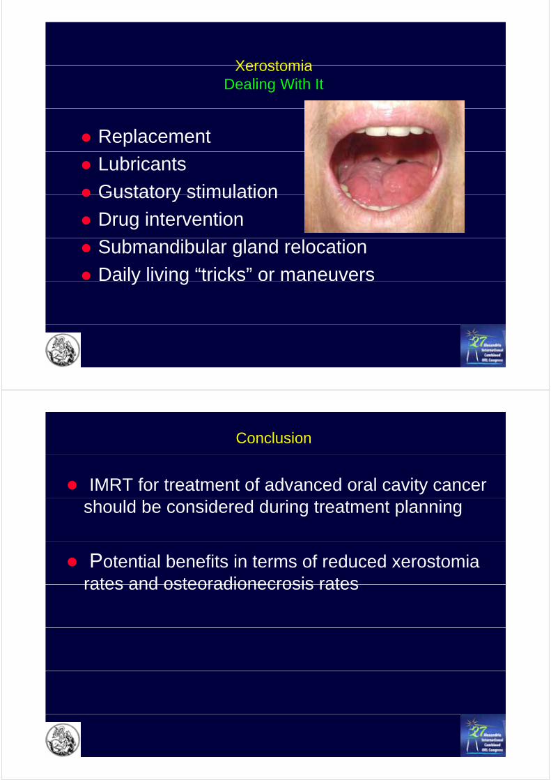
XerostomiaXerostomiaDealing With It
Replacement
Lubricants
Gustatory stimulationGustatory stimulation
Drug intervention
Submandibular gland relocation
Daily living “tricks” or maneuversy g
Conclusion
IMRT for treatment of advanced oral cavity cancer should be considered during treatment planning
Potential benefits in terms of reduced xerostomia rates and osteoradionecrosis ratesrates and osteoradionecrosis rates

Late
Radiation caries
Radiation Caries
Pathogenesis» Shift to cariogenic microflora and xerostomic» Shift to cariogenic microflora and xerostomic
environment
Clinical CharacteristicsClinical Characteristics» Cervical, cusp, & incisal decay
Coronal fractures» Coronal fractures

Late
Trismus
TrismusTrismus
• more common with high posterior fields of radiationmore common with high posterior fields of radiation as muscles of mastication are in field (10%)•retention of coronoid process•made worse by concomitant chemotherapy
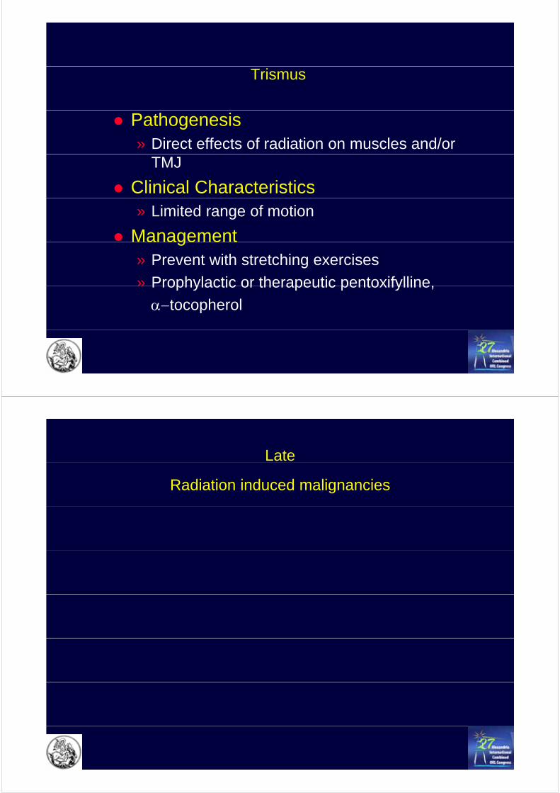
Trismus
Pathogenesis» Direct effects of radiation on muscles and/or
TMJ
Clinical Characteristics» Limited range of motion
ManagementManagement» Prevent with stretching exercises
» Prophylactic or therapeutic pentoxifylline» Prophylactic or therapeutic pentoxifylline,
α−tocopherol
Late
Radiation induced malignancies

Late complications following RT
No Event occurs above event threshold dose,
severity ↑ with dose
Event can occur at any dose level
P b bilit t it ↑ ith dProbability, not severity, ↑ with dose
increasing RT dose
Late complications following RT
Xerostomia
Soft tissue fibrosis
OsteoradionecrosisOsteoradionecrosis
Radiation associated tumors
increasing RT dose

InfectionsInfections

Erythematous candidiasis and angular cheilitis from mucositis
Infection Pseudomembranous candidiasis from ulcerative / pseudomembrane mucositis and / or HSV-1mucositis and / or HSV 1
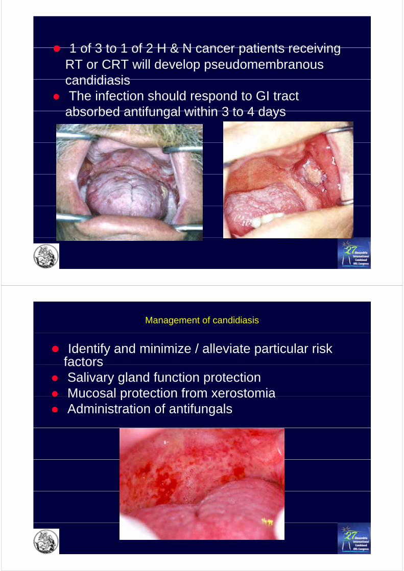
1 of 3 to 1 of 2 H & N cancer patients receiving1 of 3 to 1 of 2 H & N cancer patients receiving RT or CRT will develop pseudomembranous candidiasiscandidiasisThe infection should respond to GI tract absorbed antifungal within 3 to 4 daysabsorbed antifungal within 3 to 4 days
Management of candidiasis
Identify and minimize / alleviate particular risk factorsfactorsSalivary gland function protectionMucosal protection from xerostomiaMucosal protection from xerostomiaAdministration of antifungals

Management of candidiasis
Topical antifungal agents» Polyene compounds» Polyene compounds
Systemic antifungal agents» Azole group compounds» Azole group compounds
Late
Osteoradionecrosis
Background
Devastating complication of radiation therapy that b diffi lt t t t th i i l tcan be more difficult to treat than original tumor
Clinical definition:Devitalized, irradiated bone that is exposed through overlying mucosa or skin persisting for > 6 months
•Osteoradionecrosis presents as a broad spectrum of disease severityy
•It is rare at radiation therapy doses of less 60 Gy
•It is more common when bracytherapy is used
•The mandible must be in the treatment volume area
•Dental extractions, surgery or trauma frequently proceed its onset
•Secondary infection may be present

Pathophysiology of osteoradionecrosis
Direct radiation effects on normal tissue may be lethal or sublethal
Lethal damage is caused by
lethal or sublethal
g yionization within the desoxyribonucleinic acid (DNA)
preventing cell replication and resulting in tissue death
Sublethal damage may cause cell mutation leading to further neoplasiafurther neoplasia
3 “H” Hypothesis & Osteoradionecrosis
Hypovascularity
HypoxiaHypoxia
HypocellularityTissue injury (usually)
Tissue breakdown / nonTissue breakdown / non--healing woundhealing wound
Tissue injury (usually)
gg

The incidence of osteoradionecrosis is reported to be between 5-25% of patients receiving radiotherapy in the head and neck area.
Mendenhall WM J Clin Oncol 2004
Reuther et al, Int J Oral Maxillofac Surg 2003
There are several classifications for mandibular osteoradionecrosis and they all stage the disease according to the severity of signs and
t i ith St G d Ssymptoms in either Stages, Grades or Scores
Jereczek-Fossa BA and Orecchia R, Cancer Treatment Reviews 2002
RTOG: Radiation Therapy Oncology Group

Extractions & OsteonecrosisExtractions & OsteonecrosisTraditional Concepts
Twice the risk of ORN is seen when selected t th t t d f ll i di ti thteeth are extracted following radiation therapy
Pre-radiation extractions associated with a 3.4% risk of ORN
Risk of ORN persists for years and reduced p yhealing capacity may be considered permanentp
The role of hyperbaric oxygen

The use of Hyperbaric Oxygen (HBO)
HBO treatment in ol es the deli er of 100% o gen at highHBO treatment involves the delivery of 100% oxygen at high pressure in special chambers. The pressure of the oxygen
inhaled by the patient is usually 2.4 times more than the atmospheric pressure and can be as high as 3 times more.
Advocates of HBO therapy support the view that HBO represents the only medical treatment for osteoradionecrosis. HBO can revert the d l d di ti h i ti b ti tdelayed radiation changes in tissues by generating steep oxygen gradients between the normal and the irradiated tissues causing
oxygen to diffuse into the affected areas.

HBO has been used as an adjunctive conservative measure
The use of Hyperbaric Oxygen (HBO)
HBO has been used as an adjunctive conservative measure along with antibiotics and irrigation since the 1960s.
Using Marx’s theory that osteoradionecrosis is a result ofUsing Marx s theory that osteoradionecrosis is a result of hypoxia, hypocellularity and hypovascularity, HBO seems likely
to increase oxygen supply in hypoxic tissues, stimulating fib bl t lif ti d i ifibroblast proliferation and angiogenesis.
The role of HBO in the treatment of
osteoradionecrosis.
The Marx protocol (1982)The Marx protocol (1982)
Gal TJ et al, Arch Otolaryngol Head Neck Surg 2003

Th f HBO i th t t t f t di i d it it id dThe use of HBO in the treatment of osteoradionecrosis despite its widespread use had been largely theoretical or anecdotal because of the paucity of
controlled trials and the lack of unified assessment of symptom improvement.
Epstein J et al, Oral Surg 1997
The role of HBO in the treatment of
osteoradionecrosisosteoradionecrosis.
The study by Annane et al (2004)Annane et al (2004)
The first randomized, placebo-controlledplacebo controlled, double-blind study
assessing the efficacy and safety of HBO forand safety of HBO for the treatment of overt
mandibular osteoradionecrosis
and included 68 patients.
Annane D et al, J Clin Oncol 2004

The trial was terminated prematurely because of the failure to demonstrate any beneficial effect of HBO over placebo (19% vs. 33% respectively). They also reported the progression of disease in recovery in the arm of
HBO patients and better recovery rates in the arm of the placebo treated patients.
Annane D et al, J Clin Oncol 2004a e e a , J C O co 00
The study by Annane resulted into strong criticism and disbelief by l th ti th t it i l t d thi l i i l bseveral authors quoting that it violated an ethical principle by
exposing the control group to the potentially serious risk of acute decompression illness; a risk not present in the treatment group.
Others stated that a major error in Annane’s study was the fact that the studied group of patients with an osteoradionecrosis was g p p
not well defined.
There were though supporters of the Annane study presentingThere were though supporters of the Annane study presenting evidence that the beneficial results of HBO treatment are
equivocal and the method is time consuming and expensive.
The debate is still going on.

Management of early and advanced osteoradionecrosis
Established ORN does not regress spontaneously It either stabilizes orEstablished ORN does not regress spontaneously. It either stabilizes or gradually worsens. One of the adverse factors implemented in the
development of ORN is the Radiation Induced Fibrosis (RIF) and necrosis. It has been shown that RIF greatly regressed after antioxidant treatment with as bee s o a g ea y eg essed a e a o da ea e
the combination of pentoxifylline, tocopherol and clodronate.
Delanian S et al Head Neck 2005

With this treatment applied to 18 patients with advanced ORN, pp p16 (89%) recovered after a median 6 months of treatment.
The results of this trial raise many questions primarily about the precise mechanisms of action of the drugs used, which will remain unanswered
until further randomized clinical trials will be conducted.
Delanian S et al Head Neck 2005
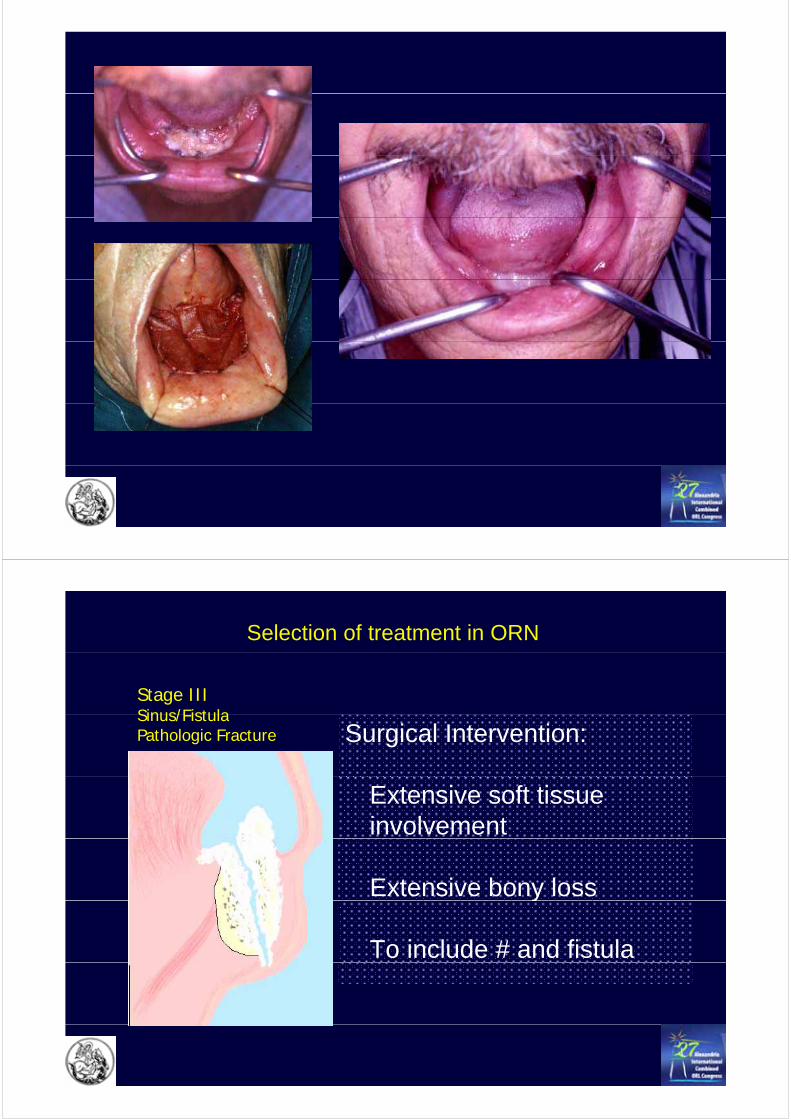
Selection of treatment in ORN
Stage I Superficial UlcerationSuperficial UlcerationExposed cortical bone Conservative management:
DebridementMeticulous oral hygieneMeticulous oral hygieneAntibiotics? HBO? HBO

Selection of treatment in ORN
Stage II Exposed medullary bone
Conservative management:+ soft tissue changes
HBO cannot revitalize dead bonebone
SequestrectomySequestrectomyin addition to other conservative measures
Selection of treatment in ORN
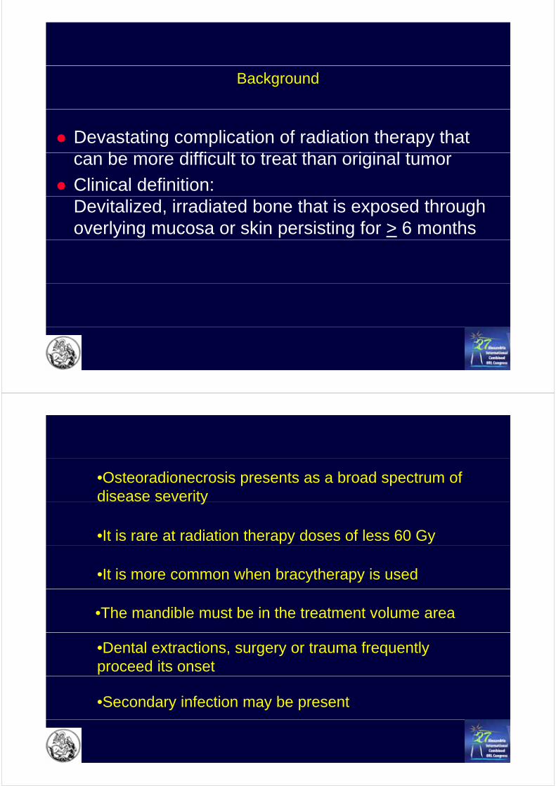
Stage III Sinus/Fistula
Surgical Intervention:Sinus/FistulaPathologic Fracture
Extensive soft tissue involvement
Extensive bony lossy
To include # and fistula


The only successful treatment of advanced y(Stage III) mandibular osteoradionecrosis is the surgical resection of diseased tissuesthe surgical resection of diseased tissues and their reconstruction with free tissue
transfertransfer.
The question whether HBO should be a precedent treatment or should be p
administered post-operatively or not at all is unansweredunanswered.
Reconstructive options in the treatment of severe (Stage III) mandibular osteoradionecrosis( g )
1. The radial forearm osteocutaneous flap
2. The fibula osteocutaneous flap
3. The use of additional flaps

Militsakh ON et al, Otolaryngol-Head and Neck Surg 2005
Shaha A et al, Head Neck 1997

Patients
47 patients between 1991 and 200647 patients between 1991 and 2006MSKCC=41; GACI=7
Age 34-81 (median 59) years
32 Males 15 Females32 Males 15 Females
Symptoms & SignsPain 75% Non-healing ulcer 66%gDraining Fistula/sinus 29%
Type of Reconstruction
OC-FRFFOther
19%Other6%
Fibula75%
n=16
Osteoradionecrosis of the mandible is a dreaded andOsteoradionecrosis of the mandible is a dreaded and devastating complication of radiation therapy for
malignant tumors of the head and neck area.malignant tumors of the head and neck area.
Various tumor, patient and treatment factors influence its development but the exact pathophysiology is still underdevelopment but the exact pathophysiology is still under
investigation.
It is especially important to prevent the development ofIt is especially important to prevent the development of osteoradionecrosis of the mandible by eliminating the so
far known risk factors.
When osteoradionecrosis reaches an advanced stage, the treatment of choice is resection of the necrosed
tissues and immediate reconstruction with free tissue transfer.
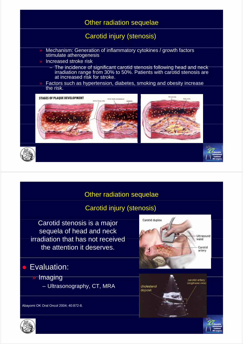
Other radiation sequelae
Carotid injury (stenosis)
Friedlander AH, Freymiller EG. J A, Dent Assoc 2003
Other radiation sequelae
Carotid injury (stenosis)
» Mechanism: Generation of inflammatory cytokines / growth factors stimulate atherogenesis
» Increased stroke riskThe incidence of significant carotid stenosis following head and neck– The incidence of significant carotid stenosis following head and neck irradiation range from 30% to 50%. Patients with carotid stenosis are at increased risk for stroke.
» Factors such as hypertension, diabetes, smoking and obesity increase y g ythe risk.
Other radiation sequelae
Carotid injury (stenosis)
Carotid stenosis is a major sequela of head and neck
irradiation that has not receivedirradiation that has not received the attention it deserves.
Evaluation:» Imaging» Imaging
– Ultrasonography, CT, MRA
Abayomi OK Oral Oncol 2004; 40:872-8.

Other radiation sequelae
Carotid injury (stenosis)
Management:» Endarterectomy; stenting
» Anticoagulation
Abayomi OK Oral Oncol 2004; 40:872-8.
Summary
Radiotherapy is an important Rx modality in oral cancersM d RT l i d t t t t h i iModern RT planning and treatment techniques improve treatment outcomeAdvances in Rx may actually increase late complicationsAdvances in Rx may actually increase late complicationsSite specific approach allows organ-sparing with high conformality RT (e.g. parotid-sparing)y ( g g)Modest benefits associated with medical treatment of established complicationsA i R f i li i i i di dAggressive Rx of certain complications is indicatedImportance of prevention of late complications through identification correction and avoidance of risk factorsidentification, correction and avoidance of risk factors
Second World Congress of the gInternational Academy of Oral Oncology
(IAOO)( )
July 8 – 11, 2009
Sheraton Centre Toronto
Invitation to Toronto!