short communication laryngeal inflammation · may cause laryngeal inflammation secondary to local...
TRANSCRIPT
Central Annals of Otolaryngology and Rhinology
Cite this article: Dworkin-Valenti JP, Sugihara E, Stern N, Naumann I, Bathula S, et al. (2015) Laryngeal Inflammation. Ann Otolaryngol Rhinol 2(9): 1058.
*Corresponding authorJ James P. Dworkin-Valenti, Department of Otolaryngology-Head & Neck Surgery, Detroit Medical Center, 5807 Cedar Ridge Dr, Ann Arbor, MI 48103, USA, Email:
Submitted: 05 August 2015
Accepted: 24 August 2015
Published: 31 August 2015
Copyright© 2015 Dworkin-Valenti et al.
OPEN ACCESS
Keywords•Laryngeal•Inflammation•Laryngitis•Granulomas
Short Communication
Laryngeal InflammationJames Paul Dworkin-Valenti*, Eric Sugihara, Noah Stern, Ilka Naumann, Samba Bathula, and Esmael Amjad Department of Otolaryngology- Head & Neck Surgery, Detroit Medical Center, USA
Abstract
Objective: To provide a synopsis of the various types, causes, and treatments of laryngeal inflammatory conditions.
Data Sources: Information contained within this paper was derived from the scientific literature and the clinical experiences of the authors. The data gathered were reinforced by the clinical and research experiences of the authors.
Review Methods: The Medline (PubMed) and Cochrane Library databases of references and abstracts were consulted for scientific information on types, causes, and treatments of laryngeal inflammation.
Conclusions: The classification of laryngitis can be divided into acute and chronic forms with a multiplicity of underlying behavioral and organic causes. These pathogenic variables are discussed, including the insidious laryngeal effects of voice abuses, infectious processes, systemic diseases, reflux disorders, various medications, and allergies. An algorithm of alternative treatments for laryngeal inflammatory conditions is presented.
INTRODUCTIONThere are many different types and causes of laryngeal
inflammation. Effective management largely depends upon the associated etiology, clinical course of the problem, and coexisting medical history of the patient afflicted. The primary objective of this article is to provide a comprehensive review of acute and chronic forms of this condition. The most common causal factors will be discussed in detail and alternative treatments will be systematically outlined.
METHODSMaterial for this review was obtained via the Medline (PubMed)
and Cochrane Library databases on benign and malignant vocal pathologies and laryngeal inflammation. The clinical and research experiences of the authors were also drawn upon to balance and organize the large data base that was obtained during this review process. Every effort was made to produce a comprehensive yet succinct paper on diagnostic and treatment implications for these pathologic conditions. Whenever conflicting or controversial information was discovered, such discrepancies were resolved via personal correspondences with other experts on this subject matter who recommended additional references to be consulted for clarification. The references included in this review were selected from nearly 300 scientific journal articles and abstracts because they provided the most salient information on the broad topic of laryngeal inflammation.
Anatomy
Historically, the respiratory tract has been arbitrarily
separated into upper and lower subdivisions. Within the past decade clinical researchers have postulated a single unified airway model; one composed of a system of organ linkages with anatomical similarities and common pathophysiologic mechanisms that mediate hyper-reactivity and inflammatory responses [1-4]. This interconnected airway begins within the ciliated epithelium of the nares, extends through the membranous tissue matrices of the nasal, oral, pharyngeal, and laryngeal cavities, and ends within the respiratory bronchioles and alveoli of the lungs. Pathologies that originate in any one site of this unified airway often concurrently affect other components.
Inflammatory Mechanism
There are many different proliferating patterns of tissue pathology within the unified airway including loco-regional inflammation, edema, hyperemia, and superficial cellular infiltration. The severity usually depends upon the gravity of the offending agent. Disruption of the laryngeal airway can occur for six major reasons: infection, inflammation, neoplasm, allergy, irritation, and trauma. Invasion of the larynx by viral pathogens, bacteria, fungal organisms, parasites, tumors, trauma, inhaled and ingested antigens, or environmental pollutants may result in significant loco-regional tissue inflammation. The biological mechanisms that contribute to the pathogenesis of this reaction are multi-varied and interactive. Most notably, plasma proteins and local cytokines leak from dilated blood vessels into the affected tissue, resulting in edema, erythema, and disruption of normal function. As blood vessel linings become more leaky, immune competent macrophage, neutrophil, and lymphocyte binding
Central
Dworkin-Valenti et al. (2015)Email:
Ann Otolaryngol Rhinol 2(9): 1058 (2015) 2/8
molecules are activated and recruited into the region; a process known as margination (pavementing), which is characterized by transportation of adhesion molecules into the site of involvement. In addition to these accumulated intracellular materials, other vasodilation biochemical mediators are transported from the intravascular space to the perivascular tissues and exacerbate the inflammatory response (diapedesis). These include 1) mast cell histamine, 2) blood platelet serotonin, and 3) arachnidonic acid prostaglandin and leukotriene metabolites [5-6].
Larynx Lymphatics
Embryologically, the larynx arises primarily from the paired branchial (visceral) arches III, IV and VI. The mesodermal layer provides the inherent blood vessels, cartilages, bones, and muscles. The lymphatic vessels are also derived from this primitive layer, and they assume significantly different patterns of drainage from the primary subsites of the larynx. It is important to understand these patterns because they influence differential loco-regional inflammatory reactions. For clinical staging purposes, the larynx can be subdivided into 3 distinct regions: supraglottis (epiglottis, arytenoid cartilages, aryepiglottic folds, and ventricular folds), glottis (true vocal folds, anterior commissure, and posterior commissure), and subglottis (5mm below free margin of true vocal folds to inferior margin of cricoid cartilage). The supraglottis contains rich lymphatics, which pass through the thyrohyoid membrane and drain to the subdigastric, midjugular, and lower jugular nodes. These vessels also communicate with the base of tongue, and this dense network establishes the common pathways of spread for infections, edema, and malignant tumors. Comparatively, the entire glottis has sparse lymphatic channels; the true vocal folds are actually devoid of such vessels. Consequently, infectious or malignant conditions that originate within the true vocal folds often remain confined to these structures without significant observable spread to adjacent lymph nodes at presentation. If and when drainage does occur from the glottis it flows to the internal jugular and paratracheal lymph nodes. The lymph vessels of the subglottis pass through the cricothyroid membrane and drain to the paralaryngeal, paratracheal, and lower deep cervical lymph nodes. Infections, traumatic, and malignant conditions involving this region of the larynx often spread to these lymph node chains. However, because such nodes occupy deep neck spaces their inflammatory response may remain occult in the patient who presents initially with significant clinical signs and symptoms of laryngeal pathology, such as dysphonia and/or odynophagia.
Laryngitis
This diagnostic term is frequently used to describe an inflamed appearance of the mucosa and tissues of the larynx, including those composing the epiglottis, arytenoid bodies, aryepiglottic folds, post-cricoid shelf, ventricular vocal folds, squamous epithelium of the true vocal folds, and immediate subglottis. Inflammation involving these structures may be acute or chronic in origin; with each form associated erythema, edema, tenderness, and dysfunction may vary in degree, course of involvement, and methods of treatment. The inflammatory process may damage the ciliated epithelium of the larynx, and it may impair mucus flow out of the tracheal-bronchial tree.
CLINICAL PRESENTATIONMucus stasis often induces episodic coughing and throat
clearing behaviors, which can lead to vocal fold edema, hyperemia, hyperkeratosis, acanthosis, and inherent cellular atypia. Patients with acute or chronic laryngitis often present with chief complaints of dysphonia and/or dysphagia. Dysphonia can present as breathiness, harshness, limited pitch range, and reduced vocal projection or loudness. Dysphagia can present as globus (foreign body) and choking sensations, pain during swallowing, regurgitation episodes, and the feeling of food getting stuck in the throat or upper esophageal region.
RISK FACTORS It has been our clinical experience that the following 15
populations may be generally most susceptible to laryngitis:
1) Individuals with habitually abusive and excessive vocal behaviors, such as screaming, yelling, singing, and throat clearing, which may results in unilateral or bilateral vocal fold inflammation (ie., polypoid corditis or Reinke’s edema), hemorrhagic polyp formation, or nodules,
2) Individuals with high and harsh vocal mileage, as may occur because of a combination of age, vocal personality, lifestyle, and job requirements,
3) Individuals who smoke cigarettes or who are frequently exposed to second-hand smoke, which often causes diffuse bilateral infiltrative laryngeal tissue inflammation, polypoid corditis, and/or malignancy,
4) Individuals who experience frequent laryngeal-pharyngeal reflux (LPR) events, which can cause diffuse laryngeal tissue irritation and inflammation secondary to erosive acid and pepsin effects as well as associated nocturnal coughing spells,
5) Individuals who experience laryngeal dehydration because they routinely drink excessive amounts of caffeine (diuretic) beverages, which often induces throat tickling sensations, reactive throat clearing, and resultant diffuse laryngeal inflammation,
6) Individuals who do not drink at least 6-8 glasses of non-caffeinated beverages daily for systemic maintenance of adequate laryngeal tissue hydration,
7) Individuals who regularly use anticholinergic medication, which often causes dry, brittle, and friable laryngeal mucosa and increases the risks of diffuse laryngeal inflammation,
8) Individuals who regularly use diuretic or angiotensin converting enzyme (ACE) inhibitor medications, which may cause laryngeal inflammation secondary to local tissue dehydration or chronic coughing behaviors, respectively,
9) Individuals with certain medical histories, such as asthma or chronic obstructive pulmonary disease (COPD), which may result in laryngeal candida, irritable laryngeal
Central
Dworkin-Valenti et al. (2015)Email:
Ann Otolaryngol Rhinol 2(9): 1058 (2015) 3/8
mucosa, and vocal fold inflammation secondary to associated use of inhaled corticosteroids and frequent coughing spells,
10) Individuals with infectious laryngeal conditions, such as laryngeal candidiasis and other fungal organisms, herpes, or vocal fold papilloma viruses, which can cause significant loco-regional tissue inflammation,
11) Individuals with fluctuating systemic medical conditions, such as Sjogrens syndrome, lupus, hyperthyroidism (Graves disease), hypothyroidism (myxedema), and sarcoidosis, which may cause unusually inflamed, waxy, dry, and/or brittle laryngeal mucosa and local inflammatory reactions,
12) Individuals with systemic otolaryngic allergy sequela, including post-nasal drip, upward migration of bronchial secretions, and responsive throat clearing and coughing, which may result in diffuse laryngeal inflammatory reactions,
13) Individuals with inflammation complications of laryngeal trauma,
14) Individuals with histories of haemophilus influenza type b (Hib) infection, which has been causally associated with epiglottitis in both children and adults,
15) Individuals with laryngeal malignancy histories (with or without radiation-induced loco-regional tissue fibrosis and inflammation) [7-17].
If and when these so-called vulnerable populations present with two or more of these co-morbid contributing factors their laryngeal findings may be advanced. These potentially confounding variables must always be surveyed and considered when determining the most efficacious treatments to employ for any given patient. Naturally, the prognosis for improvement and the types of treatments required for any given patient often depend upon the severity of the problem and the underlying etiology.
Physical exam
The most useful diagnostic tool in evaluating laryngeal inflammation is the rigid or flexible fiberoptic endoscope. Laryngeal endoscopy often reveals demonstrable yet variable loco-regional pathologies, depending upon the underlying etiology. These may include 1) diffuse glottal and supraglottal membranous erythema and edema and associated airway narrowing, 2) mucosal tissue tears, capillary ectasias, hemorrhaging, hyperemia, polyposis (Reinke’s edema), discrete polyp formation, nodules, hyperkeratosis, and/or leukoplakia involving the true vocal folds, 3) ulcerations, contact granulomas, and scar tissue along the medial surfaces of the vocal processes of the arytenoids and within the interarytenoid region, and 4) plica ventricularis. Glottal incompetency during phonation typically results as a consequence of these inflammatory conditions, the severity of which ordinarily determines the extent of associated voice and swallowing symptoms.
Acute laryngeal inflammation
In the acute form of laryngitis, the onset is usually abrupt with a self-limiting course of less than 3 weeks. Upper respiratory viruses, bacterial infections, fungi, and direct trauma, either by an external insult or a violent vocalization episode, are the most common causes. In certain systemic conditions, such as viral croup syndrome or candida albicans, acute involvement of upstream and downstream unified airway tissues may coexist and complicate the clinical course and treatment algorithm. Anaphylactic allergic reactions and malignant lesions may also result in acute laryngeal inflammation. These less common causes can be life threatening.
Chronic laryngeal inflammation
This condition has a more subdued onset than its acute counterpart. Signs and symptoms may wax and wane over very long periods of time, generally several weeks or months. The 15 most common etiopathogenic factors associated with diffuse and chronic endo-laryngeal inflammation have been described above. It should be noted that if these same etiologies coexist in patients with presumed acute laryngitis, the differential diagnosis of the actual subtype of this condition may be less certain or important. When the aforementioned vocal abuse or misuse behaviors occur, the surfaces of the vocal folds experience repetitive and powerful vibratory collision forces, the results of which include intense friction, thermal agitation, cell destruction, and whiplash [18]. Persistence of these offending exogenous or endogenous factors often leads to localized tissue necrosis, fibrosis, and scarring, secondary to the decaying effects of lymphocytes, eosinophilia, macrophages, fibroblasts, and collagen that accumulate in the chronically inflamed tissue. It has been suggested that chronic laryngitis may exist in approximately one-third of the general population, and that in more than 50% of individuals with symptoms of this condition various laryngeal inflammatory signs are discovered during laryngoscopy [19,20]. It should be acknowledged that in some instances of chronic laryngeal inflammation (eg., following radiation therapy) patients often habituate to glottal, supraglottal, or subglottal narrowing, without the need for further intervention. However, this functional adaptation is not often possible in cases of acute laryngeal inflammation, wherein emergency airway management may be necessary.
ALLERGY CONTROVERSY For the past 50 years, clinical researchers have continued
their debate about whether or not inhaled or ingested allergens or pollutants can contribute to or explain the chief complaints of intermittent voice and swallowing disorders observed in some allergic individuals. Such complaints are typically embedded in reports of recent repetitive exposure to known inciting antigens [9-13]. In these individuals, allergic laryngeal responses are usually related to late phase release of toxic proteins by eosinophilic cells rather than acute mast cell degranulation within the unified airway. Notwithstanding the conspicuous paucity of supportive scientific evidence to demonstrate such a causal interrelationship, allergic laryngitis remains a commonly assigned diagnostic classification by many otolaryngologists, allergists and primary care physicians.
Central
Dworkin-Valenti et al. (2015)Email:
Ann Otolaryngol Rhinol 2(9): 1058 (2015) 4/8
The immune response
The immunological mechanisms responsible for generalized sino-nasal inflammation in allergic patients have been extensively described [1,4,13,15, 21]. However, the cellular processes that may induce laryngitis in these same individuals after specific antigen exposure are not well understood, owing to relatively sparse human investigations on this topic and limited bench research with animal models. Acute phase, IgE-mediated, anaphylactic or angioedema allergic reactions within the larynx are not common, but when they do occur the mechanism of action is primarily induced by mast cells, found abundantly in the supraglottic and immediate subglottic tissues. The inflammatory reaction is prompt, vigorous, and often severe. The most common inciting antigens include food items, such as peanuts or shellfish, certain classes of drugs (eg., ACE-Inhibitors, penicillin), insect bites, and venoms. If rapid onset epiglottitis, generalized supraglottal edema, and/or subglottic narrowing occur, patients usually suffer from associated dyspnea, stridor, dysphonia, dysphagia, and globus sensations. These potentially life-threatening sequelae occur as a consequence of primary and secondary biomolecular and biomechanical edema of the loose areolar tissues of the laryngeal vestibule. Swellings of the tongue, floor of mouth, velum, and nasal mucosa commonly coexist, which can exacerbate all signs and symptoms.
The laryngeal allergic response
Signs and symptoms of bronchial and laryngeal hyper-reactivity following noxious antigen exposure usually arise gradually in the allergic individual, and they tend to be mild in degree [9-13,15]. When vocal fold inflammation and dysphonia occur following antigen exposure, explanations other than mast cell histamine deposition should be sought. Not infrequently, the patient exhibits forceful throat clearing behaviors because of perceived sticky-thick mucus accumulation at the base of tongue and within the laryngeal vestibule. These reactions can mechanically traumatize the vocal folds. If they persist and become chronic events, such shearing forces can lead to glottal incompetence and the aforementioned pathological laryngeal tissue reactions [21-24]. Irritation to the laryngeal membranes can originate from within the larynx, or occur from regional sites like downward post-nasal drainage, upward migration from the tracheal-bronchial tree, or supero-posterior reflux from the gastroesophageal tract. Laryngeal inflammation also may be the result of associated tissue dehydration, which, in turn, may lead to dry and brittle vocal fold mucosa, itching, and tickling sensations. These reactions typically induce reactive throat clearing and coughing, which triggers this vicious cycle of harmful pathophysiologic events. For completeness, it is important to note that episodic throat clearing, coughing, muscle tension vocalizations, and intermittent vocal fold edemas are also frequent sequelae of laryngeal-pharyngeal reflux. Because eosinophilic irritability has been etiologically linked to gastro-esophageal hyper-reactivity, clinical researchers have postulated common pathogenetic mechanisms for reflux and allergic diseases. [7, 25, 26] Gastro-esophageal reflux, upper respiratory tract infections, and allergic rhinitis have all been shown to precipitate cough, dyspnea, and throat clearing. These pulmonary responses are mediated by exaggerated parasympathetic-
cholinergic reflexes. Because the prevalence of such disease co-existence is relatively high among patients treated by internists, allergists, and otolaryngologists, it is very difficult to differentiate these conditions from one another for accurate diagnoses and accurate treatment recommendations.
Chemical mediators of allergic reaction
At a cellular level, both mast cells and eosinophils work synergistically to mediate inflammatory responses [4, 27-28]. The former population is composed largely of cytoplasmic granules containing histamine, heparin, and other immune modulators. These large leukocyte cells are located throughout the sinonasal tract mucosa. Activated by inhaled, ingested, or topical antigens mast cells act as primary mediators of the acute-phase allergic reaction. Following such responses, eosinophilic cells are typically recruited to the inflamed tissues, which results in late-phase protracted swelling and mucosal injury. As mentioned earlier, whereas mast cells and eosinophils densely populate the epiglottis and immediate subglottis, the squamous epithelium and neurovascular bundles of the vocal folds do not contain these cells, their mediators of inflammation, or their reactive peptide derivatives. Though such absence is likely an evolutionary and life-sustaining adaptation, this fact has significantly limited scientific extrapolations of a direct causal interrelationship between laryngitis and allergies [27,28]. That is to say, the lack of known chemical mediators of laryngeal inflammation helps explain why deliberate laboratory studies to induce glottic swellings in allergic subjects, using various allergens, have been largely unsuccessful [29-32].
Signs of Allergic Laryngitis
Notwithstanding inherent epidemiologic limitations, over the past decade several clinical researchers have investigated the extent to which specific allergens and environmental pollutants may induce common signs and symptoms of laryngitis in humans, rat, mice, guinea pig, and monkey animal models [9-15, 29-38]. Some of these studies employed dust mite and food provocation challenge methodologies in allergic and non-allergic individuals. Others utilized inhaled pollutants, such as iron- soot. In general, the findings of these research investigations have repeatedly demonstrated prompt both delayed and immediate lower and upper respiratory reactions in experimental subjects versus non-reactive controls. Possible causal, synergistic interrelationships between so-called eosinophilic bronchitis, tracheitis, laryngitis, and chronic exposure to specific antigens, pollutants, or both have been proposed to explain such outcomes [36-38]. Additionally, these authors suggested that several criteria and examination results must be present to support the possible diagnosis of laryngeal allergy. These include: 1) history of test positive allergic disease, 2) chronic throat clearing or dry hacking cough, 3) itching of the larynx, 4) transient vocal fold edema, 5) tenacious, thick, viscid endo-laryngeal mucus strands, 6) globus sensations, 7) abnormally pale, glistening, and edematous arytenoid mucosa, 8) unremarkable pulmonary function test, chest X-ray, and sinus images, 9) absence of contaminating laryngo-pharyngeal reflux or gastro-esophageal reflux symptoms or treatment history, and 10) acute or delayed dysphonia, odynophagia, or both, secondary to repetitive exposure to an inhaled, ingested, or topical antigen.
Central
Dworkin-Valenti et al. (2015)Email:
Ann Otolaryngol Rhinol 2(9): 1058 (2015) 5/8
Treatment Alternatives
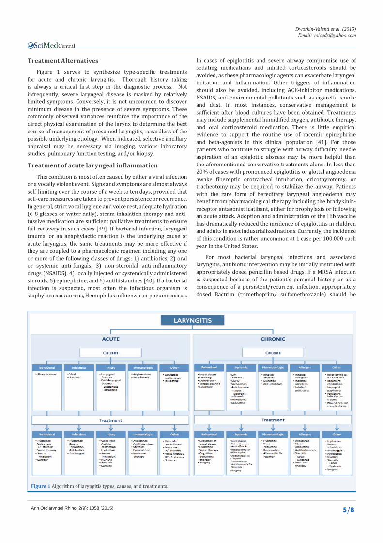
Figure 1 serves to synthesize type-specific treatments for acute and chronic laryngitis. Thorough history taking is always a critical first step in the diagnostic process. Not infrequently, severe laryngeal disease is masked by relatively limited symptoms. Conversely, it is not uncommon to discover minimum disease in the presence of severe symptoms. These commonly observed variances reinforce the importance of the direct physical examination of the larynx to determine the best course of management of presumed laryngitis, regardless of the possible underlying etiology. When indicated, selective ancillary appraisal may be necessary via imaging, various laboratory studies, pulmonary function testing, and/or biopsy.
Treatment of acute laryngeal inflammation
This condition is most often caused by either a viral infection or a vocally violent event. Signs and symptoms are almost always self-limiting over the course of a week to ten days, provided that self-care measures are taken to prevent persistence or recurrence. In general, strict vocal hygiene and voice rest, adequate hydration (6-8 glasses or water daily), steam inhalation therapy and anti-tussive medication are sufficient palliative treatments to ensure full recovery in such cases [39]. If bacterial infection, laryngeal trauma, or an anaphylactic reaction is the underlying cause of acute laryngitis, the same treatments may be more effective if they are coupled to a pharmacologic regimen including any one or more of the following classes of drugs: 1) antibiotics, 2) oral or systemic anti-fungals, 3) non-steroidal anti-inflammatory drugs (NSAIDS), 4) locally injected or systemically administered steroids, 5) epinephrine, and 6) antihistamines [40]. If a bacterial infection is suspected, most often the infectious organism is staphylococcus aureus, Hemophilus influenzae or pneumococcus.
In cases of epiglottitis and severe airway compromise use of sedating medications and inhaled corticosteroids should be avoided, as these pharmacologic agents can exacerbate laryngeal irritation and inflammation. Other triggers of inflammation should also be avoided, including ACE-inhibitor medications, NSAIDS, and environmental pollutants such as cigarette smoke and dust. In most instances, conservative management is sufficient after blood cultures have been obtained. Treatments may include supplemental humidified oxygen, antibiotic therapy, and oral corticosteroid medication. There is little empirical evidence to support the routine use of racemic epinephrine and beta-agonists in this clinical population [41]. For those patients who continue to struggle with airway difficulty, needle aspiration of an epiglottic abscess may be more helpful than the aforementioned conservative treatments alone. In less than 20% of cases with pronounced epiglottitis or glottal angioedema awake fiberoptic orotracheal intubation, cricothyrotomy, or tracheotomy may be required to stabilize the airway. Patients with the rare form of hereditary laryngeal angioedema may benefit from pharmacological therapy including the bradykinin-receptor antagonist icatibant, either for prophylaxis or following an acute attack. Adoption and administration of the Hib vaccine has dramatically reduced the incidence of epiglottitis in children and adults in most industrialized nations. Currently, the incidence of this condition is rather uncommon at 1 case per 100,000 each year in the United States.
For most bacterial laryngeal infections and associated laryngitis, antibiotic intervention may be initially instituted with appropriately dosed penicillin based drugs. If a MRSA infection is suspected because of the patient’s personal history or as a consequence of a persistent/recurrent infection, appropriately dosed Bactrim (trimethoprim/ sulfamethoxazole) should be
Figure 1 Algorithm of laryngitis types, causes, and treatments.
Central
Dworkin-Valenti et al. (2015)Email:
Ann Otolaryngol Rhinol 2(9): 1058 (2015) 6/8
considered as the first line pharmacologic regimen. Fungal infections that induce laryngeal candidiasis should be treated with appropriately dosed systemic Diflucan. Adding a swish and swallow regimen of nystatin for co-exisitng pharyngitis may be therapeutic as well. IV medications are generally reserved for acute invasive fungal infections or severely immuno-compromised patients who do not respond appropriately to oral medications.
In some cases of laryngeal trauma, surgical intervention is required either to stabilize the airway, repair a fracture, or remove an obstructive lesion, such as an intubation granuloma. All suspicious lesions identified during the physical endoscopic examination should undergo biopsy to rule out malignancy and the need for further workup (CT) or treatment (eg., surgery, radiation therapy, chemotherapy). Figure 1 lists additional treatment strategies for type-specific acute laryngitis varieties.
Treatment of acute allergic laryngitis depends upon the severity of symptoms. First line management requires removal of all suspected noxious triggers, coupled to strict avoidance behaviors. With significant signs and symptoms of airway compromise, epinephrine (epipen) should be promptly administered for rescue, followed by close observation. If indicated, inhaled Beta-antagonists should be administered for signs of wheezing. Concurrent intravenous steroids (Decadron), oral steroids (prednisone), antihistamine therapy (diphenhydramine), and H2-Blockers may also be effective ancillary prescriptions to avert progression of signs and symptoms and to prevent late phase reactions.
Treatment of chronic laryngeal inflammation
Treatments for this condition should especially focus on the presumed underlying cause, such as vocal abuses, smoking, dehydration, reflux, asthma, allergy, systemic disease, and irritating drug sequelae. In many cases, behavioral modification and vocal rest suffice to improve the condition. Instructing the patient to avoid the following conditions and behaviors is generally recommended: 1) first or second-hand smoke, 2) specific allergens like dust, pollen or environmental pollutants, 3) caffeinated beverages, 4) systemic decongestant medications, 5) inhalers that contain a steroid ingredient, 6) throat clearing activities, and 7) whispering. Adequate daily water consumption, as noted above, is often helpful, as is use of sugar free throat lozenges and a cool mist humidifier for topical throat moisturization; the soothing benefits of steam inhalation are also worth attempting with patients who do not improve significantly with these other techniques of management. The value of isotonic saline therapy may be beneficial for patients with persistent laryngitis despite alternative treatments. In certain unique circumstances, where voice improvement is urgently required, a short duration course of systemic corticosteroids (eg., Medrol dose pack), or an isolated steroid injection directly into the superficial layer of the lamina propria of the vocal folds may be administered in the office setting for prompt relief of associated inflammation and dysphonia. Methylprednisolone (40mg/mL) is recommended for the injection procedure, using a 1-mL syringe, a curved cannula, and a disposable injector needle. The recommended dose ranges from .1mL to 1.0 mL per intralesional injection. For benign vocal fold free edge lesions and inflammation smaller doses are
generally best; larger volumes should be reserved for laryngeal scars and granulomas [42]. Aggressive surgical intervention can often be delayed or avoided with steroids. Much different chronic or acute laryngeal pathology, such as persistent edema, polypoid corditis, and fusiform or hemorrhagic polyps, may be effectively treated initially via behavioral modification strategies, followed by the aforementioned pharmacologic regime if necessary. It should be noted that the duration of any benefits derived from steroid injection into the vocal folds may vary considerably from patient to patient. In general, the positive effects may last for two months or longer in some individuals, particularly those who cease engaging in the behaviors or activities that are causally related to the condition. For recurrent symptoms, repeat treatment can be attempted, but no sooner than 3 months after the initial injection. No more than 3 such injections should be administered to the same patient within a period of one year for fear of possible bowing and atrophy sequelae. Additionally, intralesional laryngeal steroid injections may be preferred in patients with risk factors for oral steroid use, such as those individuals with histories of glaucoma, peptic ulcer disease, or immunocompromised conditions (eg., diabetes, HIV, post chemo-radiation therapy for malignancies, kidney failure).
Whereas in virtually all cases of chronic laryngitis there is no role for antibiotic therapy, in a select number of individuals with recurrent or persistent bacterial or fungal infections use of antibiotic agents (+/- systemic anti-fungal medication) may be appropriate and helpful, as previously described for acute laryngitis. It is important to note that prolonged courses of these drugs may be needed to clear the infection and to prevent recurrences. In patients with chronic laryngitis whose histories include routine use of inhaled corticosteroids for asthma or COPD, either experimental cessation of this medication, dose reduction, or substitution of an alternative nonsteriodal inhaler may induce positive laryngeal reactions. [8,16]. Local nasal steroid and nasal antihistamine therapy may prove valuable for the individual whose chronic laryngeal inflammation is thought to be causally related to seasonal or perennial allergy with associated post-nasal drip and reactive chronic coughing spells. In the intractable allergic patient, immunotherapy may be required to reduce these possible adverse side effects and inflammatory laryngeal sequelae. Anti-reflux medications and proper diet restrictions can be of similar additive value for those patients with known LPR signs and symptoms. Not infrequently, patients with chronic laryngitis require a multi-pronged approach to treatment that will include behavior modifications, vocal rest, topical and systemic moisturization strategies, current drug alterations, and additional pharmaceutical prescriptions, as illustrated in the Figure 1 algorithm. On occasion, biopsy and/or surgical excision of chronically pathological laryngeal tissues may be necessary for definitive diagnosis. When edematous nodules, hemorrhagic polyps, or Reinke’s edema do not resolve with conservative measures, including voice therapy, excision using microsurgical dissection or evacuation methods may be necessary for successful treatment outcomes, followed by another stint of behavioral voice therapy to review and reinforce the techniques and importance of proper vocal hygiene.
Finally, for the patient who suffers from chronic cough behaviors and correlated laryngeal inflammation it would be
Central
Dworkin-Valenti et al. (2015)Email:
Ann Otolaryngol Rhinol 2(9): 1058 (2015) 7/8
prudent to rule out the possible, well-known causal influence of ACE-Inhibitor medications. In many such cases, prompt alleviation is achieved with an alternative class of equally effective hypertensive medication. Idiopathic coughing spells are not uncommon. If coughing is not initially significantly alleviated with standard anti-tussive medications, including cough syrups and/or Tessalon capsules or perles, an alternative pharmacologic regimen may prove helpful. For intractable coughing behaviors, usually of unknown or neurologic origin, we frequently prescribe Tramadol with very good results. Because this drug has been known to cause addictive connections in some individuals, a limited initial dose and course is recommended with hopes that the above regimen breaks the coughing cycle and the induced laryngeal inflammatory sequelae. As with all drugs that may be prescribed for a given patient, it is always wise to survey for potential hazardous drug interactions, general side effects, and use contraindications based on the medical history, before recommending any particular agent for any reason whatsoever.
CONCLUSION AND IMPLICATIONS FOR CLINICAL PRACTICE
Laryngitis is a common condition that can be sub classified into acute and chronic forms, depending upon the duration of symptoms. The etiology can be related to infectious, allergic, or irritative precursors. As is true for any medical problem, determining the underlying etiology of laryngitis is paramount to accurate treatment. A detailed history followed by physical exam usually ensures a successful diagnosis and outcome. If the patient’s complaints are rather mild, formal treatments may be unnecessary, as the problem is usually self-limiting. However, with more advanced inflammatory characteristics, regardless of the cause, signs and symptoms of clinically significant airway compromise, dysphagia, and dysphonia may coexist and require more comprehensive otorhinolaryngologic examinations and treatments. Because laryngitis may be observed in a broad spectrum of patient populations, intervention alternatives may range from simplistic methods of reassurance and avoidance behaviors to conservative or aggressive medico-surgical interventions. Detailed descriptions of such techniques were presented in the treatment algorithm for each subtype of laryngitis.
REFERENCES1. Hurwitz B. Nasal Pathophysiology impacts bronchial reactivity in
asthmatic patients with allergic rhinitis. J Asthma. 1997; 34: 427-431.
2. Grossman J. One airway, one disease. Chest. 1997; 111: 11S-16S.
3. Dworkin JP, Stachler, RJ. Management of the patient with laryngitis. In Krouse, Chadwick and Dereberry (Eds). Managing the Allergic Patient. Elsevier, Inc. 2008: 233-272.
4. Krouse JH, Altman KW. Rhinogenic laryngitis, cough, and the unified airway. Otolaryngol Clin North Am. 2010; 43: 111-121, ix-x.
5. Dworkin JP. Laryngitis: types, causes, and treatments. Otolaryngol Clin North Am. 2008; 41: 419-436, ix.
6. Krouse JH. The unified airway--conceptual framework. Otolaryngol Clin North Am. 2008; 41: 257-266, v.
7. Vaezi MF. Extraesophageal manifestations of gastroesophageal reflux disease. Clin Cornerstone. 2003; 5: 32-38.
8. DelGaudio JM. Steroid inhaler laryngitis: dysphonia caused by inhaled fluticasone therapy. Arch Otolaryngol Head Neck Surg. 2002; 128: 677-681.
9. Reidy PM, Dworkin JP, Krouse JH. Laryngeal effects of antigen stimulation challenge with perennial allergen Dermatophagoides pteronyssinus. Otolaryngol Head Neck Surg. 2003; 128: 455-462.
10. Dworkin JP, Reidy PM, Stachler RJ, Krouse JH. Effects of sequential Dermatophagoides pteronyssinus antigen stimulation on anatomy and physiology of the larynx. Ear Nose Throat J. 2009; 88: 793-799.
11. Krouse JH, Dworkin JP, Carron MA, Stachler RJ. Baseline laryngeal effects among individuals with dust mite allergy. Otolaryngol Head Neck Surg. 2008; 139: 149-151.
12. Mouadeb DA, Belafsky PC, Birchall M, Hood C, Konia T, Pinkerton KE. The effects of allergens and tobacco smoke on the laryngeal mucosa of guinea pigs. Otolaryngol Head Neck Surg. 2009; 140: 493-497.
13. Randhawa PS, Nouraei S, Mansuri S, Rubin JS. Allergic laryngitis as a cause of dysphonia: a preliminary report. Logoped Phoniatr Vocol. 2010; 35: 169-174.
14. Brightling CE, Ward R, Wardlaw AJ, Pavord ID. Airway inflammation, airway responsiveness and cough before and after inhaled budesonide in patients with eosinophilic bronchitis. Eur Respir J. 2000; 15: 682-686.
15. Naito K, Iwata S, Yokoyama N. Laryngeal symptoms in patients exposed to Japanese cedar pollen: allergic reactions and environmental pollution. Eur Arch Otorhinolaryngol. 1999; 256: 209-211.
16. Morrison M, Rammage L, Emami AJ. The irritable larynx syndrome. J Voice. 1999; 13: 447-455.
17. Tanner K, Roy N, Merrill RM, Elstad M. The effects of three nebulized osmotic agents in the dry larynx. J Speech Lang Hear Res. 2007; 50: 635-646.
18. Titze IR, Svec JG, Popolo PS. Vocal dose measures: quantifying accumulated vibration exposure in vocal fold tissues. J Speech Lang Hear Res. 2003; 46: 919-932.
19. Koufman JA. Laryngopharyngeal reflux 2002: a new paradigm of airway disease. Ear Nose Throat J. 2002; 81: 2-6.
20. Reulbach TR, Belafsky PC, Blalock PD, Koufman JA, Postma GN. Occult laryngeal pathology in a community-based cohort. Otolaryngol Head Neck Surg. 2001; 124: 448-450.
21. Corey JP. Allergy for the laryngologist. Otolaryngology Clinics of North America 1998; 31: 422-426.
22. Chadwick SJ. Allergy and the contemporary laryngologist. Otolaryngol Clin North Am. 2003; 36: 957-988.
23. Baroody FM. Allergic rhinitis: broader disease effects and implications for management. Otolaryngol Head Neck Surg. 2003; 128: 616-631.
24. Alimov AI. [Clinical symptoms in the diagnosis of allergy in acute and chronic laryngitis]. Vestn Otorinolaringol. 1968; 30: 71-75.
25. Belafsky PC, Postma GN, Amin MR, Koufman JA. Symptoms and findings of laryngopharyngeal reflux. Ear Nose Throat J. 2002; 81: 10-13.
26. Ayik SO, Başoğlu OK, Erdínç M, Bor S, Veral A, Bílgen C. Eosinophilic bronchitis as a cause of chronic cough. Respir Med. 2003; 97: 695-701.
27. Domeij S, Dahlqvist A, Eriksson A, Forsgren S. Similar distribution of mast cells and substance P- and calcitonin gene-related peptide-immunoreactive nerve fibers in the adult human larynx. Ann Otol Rhinol Laryngol. 1996; 105: 825-831.
Central
Dworkin-Valenti et al. (2015)Email:
Ann Otolaryngol Rhinol 2(9): 1058 (2015) 8/8
Dworkin-Valenti JP, Sugihara E, Stern N, Naumann I, Bathula S, et al. (2015) Laryngeal Inflammation. Ann Otolaryngol Rhinol 2(9): 1058.
Cite this article
28. Ishida H, Yoshida T, Iwae S, Amatsu M. Immunohistochemical study on distribution of mast cell phenotypes in human laryngeal mucosa: evidence for laryngeal type I allergy. Ann Otol Rhinol Laryngol. 2005; 114: 139-143.
29. Niklasson A, Dahlqvist A. Antigen challenge induces a supraglottic but not a subglottic edema in the rat larynx. Otolaryngol Head Neck Surg. 2005; 132: 694-700.
30. Ishii J, Ogawa T, Naito K, Miyata S, Ishihara M, Baba R, et al. Local eosinophilia of the nose, the larynx and the trachea in rats sensitized with Japanese cedar pollen. Arerugi. 1997; 46: 1251-1257.
31. Iwae S, Ishida H, Amatsu M. Laryngeal type 1 allergy in sensitized guinea pig. Larynx Jpn 1995;7:1-6.
32. Lidegran M, Domeij S, Forsgren S, Dahlqvist A. Mast cells in the laryngeal mucosa of the rat. Effect of compound 48/80 and dexamethasone: a quantitative and immunohistochemical study at the light- and electron-microscopic levels. Acta Anat (Basel). 1996; 157: 135-143.
33. Williams RI. Allergic laryngitis. Ann Otol Rhinol Laryngol. 1972; 81: 558-565.
34. Sala E, Hytönen M, Tupasela O, Estlander T. Occupational laryngitis with immediate allergic or immediate type specific chemical hypersensitivity. Clin Otolaryngol Allied Sci. 1996; 21: 42-48.
35. Dixon HS. Allergy and laryngeal disease. Otolaryngol Clin North Am. 1992; 25: 239-250.
36. Zhou YM, Zhong CY, Kennedy IM, Pinkerton KE. Pulmonary responses of acute exposure to ultrafine iron particles in healthy adult rats. Environ Toxicol. 2003; 18: 227-235.
37. Leonard R, Charpied G, Faddis B. Effects of chronic ozone (O3) exposure on vocal-fold mucosa in bonnet monkeys. J Voice. 1995; 9: 443-448.
38. Belafsky PC, Peake J, Smiley-Jewell S, Verma S, Dworkin-Valenti JP, Pinkerton K. Soot and house dust mite allergen cause eosinophilic laryngitis in an animal model of chronic laryngitis. Laryngoscope 2015; In Press.
39. Reveiz L, Cardona AF, Ospina EG. Antibiotics for acute laryngitis in adults. Cochrane Database Syst Rev 2015; 2.
40. Kinnari TJ, Lampikoski H, Hyyrynen T, Aarnisalo AA. Bacterial biofilm associated with chronic laryngitis. Arch Otolaryngol Head Neck Surg. 2012; 138: 467-470.
41. Shah RK, Stocks C. Epiglottitis in the United States: national trends, variances, prognosis, and management. Laryngoscope. 2010; 120: 1256-1262.
42. Mortensen M, Woo P. Office steroid injections of the larynx. Laryngoscope. 2006; 116: 1735-1739.