significant improvements in cognitive performance post
TRANSCRIPT

Significant Improvements in Cognitive PerformancePost-Transcranial, Red/Near-Infrared Light-Emitting
Diode Treatments in Chronic, Mild Traumatic Brain Injury:Open-Protocol Study
Margaret A. Naeser,1,2 Ross Zafonte,3,4 Maxine H. Krengel,1,2 Paula I. Martin,1,2 Judith Frazier,3
Michael R. Hamblin,5 Jeffrey A. Knight,6 William P. Meehan III,7 and Errol H. Baker1
Abstract
This pilot, open-protocol study examined whether scalp application of red and near-infrared (NIR) light-emitting diodes
(LED) could improve cognition in patients with chronic, mild traumatic brain injury (mTBI). Application of red/NIR light
improves mitochondrial function (especially in hypoxic/compromised cells) promoting increased adenosine triphosphate
(ATP) important for cellular metabolism. Nitric oxide is released locally, increasing regional cerebral blood flow. LED
therapy is noninvasive, painless, and non-thermal (cleared by the United States Food and Drug Administration [FDA], an
insignificant risk device). Eleven chronic, mTBI participants (26–62 years of age, 6 males) with nonpenetrating brain
injury and persistent cognitive dysfunction were treated for 18 outpatient sessions (Monday, Wednesday, Friday, for 6
weeks), starting at 10 months to 8 years post- mTBI (motor vehicle accident [MVA] or sports-related; and one participant,
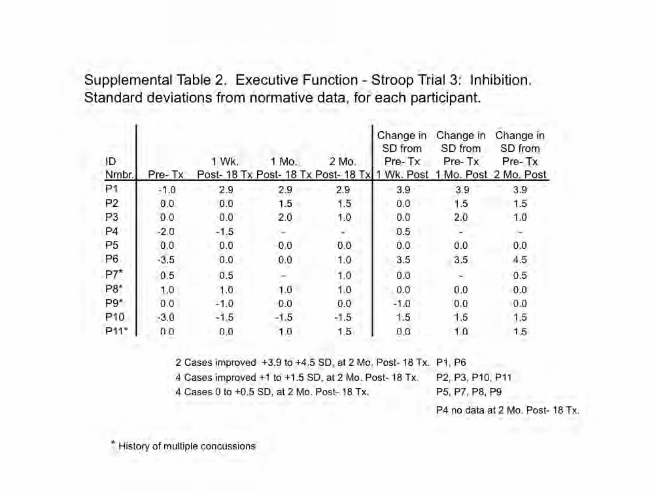
improvised explosive device [IED] blast injury). Four had a history of multiple concussions. Each LED cluster head
(5.35 cm diameter, 500 mW, 22.2 mW/cm2) was applied for 10 min to each of 11 scalp placements (13 J/cm2). LEDs were
placed on the midline from front-to-back hairline; and bilaterally on frontal, parietal, and temporal areas. Neuropsychological
testing was performed pre-LED, and at 1 week, and 1 and 2 months after the 18th treatment. A significant linear trend was
observed for the effect of LED treatment over time for the Stroop test for Executive Function, Trial 3 inhibition ( p = 0.004);
Stroop, Trial 4 inhibition switching ( p = 0.003); California Verbal Learning Test (CVLT)-II, Total Trials 1–5 ( p = 0.003);
and CVLT-II, Long Delay Free Recall ( p = 0.006). Participants reported improved sleep, and fewer post-traumatic stress
disorder (PTSD) symptoms, if present. Participants and family reported better ability to perform social, interpersonal, and
occupational functions. These open-protocol data suggest that placebo-controlled studies are warranted.
Key words: executive function; mTBI; photobiomodulation; treatment for mTBI
Introduction
Each year in the United States, *1,700,000 patients are
evaluated for traumatic brain injury (TBI); three TBIs every
minute.1 It is estimated that there are 5,300,000 Americans living with
TBI-related disabilities.1,2 The annual economic cost is estimated to
be between $60 and $76.5 billion.1,3 The majority of cases (70–85%)
are mild TBI (mTBI).1 Most civilian mTBI patients recover cognitive
abilities within 3 months;4 however, the literature reports that between
5 and 22% of individuals have persistent symptoms.5–9
Cognitive dysfunction associated with sports-related mTBI is of
increasing concern, both for males and females (including chil-
dren).10 Within the past 10 years, the diagnosis of concussion in
high school sports has increased annually, by 16.5%.11
1VA Boston Healthcare System, Boston, Massachusetts.2Department of Neurology, Boston University School of Medicine, Boston, Massachusetts.3Department of Physical Medicine and Rehabilitation, Spaulding Rehabilitation Hospital, Charlestown, Massachusetts.4Massachusetts General Hospital, Brigham and Women’s Hospital, Red Sox/MGH Home Base Program, Department of Physical Medicine and
Rehabilitation, Harvard Medical School, Boston, Massachusetts.5Wellman Center for Photomedicine, Massachusetts General Hospital, Department of Dermatology, Harvard Medical School, Harvard-MIT Division
of Health Science and Technology, Cambridge, Massachusetts.6National Center for PTSD, VA Boston Healthcare System, Boston, Massachusetts.7Micheli Center for Sports Injury Prevention, Waltham, Massachusetts and Sports Concussion Clinic, Boston Children’s Hospital, Harvard Medical
School, Boston, Massachusetts.
JOURNAL OF NEUROTRAUMA 31:1008–1017 (June 1, 2014)ª Mary Ann Liebert, Inc.DOI: 10.1089/neu.2013.3244
1008

It is estimated that 15–40% of soldiers returning from Iraq and
Afghanistan as part of Operation Enduring Freedom/Operation
Iraqi Freedom (OEF/OIF) report at least one mTBI.12–14 Closed-
head blast injury is the signature injury,12 and the cognitive se-
quelae, recovery, and rehabilitation are of increasing concern.15 It
is estimated that there are as many as 320,000 veterans who have
returned with TBI, most with mTBI.16,17 Post-traumatic stress
disorder (PTSD) is also a major concern with OEF/OIF soldiers
who have experienced mTBI.18 Therefore, a compelling need exists
to address chronic deficits in this population.
Cases of chronic mTBI often present clinically with deficits in
attention, working memory, cognitive manipulation of temporal
information, and general information processing speed.7,8,19–23 The
most common complaints are in attention/concentration and
working memory; that is, the ability to hold information in the
mind, and to manipulate it in light of incoming material.24 At 6
months post-injury, indices of executive function were found to
predict persistence of post-concussive syndrome in mild and
moderate TBI patients.25 In addition to cognitive problems, these
patients are often unable to re-establish family and work relation-
ships.26 Because of the diffuse nature of damage, however, no
single behavioral outcome measure captures the multidimensional
nature of mTBI outcome.27
Rationale for the present study
The present study examined whether the application of red and
near-infrared (NIR) light, utilizing light-emitting diodes (LED)
applied directly to the head could improve cognitive function,
particularly executive function and verbal memory, in chronic,
mTBI patients. Photons in the red and NIR wavelengths have the
potential to improve subnormal, cellular activity of brain tissue that
has been damaged by brain trauma.
Scalp application of red and NIR light is a new application for
LED technology. More than 30 years ago, however, it was observed
in human cadaver studies that red (600 nm) and NIR (800–900nm)
wavelengths could penetrate through the scalp and skull (* 1 cm).28
Two physiological changes associated with exposure of cells to red
and NIR wavelengths of light are: 1) Increased production of
adenosine triphosphate (ATP) by the mitochondria,29,30 and 2)
Increased vasodilation/regional cerebral blood flow (rCBF),31 ex-
plained subsequently.
The last enzyme complex (cytochrome c oxidase) of the electron
transport chain within the mitochondrial membrane is a photo-
acceptor for red and NIR photons.32,33 There is mitochondrial
damage and dysfunction after TBI.34,35 Increased ATP production by
the mitochondria improves cellular respiration, oxygenation, and
function. Also, in hypoxic/compromised cells, cytochrome c oxidase
is inhibited by non-covalently bound nitric oxide. When the mito-
chondria are exposed to red/NIR photons, nitric oxide is released and
diffused outside the cell wall, promoting local vasodilation and in-
creased blood flow. The effect of the light is non-thermal.36
Multiple animal studies using mice, show significantly better
recovery of motor and cognitive function after NIR transcranial
low-level laser therapy when treated in the acute post-injury
phase.37–40 Most of this work has suggested improved energy ki-
netics and decreased inflammation as possible mechanisms for
acute neuroprotection. Energetics may also have a role in the
chronic phase of injury. We have reported that midline and bilateral
scalp application of red/NIR LED therapy improved executive
function and verbal memory in two patients with chronic TBI.41
One TBI patient with a history of multiple concussions (retired
military) who had been on medical disability for 5 months prior to
transcranial LED treatments, returned to full-time employment
after 4 months of nightly, home LED treatments.
We undertook a pilot, open-protocol study among patients with
chronic mTBI who received the same number of specific, tran-
scranial LED treatments. Pre- and post- neuropsychological (NP)
testing and psychological measurements were acquired up to 2
months after the last LED treatment, in order to evaluate its po-
tential to improve chronic symptomatology.
Methods
Design
We utilized a case series design.42
Participants
Eleven, chronic, mTBI cases with nonpenetrating brain injury(6 males) participated. Their ages ranged from 26 to 62 years attime of entry (mean, 44.3 years; SD, 13.7) and the time post- mTBIranged from 10 months to 8 years (mean, 38.2 months., SD, 29.4).Their demographics, including medical history for mTBI, years ofeducation, and work status, are provided in Table 1.
All participants had previously been diagnosed as having mTBI,with loss of consciousness (LOC) lasting £ 30 min (or no LOC);and with a period of altered mental status that could include post-traumatic amnesia: ‘‘memory gaps’’ or confusion lasting up to 24 h.To be included in the study, persistent cognitive problems consis-tent with a diagnosis of mTBI had to have been present for at least 6months prior to screening cognitive testing. Exclusion criteria in-cluded moderate or severe TBI, penetrating brain injury, or historyof craniotomy or craniectomy. Medical records were obtained, andeach participant was examined by a single experienced study cli-nician prior to referral for screening cognitive testing. Participantswere requested not to change their medications or dosages duringparticipation in the study.
Prior to official enrollment into the study, all subjects signedinformed consent forms approved by the Spaulding RehabilitationHospital, Institutional Review Board (IRB). All study procedurescomplied with the IRB and with The Health Insurance Portabilityand Accountability Act (HIPAA) standards.
Screening cognitive testing
In addition to the criteria mentioned, in order to qualify for entryinto the study, each participant needed to score at least 2 SD belowaverage on one, or 1 SD below average on at least two of thefollowing NP tests (using age and education-adjusted norms). 1)Trail Making Test, Trails A and Trails B, measuring problem-solving, thinking flexibility, and planning.43 2) Controlled OralWord Association Test (COWAT)/FAS Test, total words generatedfor the letters F, A, and S (1 min per letter), measuring verbalfluency and categorical generative capacity.44,45 3) CaliforniaVerbal Learning Test-II, examining aspects of verbal learning,organization, and memory.46 4) Stroop Test for Executive Func-tion, examining attention, mental speed, mental control, inhibition,and inhibition switching.47
NP tests administered pre-/post- LED treatment series
The participants were tested four times during participation inthe study: 1) pre- testing, within 1 week before the first LEDtreatment; 2) post- testing, within 1 week after the final (18th) LEDtreatment; 3) at 1 month after the final LED treatment; and 4) at 2months after the final LED treatment. The tests administered at allfour testing times included the following: 1) Stroop Test for Ex-ecutive Function;47 2) California Verbal Learning Test-II (CVLT-II)with Alternating Versions, with Short Delay Free Recall, Short
TRANSCRANIAL LED THERAPY TO TREAT MILD TBI 1009

Delay Cued Recall, Long Delay (20 min later) Free Recall, andLong Delay Cued Recall, for each testing session;46 3) Delis–Kaplan Executive Function (D-KEF) - Trails Test;47 4) ControlledOral Word Association Test (COWAT)/FAS Test, total wordsgenerated, for the letters F, A and S;44,45 and Digit Span, Forwardsand Backwards, Wechsler Adult Intelligence Scale (WAIS)-IV.48
Some of the tests administered at screening were carried over toserve as pretreatment/baseline scores at Time 1.
In order to avoid practice effects, alternating versions of the NPtests were used at the post-LED testing times, when possible. Thisincluded the CVLT-II, Alternating Versions for the 16 words pre-sented;46 and the alternate form of the FAS test, using the threedifferent letters B, H, and R.47 A consistent practice effect for re-peated presentations of the Stroop test has not been demonstrated.49
In addition, psychological measurements included the PTSDChecklist–Civilian (PCL-C);50 the Beck Depression Inventory-II(BDI);51 and the Visual Analog Scale (VAS) for pain (0–10, verbalreport).52
LED device and LED treatment method
Two identical, LED Console Units were used (MedX Health,Model 1100, Toronto). Each Console Unit had three LED clusterheads. This device was cleared by the United States Food and DrugAdministration (FDA) in 2003 as posing an insignificant risk(FDA-cleared for home treatment, 2005). A sample LED clusterhead is shown in Figure 1. Each LED cluster head had a 5.35 cmdiameter (9 red diodes, 633 nm, and 52 NIR diodes, 870 nm wereembedded into each LED cluster head); 22.48 cm2 in size; 500 mWtotal power; 22.2 mW/cm2 power density; continuous wave. Thepower output (500 mW) for each LED cluster head was verifiedbefore and after the LED treatments using the MedX ME-DRAD200X Radiometer System.
Table 2 lists the LED cluster head placements on the head thatwere used at each visit (Set A for 10 min, followed immediately bySet B for 10 min), and approximate surface brain cortex areas that
were impacted with the red/NIR LEDs. The LED placement lociincluded, in part, a proposed paradigm to target nodes within thedefault mode network (DMN) and the salience network (SN), areaswhere functional connectivity MRI studies have reported abnor-malities in TBI.53 The LED placement loci also included dorso-lateral prefrontal cortex (DLPFC), part of the central executivenetwork (CEN).54 Supplementary Figure 1 (see online supplemen-tary material at http://www.liebertonline.com) shows the location ofextracranial bone and suture landmarks on the skull, in relationshipto approximate surface brain cortex areas. In the present study, theLED placements were hypothesized to impact the immediate sub-jacent, surface cortical areas, although this is unknown.
At each visit, six, 5.35 cm diameter LED cluster heads wereapplied simultaneously for 10 min during Set A, and then imme-diately after Set A, the LED placement loci were changed, and Set Bwas treated for 10 min (energy density, 13 Joules/cm2 [J/cm2] pereach LED cluster head placement). It was estimated that up to 3%(0.4 J/cm2) could reach the surface brain cortex, although this is un-known in humans (M. Hamblin, personal observation).28,55 Patientswere treated in a recliner chair, and the total LED treatment time pervisit was 20 min. The LED cluster heads were held in place with a softnylon cap. Because of the elastic tension from the cap, the locationof the LED placements did not shift during each 10 min treatment(Fig. 1). LED therapy is noninvasive, painless and non-thermal.36
Each participant received 18 treatments (Monday, Wednesday, andFriday, minimum of 48 h between treatments), for 6 weeks.
Statistical analysis
The effect of LED treatments over time was examined for thefollowing NP tests: Stroop Test for Executive Function (D-KEF);CVLT-II, Alternating Versions; D-KEF Trails Test; COWAT/FASTest; and Digit Span Forwards and Backwards. The psychologicalmeasures including BDI, PCL-C, and VAS for pain were also ex-amined. These data were analyzed in a series of univariate one way,repeated measures analyses of variance (ANOVA) with trend
Table 1. Demographics for 11 Chronic, Mild Traumatic Brain Injury (mTBi) Cases Treated
with Transcranial, Red/Near-Infrared Light-Emitting Diode (LED) Therapy
Time post-TBIIDnumber
Age atentry Gender Yr. Mo. Medical history for mTBI
Yearseducation Work status
P1 52 M 5 - Motor vehicle accident (MVA) 16 DisabledP2 59 M 2 6 MVA 22 Partially disabled; working only
22 hours/week at entryP3 50 F 1 8 Pedestrian hit by a car. 18 Disabled
Unable to return to workP4 26 F 1 4 Multiple-car MVA, hit from behind. 14 Unemployed, disabledP5 58 M 1 - Sports injury, close-range impact.
Hit in the head by a baseball.16 Disabled
P6 62 F 7 - MVA 12 Partial employment; disabledP7a 49 F 3 - Ski accident. History of multiple
concussions and a prior stroke.16 Disabled
P8a 32 M 3 - Blast Injury. Additional improvisedexplosive device (IED), TBIs.
13 Disabled, active duty military.Unable to return to unit,since 3 years previously
P9a 44 M - 10 MVA. Multiple ( > 10) concussions(falls, sports injuries, accidents).
16 Disabled
P10 27 M 1 - Industrial/work accident. ComplicatedmTBI, maxillo-orbital fractures withfragments into left infratemporal fossa.
No craniotomy.
12 Disabled
P11a 28 F 8 - Fell off a chair; also a concussion,12 years prior to entry.
18 Working full time.
aHistory of multiple concussions.
1010 NAESER ET AL.

analysis to examine changes following treatment and the pattern ofchange over time: pre-LED, and at 1 week, 1 month, and 2 monthsafter the 18th LED treatment (SPSS, v.20). In the few instanceswhere there was a missing data point, the group mean for that timepoint was used to estimate that score. To reduce the number ofdependent variables, the collinearity of those measures was com-puted (see Supplementary Table 1 for all bivariate correlations)(see online supplementary material at http://www.liebertonline.com). When r ‡ 0.8, only one of that pair of variables was analyzed.In order to correct for the number of comparisons, a conservativep value ( p < 0.025) was adopted. As this was a pilot study, several
NP tests were used in order to identify a subset of outcome mea-sures for a future, more statistically robust controlled study.
Results
There were no significant correlations between age or years of
education, and the pre-LED scores on the NP tests or psychological
measures. Results showed a significant linear trend for the effect of
LED treatment over time for the following NP tests: inhibition
(Stroop test for executive function, Trial 3; F = 14.228, df = 1,
p = 0.004); and inhibition switching (Stroop Trial 4; F = 16.091,
df = 1, p = 0.003); verbal learning and memory (CVLT-II, Total
Trials 1–5; F = 14.470, df = 1, p = 0.003); and CVLT-II, Long Delay
Free Recall (F = 11.873, df = 1, p = 0.006) (see Figure 2A–D). Pre-
and post-treatment NP data for each participant are provided
in Supplementary Tables 2–5, and Supplementary Figures 2 and
3 (see online supplementary material at http://www.liebertonline
.com).
No other NP measures showed a significant effect of treatment
over time (Supplementary Figure 4A,B) (see online supplementary
material at http://www.liebertonline.com).
A trend towards significance was observed for the psychological
measure of depression (BDI) at 1-week post-treatment (F = 5.412,
df = 1, p = 0.045) (Supplementary Fig. 4C and Supplementary Ta-
ble 6) (see online supplementary material at http://www.lie
bertonline.com).
Using PTSD criteria established by Monson,56,57 only four
participants initially reported symptoms suggestive of PTSD
(scores ‡ 36/85; for specialized medical clinics, such as TBI or
pain). All four cases showed a clinically meaningful decrease or a
reliable decrease post-LED (Supplementary Table 7)(see online
supplementary material at http://www.liebertonline.com).
No significant decrease or increase in VAS Pain Scale scores
across time were observed for the five participants who reported
pain at pretreatment, when scores ranged from 2 to 4.5 on a 0–10
scale (F = 1.398, df = 1, p = 0.303). Reported changes in psycho-
social adjustments post-LED from the participants and families are
provided in Table 3. There were no adverse events or negative side
effects.
Case analyses
For each participant, Supplementary Table 8(see online sup-
plementary material at http://www.liebertonline.com) shows the
pre-LED scores and the amount of change at 2 months post-LED,
for three NP tests and the psychological measures of depression and
PTSD.
NP tests
The level of severity at entry (pre-LED) for each participant was
examined in relationship to the amount of change present at 2 months
post-LED treatment. For Stroop, Trial 4 inhibition switching, five
participants entered with pre-LED scores of - 1 to - 3 SD below
their age- and education-adjusted norms, and all five improved by
+ 1 to + 4 SD at 2 months post-LED (Supplementary Table 8). Four
of the nine participants entered with pre-LED scores of 0 or + 0.5 SD
(average scores for their age and education), and two of these par-
ticipants improved by + 1.5 and + 2 SD; two participants showed no
change. Therefore, five out of five participants who entered the study
with more severe deficits on Stroop, Trial 4 inhibition switching ( - 1
to - 3 SD) improved by at least 1 SD. Only half of the participants
(two out of four participants) who entered with average scores (0–0.5
SD) improved by at least 1 SD.
FIG. 1. (a) Sample light-emitting diode (LED) cluster head,showing the side that was applied to the skin. The ‘‘X’’ showslocation of the nine red diodes embedded within the LED clusterhead. The 52 near-infrared (NIR) diodes surrounding the ‘‘X’’are not visible to the eye. Each red/NIR LED cluster head had a5.35 cm diameter, and the total power output was 500 mW. (b)View of subject being treated, and example of three LED place-ment areas on the head from Set A (first, second, and third LEDplacements described in Table 2). During each treatment, sixLED cluster heads were used simultaneously (13 J/cm2, 10 min perLED placement). Immediately after treatment using the Set A LEDplacements, the LED cluster heads were moved to other placementson the scalp (Set B) for 10 min. The LED cluster heads were held inplace with a soft, nylon cap. The total treatment time per visit was20 min; it was painless, noninvasive, and non-thermal.
TRANSCRANIAL LED THERAPY TO TREAT MILD TBI 1011

For CVLT-II, Total Trials 1–5, 3 of the 10 participants entered
with pre-LED scores of - 1 to - 1.5 SD below their norms, and all
three improved by + 1 to + 2 SD at 2 months post-LED (Supple-
mentary Table 8). Six of the 10 participants entered with pre-LED
scores of 0 or - 0.5 SD (average scores), and three improved by
+ 1.5 to + 3 SD; three participants showed no change. One partic-
ipant entered at the level of + 1 SD, and remained at that level, post-
LED (P1). Therefore, three out of three participants who entered the
study with more severe deficits on CVLT-II, Total Trials 1–5 ( - 1
to - 1.5 SD) improved by at least 1 SD. Half of the participants
(three out of six) who entered with average scores (0 to - 0.5 SD),
improved by at least 1 SD.
For CVLT-II, Long Delay Free Recall, seven of the participants
entered with pre-LED scores of - 1 to - 3.5 SD below their norms,
and five of these improved by + 1 to + 3.5 SD at 2 months post-
LED; two participants showed no change (Supplementary Table 8).
Two of the 10 participants entered with pre-LED scores of 0 SD
(average scores), and both of these participants improved by + 1 to
+ 1.5 SD. One participant entered at the level of + 1.5 SD, and
remained at that level, post- LED (P1). Therefore, five out of seven
participants who entered the study with more severe deficits on
CVLT-II, Long Delay Free Recall (-1 to - 3.5 SD), improved by at
least + 1 SD. Also, both participants who entered with average
scores (0 SD), improved by + 1 or + 1.5 SD. On each of the
abovementioned measures, not a single participant worsened.
Multiple concussions
There were four participants who had a history of multiple
concussions. At 2 months post-LED, two participants (P8, P11)
improved on Stroop, Trial 4 inhibition switching, by + 1 and + 4
SD. Three participants (P7, P9, P11) improved on CVLT, Total
Trials 1–5 by + 1.5 or + 3 SD. All four improved on CVLT, Long
Delay Free Recall by + 1 to + 2.5 SD. None of these participants
with multiple concussions reported moderate or severe depression,
or presence of PTSD.
Psychological measures
Depression. On the BDI, 5 of the 10 participants entered the
study with moderate or severe depression scores (Supplementary
Tables 6 and 8). Of these five participants, three had a reduced level
of depression at 2 months post-LED (P2, P6, P10), either from a
severe level to moderate; or from moderate level to minimal or mild.
At the 1 week post-LED testing, four participants had reported a
reduced level of depression (P2, P3, P6, P10). Three of them con-
tinued to report a reduced level at 1 month and 2 months post-LED
Table 2. List of LED Cluster Head Placements on the Forehead and Scalp Treated at Each Visit
Placement order LED placement loci for each LED cluster head
Approximate, surfacebrain cortex areas hypothesized
to be impacted with the LED cluster heads
Set A LED placements
1st Midline of face, centered over front hairline (half of LEDplacement was anterior to hairline on forehead; half ofLED placement, posterior to hairline).
L & R dACC, part of SNL & R vmPFC, part of DMN
2nd and 3rd L & R forehead, between eyebrow and front hairline,centered on pupil line
L & R orbitofrontal cortex; andmost anterior, MFG areas
4th Midline, superior to external occipital protuberance(half-way to vertex); and on alternate treatment days,midline, inferior to external occipital protuberance.
L & R precuneus, with midlineplacement superior to occipitalprotuberance. Precuneus areasare part of DMN. Placementinferior to occipital protuberancewas used to promote neck musclerelaxation, and treat headachepain, if present.
5th Midline, vertex of the head L & R SMA, and PreSMA.PreSMAs are part of SN
6th Sole of foot (proximal to toes), alternating L & R on differenttreatment days, as well as alternating with a placementon dorsum of foot (proximal to toes), also alternatingL & R on different treatment days.
Red wavelength of low-level laserlight applied to a point on thefoot has been observed to increaseipsilateral, regional cerebral bloodflow to occipital cortex.72
Set B LED placements
7th and 8th Immediately posterior to L & R front hairline,centered on pupil line
L & R MFG areas, including DLPFC, part of CEN
9th and 10th L & R temple areas L & R IFG, possibly including L & R AI, butunknown because of greater depth of AI, part of SN
11th and 12th L & R posterior, superior to each ear Posterolateral IPC, part of DMN
Each participant received Set A, followed immediately by Set B, at each treatment session. Each LED cluster head had a 5.35 cm diameter. (See alsoFig. S1 showing location of external bone and suture landmarks on the skull in relationship to surface brain cortex areas.)
LED, light-emitting diode; dACC, dorsal, anterior cingulate cortex; DMN, default mode network; vmPFC, ventral medial prefrontal cortex; SN,salience network; MFG, middle frontal gyrus; SMA, supplementary motor area; DLPFC, dorsolateral prefrontal cortex; CEN, central executive network;IFG, inferior frontal gyrus; AI, anterior insula; IPC, inferior parietal cortex (PPC, posterior parietal cortex), includes angular gyrus.
1012 NAESER ET AL.

(P2, P6, P10). For P3, however, the initial reduction in depression at 1
week and 1 month from severe to moderate, reverted back to severe
at 2 months post-LED. In summary, two out of five participants
remained with severe or moderate depression at 2 months post-LED.
PTSD. Four participants entered the study with PCL-C scores
suggestive of PTSD (scores ‡ 36/85; for specialized medical
clinics, such as TBI or pain).56,57 Using criteria established by
Monson,56,57 a clinically meaningful decrease in PTSD severity is
defined as a change of 10–20 points, and a reliable decrease is
defined as a change of 5–10 points. All four participants who ini-
tially reported symptoms suggestive of PTSD showed a clinically
meaningful decrease or a reliable decrease post-LED, however,
data were available for only three of these participants at 2 months
post-LED (Supplementary Tables 7 and 8). These three participants
first showed reduction in PCL-C scores at 1 week (P5, P10), or at 1
month post-LED testing (P3) and those improvements were still
present at 2 months post-LED. It is of note that P4 only had post-
LED testing at 1 week; however, at that time she, too, reported a
clinically meaningful decrease in PCL-C scores. In summary, all
four participants who entered with scores suggestive of PTSD re-
ported a reduced level of PTSD post-LED.
PTSD plus depression. Three participants entered with
PTSD plus moderate or severe depression (P3, P5, P10). All three
of these participants had a reduction in PCL-C scores, as first re-
ported at the 1 week or 1 month post-LED testing; which was
retained at 2 months post-LED. However, only one of these three
participants (P10) also reported a reduced level of depression at 1
week, and 1 and 2 months post-LED. Therefore, when PTSD and
depression co-occurred, the improvements post-LED were not
parallel. Better results were obtained for reducing PTSD, than for
decreasing depression at 2 months post-LED in this small sample
(Supplementary Tables 6 and 7).
Discussion
This small pilot, open-protocol study using transcranial red/NIR
LED therapy noted significant improvements in executive function
(Stroop, Trial 3 inhibition; and Stroop, Trial 4 inhibition switching)
and in verbal learning and memory (CVLT Total Trials 1–5; and
Long Delay Free Recall), in chronic mTBI patients. These partic-
ipants had experienced persistent cognitive dysfunction, ranging
from 10 months to 8 years. As is common with mTBI, heteroge-
neity was present among the 11 participants,27 including 4 with a
history of multiple concussions. These findings are discussed sep-
arately, and possible mechanisms associated with beneficial effects
post-LED are presented.
Executive function
In the area of executive function (Stroop, Trial 4 inhibition
switching) there was variability in the entry levels across our mTBI
FIG. 2. Graphs showing a significant linear trend over time for the effect of LED treatments on specific neuropsychological tests. (A)Stroop Test for Executive Function, Trial 3 inhibition ( p = 0.004); (B) Stroop, Trial 4 inhibition switching ( p = 0.003); (C) CaliforniaVerbal Learning Test (CVLT)-II, Total Trials 1–5 ( p = 0.003); and (D) CVLT-II, Long Delay Free Recall ( p = 0.006).
TRANSCRANIAL LED THERAPY TO TREAT MILD TBI 1013

participants. For example, in five out of nine participants (56%), the
pre-LED levels were at least - 1 SD below average; whereas four out
of nine entered with average scores (age- and education-adjusted
norms). All five participants who entered with below-average scores
on the Stroop, Trial 4 inhibition switching, improved by + 1 to + 4.5
SD at 2 months post-LED. Variability in performance on Stroop in-
hibition switching was also recently observed among a large num-
ber of TBI cases who were studied with resting state functional
connectivity MRI (rs-fMRI), task-oriented functional MRI (fMRI)
and diffusion tensor imaging (DTI).53 In that study, 20/46, (43%)
performed poorly on the stop signal reaction time (SSRT) task,
with slower response inhibition (higher SSRT). These cases with
slower reaction times in the NoGo condition were observed to have
failure deactivating the DMN, particularly the precuneus/posterior
cingulate cortex (precu/PCC) portion. Failure to properly modulate
the DMN during cognitive tasks that require rapid shifting of at-
tention and inhibition has also been observed in other studies with
TBI cases.58–61
For the five mTBI participants in the present study who entered
with below-average Stroop inhibition switching scores, but who
also improved by at least + 1 to + 4.5 SD post-LED, it is possible
that nodes within the SN and/or the DMN were impacted post-
LED, thus improving function and/or connections among these
nodes. It is also possible that the red/NIR photons affected the
DLPFC as well as the anterior cingulate cortex (ACC), both of
which have been shown to be active during functional imaging
studies of the Stroop effect.62 Further mechanistic rs-fMRI and
task-oriented fMRI studies would be warranted to explore these
potential relationships.
Verbal memory
The CVLT is a verbal working memory task in which increased
activation on task-related fMRI is associated with DLPFC, and/or
frontoparietal areas.63,64 In the present study, all three participants
who entered with scores at least - 1 SD below average on the
CVLT Total Trials 1–5 improved by + 1 to + 2 SD at 2 months post-
LED. Also, a total of five out of seven participants who entered with
scores at least - 1 SD below average on the CVLT Long Delay Free
Recall, improved by + 1 to + 3.5 SD at 2 months post-LED. Al-
though no rs-fMRI, or task-specific fMRI studies were part of this
pilot study, specific LED placements may have had a beneficial
focal effect on specific nodes within the CEN.
Summary, NP cognitive tests
Each participant who entered this study (regardless of severity
level at entry) improved by at least + 1 SD on either the Stroop
and/or the CVLT post- LED therapy. For example, on the Stroop
(Trial 3 or 4), 9/11 cases improved by at least + 1 SD, at the
maximum post-LED testing time available (Supplementary Ta-
ble 8). Also, on the CVLT, 7/11 cases improved by at least + 1 SD
(Supplementary Table 8). Both of the two participants who did
not improve by at least + 1 SD on the Stroop post-LED (P7, P9)
did improve on the CVLT by at least + 1 SD. Therefore, all pa-
tients improved by at least + 1 SD on either the Stroop and/or the
CVLT, post-LED; and 9/11 cases improved by at least + 1 SD on
both the Stroop (executive function) and the CVLT (verbal
memory).
Depression
There was only a trend for significant change in depression at the
1 week post-LED testing ( p = 0.045), and not an overall linear trend
effect at 2 months post-LED. Only five participants had entered the
study with moderate or severe depression. The pattern of initial
reduction in depression at 1 week post-LED in 4/5 of these par-
ticipants (but not an overall lasting change at 1 or 2 months post-
LED), is similar to results observed in the Schiffer et al. study,65
with 10 severe depression cases, in whom depression was signifi-
cantly reduced at 2 weeks after a single NIR LED treatment to the
left and right forehead areas; however, scores returned toward
baseline at 4 weeks post- LED. In both the Schiffer study,65 and our
study, however, the post-LED depression scores did not return to
the pre-LED levels.
Potential mechanisms
Our study suggests a potential cognitive benefit (and reduction
in PTSD symptoms) post-transcranial red/NIR LED therapy in
chronic mTBI. Specific underlying physiological changes that
occur post-LED therapy in this patient population are largely
unknown. Data from animal and cellular studies, however, would
suggest increase in ATP,29,33,66,67 diffusion of nitric oxide pro-
moting vasodilation38 and rCBF in cortical areas,31 an increase in
antioxidants,68 and decreased inflammation39,69,70 as possible
supporting mechanisms for improved function in the chronic
stage.
Table 3. Psychosocial Changes After Light-Emitting
Diode (LED), Reported by Participants and Families
IDnumber Psychosocial changes post-LED
P1 Able to sort bills, write checks and read essays,tasks he had been unable to perform for5 years, since the MVA.
P2 Able to continue work 22 hours/week, and later,full-time. Headache pain was reduced; nolonger required medication for headache pain.
P3 Non-talkative at entry, but became quite verbaland talkative after LED Tx. Husbandreported that she was ‘‘better adjusted’’at home. Beck Depression Index (BDI) remainedat moderate level.
P4 Clinically meaningful decrease in post-traumaticstress disorder (PTSD).
P5 Clinically meaningful decrease in PTSD. Wifereported that he was more active around thehome and was able to perform errands. Wenton a job interview.
P6 Remained disabled.P7a Remained disabled.P8a Post- LED treatment series, able to return to the
military for further evaluation.P9a Remained disabled.P10 Clinically meaningful decrease in PTSD. Pre-
LED treatment, the patient reported recurrentnightmares of the mTBI event. After a fewweeks of LED treatments, he reported thatthe nightmares had stopped.
P11a Prior to the post-testing at 1 week, she waspromoted to a new position, causing distress.PTSD and BDI were minimal at pre-Tx.,and at 2 months post-LED. She reportedbetter sleep.
aHistory of multiple concussions.
1014 NAESER ET AL.

Limitations of the present study
The results of this work should be interpreted with caution. This
was a small-sample, open-protocol pilot study with 11 chronic
mTBI participants; no controls were studied. Although there was
heterogeneity for etiology across the 11 participants, and 4 had a
history of multiple concussions, all met the inclusion criteria for
persistent cognitive deficits (at least 6 months post-injury), as tested
at entry. It is possible that these deficits present at entry could have
spontaneously improved without intervention because of the pas-
sage of time. A recent study, however, with > 140 TBI patients, has
reported that the overall problems present at 2 years post-injury
(cognitive, communication, behavioral, and emotional problems
that were present in 60% of the cases) persisted even at 10 years
post-injury.71 Therefore, in a chronic mTBI group such as ours,
where 6/11 were > 2 years post-TBI, significant improvements in
cognition would not be expected. (Each of our 11 participants
improved by at least + 1 SD, on the Stroop and/or the CVLT, post-
LED.) The potential impact of a placebo effect in this chronic mTBI
population, however, should not be underestimated. The potential
for placebo to impact anxiety and symptoms of well-being is clearly
present, and could have impacted the post-LED test results. The
unusual cognitive improvement in this chronic mTBI sample,
however, suggests that further exploration of the possible efficacy
of transcranial red/NIR LED therapy for TBI in a larger, controlled
study would be warranted.
Conclusion
A small number of chronic mTBI cases (n = 11), with non-
penetrating brain injury from diverse etiologies (motor vehicle
accident [MVA], sports-related accident, work or home acci-
dent, and blast TBI) all improved by at least + 1 SD on the Stroop
test for executive function, and/or verbal learning and memory
on the CVLT, post-LED therapy. Group statistical analyses
with linear trend analysis showed significant improvements over
time (out to 2 months post-LED) on the Stroop test for execu-
tive function – inhibition ( p < 0.004); inhibition switching
( p < 0.003); and verbal learning and memory – CVLT-II, Total
Trials 1–5 ( p < 0.003) and Long Delay Free Recall ( p < 0.006).
In addition, patients who had symptoms compatible with PTSD
at entry into the study reported either a clinically meaningful
decrease, or a reliable decrease in symptoms post-LED therapy.
These results should be interpreted with caution, however, be-
cause this was a small, open-protocol study with potential for a
placebo effect. Future studies with a larger number of patients,
including a control arm, are needed to determine the true effect
of LED therapy.
Tests recommended as primary outcome measures in future
studies include the Stroop test for executive function, and the
CVLT-II (Alternating Versions) for verbal memory and learning;
the COWAT/FAS test should also be considered. Future studies
should segregate participants with separate mechanisms of trau-
matic injury, and consider including groups with and without
concurrent PTSD.
The optimum transcranial LED placements, as well as optimum
LED treatment parameters such as wavelength, power density, and
J/cm2 delivered to the scalp should be studied. A series of fMRI
studies before and after the LED treatments would help to refine the
LED placements, and examine whether changes had been made
post-LED therapy in the functional connectivity networks often
negatively impacted with TBI, including SN, DMN, and CEN.
Additional task-oriented fMRI and DTI studies would also provide
invaluable information regarding the possible effects of tran-
scranial red/NIR LED in the treatment of TBI.
Acknowledgments
M. Naeser was supported by the Clinical Sciences Research and
Development, Department of Veterans Affairs. M.R. Hamblin was
supported by United States National Institutes of Health (NIH)
grant R01AI050875. W.P. Meehan was supported by an American
Medical Society for Sports Medicine (AMSSM) Young In-
vestigator Award and an American College of Sports Medicine-
American Medical Society for Sports Medicine Foundation Award.
The authors thank Laura Burns for assistance with participant en-
rollment, Iris Monge for the LED treatments, Anita Saltmarche for
assistance with acquisition of the LED units and LED methodol-
ogy, and Michael D. Ho for assistance with manuscript preparation.
Author Disclosure Statement
No competing financial interests exist.
References
1. Faul, M., Xu, L., Wald, M.M. and Coronado, V.G. (2013). TraumaticBrain Injury in the United States: Emergency Department Visits,Hospitalizations, and Deaths. Center for Disease Control and Pre-vention: Atlanta.
2. Zaloshnja, E., Miller, T., Langlois, J.A., and Selassie, A.W. (2008).Prevalence of long-term disability from traumatic brain injury in thecivilian population of the United States, 2005. J. Head Trauma Re-habil. 23, 394–400.
3. Maas, A.I., and Menon, D.K. (2012). Traumatic brain injury: re-thinking ideas and approaches. Lancet Neurol. 11, 12–13.
4. Carroll, L.J., Cassidy, J.D., Peloso, P.M., Borg, J., von Holst, H.,Holm, L., Paniak, C., and Pepin, M. (2004). Prognosis for mildtraumatic brain injury: results of the WHO Collaborating CentreTask Force on Mild Traumatic Brain Injury. J. Rehabil. Med. 84–105.
5. Rimel, R.W., Giordani, B., Barth, J.T., Boll, T.J., and Jane, J.A.(1981). Disability caused by minor head injury. Neurosurgery 9, 221–228.
6. Frenchmen, K.A., Fox, A.M., and Mayberry, M.T. (2005). Neu-ropsychological studies of mild traumatic brain injury: a meta-analyticreview of research since 1995. J. Clin. Exp. Neuropsychol. 27, 334–351.
7. Binder, L.M. (1997). A review of mild head trauma. Part II: Clinicalimplications. J. Clin. Exp. Neuropsychol. 19, 432–457.
8. Binder, L.M., Rohling, M.L., and Larrabee, G.J. (1997). A review ofmild head trauma. Part I: Meta-analytic review of neuropsychologicalstudies. J. Clin. Exp. Neuropsychol. 19, 421–431.
9. McMahon, P., Hricik, A., Yue, J.K., Puccio, A.M., Inoue, T., Lings-ma, H.F., Beers, S.R., Gordon, W.A., Valadka, A.B., Manley, G.T.,Okonkwo, The Track-Tbi Investigators Including, D.O., Casey, S.S.,Cooper, S.R., Dams–O’Connor, K., Menon, D.K., Sorani, M.D., Yuh,E.L., Mukherjee, P., Schnyer, D.M., and Vassar, M.J. (2014). Symp-tomatology and functional outcome in mild traumatic brain injury:results from the prospective TRACK-TBI Study. J. Neurotrauma 31,26–33.
10. McCrea, M., Guskiewicz, K.M., Marshall, S.W., Barr, W.,Randolph, C., Cantu, R.C., Onate, J.A., Yang, J., and Kelly, J.P.(2003). Acute effects and recovery time following concussion incollegiate football players: the NCAA Concussion Study. JAMA290, 2556–2563.
11. Lincoln, A.E., Caswell, S.V., Almquist, J.L., Dunn, R.E., Norris, J.B.,and Hinton, R.Y. (2011). Trends in concussion incidence in highschool sports: a prospective 11-year study. Am. J. Sports Med. 39,958–963.
12. Hoge, C.W., McGurk, D., Thomas, J.L., Cox, A.L., Engel, C.C., andCastro, C.A. (2008). Mild traumatic brain injury in U.S. Soldiers re-turning from Iraq. N. Engl. J. Med. 358, 453–463.
13. Terrio, H., Brenner, L.A., Ivins, B.J., Cho, J.M., Helmick, K., Schwab,K., Scally, K., Bretthauer, R., and Warden, D. (2009). Traumatic brain
TRANSCRANIAL LED THERAPY TO TREAT MILD TBI 1015

injury screening: preliminary findings in a US Army Brigade CombatTeam. J. Head Trauma Rehabil. 24, 14–23.
14. Morissette, S.B., Woodward, M., Kimbrel, N.A., Meyer, E.C., Kruse,M.I., Dolan, S., and Gulliver, S.B. (2011). Deployment-related TBI,persistent postconcussive symptoms, PTSD, and depression in OEF/OIF veterans. Rehabil. Psychol. 56, 340–350.
15. Bogdanova, Y., and Verfaellie, M. (2012). Cognitive sequelae ofblast-induced traumatic brain injury: recovery and rehabilitation.Neuropsychol. Rev. 22, 4–20.
16. Lew, H.L., Vanderploeg, R.D., Moore, D.F., Schwab, K., Friedman,L., Yesavage, J., Keane, T.M., Warden, D.L., and Sigford, B.J. (2008).Overlap of mild TBI and mental health conditions in returning OIF/OEF service members and veterans. J. Rehabil. Res. Dev. 45, xi–xvi.
17. Tanielian, T., and Jaycox, L.H. (eds) 2008. Invisible Wounds of War:Psychological and Cognitive Injuries, Their Consequences, and Ser-vices to Assist Recovery. RAND Corporation: Santa Monica, CA.
18. Vasterling, J.J., Verfaellie, M., and Sullivan, K.D. (2009). Mildtraumatic brain injury and posttraumatic stress disorder in returningveterans: perspectives from cognitive neuroscience. Clin. Psychol.Rev. 29, 674–684.
19. Levin, H.S. (1990). Memory deficit after closed head injury. J. Clin.Exp. Neuropsychol. 12, 129–153.
20. Levin, H.S., Li, X., McCauley, S.R., Hanten, G., Wilde, E.A., andSwank, P. (2013). Neuropsychological outcome of mTBI: a principalcomponent analysis approach. J. Neurotrauma 30, 625–632.
21. Stuss, D.T., Ely, P., Hugenholtz, H., Richard, M.T., LaRochelle, S.,Poirier, C.A., and Bell, I. (1985). Subtle neuropsychological deficits inpatients with good recovery after closed head injury. Neurosurgery 17,41–47.
22. Fork, M., Bartels, C., Ebert, A.D., Grubich, C., Synowitz, H., andWallesch, C.W. (2005). Neuropsychological sequelae of diffusetraumatic brain injury. Brain Inj. 19, 101–108.
23. Himanen, L., Portin, R., Isoniemi, H., Helenius, H., Kurki, T., andTenovuo, O. (2006). Longitudinal cognitive changes in traumaticbrain injury: a 30-year follow-up study. Neurology 66, 187–192.
24. McAllister, T.W., Flashman, L.A., McDonald, B.C., and Saykin, A.J.(2006). Mechanisms of working memory dysfunction after mild andmoderate TBI: evidence from functional MRI and neurogenetics. J.Neurotrauma 23, 1450–1467.
25. Hartikainen, K.M., Waljas, M., Isoviita, T., Dastidar, P., Liimatainen,S., Solbakk, A.K., Ogawa, K.H., Soimakallio, S., Ylinen, A., andOhman, J. (2010). Persistent symptoms in mild to moderate traumaticbrain injury associated with executive dysfunction. J Clin Exp Neu-ropsychol 32, 767–774.
26. Chew, E., and Zafonte, R.D. (2009). Pharmacological management ofneurobehavioral disorders following traumatic brain injury—a state-of-the-art review. J. Rehabil. Res. Dev. 46, 851–879.
27. Zafonte, R., Friedewald, W.T., Lee, S.M., Levin, B., Diaz–Arrastia,R., Ansel, B., Eisenberg, H., Timmons, S.D., Temkin, N., Novack, T.,Ricker, J., Merchant, R., and Jallo, J. (2009). The citicoline braininjury treatment (COBRIT) trial: design and methods. J. Neurotrauma26, 2207–2216.
28. Wan, S., Parrish, J.A., Anderson, R.R., and Madden, M. (1981).Transmittance of nonionizing radiation in human tissues. Photochem.Photobiol. 34, 679–681.
29. Karu, T.I., Pyatibrat, L.V., and Afanasyeva, N.I. (2005). Cellular ef-fects of low power laser therapy can be mediated by nitric oxide.Lasers Surg. Med. 36, 307–314.
30. Lane, N. (2006). Cell biology: power games. Nature 443, 901–903.31. Nawashiro, H., Wada, K., Nakai, K., and Sato, S. (2012). Focal in-
crease in cerebral blood flow after treatment with near-infrared light tothe forehead in a patient in a persistent vegetative state. Photomed.Laser. Surg. 30, 231–233.
32. Eells, J.T., Henry, M.M., Summerfelt, P., Wong–Riley, M.T., Buch-mann, E.V., Kane, M., Whelan, N.T., and Whelan, H.T. (2003).Therapeutic photobiomodulation for methanol-induced retinal toxic-ity. Proc. Natl. Acad. Sci. U. S. A. 100, 3439–3444.
33. Wong–Riley, M.T., Liang, H.L., Eells, J.T., Chance, B., Henry, M.M.,Buchmann, E., Kane, M., and Whelan, H.T. (2005). Photo-biomodulation directly benefits primary neurons functionally in-activated by toxins: role of cytochrome c oxidase. J. Biol. Chem. 280,4761–4771.
34. Verweij, B.H., Muizelaar, J.P., Vinas, F.C., Peterson, P.L., Xiong, Y.,and Lee, C.P. (2000). Impaired cerebral mitochondrial function aftertraumatic brain injury in humans. J. Neurosurg. 93, 815–820.
35. Lifshitz, J., Sullivan, P.G., Hovda, D.A., Wieloch, T., and McIntosh,T.K. (2004). Mitochondrial damage and dysfunction in traumatic braininjury. Mitochondrion 4, 705–713.
36. Uozumi, Y., Nawashiro, H., Sato, S., Kawauchi, S., Shima, K., andKikuchi, M. (2010). Targeted increase in cerebral blood flowby transcranial near-infrared laser irradiation. Lasers Surg. Med. 42,566–576.
37. Oron, A., Oron, U., Streeter, J., de Taboada, L., Alexandrovich,A., Trembovler, V., and Shohami, E. (2007). low-level laser therapyapplied transcranially to mice following traumatic brain injury sig-nificantly reduces long-term neurological deficits. J. Neurotrauma 24,651–656.
38. Khuman, J., Zhang, J., Park, J., Carroll, J.D., Donahue, C., andWhalen, M.J. (2012). Low-level laser light therapy improves cognitivedeficits and inhibits microglial activation after controlled corticalimpact in mice. J. Neurotrauma 29, 408–417.
39. Wu, Q., Xuan, W., Ando, T., Xu, T., Huang, L., Huang, Y.Y., Dai, T.,Dhital, S., Sharma, S.K., Whalen, M.J., and Hamblin, M.R. (2012).Low-level laser therapy for closed-head traumatic brain injury inmice: effect of different wavelengths. Lasers Surg. Med. 44, 218–226.
40. Xuan, W., Vatansever, F., Huang, L., Wu, Q., Xuan, Y., Dai, T.,Ando, T., Xu, T., Huang, Y.Y., and Hamblin, M.R. (2013). Tran-scranial low-level laser therapy improves neurological performance intraumatic brain injury in mice: effect of treatment repetition regimen.PLoS One 8, e53454.
41. Naeser, M.A., Saltmarche, A., Krengel, M.H., Hamblin, M.R., andKnight, J.A. (2011). Improved cognitive function after transcranial,light-emitting diode treatments in chronic, traumatic brain injury: twocase reports. Photomed. Laser Surg. 29, 351–358.
42. Whitaker, H.J., Farrington, C.P., Spiessens, B., and Musonda, P.(2006). Tutorial in biostatistics: the self-controlled case series method.Stat. Med. 25, 1768–1797.
43. Reynolds, C.R. (2002). Comprehensive Trail-Making Test (CTMT).Psychological Assessment Resources, Austin, TX.
44. Spreen, O., and Benton, A. (1977). Neurosensory Center Compre-hensive Examination for Aphasia (NCCEA). University of VictoriaNeuropsychology Laboratory: Victoria, Australia.
45. Benton, A., and Hamsher, K. (1989). Multilingual Aphasia Ex-amination. AJA Associates: Iowa City, IA.
46. Delis, D.C., Kramer, J.H., Kaplan, E., and Ober, B.A. (2000). Cali-fornia Verbal Learning Test-2nd ed. Adult Version. Manual. ThePsychological Corporation: San Antonio.
47. Delis, D.C., Kaplan, E., and Kramer, J.H. (2001). Delis–Kaplan Ex-ecutive Function System (D-KEFS): Examiner’s Manual. The Psy-chological Corporation: San Antonio.
48. Wechsler, D. (2008). WAIS-IV-Wechsler Adult Intelligence Scale-4thed.. Pearson Education, Inc.: San Antonio.
49. MacLeod, C.M. (1991). Half a century of research on the Stroopeffect: an integrative review. Psychol. Bull. 109, 163–203.
50. Blanchard, E.B., Jones–Alexander, J., Buckley, T.C., and Forneris,C.A. (1996). Psychometric properties of the PTSD Checklist (PCL).Behav. Res. Ther. 34, 669–673.
51. Beck, A.T., and Steer, R.A. (1993). Manual for Beck Depression In-ventory. The Psychological Corporation: San Antonio.
52. Pain Assessment Visual Analog Scale. (2008). FibroAction. Available at:http://www.fibroaction.org/Articles/Scales-and-Assessments-for-Measuring-and-Recording-Pain.aspx (last accessed April 16, 2014).
53. Bonnelle, V., Ham, T.E., Leech, R., Kinnunen, K.M., Mehta, M.A.,Greenwood, R.J., and Sharp, D.J. (2012). Salience network integritypredicts default mode network function after traumatic brain injury.Proc. Natl. Acad. Sci. U. S. A. 109, 4690–4695.
54. Menon, V. (2011). Large-scale brain networks and psychopathology: aunifying triple network model. Trends Cogn. Sci. 15, 483–506.
55. Detaboada, L., Ilic, S., Leichliter–Martha, S., Oron, U., Oron, A., andStreeter, J. (2006). Transcranial application of low-energy laser irra-diation improves neurological deficits in rats following acute stroke.Lasers Surg. Med. 38, 70–73.
56. Monson, C.M., Gradus, J.L., Young–Xu, Y., Schnurr, P.P., Price, J.L.,and Schumm, J.A. (2008). Change in posttraumatic stress disor-der symptoms: do clinicians and patients agree? Psychol. Assess. 20,131–138.
57. Weathers, F.W. (1993). Using the PTSD Checklist (PCL). NationalCenter for PTSD, Posttraumatic Stress Disorder, PTSD Checklist andHandout. Available at: http://www.ptsd.va.gov/professional/pages/assessments/ptsd-checklist.asp (last accessed April 16, 2014).
1016 NAESER ET AL.

58. Bonnelle, V., Leech, R., Kinnunen, K.M., Ham, T.E., Beckmann, C.F.,De Boissezon, X., Greenwood, R.J., and Sharp, D.J. (2011). Defaultmode network connectivity predicts sustained attention deficits aftertraumatic brain injury. J. Neurosci. 31, 13,442–13,451.
59. Mayer, A.R., Mannell, M.V., Ling, J., Gasparovic, C., and Yeo, R.A.(2011). Functional connectivity in mild traumatic brain injury. Hum.Brain Mapp. 32, 1825–1835.
60. Mayer, A.R., Yang, Z., Yeo, R.A., Pena, A., Ling, J.M., Mannell,M.V., Stippler, M., and Mojtahed, K. (2012). A functional MRI studyof multimodal selective attention following mild traumatic brain in-jury. Brain Imaging Behav. 6, 343–354.
61. Johnson, B., Zhang, K., Gay, M., Horovitz, S., Hallett, M., Sebastia-nelli, W., and Slobounov, S. (2012). Alteration of brain default net-work in subacute phase of injury in concussed individuals: resting-state fMRI study. Neuroimage 59, 511–518.
62. Carter, C.S., and van Veen, V. (2007). Anterior cingulate cortex andconflict detection: an update of theory and data. Cogn. Affect. Behav.Neurosci. 7, 367–379.
63. Smith, E.E., and Jonides, J. (1998). Neuroimaging analyses of humanworking memory. Proc. Natl. Acad. Sci. U. S. A. 95, 12,061–12,068.
64. Curtis, C.E., and D’Esposito, M. (2003). Persistent activity in the pre-frontal cortex during working memory. Trends Cogn. Sci. 7, 415–423.
65. Schiffer, F., Johnston, A.L., Ravichandran, C., Polcari, A., Teicher,M.H., Webb, R.H., and Hamblin, M.R. (2009). Psychological benefits2 and 4 weeks after a single treatment with near infrared light to theforehead: a pilot study of 10 patients with major depression andanxiety. Behav. Brain Funct. 5, 46.
66. Eells, J.T., Wong–Riley, M.T., VerHoeve, J., Henry, M., Buchman,E.V., Kane, M.P., Gould, L.J., Das, R., Jett, M., Hodgson, B.D.,Margolis, D., and Whelan, H.T. (2004). Mitochondrial signal trans-duction in accelerated wound and retinal healing by near-infrared lighttherapy. Mitochondrion 4, 559–567.
67. Pastore, D., Greco, M., and Passarella, S. (2000). Specific helium-neonlaser sensitivity of the purified cytochrome c oxidase. Int. J. Radiat.Biol. 76, 863–870.
68. Sompol, P., Xu, Y., Ittarat, W., Daosukho, C., and St Clair, D.(2006). NF-kappaB-associated MnSOD induction protects againstbeta-amyloid-induced neuronal apoptosis. J. Mol. Neurosci. 29,279–288.
69. Rojas, J.C., and Gonzalez–Lima, F. (2011). Low-level light therapy ofthe eye and brain. Eye Brain 3, 49–67.
70. Aimbire, F., Albertini, R., Pacheco, M.T., Castro–Faria–Neto, H.C.,Leonardo, P.S., Iversen, V.V., Lopes–Martins, R.A., and Bjordal, J.M.(2006). Low-level laser therapy induces dose-dependent reduction ofTNFalpha levels in acute inflammation. Photomed. Laser Surg. 24,33–37.
71. Ponsford, J.L., Downing, M.G., Olver, J., Ponsford, M., Acher, R.,Carty, M., and Spitz, G. (2014). Longitudinal follow-up of patientswith traumatic brain injury: outcome at two, five, and ten years post-injury. J. Neurotrauma 31, 64–77.
72. Siedentopf, C.M., Golaszewski, S.M., Mottaghy, F.M., Ruff, C.C.,Felber, S., and Schlager, A. (2002). Functional magnetic resonanceimaging detects activation of the visual association cortex during laseracupuncture of the foot in humans. Neurosci. Lett. 327, 53–56.
Address correspondence to:
Margaret A. Naeser, PhD
VA Boston Healthcare System (12-A)
150 So. Huntington Avenue
Boston, MA 02130
E-mail: [email protected]
TRANSCRANIAL LED THERAPY TO TREAT MILD TBI 1017

Supplementary Data
SUPPLEMENTARY FIG. S1. Approximate locations of sur-face brain cortex areas in relation to bone suture lines and ex-tracranial landmarks on the skull (reproduction of a lithograph,Gray 1197.png). In the present study, the 5.35 cm diameter LEDcluster heads were placed on the forehead and scalp areas basedon these surface landmarks. Source: Gray’s Anatomy of the Hu-man Body (originally published in 1918) is now in the publicdomain; permission to reprint is not required.
NEU-2013-3244-ver9-Naeser-Suppl_2P.3d 04/21/14 5:19pm Page 1

SUPPLEMENTARY FIG. S2. Executive Function, StroopTrial 4 inhibition switching: Graph showing pre- and post-LEDtest scores for each participant (SD adjusted for age and educa-tion). P8 was an active duty member of the military with an im-provised explosive device (IED) blast traumatic brain injury (TBI)(and other mild TBIs [mTBIs]) 3 years before entry. He wastreated with transcranial LED and then returned for further eval-uation by his unit.
NEU-2013-3244-ver9-Naeser-Suppl_2P.3d 04/21/14 5:20pm Page 2

SUPPLEMENTARY FIG. S3. Verbal Memory, CaliforniaVerbal Learning Test-II, Alternating Versions, Total Trials 1–5:Graph showing pre- and post-LED test scores for each participant(SD adjusted for age and education).
NEU-2013-3244-ver9-Naeser-Suppl_2P.3d 04/21/14 5:20pm Page 3

SUPPLEMENTARY FIG. S4. Graphs for two additional NP tests and one psychological measure. There was no significant trendover time for the NP tests. (A) FAS Test (p = .087) and (B) Digital Span, Forwards and Backwards, Total (p = .119). There was a trendtoward significant improvement on the psychological measure for depression (BDI) at 1 week post–the 18th LED treatment vs. pre-LED(p = .045) (C).
NEU-2013-3244-ver9-Naeser-Suppl_2P.3d 04/21/14 5:20pm Page 4