signs and symptoms of urinary system...
TRANSCRIPT
SIGNS AND SYMPTOMS
OF URINARY SYSTEM DISEASES (urinary syndrome, nephrotic syndrome, nephritic syndrome,
urinary tract obstruction syndrome, hypertensive syndrome)
LECTURE IN INTERNAL MEDICINE PROPAEDEUTICS
M. Yabluchansky, L. Bogun, L.Martymianova, O. Bychkova, N. Lysenko
V.N. Karazin National University Medical School’ Internal Medicine Dept.
Plan of the lecture • The importance(value) of a human kidney
• Reminder (how do kidneys work, the primary function, purpose)
• History-taking & patient’s examination
• Spectrum of urinary system diseases
• Urinary system diseases’ symptoms and syndromes (symptoms, urinary syndrome, nephrotic syndrome, nephritic syndrome, urinary tract obstruction syndrome, hypertensive syndrome)
http://images.emedicinehealth.com/images/illustrations/urinary_structures.jpg
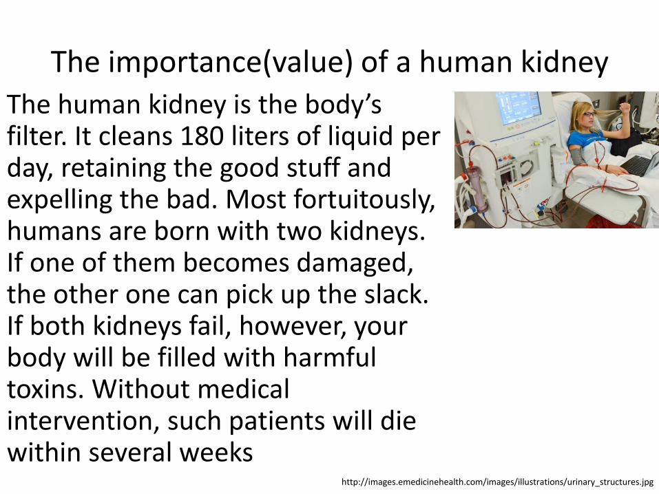
The importance(value) of a human kidney
The human kidney is the body’s filter. It cleans 180 liters of liquid per day, retaining the good stuff and expelling the bad. Most fortuitously, humans are born with two kidneys. If one of them becomes damaged, the other one can pick up the slack. If both kidneys fail, however, your body will be filled with harmful toxins. Without medical intervention, such patients will die within several weeks
http://images.emedicinehealth.com/images/illustrations/urinary_structures.jpg
Reminder: the primary urinary system functions
• maintain homeostasis
• regulate fluids and electrolytes
• eliminate waste products
• maintain blood pressure (BP)
• involved with red blood cell (RBC) production
• involved with bone metabolism
Reminder: purpose
• General evaluation of health
• Diagnosis of disease or disorders of the kidneys or urinary tract
• Diagnosis of other systemic diseases that affect kidney function
• Monitoring of patients with diabetes
• Screening for drug toxicity (eg. sulfonamide or aminoglycosides)
History-taking (patient’s interviewing )
gathering of information
patient’s narrative
biomedical perspective
psychosocial perspective
context
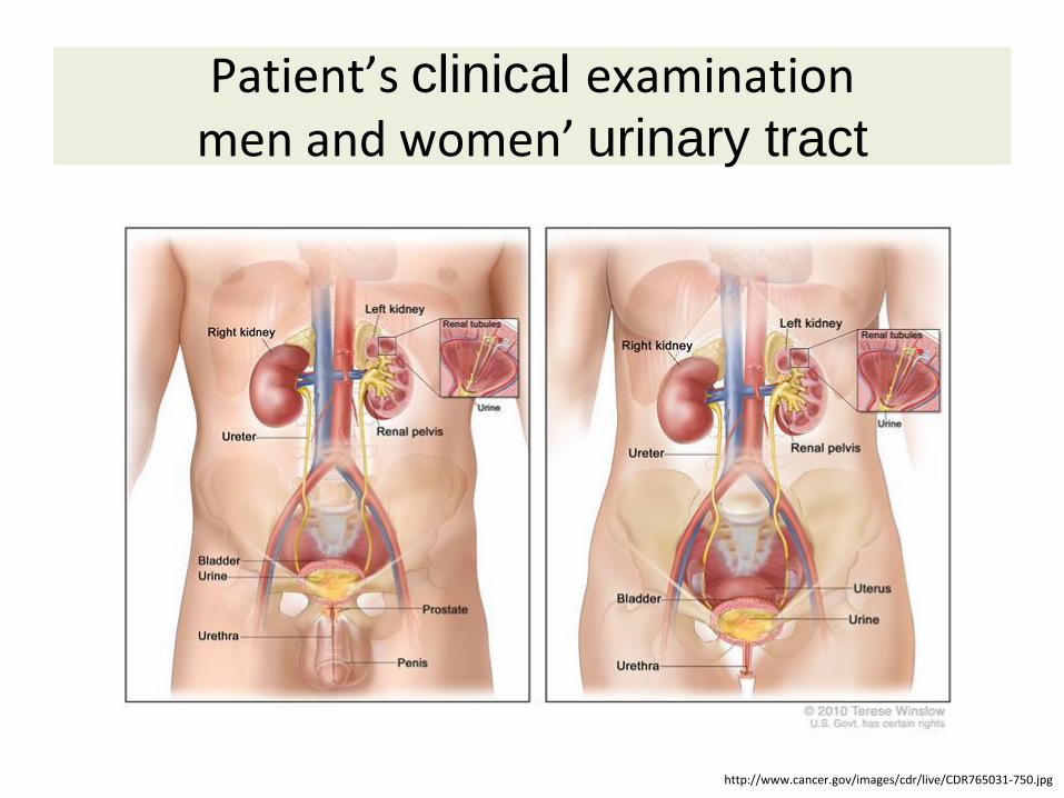
Patient’s clinical examination men and women’ urinary tract
http://www.cancer.gov/images/cdr/live/CDR765031-750.jpg
Anterior view Posterior view
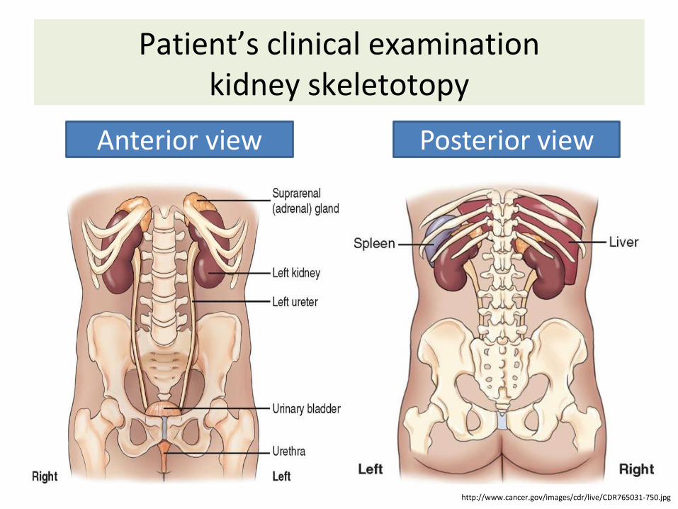
Patient’s clinical examination kidney skeletotopy
http://www.cancer.gov/images/cdr/live/CDR765031-750.jpg
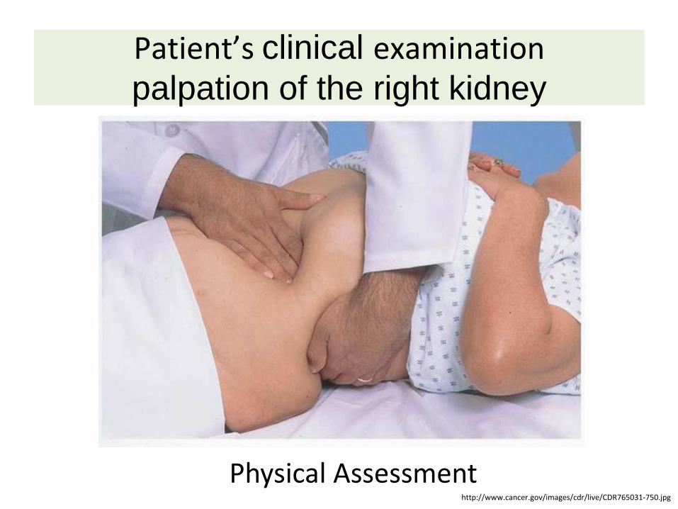
Patient’s clinical examination palpation of the right kidney
Physical Assessment http://www.cancer.gov/images/cdr/live/CDR765031-750.jpg

Patient’s clinical examination kidney pain localization
Kidney Pain
http://www.cancer.gov/images/cdr/live/CDR765031-750.jpg
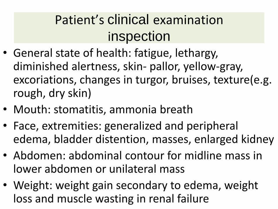
• General state of health: fatigue, lethargy, diminished alertness, skin- pallor, yellow-gray, excoriations, changes in turgor, bruises, texture(e.g. rough, dry skin)
• Mouth: stomatitis, ammonia breath
• Face, extremities: generalized and peripheral edema, bladder distention, masses, enlarged kidney
• Abdomen: abdominal contour for midline mass in lower abdomen or unilateral mass
• Weight: weight gain secondary to edema, weight loss and muscle wasting in renal failure
Patient’s clinical examination inspection

• Kidney: percussion (to detect areas of tenderness by costovertebral test) and palpation (contour, size, tenderness, and lump); presence of tenderness and pain indicates a kidney infection or polycystic kidney disease
• Bladder: percussion of the area over the bladder (5cm) above the symphysis pubis to detect difference in sound, percussion toward the base of the bladder
12
Patient’s clinical examination percussion, palpation, auscultation 1
• urethral meatus: inspection for swelling, discharge and inflammation
• Auscultation: the abdominal aorta & renal arteries are auscultated for a bruits, which indicate impaired blood flow to the kidneys
13
Patient’s clinical examination percussion, palpation, auscultation 2
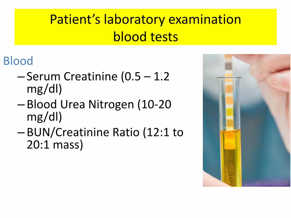
Blood – Serum Creatinine (0.5 – 1.2
mg/dl) –Blood Urea Nitrogen (10-20
mg/dl) –BUN/Creatinine Ratio (12:1 to
20:1 mass)
Patient’s laboratory examination blood tests
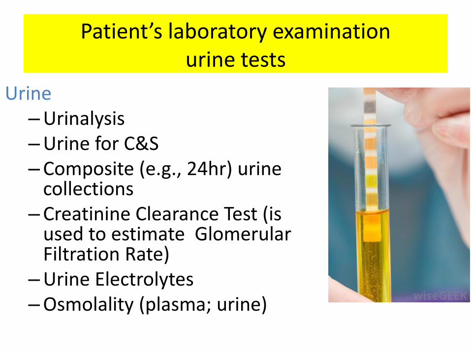
Urine –Urinalysis –Urine for C&S –Composite (e.g., 24hr) urine
collections –Creatinine Clearance Test (is
used to estimate Glomerular Filtration Rate)
–Urine Electrolytes –Osmolality (plasma; urine)
Patient’s laboratory examination urine tests
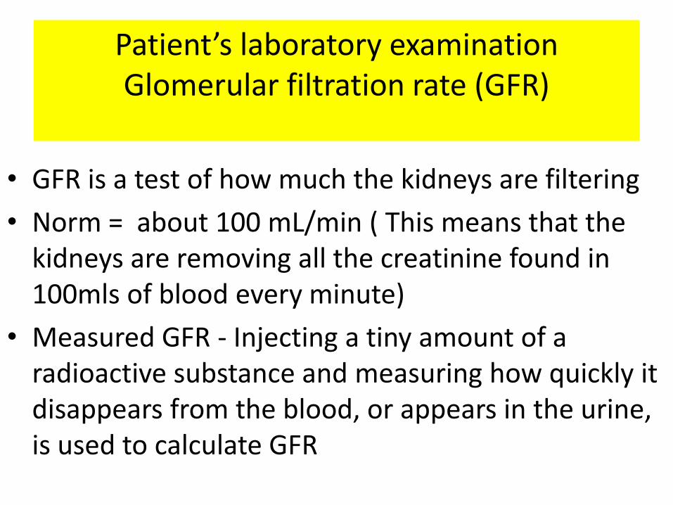
• GFR is a test of how much the kidneys are filtering
• Norm = about 100 mL/min ( This means that the kidneys are removing all the creatinine found in 100mls of blood every minute)
• Measured GFR - Injecting a tiny amount of a radioactive substance and measuring how quickly it disappears from the blood, or appears in the urine, is used to calculate GFR
Patient’s laboratory examination Glomerular filtration rate (GFR)

• eGFR - Using blood tests, age, sex, and sometimes other information to estimate the GFR from the MDRD equation (eGFR). This isn't as good as measuring it, but is much simpler as it requires just one blood test.
• Creatinine clearance (blood creatinine measurements by collecting urine for 24 hours and measuring how much creatinine is in the urine at the same time as finding out how much is in the blood – Ccr)
• (If any urine produced during the 24 hours is not collected the result will not be accurate)
Patient’s laboratory examination estimated GFR (eGFR) and – Ccr
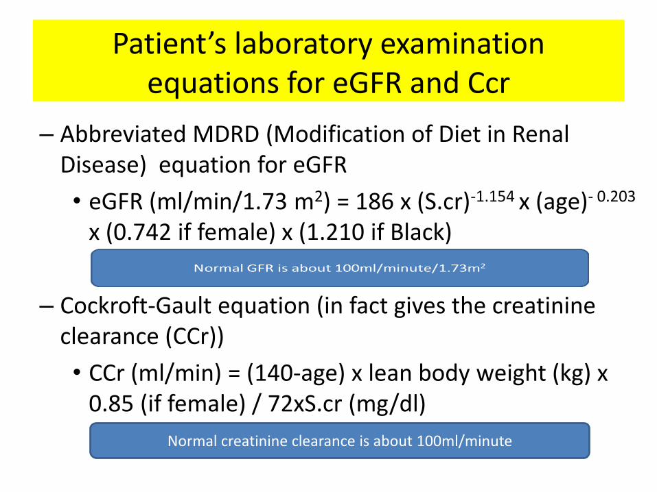
– Abbreviated MDRD (Modification of Diet in Renal Disease) equation for eGFR
• eGFR (ml/min/1.73 m2) = 186 x (S.cr)-1.154 x (age)- 0.203
x (0.742 if female) x (1.210 if Black)
– Cockroft-Gault equation (in fact gives the creatinine clearance (CCr))
• CCr (ml/min) = (140-age) x lean body weight (kg) x 0.85 (if female) / 72xS.cr (mg/dl)
Normal creatinine clearance is about 100ml/minute
Patient’s laboratory examination equations for eGFR and Ccr
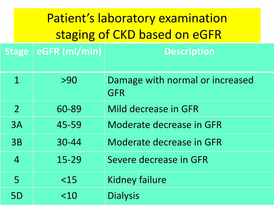
Stage eGFR (ml/min)
Description
1 >90 Damage with normal or increased GFR
2 60-89 Mild decrease in GFR
3A 45-59 Moderate decrease in GFR
3B 30-44 Moderate decrease in GFR
4 15-29 Severe decrease in GFR
5 <15 Kidney failure
5D <10 Dialysis
Patient’s laboratory examination staging of CKD based on eGFR
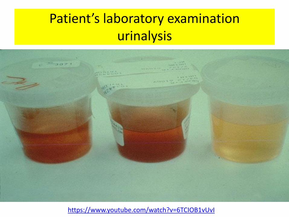
Patient’s laboratory examination urinalysis
https://www.youtube.com/watch?v=6TCIOB1vUvI
Patient’s laboratory examination
urinalysis
• Collection of urine specimens – first voided morning (most common)
– random (for emergency)
– clean-catch, midstream (for urine culture)
Attention: need to be examined within 1 hour
• Urine specimens examination – physical (appearance, volume, specific gravity (SG)
– chemical
– microscopic examination
– urine for culture and sensitivity
Urine specimens examination physical appearance 1
• Color
–normal, pale to dark yellow (urochrome)
– abnormal
• some drugs cause color changes
• red urine (hematuria, hemoglobinuria, myoglobinuria, pseudohematuria)
• yellow-brown or green-brown urine (bilirubin: obstructive jaundice)
Urine specimens examination physical appearance 2
• Clarity
–normal, clear
– abnormal, cloudy
• crystals or nonpathologic salts
• phosphate, carbonate in alkaline urine
• uric acid in acid urine
• various cellular elements ( leukocytes, RBCs, epithelial cells)
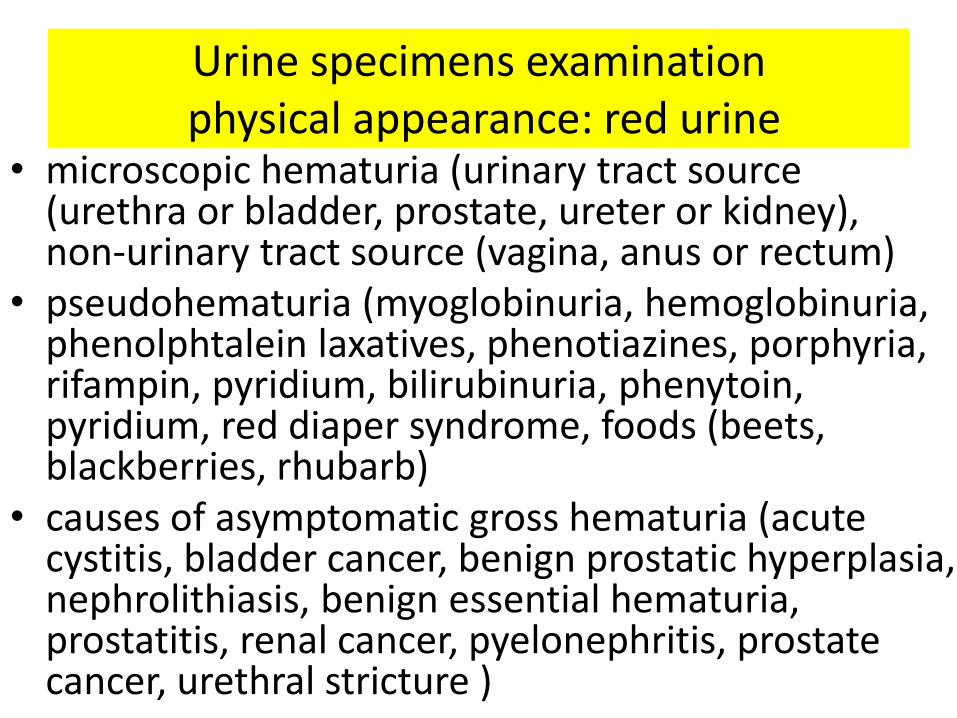
Urine specimens examination physical appearance: red urine
• microscopic hematuria (urinary tract source (urethra or bladder, prostate, ureter or kidney), non-urinary tract source (vagina, anus or rectum)
• pseudohematuria (myoglobinuria, hemoglobinuria, phenolphtalein laxatives, phenotiazines, porphyria, rifampin, pyridium, bilirubinuria, phenytoin, pyridium, red diaper syndrome, foods (beets, blackberries, rhubarb)
• causes of asymptomatic gross hematuria (acute cystitis, bladder cancer, benign prostatic hyperplasia, nephrolithiasis, benign essential hematuria, prostatitis, renal cancer, pyelonephritis, prostate cancer, urethral stricture )

Urine specimens examination physical: urine volume 1
• normal adult average – (400 – 2000) ml/24h
• increase average (polyuria) – > 2000 ml/24h
–physiological (water intake, some drugs, intravenous solutions)
–pathologic (CKD, diabetes mellitus, diabetes insipidus)
• decrease average ( oliguria - < 400 ml/24h, anuria - < 100ml /24h)
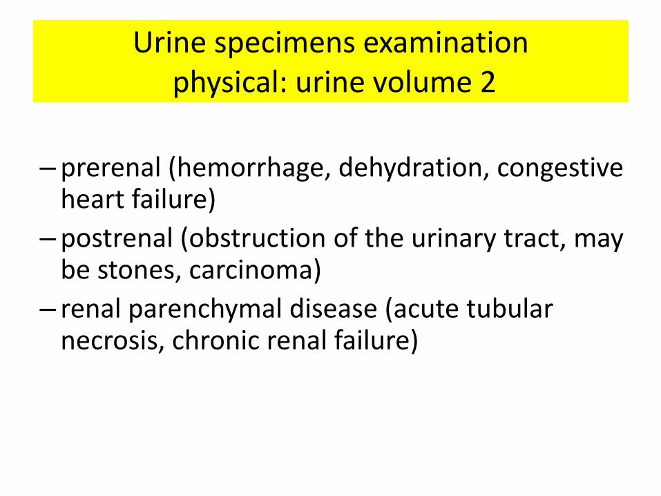
Urine specimens examination physical: urine volume 2
–prerenal (hemorrhage, dehydration, congestive heart failure)
–postrenal (obstruction of the urinary tract, may be stones, carcinoma)
– renal parenchymal disease (acute tubular necrosis, chronic renal failure)
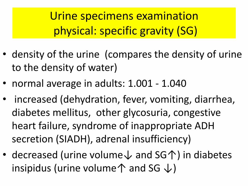
Urine specimens examination physical: specific gravity (SG)
• density of the urine (compares the density of urine to the density of water)
• normal average in adults: 1.001 - 1.040
• increased (dehydration, fever, vomiting, diarrhea, diabetes mellitus, other glycosuria, congestive heart failure, syndrome of inappropriate ADH secretion (SIADH), adrenal insufficiency)
• decreased (urine volume↓ and SG↑) in diabetes insipidus (urine volume↑ and SG ↓)
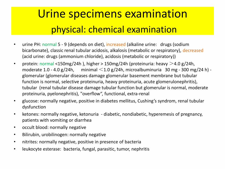
Urine specimens examination physical: chemical examination
• urine PH: normal 5 - 9 (depends on diet), increased (alkaline urine: drugs (sodium bicarbonate), classic renal tubular acidosis, alkalosis (metabolic or respiratory), decreased (acid urine: drugs (ammonium chloride), acidosis (metabolic or respiratory))
• protein: normal <150mg/24h ), higher > 150mg/24h (proteinuria: heavy >4.0 g/24h, moderate 1.0 - 4.0 g/24h, minimal <1.0 g/24h, microalbuminuria 30 mg - 300 mg/24 h) -glomerular (glomerular diseases damage glomerular basement membrane but tubular function is normal, selective proteinuria, heavy proteinuria, acute glomerulonephritis), tubular (renal tubular disease damage tubular function but glomerular is normal, moderate proteinuria, pyelonephritis), "overflow“, functional, extra-renal
• glucose: normally negative, positive in diabetes mellitus, Cushing’s syndrom, renal tubular dysfunction
• ketones: normally negative, ketonuria - diabetic, nondiabetic, hyperemesis of pregnancy, patients with vomiting or diarrhea
• occult blood: normally negative
• Bilirubin, urobilinogen: normally negative
• nitrites: normally negative, positive in presence of bacteria
• leukocyte esterase: bacteria, fungal, parasitic, tumor, nephritis
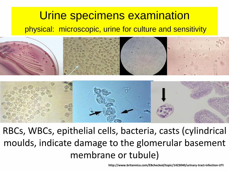
Urine specimens examination physical: microscopic, urine for culture and sensitivity
http://www.britannica.com/EBchecked/topic/1423040/urinary-tract-infection-UTI
RBCs, WBCs, epithelial cells, bacteria, casts (cylindrical moulds, indicate damage to the glomerular basement
membrane or tubule)
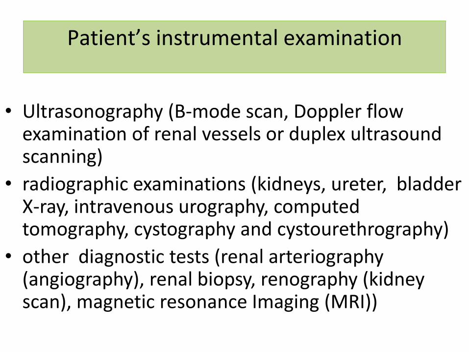
• Ultrasonography (B-mode scan, Doppler flow examination of renal vessels or duplex ultrasound scanning)
• radiographic examinations (kidneys, ureter, bladder X-ray, intravenous urography, computed tomography, cystography and cystourethrography)
• other diagnostic tests (renal arteriography (angiography), renal biopsy, renography (kidney scan), magnetic resonance Imaging (MRI))
Patient’s instrumental examination
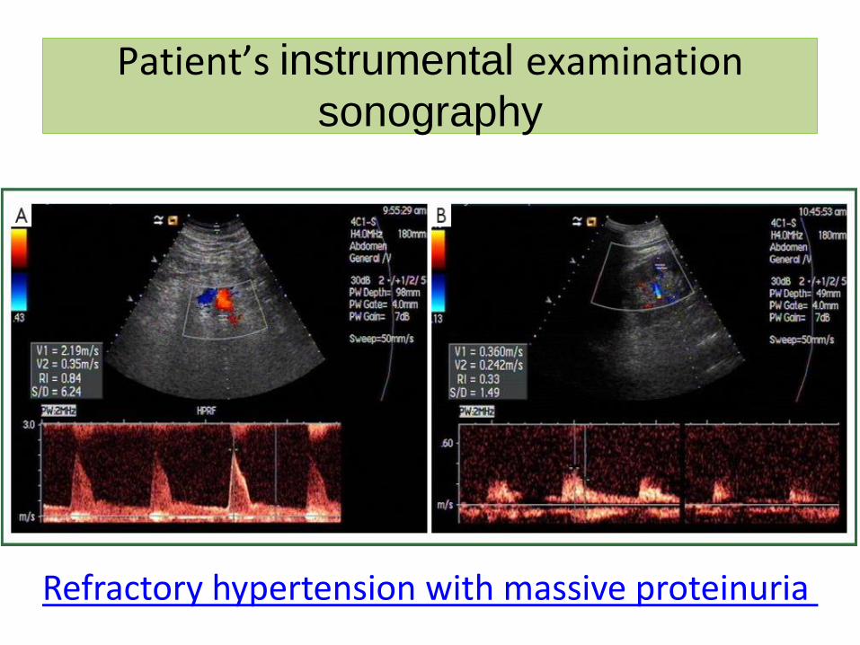
Patient’s instrumental examination sonography
Refractory hypertension with massive proteinuria
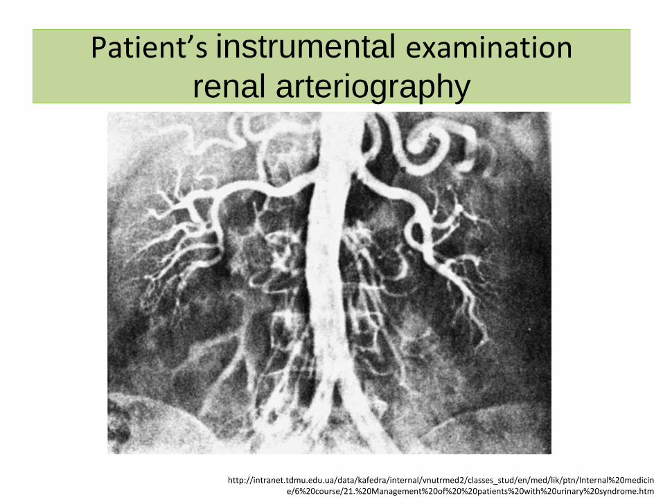
Patient’s instrumental examination renal arteriography
http://intranet.tdmu.edu.ua/data/kafedra/internal/vnutrmed2/classes_stud/en/med/lik/ptn/Internal%20medicine/6%20course/21.%20Management%20of%20%20patients%20with%20urinary%20syndrome.htm
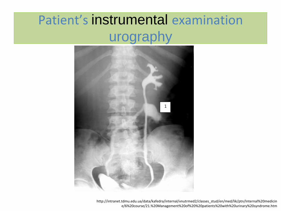
Patient’s instrumental examination urography
http://intranet.tdmu.edu.ua/data/kafedra/internal/vnutrmed2/classes_stud/en/med/lik/ptn/Internal%20medicine/6%20course/21.%20Management%20of%20%20patients%20with%20urinary%20syndrome.htm

Patient’s instrumental examination radiolucent stones
Bladder calculi (stones )
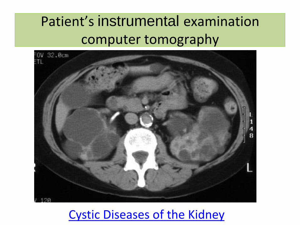
Patient’s instrumental examination computer tomography
Cystic Diseases of the Kidney
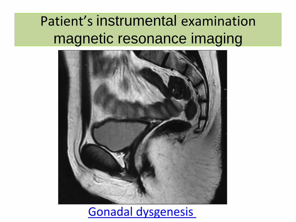
Patient’s instrumental examination magnetic resonance imaging
Gonadal dysgenesis
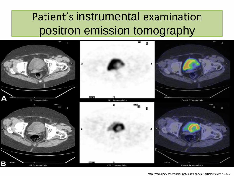
Patient’s instrumental examination positron emission tomography
http://radiology.casereports.net/index.php/rcr/article/view/479/805
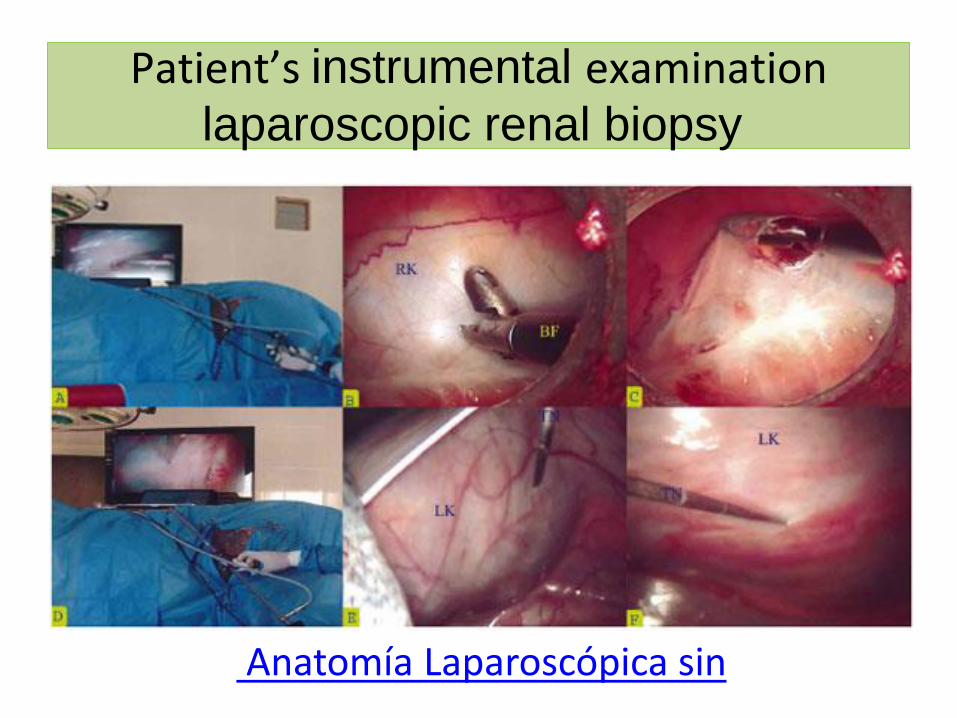
Patient’s instrumental examination laparoscopic renal biopsy
Anatomía Laparoscópica sin
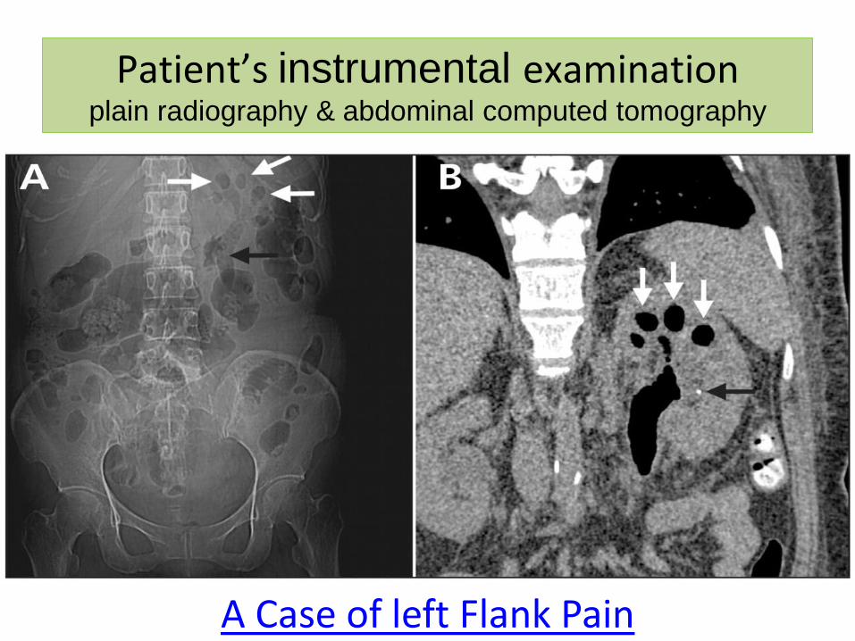
Patient’s instrumental examination plain radiography & abdominal computed tomography
A Case of left Flank Pain
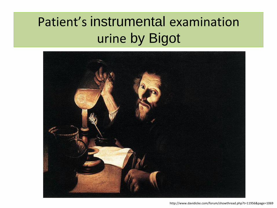
Patient’s instrumental examination urine by Bigot
http://www.davidicke.com/forum/showthread.php?t=11956&page=1069
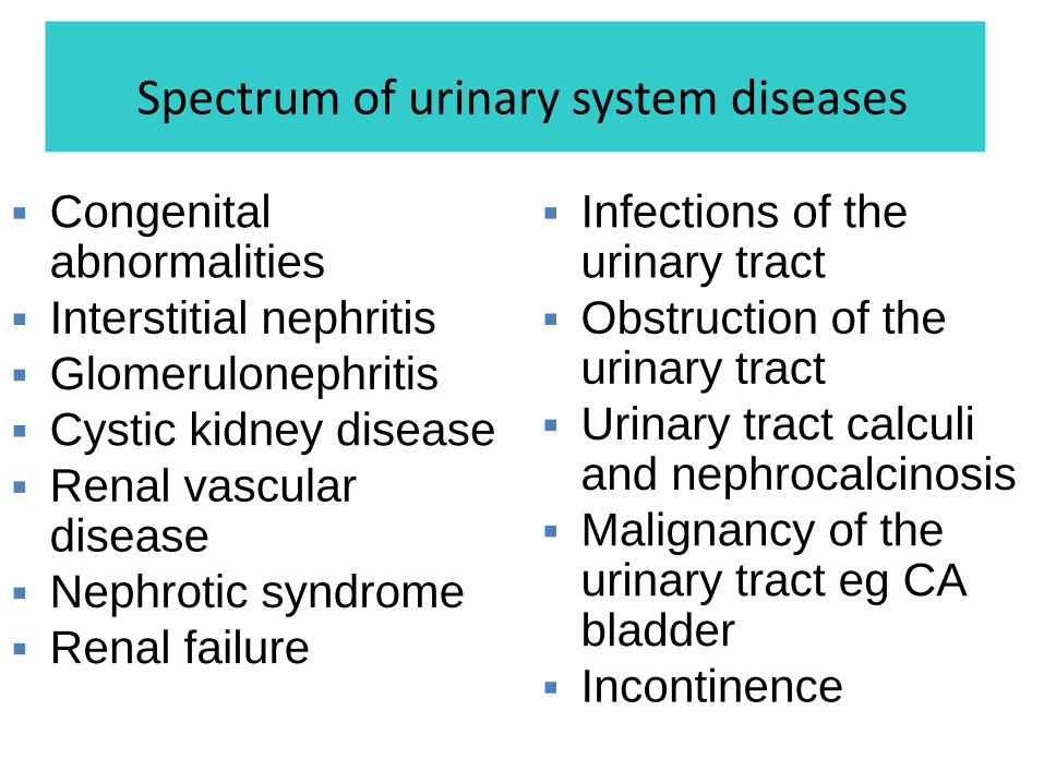
Spectrum of urinary system diseases
Congenital abnormalities
Interstitial nephritis
Glomerulonephritis
Cystic kidney disease
Renal vascular disease
Nephrotic syndrome
Renal failure
Infections of the urinary tract
Obstruction of the urinary tract
Urinary tract calculi and nephrocalcinosis
Malignancy of the urinary tract eg CA bladder
Incontinence
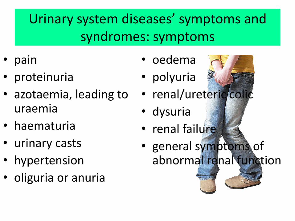
Urinary system diseases’ symptoms and syndromes: symptoms
• pain
• proteinuria
• azotaemia, leading to uraemia
• haematuria
• urinary casts
• hypertension
• oliguria or anuria
• oedema
• polyuria
• renal/ureteric colic
• dysuria
• renal failure
• general symptoms of abnormal renal function
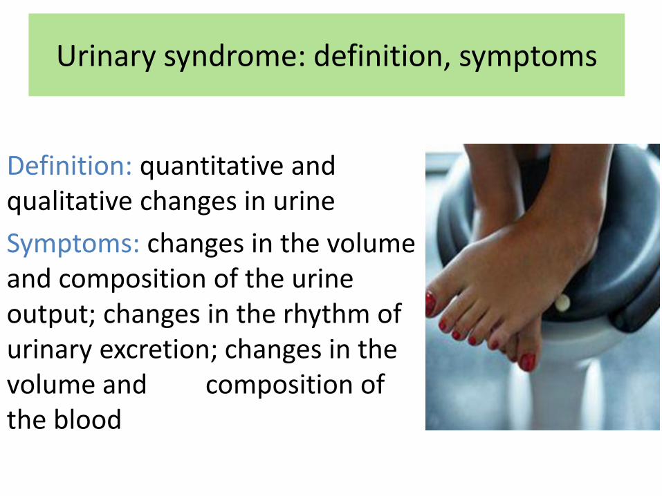
Urinary syndrome: definition, symptoms
Definition: quantitative and qualitative changes in urine
Symptoms: changes in the volume and composition of the urine output; changes in the rhythm of urinary excretion; changes in the volume and composition of the blood
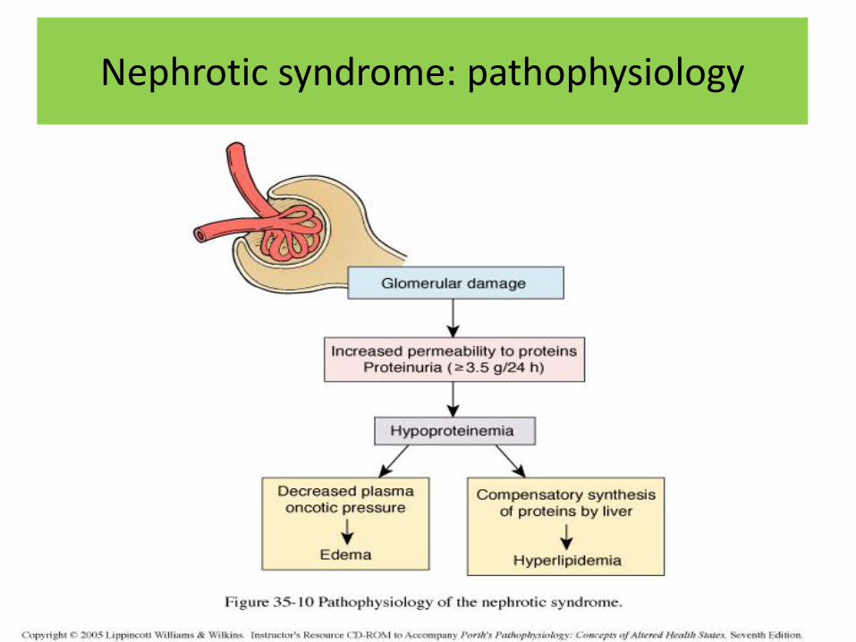
Nephrotic syndrome: definition
Clinical and laboratory syndrome characterized by massive proteinuria, which lead to hypoproteinemia ( hypoalbuminemia), hyperlipidemia and pitting edema in results from increased permeability of glomerular basement membrane (GBM) to plasma protein
Nephrotic syndrome: criteria
• hematuria (RBC in urine, gross hematuria)
• hypertension (≥140 /90 mmHg)
• azotemia(renal insufficiency - Increased level of serum BUN , Cr)
• hypocomplementemia (decreased level of serum c3)
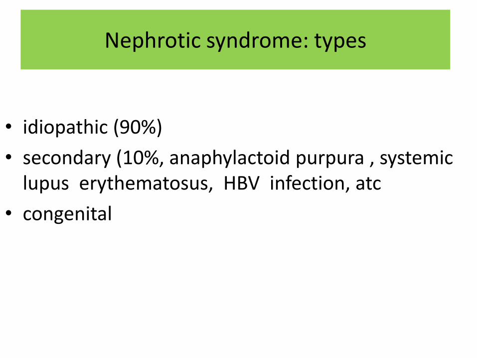
Nephrotic syndrome: types
• idiopathic (90%)
• secondary (10%, anaphylactoid purpura , systemic lupus erythematosus, HBV infection, atc
• congenital
How many pathological types causes nephrotic syndrome?
Nephrotic syndrome: pathophysiology
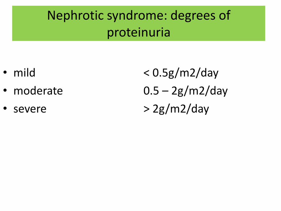
• mild < 0.5g/m2/day
• moderate 0.5 – 2g/m2/day
• severe > 2g/m2/day
Nephrotic syndrome: degrees of proteinuria
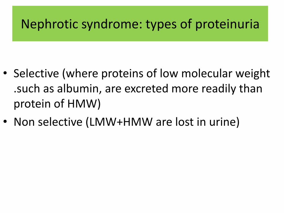
• Selective (where proteins of low molecular weight .such as albumin, are excreted more readily than protein of HMW)
• Non selective (LMW+HMW are lost in urine)
Nephrotic syndrome: types of proteinuria
Edema (varying degrees): local (edema of face (facial edema), edema around eyes (periorbital swelling) , in lower extremities), generalized (anasarca), edema of penis and scrotum
Other clinical symptoms: fatigue, lethargy, loss of appetite, nausea and vomiting ,abdominal pain , diarrhea, body weight increase, urine output decrease, pleural effusion (respiratory distress)
Nephrotic syndrome: symptoms
Blood tests (serum protein >5.5gm/dL , albumin <2.5gm/dL, cholesterol >220mg/dl)
Urine tests (proteinuria, oliguria (during stage of edema formation), microscopic hematuria 20%, large number of hyaline casts)
Differential diagnosis of generalized edema
Nephrotic syndrome: tests, differential diagnosis

1. Massive prOteinuria
2. HypOprOteinemia (peeing out albumin)
3. Oedema (Oncotic pressure in the blood goes down)
4. HyperchOlesterolemia (hyperlipidemia/hyperlipiduria)
5. HypercOagulable state (thrOmbotic and thrOmboembolic complications)
Main in nephrOtic syndrome (all words contain letter O)
Nephritic syndrome: definition
Clinical and laboratory syndrome associated with disorders affecting the kidneys, more specifically glomerular structures, and characterized by having a thin glomerular basement membrane and small pores in the podocytes of the glomerulus, large enough to permit proteins (proteinuria) and red blood cells (hematuria) to pass into the urine
Nephritic syndrome: criteria
• hematuria , with red blood cell (RBC) casts present in the urine
• proteinuria (<3.5 g/day)
• hypertension
• uremia, due to retention of waste products
• variable renal insufficiency, with azotemia, oliguria (low urine output <400 mL/day)

Nephritic syndrome: types
• post-streptococcal glomerulonephritis
• crescentic glomerulonephritis (rapidly progressive glomerulonephritis)
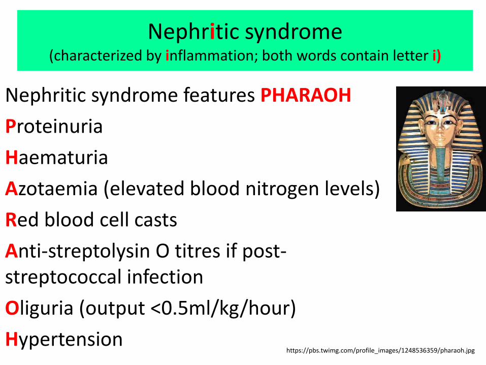
Nephritic syndrome (characterized by inflammation; both words contain letter i)
Nephritic syndrome features PHARAOH
Proteinuria
Haematuria
Azotaemia (elevated blood nitrogen levels)
Red blood cell casts
Anti-streptolysin O titres if post-streptococcal infection
Oliguria (output <0.5ml/kg/hour)
Hypertension
https://pbs.twimg.com/profile_images/1248536359/pharaoh.jpg
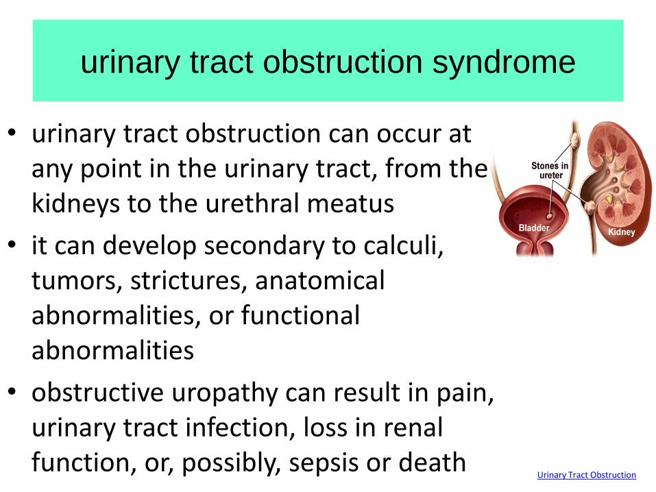
urinary tract obstruction syndrome
• urinary tract obstruction can occur at any point in the urinary tract, from the kidneys to the urethral meatus
• it can develop secondary to calculi, tumors, strictures, anatomical abnormalities, or functional abnormalities
• obstructive uropathy can result in pain, urinary tract infection, loss in renal function, or, possibly, sepsis or death
Urinary Tract Obstruction
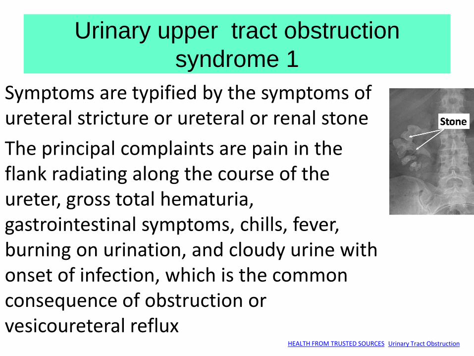
Urinary upper tract obstruction
syndrome 1
Symptoms are typified by the symptoms of ureteral stricture or ureteral or renal stone
The principal complaints are pain in the flank radiating along the course of the ureter, gross total hematuria, gastrointestinal symptoms, chills, fever, burning on urination, and cloudy urine with onset of infection, which is the common consequence of obstruction or vesicoureteral reflux
Urinary Tract Obstruction HEALTH FROM TRUSTED SOURCES
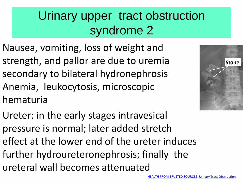
Urinary upper tract obstruction
syndrome 2
Nausea, vomiting, loss of weight and strength, and pallor are due to uremia secondary to bilateral hydronephrosis Anemia, leukocytosis, microscopic hematuria
Ureter: in the early stages intravesical pressure is normal; later added stretch effect at the lower end of the ureter induces further hydroureteronephrosis; finally the ureteral wall becomes attenuated
Urinary Tract Obstruction HEALTH FROM TRUSTED SOURCES
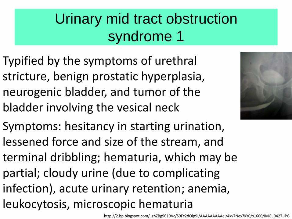
Urinary mid tract obstruction
syndrome 1
Typified by the symptoms of urethral stricture, benign prostatic hyperplasia, neurogenic bladder, and tumor of the bladder involving the vesical neck
Symptoms: hesitancy in starting urination, lessened force and size of the stream, and terminal dribbling; hematuria, which may be partial; cloudy urine (due to complicating infection), acute urinary retention; anemia, leukocytosis, microscopic hematuria
http://2.bp.blogspot.com/_zhZBg9019Vc/S9Fc2dOlp9I/AAAAAAAAAeI/4kv7Nex7kY0/s1600/IMG_0427.JPG

Urinary mid tract obstruction
syndrome 2
Stages
• compensation - the bladder musculature becomes hypertrophied the thickness may double or triple, hypertrophied muscle may be seen endoscopically superimposed with secondary infection
• decompensation - large obstructing gland can be palpated rectally and observed cystoscopically, may appears as a mild obstruction cystoscopically
http://2.bp.blogspot.com/_zhZBg9019Vc/S9Fc2dOlp9I/AAAAAAAAAeI/4kv7Nex7kY0/s1600/IMG_0427.JPG
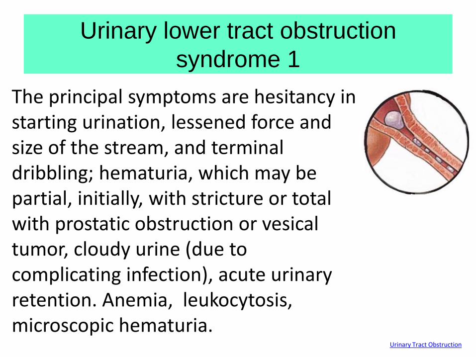
Urinary lower tract obstruction
syndrome 1
The principal symptoms are hesitancy in starting urination, lessened force and size of the stream, and terminal dribbling; hematuria, which may be partial, initially, with stricture or total with prostatic obstruction or vesical tumor, cloudy urine (due to complicating infection), acute urinary retention. Anemia, leukocytosis, microscopic hematuria.
Urinary Tract Obstruction
Urinary lower tract obstruction
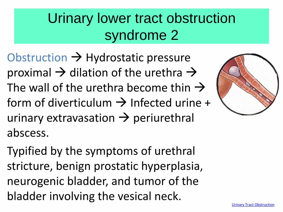
syndrome 2
Obstruction Hydrostatic pressure proximal dilation of the urethra The wall of the urethra become thin form of diverticulum Infected urine + urinary extravasation periurethral abscess.
Typified by the symptoms of urethral stricture, benign prostatic hyperplasia, neurogenic bladder, and tumor of the bladder involving the vesical neck.
Urinary Tract Obstruction
Hypertensive syndrome 1
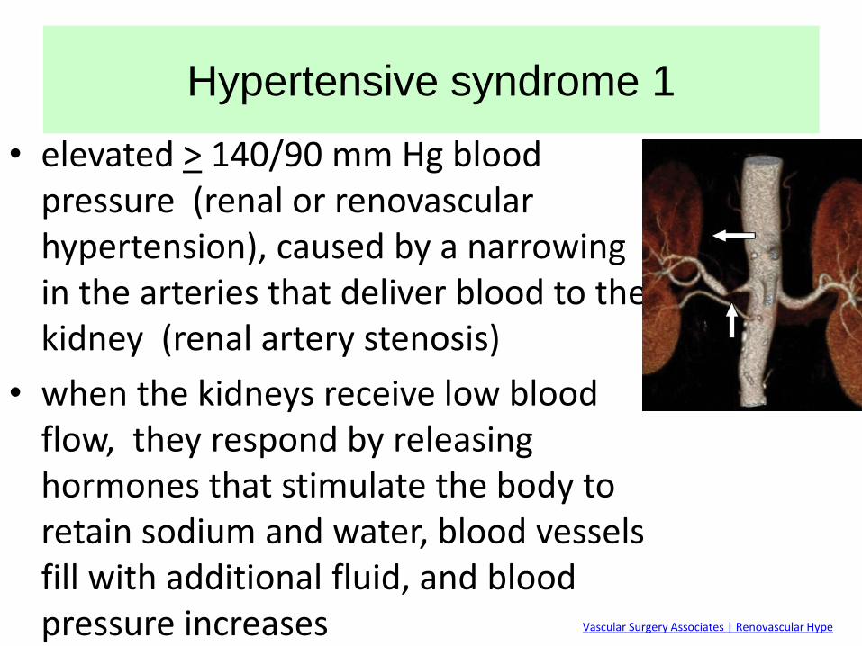
• elevated > 140/90 mm Hg blood pressure (renal or renovascular hypertension), caused by a narrowing in the arteries that deliver blood to the kidney (renal artery stenosis)
• when the kidneys receive low blood flow, they respond by releasing hormones that stimulate the body to retain sodium and water, blood vessels fill with additional fluid, and blood pressure increases Vascular Surgery Associates | Renovascular Hype

Hypertensive syndrome 2
• the narrowing in one or both renal arteries is most often caused by atherosclerosis, or hardening of the arteries
• symptoms: headache, confusion, blurred or double vision, bloody (pink-colored) urine, nosebleed, bruits over affected renal artery
• hypertension can cause chronic kidney disease
Vascular Surgery Associates | Renovascular Hype