silibinin is a direct inhibitor of stat3
TRANSCRIPT
Contents lists available at ScienceDirect
Food and Chemical Toxicology
journal homepage: www.elsevier.com/locate/foodchemtox
Silibinin is a direct inhibitor of STAT3Sara Verduraa,b,1, Elisabet Cuyàsa,b,1, Laura Llorach-Parésc, Almudena Pérez-Sánchezd,Vicente Micold,e, Alfons Nonell-Canalsc, Jorge Jovenf, Manuel Valienteg,Melchor Sánchez-Martínezc, Joaquim Bosch-Barrerah,i,∗∗, Javier A. Menendeza,b,∗a Program Against Cancer Therapeutic Resistance (ProCURE), Metabolism and Cancer Group, Catalan Institute of Oncology, Girona, SpainbMolecular Oncology Group, Girona Biomedical Research Institute (IDIBGI), Girona, SpaincMind the Byte, Barcelona, Spaind Instituto de Biología Molecular y Celular (IBMC), Miguel Hernández University (UMH), Elche, Alicante, Spaine CIBER, Fisiopatología de la Obesidad y la Nutrición, CIBERobn, Instituto de Salud Carlos III (CB12/03/30038), SpainfUnitat de Recerca Biomèdica, Hospital Universitari de Sant Joan, IISPV, Rovira i Virgili University, Reus, Spaing Brain Metastasis Group, Molecular Oncology Program, Spanish National Cancer Research Centre (CNIO), Madrid, SpainhDepartment of Medical Oncology, Catalan Institute of Oncology, Girona, Spaini Department of Medical Sciences, Medical School, University of Girona, Girona, Spain
A R T I C L E I N F O
Keywords:SilibininSTAT3CancerMetastasis
A B S T R A C T
We herein combined experimental and computational efforts to delineate the mechanism of action throughwhich the flavonolignan silibinin targets STAT3. Silibinin reduced IL-6 inducible, constitutive, and acquiredfeedback activation of STAT3 at tyrosine 705 (Y705). Silibinin attenuated the inducible phospho-activation ofY705 in GFP-STAT3 genetic fusions without drastically altering the kinase activity of the STAT3 upstream ki-nases JAK1 and JAK2. A comparative computational study based on docking and molecular dynamics simulationover 14 different STAT3 inhibitors (STAT3i) predicted that silibinin could directly bind with high affinity to boththe Src homology-2 (SH2) domain and the DNA-binding domain (DBD) of STAT3. Silibinin partially overlappedwith the cavity occupied by other STAT3i in the SH2 domain to indirectly prevent Y705 phosphorylation, yetshowing a unique binding mode. Moreover, silibinin was the only STAT3i predicted to establish direct inter-actions with DNA in its targeting to the STAT3 DBD. The prevention of STAT3 nuclear translocation, theblockade of the binding of activated STAT3 to its consensus DNA sequence, and the suppression of STAT3-directed transcriptional activity confirmed silibinin as a direct STAT3i. The unique characteristics of silibinin asa bimodal SH2- and DBD-targeting STAT3i make silibinin a promising lead for designing new, more effectiveSTAT3i.
1. Introduction
The aberrant activation of signal transducer and activator of tran-scription 3 (STAT3) contributes to cancer initiation and progression in amulti-faceted manner via promotion of cell proliferation/survival, in-vasion/migration, angiogenesis, and immune-evasion (Chang et al.,2013; Sansone and Bromberg, 2012; Yu et al., 2009, 2014). Feedbackactivation of STAT3 additionally mediates tumor resistance to a broadspectrum of cancer therapies, including radiotherapy, conventionalchemotherapy, and modern targeted therapies (Lee et al., 2014; Poliand Camporeale, 2015; Tan et al., 2014; Zhao et al., 2016). STAT3activation associates also with the generation and maintenance of
cancer stem cells (CSC), a particularly aggressive type of malignant celldefined in terms of functional traits including tumor/metastasis-in-itiating capacity and therapy resistance (Kroon et al., 2013; Misra et al.,2018; Schroeder et al., 2014; Wang et al., 2018). Not surprisingly, theactivation status of STAT3 is a strong predictor of poor prognosis and isan independent risk factor for tumor recurrence and post-therapy pro-gression (Chen et al., 2013; Liu et al., 2012; Tong et al., 2017; Wu et al.,2016). These observations have motivated great efforts over the lastdecade to clinically exploit the beneficial effects of inhibiting STAT3 inhuman malignancies. Accordingly, a large number of STAT3 inhibitors(STAT3i) have been developed as potential cancer therapeutics (Fagardet al., 2013; Furtek et al., 2016a; b; Jin et al., 2016; Miklossy et al.,
https://doi.org/10.1016/j.fct.2018.04.028Received 8 March 2018; Received in revised form 3 April 2018; Accepted 13 April 2018
∗ Corresponding author. Catalan Institute of Oncology (ICO), Girona Biomedical Research Institute (IDIBGI), Edifici M2, Parc Hospitalari Martí i Julià, E-17190 Salt, Girona, Spain.∗∗ Corresponding author. Catalan Institute of Oncology (ICO), Hospital Dr. Josep Trueta de Girona, Avda. de França s/n, 17007, Girona, Spain
1 These authors contributed equally.E-mail addresses: [email protected] (J. Bosch-Barrera), [email protected], [email protected] (J.A. Menendez).
)RRG�DQG�&KHPLFDO�7R[LFRORJ\���������������²���
$YDLODEOH�RQOLQH����$SULO�����������������������3XEOLVKHG�E\�(OVHYLHU�/WG�
7

2013; Siveen et al., 2014; Yue and Turkson, 2009).STAT3i can be classified as indirect or direct according to their
mode of action. Indirect STAT3i interfere with cytokine- and growthfactor receptor-activated upstream kinases such as the Janus kinases(JAK) that phosphorylate STAT3. Direct STAT3i bind to STAT3 proteindomains critically involved in STAT3 activation/dimerization (Srchomology 2 domain, SH2) or DNA binding (DNA-binding domain,DBD). The usage of broad-spectrum indirect STAT3i (e.g., JAK in-hibitors), however, often results in undesirable off-target effects.Research into direct STAT3i has focused mainly on targeting the SH2domain, the protein-protein interface responsible for the formation ofSTAT3 dimers by reciprocal phosphotyrosine-SH2 interactions fol-lowing activation of the tyrosine 705 (Y705) residue. Unfortunately,only a limited number of direct, SH2-targeted STAT3i have reached pre-clinical and clinical trials. This is due mostly to the intrinsic difficulty indeveloping small molecules capable of efficaciously disrupting protein-protein interactions over a large surface such as those involving SH2-mediated STAT3 dimerization, while maintaining drug-like propertiesin vivo. Moreover, the sole blockade of active STAT3 dimers might notbe sufficient to fully abrogate STAT3 signaling (Nkansah et al., 2013;Timofeeva et al., 2012). Although targeting of the STAT3 DBD anddisruption of its DNA binding activity has the potential to circumventthe transcriptional activation of STAT3 irrespective of its activation/dimerization status (Huang et al., 2016), very few small molecules havebeen reported to date as STAT3 DBD inhibitors. This is mainly due tothe previously thought undruggable nature of the DBD and potentiallylimited selectivity (Huang et al., 2016), and also the lack of adequateassay systems (Furtek et al., 2016a; b). Furthermore, there are onlythree crystal structures available [PDB ID: 4E68 (Nkansah et al., 2013),3CWG (Ren et al., 2008), and 1BG1 (Becker et al., 1998)] of the mousebut not human core STAT3 fragment containing the SH2 and DBD do-mains in the Protein Data Bank (http://www.rcsb.org), and co-crystalstructures of STAT3i bound to STAT3 are lacking.
There is ever-growing evidence that the flavonolignan silibinin, themajor bioactive constituent of the seed extract of the plant Milk thistle(Silybum marianum) (Agarwal et al., 2006; Cufí et al., 2013a,b; Gažáket al., 2007), possesses drug-like properties with proven clinical activityvia inhibition of STAT3 signaling (Chittezhath et al., 2008; Cuyàs et al.,2016; Shukla et al., 2015; Singh et al., 2009). Although initial clinicalexperiences with silibinin supplementation in cancer patients have beendisappointing (Flaig et al., 2010; Hoh et al., 2006; Siegel et al., 2014),new silibinin formulations with improved water solubility, absorption,and bioavailability appear to translate into proven therapeutic benefits(Bosch-Barrera et al., 2014, 2016). Unfortunately, whereas the possi-bility of providing oncologists with new silibinin formulations or sili-binin derivatives capable of functioning as STAT3i in a clinical settingmay broaden their therapeutic armamentarium (Bosch-Barrera andMenendez, 2015; Bosch-Barrera et al., 2017), the precise mechanismthrough which silibinin targets STAT3 remains unknown. Here, weaimed to combine experimental and in silico efforts to clearly delineatethe molecular bases of the silibinin-STAT3 interaction.
2. Materials and methods
2.1. Reagents
Recombinant IL-6 (Cat. No. 7270-IL-25) was obtained from R&D.ONE-Glo™ Luciferase Assay System (Cat. No E6110) and the pGL4.47(luc2P/STAT-3 inducible element [SIE]/Hygro) vector (Cat. No E4041)were purchased from Promega (Madison, WI, USA). TransAM®
Transcription Factor ELISA (Cat. No 45196) was obtained from ActiveMotif (Carlsbad, CA, USA). Antibodies against total STAT3 (124H6, Cat.No 9139) and phospho-STAT3 Tyr705 (D3A7, Cat. No 9145S) werepurchased from Cell Signaling Technology (Beverly, MA, USA). H2228and H2228/CR cells were generously provided by Daniel B. Costa(Division of Hematology/Oncology, Beth Israel Deaconess Medical
Center, Boston, USA).
2.2. LanthaScreen STAT3 GripTite inhibitor screen
To characterize the STAT3 inhibitory potency of silibinin, IC50 de-terminations for phospho-STAT3Y705 were outsourced to Invitrogen(Life Technologies) using the LanthaScreen STAT3 GripTite inhibitorscreen service. Briefly, cells were thawed and resuspended in AssayMedium (OPTI-MEM, 1% csFBS, 0.1 mmoL/L NEAA, 1mmoL/L sodiumpyruvate, 100 U/mL/100 μg/mL Pen/Strep) to a concentration of625,000 cells/mL. Thirty-two microliters of the cell suspension wereadded to each well of a white tissue culture-treated assay plate(20,000 cells/well) and incubated for 16–26 h at 37 °C/5% CO2 in ahumidified incubator. Then, 4 μL of the control inhibitor JAK Inhibitor Ior silibinin was added to the appropriate assay wells followed by theaddition of 4 μL of Assay Medium. The assay plate was incubated for30–60min at 37 °C/5% CO2 in a humidified incubator. Then 4 μL of a10× control activator, IFN-α or IL-6 at the pre-determined EC80 con-centration, was added to wells containing the control inhibitor or sili-binin. The assay plate was then incubated as before for 30min. Next,the assay medium was aspirated from the wells and 20 μL ofLanthaScreen Cellular Assay Lysis Buffer containing 5 nmoL/L ofLanthaScreen Tb-anti-STAT3Y705 antibody was added. The assay platewas incubated for 60min at room temperature and then read with afluorescent plate reader.
2.3. Z′-LYTE JAK kinase activity assay
To characterize the JAK1/JAK2 kinase inhibitory potency of sili-binin, IC50 determinations for JAK1/JAK2 kinase activity were out-sourced to Invitrogen (Life Technologies) using the FRET-based Z-LYTE™ SelectScreen Kinase Profiling Service. The 2× JAK1/Tyr 06 orJAK2/Tyr 06 mixture was prepared in 50mmoL/L HEPES pH 6.5,0.01% BRIJ-35, 10mmoL/L MgCl2, 1mmoL/L EGTA, and 0.02% NaN3.The final 10 μL Kinase Reaction consisted of 21.2–91.5 ng JAK1 (orJAK2) and 2 μmoL/L Tyr 06 in 50mmoL/L HEPES pH 7.0, 0.01% BRIJ-35, 10mmoL/L MgCl2, 1 mmoL/L EGTA, and 0.01% NaN3. After in-cubation for 1 h, 5 μL of a 1:128 dilution of Development Reagent A wasadded.
SelectScreen Kinase Profiling Service uses XLfit software from IDBusiness Solutions (UK). The dose response curve is fitted to modelnumber 205 (sigmoidal dose-response model). If the bottom of thecurve does not fit between −20% and 20% inhibition, it is set to 0%inhibition. If the top of the curve does not fit between 70% and 130%inhibition, it is set to 100% inhibition.
2.4. Computational modeling of human STAT3
The homology-modeling software tools SWISS-MODEL and I-TASSER were employed to generate a computational homology modelof human STAT3. The human amino acidic sequence [UniprotIDP40763] was extracted from Uniprotkb and, in both cases, the three-dimensional crystal structure of the mouse STAT3 homodimer bound toDNA [PDB ID 1BG1 (Becker et al., 1998)] was employed as template.Whereas SWISS-MODEL generated a homology model that failed tocover a few residues on the SH2 domain, I-TASSER employed 1BG1 andother templates including 4E68 (unphosphorylated mouse STAT3 coreprotein binding to double-stranded DNA (Nkansah et al., 2013)), 3CWG(unphosphorylated mouse STAT3 core fragment (Ren et al., 2008)), and1YVL (unphosphorylated mouse STAT1 (Mao et al., 2005)) to cover alarger extent of the protein. All the PDB entries used to generatestructures were constructed as monomers and then assembled as dimersusing 1BG1 as template.
S. Verdura et al. )RRG�DQG�&KHPLFDO�7R[LFRORJ\���������������²���
���

2.5. Docking calculations
All docking calculations were performed using Itzamna and Kin(www.mindthebyte.com), classical docking, and blind-docking soft-ware tools. Protein structures from RSCB PDB as well as the above-mentioned human homology models were directly employed fordocking calculations using the SH2 and DBD cavities defined in theliterature as STAT3 binding regions. Two runs were carried out for eachcalculation to avoid false positives.
2.6. Molecular dynamics (MD) simulations
Docking post-processing allowing conformational selections/in-duced fit events to optimize the interactions were performed via short(1 ns) MD simulations using NAMD version 2.10 over the best-dockedcomplexes, which were selected based on the interaction energy. TheAmbers99SB-ILDN and the GAFF forcefield set of parameters wereemployed for STAT3 and STAT3i including silibinin, respectively. TheGAFF parameters were obtained using Acpype software, whereas theSTAT3 structures were modeled using the leap module of Amber Tools.Simulations were carried out in explicit solvent using the TIP3P watermodel with the imposition of periodic boundary conditions via a cubicbox. Electrostatic interactions were calculated by the particle-meshEwald method using constant pressure and temperature conditions.Each complex was solvated with a minimum distance of 10 Å from thesurface of the complex to the edge of the simulation box. Na+ or Cl−
ions were also added to the simulation to neutralize the overall chargeof the systems. The temperature was maintained at 300 K using aLangevin thermostat, and the pressure was maintained at 1 atm using aLangevin Piston barostat. The time step employed was 2 fs. Bondlengths to hydrogens were constrained with the SHAKE algorithm.Before production runs, the structure was energy minimized followedby a slow heating-up phase using harmonic position restraints on theheavy atoms of the protein. Subsequently, the system was energyminimized until volume equilibration, followed by the production runwithout any position restraints.
2.7. Binding free energy analysis
Molecular Mechanics/Generalized Borne Surface Area (MM/GBSA)calculations were performed to calculate the alchemical binding freeenergy (ΔGbind) of silibinin and direct STAT3i against STAT3. MM/GBSA rescoring was performed using the MMPBSA.py algorithm withinAmberTools. The snapshots generated in the 1 ns MD simulation wereimputed into the post-simulation MM/GBSA calculations of binding freeenergy. Graphical representations were prepared using PyMOL programand PLIP version 1.3.0.
2.8. Interaction analysis
The predicted binding site residues of silibinin to the SH2 and DBDdomains of STAT3 were defined using evidence-based interactionanalyses of known STAT3 inhibitors with well-defined binding residuesin the SH2 and DBS sites.
2.9. Indirect immunofluorescence microscopy
Cells seeded on a glass plate were fixed with methanol and in-cubated with the respective antibodies against STAT3 and phospho-STAT3 Tyr705. Antibody binding was localized with either a goat anti-rabbit IgG (H + L) secondary antibody, Alexa Fluor® 594 conjugate or agoat anti-mouse IgG (H + L) secondary antibody, Alexa Fluor® 488conjugate (both from Invitrogen). Nuclei were counterstained withHoechst 33342. Images were obtained with a Nikon Eclipse 50i fluor-escence microscope including NIS-Elements imaging software.
2.10. STAT3 luciferase reporter assay
Luciferase activities of HEK293T cells transfected with the pGL4.47reporter, in which five copies of the STAT3 DNA binding site-containingthe SIE drives transcription of the luciferase-reporter gene luc2P(Photinus pyralis, 2), were measured using a Dual-Luciferase ReporterAssay System (ONE-Glo™, Promega).
2.11. STAT3 TransAM™ enzyme-linked immunosorbent assay
The STAT3 DNA-binding assay was performed using the TransAM™Transcription Factor ELISA. Briefly, nuclear extracts from IL-6-stimu-lated cells containing activated STAT3 were directly added with gradedconcentrations of silibinin and complete binding buffer to microtiterwells coated with the STAT3 consensus sequence (5′-TTCCCGGAA-3′)for 3 h at room temperature. The wells were washed three times with1×wash buffer, and incubated with STAT3 antibody for 1 h. The wellswere then washed as before and incubated with a horseradish peroxide-conjugated secondary antibody at room temperature for 1 h. Afterwashing again, 100 μL of developing solution was added to the wells,which was quenched with 100 μL of stop solution, and the absorbancewas measured at 450 nm.
2.12. Statistical analysis
All statistical analyses were performed using XLSTAT 2010(Addinsoft™). For all experiments, at least three independent biologicalreplicates were performed with n≥ 3 technical replicates per experi-ment. No statistical method was used to predetermine sample size.Investigators were not blinded to data allocation. Experiments were notrandomized. Data are presented as mean ± S.D. Two-group compar-isons were performed using Student's t-test for paired and unpairedvalues. Comparisons of means of ≥3 groups were performed byANOVA, and the existence of individual differences, in case of sig-nificant F values at ANOVA, were tested by Scheffé's multiple contrasts.P values < 0.05 were considered to be statistically significant (denotedas *). All statistical tests were two-sided.
3. Results
3.1. Silibinin inhibits Y705 STAT3 phosphorylation in cell-based assays
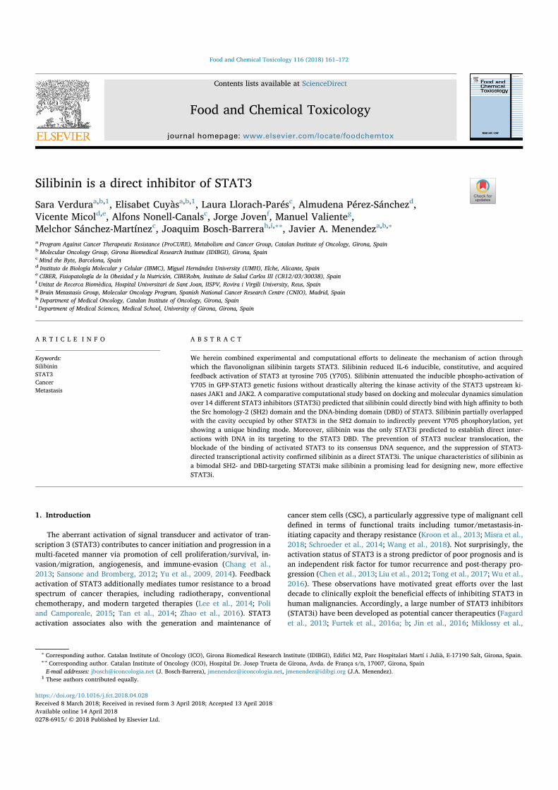
We initially assessed the ability of silibinin to interfere with thethree known activating modes of Y705 STAT3 phosphorylation, namelyIL-6-inducible, constitutive, and acquired (feedback hyperactivation),in a panel of non-small cell lung carcinoma (NSCLC) cell lines (Fig. 1A).H460 and PC9 cell lines, which do not express persistently hyperpho-sphorylated STAT3, were used to determine whether silibinin couldinhibit Y705 STAT3 phosphorylation induced by the pro-inflammatorycytokine IL-6. Immunoblotting procedures revealed that silibinintreatment completely prevented the capacity of IL-6 to induce thephosphorylation of Y705 STAT3 in H460 cells (Fig. 1B). Moreover, theability of IL-6 to augment by 4.0-fold the phosphorylation of Y705STAT3 in PC9 cells was reduced to 2.1-fold in the presence of silibinin(Fig. 1B). This prevention of IL-6-inducible Y705 STAT3 phosphoryla-tion was accompanied by a rapid and noteworthy reduction in theprotein level of the key STAT3 target gene c-MYC in H460 and PC9 cells(Fig. 1B).
Treatment with graded concentrations of silibinin dose-dependentlyabrogated Y705 STAT3 phosphorylation in the H2228 cell line, whichexhibits constitutive hyperphosphorylation of STAT3 (Fig. 1C). Silibininalso suppressed, in a dose-dependent manner, the acquired feedbackhyperactivation of Y705 STAT3 in H3122CR cells, which has beenshown to occur as a non-genetic mechanism of acquired resistance tothe ALK-tyrosine kinase inhibitor crizotinib in ALK-rearranged H3122parental cells (Cuyàs et al., 2016) (Fig. 1D).
S. Verdura et al. )RRG�DQG�&KHPLFDO�7R[LFRORJ\���������������²���
���
3.2. Silibinin inhibits Y705 STAT3 phosphorylation in a JAK1/JAK2-independent manner
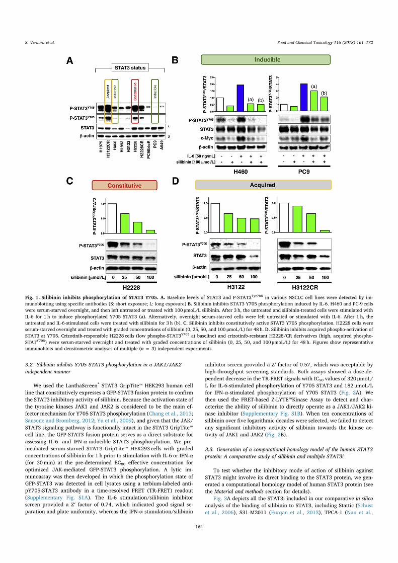
We used the LanthaScreen® STAT3 GripTite™ HEK293 human cellline that constitutively expresses a GFP-STAT3 fusion protein to confirmthe STAT3 inhibitory activity of silibinin. Because the activation state ofthe tyrosine kinases JAK1 and JAK2 is considered to be the main ef-fector mechanism for Y705 STAT3 phosphorylation (Chang et al., 2013;Sansone and Bromberg, 2012; Yu et al., 2009), and given that the JAK/STAT3 signaling pathway is functionally intact in the STAT3 GripTite™cell line, the GFP-STAT3 fusion protein serves as a direct substrate forassessing IL-6- and IFN-α-inducible STAT3 phosphorylation. We pre-incubated serum-starved STAT3 GripTite™ HEK293 cells with gradedconcentrations of silibinin for 1 h prior to stimulation with IL-6 or IFN-α(for 30min) at the pre-determined EC80 effective concentration foroptimized JAK-mediated GFP-STAT3 phosphorylation. A lytic im-munoassay was then developed in which the phosphorylation state ofGFP-STAT3 was detected in cell lysates using a terbium-labeled anti-pY705-STAT3 antibody in a time-resolved FRET (TR-FRET) readout(Supplementary Fig. S1A). The IL-6 stimulation/silibinin inhibitorscreen provided a Z′ factor of 0.74, which indicated good signal se-paration and plate uniformity, whereas the IFN-α stimulation/silibinin
inhibitor screen provided a Z’ factor of 0.57, which was acceptable byhigh-throughput screening standards. Both assays showed a dose-de-pendent decrease in the TR-FRET signals with IC50 values of 320 μmoL/L for IL-6-stimulated phosphorylation of Y705 STAT3 and 182 μmoL/Lfor IFN-α-stimulated phosphorylation of Y705 STAT3 (Fig. 2A). Wethen used the FRET-based Z-LYTE™Kinase Assay to detect and char-acterize the ability of silibinin to directly operate as a JAK1/JAK2 ki-nase inhibitor (Supplementary Fig. S1B). When ten concentrations ofsilibinin over five logarithmic decades were selected, we failed to detectany significant inhibitory activity of silibinin towards the kinase ac-tivity of JAK1 and JAK2 (Fig. 2B).
3.3. Generation of a computational homology model of the human STAT3protein: A comparative study of silibinin and multiple STAT3i
To test whether the inhibitory mode of action of silibinin againstSTAT3 might involve its direct binding to the STAT3 protein, we gen-erated a computational homology model of human STAT3 protein (seethe Material and methods section for details).
Fig. 3A depicts all the STAT3i included in our comparative in silicoanalysis of the binding of silibinin to STAT3, including Stattic (Schustet al., 2006), S31-M2011 (Furqan et al., 2013), TPCA-1 (Nan et al.,
Fig. 1. Silibinin inhibits phosphorylation of STAT3 Y705. A. Baseline levels of STAT3 and P-STAT3Tyr705 in various NSCLC cell lines were detected by im-munoblotting using specific antibodies (S: short exposure; L: long exposure) B. Silibinin inhibits STAT3 Y705 phosphorylation induced by IL-6. H460 and PC-9 cellswere serum-starved overnight, and then left untreated or treated with 100 μmoL/L silibinin. After 3 h, the untreated and silibinin-treated cells were stimulated withIL-6 for 1 h to induce phosphorylated Y705 STAT3 (a). Alternatively, overnight serum-starved cells were left untreated or stimulated with IL-6. After 1 h, theuntreated and IL-6-stimulated cells were treated with silibinin for 3 h (b). C. Silibinin inhibits constitutively active STAT3 Y705 phosphorylation. H2228 cells wereserum-starved overnight and treated with graded concentrations of silibinin (0, 25, 50, and 100 μmoL/L) for 48 h. D. Silibinin inhibits acquired phospho-activation ofSTAT3 at Y705. Crizotinib-responsible H2228 cells (low phospho-STAT3Y705 at baseline) and crizotinib-resistant H2228/CR derivatives (high, acquired phospho-STATY705) were serum-starved overnight and treated with graded concentrations of silibinin (0, 25, 50, and 100 μmoL/L) for 48 h. Figures show representativeimmunoblots and densitometric analyses of multiple (n = 3) independent experiments.
S. Verdura et al. )RRG�DQG�&KHPLFDO�7R[LFRORJ\���������������²���
���
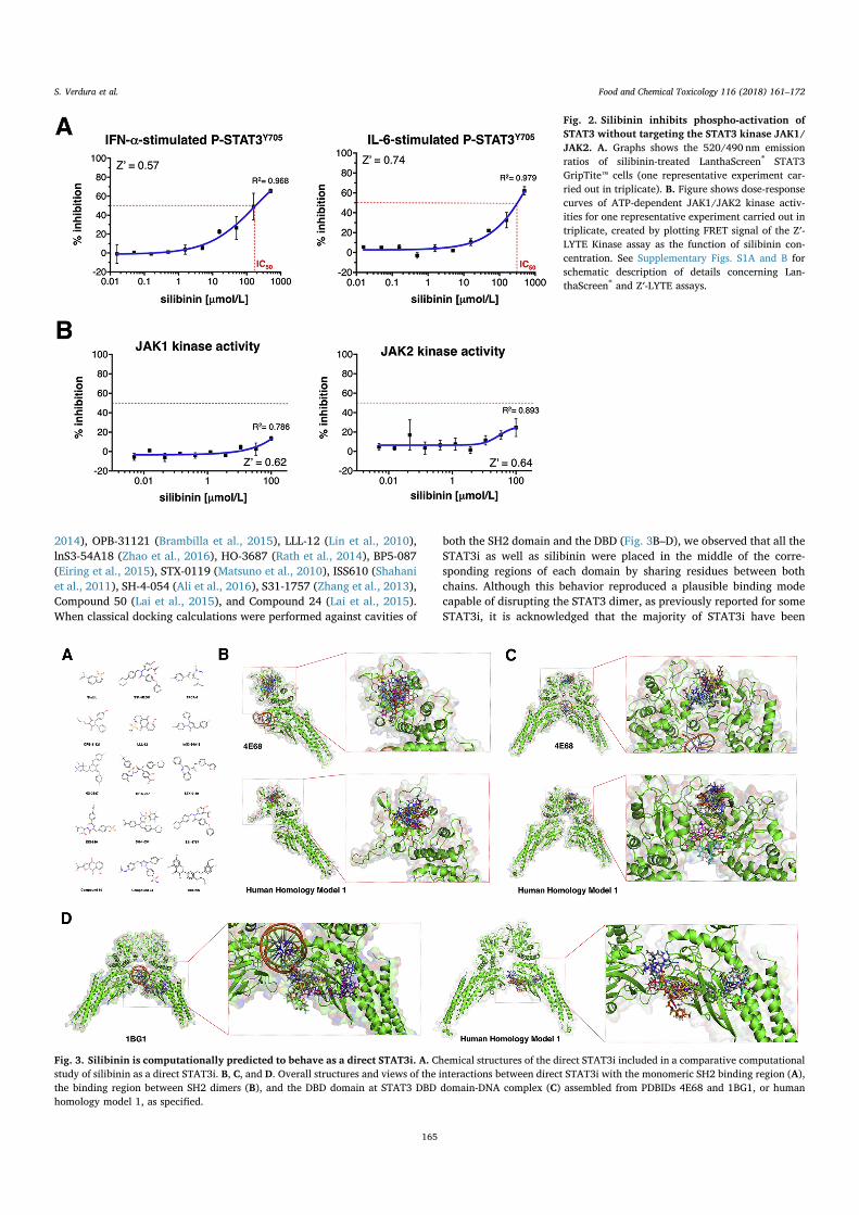
2014), OPB-31121 (Brambilla et al., 2015), LLL-12 (Lin et al., 2010),lnS3-54A18 (Zhao et al., 2016), HO-3687 (Rath et al., 2014), BP5-087(Eiring et al., 2015), STX-0119 (Matsuno et al., 2010), ISS610 (Shahaniet al., 2011), SH-4-054 (Ali et al., 2016), S31-1757 (Zhang et al., 2013),Compound 50 (Lai et al., 2015), and Compound 24 (Lai et al., 2015).When classical docking calculations were performed against cavities of
both the SH2 domain and the DBD (Fig. 3B–D), we observed that all theSTAT3i as well as silibinin were placed in the middle of the corre-sponding regions of each domain by sharing residues between bothchains. Although this behavior reproduced a plausible binding modecapable of disrupting the STAT3 dimer, as previously reported for someSTAT3i, it is acknowledged that the majority of STAT3i have been
Fig. 2. Silibinin inhibits phospho-activation ofSTAT3 without targeting the STAT3 kinase JAK1/JAK2. A. Graphs shows the 520/490 nm emissionratios of silibinin-treated LanthaScreen® STAT3GripTite™ cells (one representative experiment car-ried out in triplicate). B. Figure shows dose-responsecurves of ATP-dependent JAK1/JAK2 kinase activ-ities for one representative experiment carried out intriplicate, created by plotting FRET signal of the Z′-LYTE Kinase assay as the function of silibinin con-centration. See Supplementary Figs. S1A and B forschematic description of details concerning Lan-thaScreen® and Z′-LYTE assays.
Fig. 3. Silibinin is computationally predicted to behave as a direct STAT3i. A. Chemical structures of the direct STAT3i included in a comparative computationalstudy of silibinin as a direct STAT3i. B, C, and D. Overall structures and views of the interactions between direct STAT3i with the monomeric SH2 binding region (A),the binding region between SH2 dimers (B), and the DBD domain at STAT3 DBD domain-DNA complex (C) assembled from PDBIDs 4E68 and 1BG1, or humanhomology model 1, as specified.
S. Verdura et al. )RRG�DQG�&KHPLFDO�7R[LFRORJ\���������������²���
���

suggested to bind the corresponding SH2 or DBD domain solely in oneof the monomers, without sharing residues with the other one. To ex-plore in more detail the latter behavior, we performed docking simu-lations to the monomeric structures, to mimic the desired binding ofknown STAT3i. The binding energies obtained from in silico bindingexperiments using rigid docking calculations, which were run twice toavoid false positives, are summarized in Table S1 (SH2 domain ofmonomeric structures), Table S2 (SH2 domain of dimeric structures),and Table S3 (DBD of dimeric structures). This approach predicted theability of silibinin to directly bind the mouse and human STAT3structures, with energy values ranging from −5.9 kcaL/mol to−8.5 kcaL/mol when using the mouse crystal structures 1BG1, 3CWG,4E68, and from −5.6 kcaL/mol to −9.0 kcaL/mol when using thehuman homology models 1 and 2.
To add protein flexibility to the analysis and to better test the sta-bility of the silibinin-STAT3 complexes, we carried out short MD si-mulations of 1 ns and applied MM/GBSA calculations to estimate morereliable binding energies, which are summarized in Table S4, S5, andS6. For MD simulations and MM/GBSA calculations, we selected mouse4E68 and human homology model 1 to investigate the interactions withthe SH2 domain, whereas mouse 1BG1 and human homology model 1were selected to investigate the interactions with the DBD. Such ap-proaches predicted the capacity of silibinin to bind mouse and humanSTAT3 structures with energy values ranging from −24.5797 kcaL/molto −40.5752 kcaL/mol when using the mouse crystal structures 1BG1and 4E68, and from −20.0086 kcaL/mol to −36.4145 kcaL/mol whenusing the human homology model 1.
3.4. Silibinin is predicted to bind to the SH2 and the DBD domains ofSTAT3
The binding modes of well-characterized direct STAT3i were sig-nificantly shared between the mouse PDB crystal structures and thehuman homology models, highlighting a high degree of conservation ofthe SH2 and DBD domains between mouse and human STAT3 proteins.The evaluation of the binding mode of silibinin to the monomeric formof the SH2 domain revealed a common group of predicted interactingresidues shared with other direct STAT3i (Table S7); namely, M660,E638, K626, 7620, P639, V637, Y657, W623, and T714 in the mousecrystal structure 4E68, and S613, K626, P639, Q635, W623, and E638in the human homology model 1. Silibinin was predicted not to shareany interacting residue with S31-757 in the human homology model 1of the monomeric form of the SH2 domain.
When evaluating the binding of silibinin to the dimeric form of theSH2 domain, we observed that silibinin was predicted to place differ-ently to the remainder of the direct STAT3i (Table S8). Accordingly,silibinin was predicted to share with other direct STAT3 inhibitors asignificant number of interacting residues in the human homologymodel 1 (K1658, M655, I1711, P1715, K709, V713, E652, V1713,L1666, I711, L666), but only a few interacting residues in the mousecrystal structure 4E68 (Q4644, E4638, and M648). Silibinin was pre-dicted not to share any interacting residues with S31-M2001 and STX-0119 in the human homology model 1 of the dimeric form of the SH2domain.
The evaluation of the binding mode of silibinin to the DBD revealeda common group of putative interacting residues shared with other
Fig. 4. Mode of binding of silibinin to SH2 activation/dimerization and DNA binding (DBD) domains of STAT3. Figure shows in sticks all the pharmacophoricinteraction residues involved in the in silico binding of silibinin to the SH2 and DBD domains of STAT3, using PLIP. Orange dashed lines represent hydrogen bondinteractions; grey dashed lines represent hydrophobic interactions. The main residues involved in silibinin interaction with the protein backbone are shown in black;the residue numbers shown correspond to the original PDB file numbering. Left panels correspond to binding poses resulting from simple, rigid docking studies; rightpanels correspond to self-docking poses under molecular dynamics (MD) simulations modeling the backbone and ligand (silibinin) flexibility. (For interpretation ofthe references to colour in this figure legend, the reader is referred to the Web version of this article.)
S. Verdura et al. )RRG�DQG�&KHPLFDO�7R[LFRORJ\���������������²���
���

direct STAT3i (Table S9); namely, R423, K383, G419, R382, V432, andE415 in the mouse crystal structure 1BG1, and G419, G422, K383,G390, Q416, and R423 in the human homology model 1. Silibinin waspredicted not to share any interacting residues with Stattic, S31-M2001,ln53–5418, S31-1757, and Compound 24 in the mouse crystal structure1BG1, or S31-M2011, ln53-54A18, STX-0119, Compound 50, andCompound 24, in the human homology model 1.
3.5. The predicted binding mode of silibinin to STAT3 domains is differentto other STAT3i
Silibinin was predicted to establish hydrogen bond interactions withS613, K626, E638, and M660 within the binding pocket of the mono-meric SH2 domain of STAT3 (Fig. 4). Silibinin was predicted to ad-ditionally establish hydrophobic interactions with T620, W623, Q635,V637, E638, P639, Y657, and T714 (Fig. 4). The binding and putativeinhibitory capacity of silibinin against the SH2 domain of STAT3 isunderscored by the fact that it was predicted to share a significantnumber of interacting residues (W623, K626, Q635, V637, E638, Y657,and T714), or interact with those placed nearby or adjacent to thosethat were identified upon an extensive bibliographic search for keyinteracting residues employed by existing SH2-targeted STAT3i (F588,I589, S590, K591, E594, R595, R609, S611, E612, W623, K626, Q635,S636, V637, E638, Y657, I659, C687, Y705, T714, P715, T716, T717,and S727) (Fig. 4).
Silibinin was predicted to establish hydrogen bond interactions withS649, F710, and C722 (α chain), and T641 (β chain) within the bindingpocket of the dimeric SH2 domains of STAT3 (Fig. 4). Silibinin waspredicted to additionally establish hydrophobic interactions withM648, S649, L666, T708, F710, I711, and V713 (α chain) and withE638, P639, Y640, K658, I711, and V713 (β chain) (Fig. 4). Althoughmost of these residues were placed nearby or adjacent to the above-mentioned key interacting residues employed by existing SH2-targetedSTAT3 inhibitors (Supplementary Fig. S2), E638 was identified as thesole key interacting residue shared with silibinin, thus supporting thenotion that direct STAT3i including silibinin might employ the bindingpocket within the monomeric, but not the dimeric, SH2 domain ofSTAT3.
Silibinin was predicted to establish hydrogen bond interactions withR382, K383, G419, G422, R423, and G380 within the DBD of dimericSTAT3 (Fig. 4). Silibinin was predicted to additionally establish hy-drophobic interactions with R382, K383, E415, R423, and V432. Itshould be noted that all these residues were included in the list of keyinteracting DBD residues that were identified upon an extensive bib-liographic search of direct STAT3i; namely, Q326, P327, P330, M331,H332, K340, T341, V343, F345, T412, E415, N420, R423, I431, V432,S465, N466, I467, Q469, M470, W474, and N485. Moreover, despitethe fact that DNA was complexed in the 1BG1 crystal structure for allthe docking and MD simulations, silibinin was the sole STAT3i that waspredicted to establish a hydrogen bond interaction with DT1005(Fig. 4).
3.6. Silibinin prevents nuclear accumulation of activated STAT3
Since Y705 phosphorylation and dimerization of STAT3 is a pre-requisite for its cytokine-induced nuclear translocation, we would ex-pect a direct STAT3 SH2 domain inhibitor such as silibinin to inhibit IL-6-induced nuclear translocation of phospho-active STAT3. To test this,PC9 cells were seeded on coverslips and stimulated for 24 h with IL-6 inthe absence or presence of silibinin. In untreated conditions, immuno-fluorescence microscopy showed that STAT3 was uniformly distributedbetween the cytoplasm and nucleus in PC9 cells; conversely, a greaternumber of STAT3 molecules appeared to be more prominently nuclearfollowing IL-6 stimulation (Fig. 5A). The presence of silibinin failed tosignificantly alter the distribution pattern of total STAT3 in the absenceof IL-6 stimulation; however, silibinin co-treatment suppressed IL-6-
induced nuclear accumulation of STAT3. Moreover, when cells werestained for phosphorylated Y705 STAT3, we confirmed that IL-6-mediated nuclear accumulation of STAT3 is a molecular event largelydependent on the Y705 phosphorylation, which permits STAT3 to formdimers and enter the nuclei. Such IL-6-induced conspicuous STAT3Y705 phosphorylation and translocation into the nucleus was com-pletely prevented in the presence of silibinin (Fig. 5A).
3.7. Silibinin blocks transcriptional activity of STAT3
We then examined whether silibinin suppresses the transcriptionalactivity of STAT3 after IL-6 stimulation using a dual-luciferase assaysystem. HEK293 cells were transiently transfected with a reporterplasmid containing the STAT3-binding response element driving theexpression of the luciferase gene. The STAT3-luciferase reporter con-struct responded exquisitely, in a dose-dependent manner, to gradedconcentrations of IL-6 (Fig. 5B). A concentration of silibinin as low as100 μmoL/L completely prevented the transcriptional activity of STAT3after stimulation with graded concentrations of IL-6. Moreover, whencells transiently transfected with the STAT3-luciferase reporter con-struct were stimulated with an optimal STAT3 activating concentrationof IL-6 (50 ng/mL) in the presence of graded concentrations of silibinin,we confirmed the ability of silibinin to dramatically inhibit STAT3-dependent luciferase activity in a dose-dependent manner, with IC50
values lower than 25 μmoL/L (Fig. 5B).
3.8. Silibinin reduces the DNA binding activity of STAT3
Such a potent inhibitory effect of silibinin on the transcriptionalactivity of STAT3 might reflect not only its ability to influence tyrosinephosphorylation and nuclear accumulation of STAT3, but also the insilico predicted capability of silibinin to establish direct interactionswith DNA in its inhibitory targeting to the DBD of STAT3. To evaluatethe hypothesis that silibinin might also alter STAT3 retention via DNAbinding, we employed the ELISA-based TransAM™ method to quanti-tatively evaluate the ability of the STAT3 residing in cellular nuclearextracts to bind its corresponding DNA consensus sequence (im-mobilized on the 96-well plate) when exposed to silibinin(Supplementary Fig. S1C). Nuclear extracts from IL-6-stimulated H460and PC9 cells containing Y705-phosphorylated STAT3 were incubatedwith increasing concentrations of silibinin to directly determine thepotency of silibinin to inhibit the DNA-binding activity of STAT3. Adose-dependent reduction in the DNA-binding activity of STAT3 wasobserved in the presence of silibinin (up to 60% at 200 μmoL/L sili-binin; Fig. 6). When nuclear extracts obtained from H2228 cells, whichexhibit constitutive activation of STAT3, were incubated in the pre-sence of graded concentrations of silibinin, their STAT3 DNA bindingactivity was similarly reduced by up to 60% at 200 μmoL/L silibinincompared with vehicle-treated controls (Fig. 6).
4. Discussion
Although silibinin is known to be an inhibitor of STAT3 signaling, itremained to be clarified whether silibinin should be classified as anindirect STAT3i via kinase inhibition of the JAK/STAT pathway or as adirect STAT3i capable of binding and interfering with specific domainsof the STAT3 protein. As with many other plant-derived secondarymetabolites including cucurbitacin, curcumin, indirubin, cryptotan-shinone, resveratrol, flavopiridol, and galiellalactone (Schust et al.,2006), silibinin has repeatedly been shown to inhibit STAT3 signalingin cancer cells (Jin et al., 2016; Bosch-Barrera and Menendez, 2015).While some of these natural products might operate as STAT3i throughunknown targets, or have been shown to inhibit kinases upstream ofSTAT3 (JAK1/2, Src), others have been suggested to directly bind toSTAT3 functional domains; for example, the SH2 domain, blockingSTAT3 dimerization, or the STAT3 DBD, preventing sequence-specific
S. Verdura et al. )RRG�DQG�&KHPLFDO�7R[LFRORJ\���������������²���
���
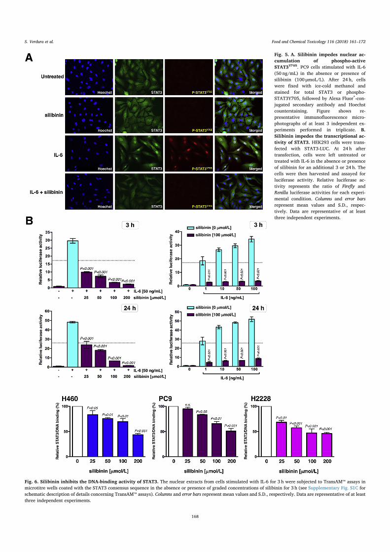
Fig. 5. A. Silibinin impedes nuclear ac-cumulation of phospho-activeSTAT3Y705. PC9 cells stimulated with IL-6(50 ng/mL) in the absence or presence ofsilibinin (100 μmoL/L). After 24 h, cellswere fixed with ice-cold methanol andstained for total STAT3 or phospho-STAT3Y705, followed by Alexa Fluor®-con-jugated secondary antibody and Hoechstcounterstaining. Figure shows re-presentative immunofluorescence micro-photographs of at least 3 independent ex-periments performed in triplicate. B.Silibinin impedes the transcriptional ac-tivity of STAT3. HEK293 cells were trans-fected with STAT3-LUC. At 24 h aftertransfection, cells were left untreated ortreated with IL-6 in the absence or presenceof silibinin for an additional 3 or 24 h. Thecells were then harvested and assayed forluciferase activity. Relative luciferase ac-tivity represents the ratio of Firefly andRenilla luciferase activities for each experi-mental condition. Columns and error barsrepresent mean values and S.D., respec-tively. Data are representative of at leastthree independent experiments.
Fig. 6. Silibinin inhibits the DNA-binding activity of STAT3. The nuclear extracts from cells stimulated with IL-6 for 3 h were subjected to TransAM™ assays inmicrotitre wells coated with the STAT3 consensus sequence in the absence or presence of graded concentrations of silibinin for 3 h (see Supplementary Fig. S1C forschematic description of details concerning TransAM™ assays). Columns and error bars represent mean values and S.D., respectively. Data are representative of at leastthree independent experiments.
S. Verdura et al. )RRG�DQG�&KHPLFDO�7R[LFRORJ\���������������²���
���

DNA binding ability and STAT3 transactivation activity. We now reportthat silibinin appears to work synergistically on STAT3 functionthrough a bimodal mechanism of action involving blockade of thefunction of the STAT3 SH2 domain, which is crucial for both STAT3activation and nuclear translocation, and of STAT3 transcriptional ac-tivity, which might involve not only disruption of STAT3 dimerization,but also a direct inhibition of the ability of STAT3 to bind DNA(Fig. 7A).
The STAT3 inhibitory activity of silibinin was not influenced by thepre-existing phosphorylation status of STAT3, as significant inhibitoryeffects were observed in cells with inducible, constitutive, and acquiredphosphorylation at the Y705 site. LanthaScreen™-based cellular pro-filing assays revealed that silibinin attenuates the induced phospho-activation of Y705 in GFP-STAT3 genetic fusions without drasticallyaltering the in vitro kinase activity of the STAT3 upstream kinases JAK1and JAK2. Although these findings are consistent with the notion thatsilibinin exerts its pY705 STAT3 inhibitory effects by directly pre-venting the activating kinases from binding to the STAT3 SH2 domain,we acknowledge that further experimentation testing the direct effectsof silibinin against other up-stream STAT3 kinases (e.g., SRC, ABL) and
non-canonical STAT3 activators is needed before unambiguously con-cluding that silibinin exclusively operates as a direct STAT3i.
When we modeled the atomic details for the silibinin-driven in-hibition of the activating phosphorylation Y705 on the SH2 domain, ourfirst-in-class computational homology model of the human STAT3protein allowing comparative docking and molecular dynamics simu-lation studies over fourteen different STAT3i, predicted that silibininshould molecularly behave as a direct STAT3i capable of establishinghigh-affinity interactions with the SH2 domain of STAT3. Using thebinding site of the direct STAT3i OPB-31121, for which we dispose ofdetailed structural information explaining its inhibitory activity on theSTAT3 SH2 domain (Brambilla et al., 2015), one could visualize thepredicted ability of silibinin to interact with up to 60% of all the re-sidues involved in the binding mode of a wide variety of structurallydiverse STAT3i (Fig. 7A). The predicted ability of silibinin to bind theSH2 activation/dimerization domain therefore appears to rely on itscapacity to overlap with the same cavity occupied by the majority ofdirect STAT3i to indirectly prevent Y705 phosphorylation in themonomeric SH2 domain of STAT3, but showing a unique binding mode.
Silibinin treatment is known to diminish nuclear DNA binding of
Fig. 7. Silibinin is a bimodal SH2- andDBD-targeted STAT3i. A. Global view ofthe STAT3 homodimer structure containingDNA (human homology model 2) and lo-cation of silibinin at the SH2 activation/di-merization and DNA-binding domains. B.Silibinin targeting of the SH2 domain ofSTAT3 monomers might prevent binding ofSTAT3 to activated cell surface receptorsbut also block dimerization (and subsequenttrans-phosphorylation) of STAT3 moleculesin the cytosol, thereby impeding nuclearaccumulation of phospho-active STAT3.Silibinin additionally establishes direct in-teractions with DNA in its direct targetingof the DBD of STAT3, resulting in a sig-nificant inhibitory effect on STAT3-DNAbinding. The bimodal SH2- and DBD-tar-geted behavior of silibinin might explainthe proven therapeutic activity of silibininin areas of unmet clinical need such asSTAT3-dependent lung cancer and mela-noma brain metastasis.
S. Verdura et al. )RRG�DQG�&KHPLFDO�7R[LFRORJ\���������������²���
���

constitutively active STAT3 homodimers (Agarwal et al., 2007). Be-cause Y705 phosphorylation is required for STAT3 to bind to specificDNA target sites but nuclear import of STAT3 takes place constitutivelyand independently of tyrosine phosphorylation (Liu et al., 2005; Reichand Liu, 2006), we employed immunofluorescence microscopy to vi-sualize whether the in silico predicted ability of silibinin to operate as adirect STAT3i of the SH2 activation/dimerization domain translatedinto an altered intracellular localization of STAT3/phospho-activeSTAT3 in cellulo. The ability of silibinin to prevent the nuclear con-centration of unphosphorylated STAT3 and Y705-phosphorylatedSTAT3 in response to IL-6 stimulation occurred without apparent ac-cumulation of STAT3 in the cytoplasmic compartment. Although fur-ther work is needed to unambiguously exclude any indirect effect ofsilibinin in the importins-driven STAT3 trafficking to the nucleus (Liuet al., 2005; Cimica et al., 2011), our findings are compatible with amechanism of action involving direct targeting of silibinin to the SH2domain of STAT3 monomers, capable of preventing not only binding ofSTAT3 to activated cell surface receptors, but also to block dimerization(and subsequent trans-phosphorylation) of STAT3 molecules in thecytosol, thereby impeding nuclear accumulation of phospho-activeSTAT3.
To further evaluate the physiological role of silibinin on STAT3-mediated transactivation, we examined whether silibinin-drivenchanges in the sub-cellular accumulation of STAT3 correlated withchanges in its transcriptional regulatory activity. Silibinin treatmentwas found to elicit the complete suppression of the IL-6-stimulatedSTAT3 transcriptional activity in living cells and, remarkably, suchstrong capability of silibinin to block STAT3-driven luciferase expres-sion was evident even at concentrations that failed to completelyshutdown the activating phosphorylation Y705 at the SH2 dimerizationdomain. Moreover, although in vitro experiments based on the detectionof an STAT3 epitope that is accessible only when STAT3 is activatedand bound to its DNA consensus binding site confirmed the in silicoprediction of the capacity of silibinin to establish direct interactionswith DNA in its targeting to the DBD of STAT3, once again the STAT3DNA-binding inhibitory activity of silibinin took place at significantlyhigher concentrations than those needed to inhibit STAT3-driventranscriptional activity. Because parallel immunoblotting experimentswith IL-6-stimulated nuclear extracts showed that silibinin can blockthe binding of activated STAT3 to its consensus DNA sequence in iso-lated nuclear extracts without altering the phosphorylation status ofY705 (data not shown), these findings altogether suggest that theability of silibinin to inhibit STAT3-directed transcription in living cellsdoes not rely exclusively on the SH2 domain-related inhibition ofSTAT3 dimerization in the cytosol, but also involves direct inhibition ofSTAT3 via binding to the DBD regardless of the STAT3 dimerizationstatus. Nevertheless, the unique behavior of silibinin as a bimodal SH2-and DBD-STAT3i that strongly disrupts STAT3 transcriptional activity isdefinitively supported by the fact that cells engineered to overexpress aconstitutively active form of STAT3 (Bromberg et al., 1999), whichdimerizes spontaneously, binds to DNA and activates transcription,remain largely unresponsive to the inhibitory effects of silibinin in keytranscriptional targets of STAT3 (Shukla et al., 2015; Priego et al.,2018). The so-called STAT3C mutant, in which the SH2 domain A661and N663 residues are substituted with cysteine residues allowing adisulfide bond to form between two unphosphorylated STAT3 mono-mers, still requires Y705 phosphorylation for functional activation viapromotion of maximal DNA binding affinity, slower off-rate, and pro-tection from inactivation from phosphatases, resulting in the accumu-lation of transcriptionally active STAT3 dimer complexes (Liddle et al.,2006). We recently reported that the decreased ability of silibinin tobind the STAT3C mutant translates into refractoriness of STAT3C-ex-pressing cells to silibinin (Priego et al., 2018), demonstrating theSTAT3-dependency on the phenotypic effects of silibinin.
Beyond common issues in the development of other anti-cancerdrug families such as rapid degradation, lack of cell penetrance or lack
of binding specificity, the observation that inhibition of active STAT3dimers alone via targeting to the SH2 domain may not be sufficient inefficaciously preventing STAT3 activity (Huang et al., 2016) togetherwith the preliminary support to the notion that targeting the DBD mayprove more efficient in abrogating STAT3 activity than targeting theSH2 domain in cellular systems (Furtek et al., 2016b), can largely ex-plain why the majority of direct STAT3i have yet to enter clinicalevaluation. We have recently reported that silibinin-driven STAT3blocking translates into proven therapeutic activity in areas of unmetclinical need such as lung cancer and melanoma brain metastasis,which portend a poor prognosis and have few therapeutic options(Bosch-Barrera et al., 2016; Priego et al., 2018). We now report theunique characteristics of silibinin as a promising lead of a new gen-eration of bimodal SH2- and DBD-targeting STAT3i (Fig. 7B) that maytransform the clinical management of secondary brain tumors.
5. Conclusion
To the best of our knowledge, this is the first report establishing thatthe flavonolignan silibinin is a novel direct STAT3i. Our systematicapproach performed at multiple levels of integration including in vitro,in silico computational modeling, and in cellulo experimentation, de-monstrates that: a.) silibinin could directly bind the SH2 domain ofSTAT3 to prevent Y705 phosphorylation-related STAT3 activation anddimerization; b.) silibinin could establish direct interactions with DNAin its targeting to the STAT3 DNA-binding domain (DBD); and c.) sili-binin impedes the activation, dimerization, nuclear translocation, DNA-binding, and transcriptional activity of STAT3. Our findings showingthe unique features and putative direct modes of action of silibininagainst STAT3 will be highly relevant for further development anddesign of new, more effective silibinin-based STAT3i.
Conflicts of interest
L.L-P, A. N-C, and M. S-M are employees of Mind the Byte S.L. Allother authors have no competing interests to declare.
Acknowledgments
This work was supported by grants from the Ministerio de Ciencia eInnovación (Grant SAF2016-80639-P to J. A. Menendez), Plan Nacionalde I + D+ I, Spain, and the Agència de Gestió d’Ajuts Universitaris i deRecerca (AGAUR) (Grant 2014 SGR229 to J. A. Menendez). This studywas supported also by unrestricted research grants from Roche Pharma(Spain) and Astellas Pharma (Spain) to the Program Against CancerTherapeutic Resistance (ProCURE, Catalan Institute of Oncology).Joaquim Bosch-Barrera is supported by SEOM, Pfizer (GrantWI190764), Boehringer Ingelheim, Meda Pharma, and Pla strategic derecerca i innovació en salut 2016–2020 de la Generalitat de Catalunya(SLT006/17/114). Elisabet Cuyàs is supported by a Sara Borrell post-doctoral contract (CD15/00033) from the Ministerio de Sanidad yConsumo, Fondo de Investigación Sanitaria (FIS), Spain. The authorswould like to thank Dr. Kenneth McCreath for editorial support.
Appendix A. Supplementary data
Supplementary data related to this article can be found at http://dx.doi.org/10.1016/j.fct.2018.04.028.
Transparency document
Transparency document related to this article can be found online athttp://dx.doi.org/10.1016/j.fct.2018.04.028.
S. Verdura et al. )RRG�DQG�&KHPLFDO�7R[LFRORJ\���������������²���
���

References
Agarwal, C., Tyagi, A., Kaur, M., Agarwal, R., 2007. Silibinin inhibits constitutive acti-vation of Stat3, and causes caspase activation and apoptotic death of human prostatecarcinoma DU145 cells. Carcinogenesis 28, 1463–1470. https://doi.org/10.1093/carcin/bgm042.
Agarwal, R., Agarwal, C., Ichikawa, H., Singh, R.P., Aggarwal, B.B., 2006. Anticancerpotential of silymarin: from bench to bed side. Anticancer Res. 26, 4457–4498.
Ali, A.M., Gómez-Biagi, R.F., Rosa, D.A., Lai, P.-S., Heaton, W.L., Park, J.S., Eiring, A.M.,Vellore, N.A., de Araujo, E.D., Ball, D.P., Shouksmith, A.E., Patel, A.B., Deininger,M.W., O'Hare, T., Gunning, P.T., 2016. Disarming an electrophilic warhead: retainingpotency in tyrosine kinase inhibitor (TKI)-Resistant CML lines while circumventingpharmacokinetic liabilities. ChemMedChem 11, 850–861. https://doi.org/10.1002/cmdc.201600021.
Becker, S., Groner, B., Muller, C.W., 1998. Three-dimensional structure of the Stat3betahomodimer bound to DNA. Nature 394, 145–151. https://doi.org/10.1038/28101.
Bosch-Barrera, J., Corominas-Faja, B., Cuyàs, E., Martin-Castillo, B., Brunet, J.,Menendez, J.A., 2014. Silibinin administration improves hepatic failure due to ex-tensive liver infiltration in a breast cancer patient. Anticancer Res. 34, 4323–4328.
Bosch-Barrera, J., Menendez, J.A., 2015. Silibinin and STAT3: a natural way of targetingtranscription factors for cancer therapy. Canc. Treat Rev. 41, 540–546. https://doi.org/10.1016/j.ctrv.2015.04.008.
Bosch-Barrera, J., Queralt, B., Menendez, J.A., 2017. Targeting STAT3 with silibinin toimprove cancer therapeutics. Canc. Treat Rev. 58, 61–69. https://doi.org/10.1016/j.ctrv.2017.06.003.
Bosch-Barrera, J., Sais, E., Cañete, N., Marruecos, J., Cuyàs, E., Izquierdo, A., Porta, R.,Haro, M., Brunet, J., Pedraza, S., Menendez, J.A., 2016. Response of brain metastasisfrom lung cancer patients to an oral nutraceutical product containing silibinin.Oncotarget 7, 32006–32014. https://doi.org/10.18632/oncotarget.7900.
Brambilla, L., Genini, D., Laurini, E., Merulla, J., Perez, L., Fermeglia, M., Carbone, G.M.,Pricl, S., Catapano, C.V., 2015. Hitting the right spot: mechanism of action of OPB-31121, a novel and potent inhibitor of the Signal Transducer and Activator ofTranscription 3 (STAT3). Mol. Oncol 9, 1194–1206. https://doi.org/10.1016/j.molonc.2015.02.012.
Bromberg, J.F., Wrzeszczynska, M.H., Devgan, G., Zhao, Y., Pestell, R.G., Albanese, C.,Darnell, J.E., 1999. Stat3 as an oncogene. Cell 98, 295–303. https://doi.org/10.1016/S0092-8674(00)81959-5.
Chang, Q., Bournazou, E., Sansone, P., Berishaj, M., Gao, S.P., Daly, L., Wels, J., Theilen,T., Granitto, S., Zhang, X., Cotari, J., Alpaugh, M.L., de Stanchina, E., Manova, K., Li,M., Bonafe, M., Ceccarelli, C., Taffurelli, M., Santini, D., Altan-Bonnet, G., Kaplan, R.,Norton, L., Nishimoto, N., Huszar, D., Lyden, D., Bromberg, J., 2013. The IL-6/JAK/Stat3 feed-forward loop drives tumorigenesis and metastasis. Neoplasia 15, 848–862.
Chen, Y., Wang, J., Wang, X., Liu, X., Li, H., Lv, Q., Zhu, J., Wei, B., Tang, Y., 2013.STAT3, a poor survival predicator, Is associated with lymph node metastasis frombreast cancer. J. Breast Cancer 16, 40–49. https://doi.org/10.4048/jbc.2013.16.1.40.
Chittezhath, M., Deep, G., Singh, R.P., Agarwal, C., Agarwal, R., 2008. Silibinin inhibitscytokine-induced signaling cascades and down-regulates inducible nitric oxide syn-thase in human lung carcinoma A549 cells. Mol. Canc. Therapeut. 7, 1817–1826.https://doi.org/10.1158/1535-7163.MCT-08-0256.
Cimica, V., Chen, H.-C., Iyer, J.K., Reich, N.C., 2011. Dynamics of the STAT3 transcriptionfactor: nuclear import dependent on Ran and importin-β1. PLoS One 6, e20188.https://doi.org/10.1371/journal.pone.0020188.
Cufí, S., Bonavia, R., Vazquez-Martin, A., Corominas-Faja, B., Oliveras-Ferraros, C.,Cuyàs, E., Martin-Castillo, B., Barrajón-Catalán, E., Visa, J., Segura-Carretero, A.,Bosch-Barrera, J., Joven, J., Micol, V., Menendez, J.A., 2013a. Silibinin meglumine, awater-soluble form of milk thistle silymarin, is an orally active anti-cancer agent thatimpedes the epithelial-to-mesenchymal transition (EMT) in EGFR-mutant non-small-cell lung carcinoma cells. Food Chem. Toxicol. 60, 360–368. https://doi.org/10.1016/j.fct.2013.07.063.
Cufí, S., Bonavia, R., Vazquez-Martin, A., Oliveras-Ferraros, C., Corominas-Faja, B.,Cuyàs, E., Martin-Castillo, B., Barrajón-Catalán, E., Visa, J., Segura-Carretero, A.,Joven, J., Bosch-Barrera, J., Micol, V., Menendez, J.A., 2013b. Silibinin suppressesEMT-driven erlotinib resistance by reversing the high miR-21/low miR-200c sig-nature in vivo. Sci. Rep. 3, 2459. https://doi.org/10.1038/srep02459.
Cuyàs, E., Pérez-Sánchez, A., Micol, V., Menendez, J.A., Bosch-Barrera, J., 2016. STAT3-targeted treatment with silibinin overcomes the acquired resistance to crizotinib inALK-rearranged lung cancer. Cell Cycle 1–6. https://doi.org/10.1080/15384101.2016.1245249.
Eiring, A.M., Page, B.D.G., Kraft, I.L., Mason, C.C., Vellore, N.A., Resetca, D., Zabriskie,M.S., Zhang, T.Y., Khorashad, J.S., Engar, A.J., Reynolds, K.R., Anderson, D.J.,Senina, A., Pomicter, A.D., Arpin, C.C., Ahmad, S., Heaton, W.L., Tantravahi, S.K.,Todic, A., Colaguori, R., Moriggl, R., Wilson, D.J., Baron, R., O'Hare, T., Gunning,P.T., Deininger, M.W., 2015. Combined STAT3 and BCR-ABL1 inhibition inducessynthetic lethality in therapy-resistant chronic myeloid leukemia. Leukemia 29,586–597. https://doi.org/10.1038/leu.2014.245.
Fagard, R., Metelev, V., Souissi, I., Baran-Marszak, F., 2013. STAT3 inhibitors for cancertherapy: have all roads been explored? JAK-STAT 2, e22882. https://doi.org/10.4161/jkst.22882.
Flaig, T.W., Glodé, M., Gustafson, D., Van Bokhoven, A., Tao, Y., Wilson, S., Su, L.J., Li,Y., Harrison, G., Agarwal, R., Crawford, E.D., Lucia, M.S., Pollak, M., 2010. A study ofhigh-dose oral silybin-phytosome followed by prostatectomy in patients with loca-lized prostate cancer. Prostate 70, 848–855. https://doi.org/10.1002/pros.21118.
Furqan, M., Akinleye, A., Mukhi, N., Mittal, V., Chen, Y., Liu, D., 2013. STAT inhibitorsfor cancer therapy. J. Hematol. Oncol. 6, 90. https://doi.org/10.1186/1756-8722-
6-90.Furtek, S.L., Backos, D.S., Matheson, C.J., Reigan, P., 2016a. Strategies and approaches of
targeting STAT3 for cancer treatment. ACS Chem. Biol. 11, 308–318. https://doi.org/10.1021/acschembio.5b00945.
Furtek, S.L., Matheson, C.J., Backos, D.S., Reigan, P., 2016b. Evaluation of quantitativeassays for the identification of direct signal transducer and activator of transcription3 (STAT3) inhibitors. Oncotarget 7, 77998–78008. https://doi.org/10.18632/oncotarget.12868.
Gažák, R., Walterová, D., Křen, V., 2007. Silybin and silymarin–new and emerging ap-plications in medicine. Curr. Med. Chem. 14, 315–338. https://doi.org/10.2174/092986707779941159.
Hoh, C., Boocock, D., Marczylo, T., Singh, R., Berry, D.P., Dennison, A.R., Hemingway, D.,Miller, A., West, K., Euden, S., Garcea, G., Farmer, P.B., Steward, W.P., Gescher, A.J.,2006. Pilot study of oral silibinin, a putative chemopreventive agent, in colorectalcancer patients: silibinin levels in plasma, colorectum, and liver and their pharma-codynamic consequences. Clin. Canc. Res. 12, 2944–2950. https://doi.org/10.1158/1078-0432.CCR-05-2724.
Huang, W., Dong, Z., Chen, Y., Wang, F., Wang, C.J., Peng, H., He, Y., Hangoc, G., Pollok,K., Sandusky, G., Fu, X.-Y., Broxmeyer, H.E., Zhang, Z.-Y., Liu, J.-Y., Zhang, J.-T.,2016. Small-molecule inhibitors targeting the DNA-binding domain of STAT3 sup-press tumor growth, metastasis and STAT3 target gene expression in vivo. Oncogene35, 783–792. https://doi.org/10.1038/onc.2015.215.
Jin, Y., Kim, Y., Lee, Y.-J., Han, D.C., K.B, M., 2016. Natural products targeting STAT3signaling pathways in cancer cells. Biodesign 4, 1–17.
Kroon, P., Berry, P.A., Stower, M.J., Rodrigues, G., Mann, V.M., Simms, M., Bhasin, D.,Chettiar, S., Li, C., Li, P.K., Maitland, N.J., Collins, A.T., 2013. JAK-STAT blockadeinhibits tumor initiation and clonogenic recovery of prostate cancer stem-like cells.Canc. Res. 73, 5288–5298. https://doi.org/10.1158/0008-5472.CAN-13-0874.
Lai, P.-S., Rosa, D.A., Magdy Ali, A., Gómez-Biagi, R.F., Ball, D.P., Shouksmith, A.E.,Gunning, P.T., 2015. A STAT inhibitor patent review: progress since 2011. ExpertOpin. Ther. Pat. 25, 1397–1421. https://doi.org/10.1517/13543776.2015.1086749.
Lee, H.-J., Zhuang, G., Cao, Y., Du, P., Kim, H.-J., Settleman, J., 2014. Drug resistance viafeedback activation of Stat3 in oncogene-addicted cancer cells. Canc. Cell 26,207–221. https://doi.org/10.1016/j.ccr.2014.05.019.
Liddle, F.J., Alvarez, J.V., Poli, V., Frank, D.A., 2006. Tyrosine phosphorylation is re-quired for functional activation of disulfide-containing constitutively active STATmutants. Biochemistry 45, 5599–5605. https://doi.org/10.1021/bi0525674.
Lin, L., Hutzen, B., Li, P.-K., Ball, S., Zuo, M., DeAngelis, S., Foust, E., Sobo, M., Friedman,L., Bhasin, D., Cen, L., Li, C., Lin, J., 2010. A novel small molecule, LLL12, inhibitsSTAT3 phosphorylation and activities and exhibits potent growth-suppressive ac-tivity in human cancer cells. Neoplasia 12, 39–50.
Liu, L., McBride, K.M., Reich, N.C., 2005. STAT3 nuclear import is independent of tyr-osine phosphorylation and mediated by importin- 3. Proc. Natl. Acad. Sci. Unit. StatesAm. 102, 8150–8155. https://doi.org/10.1073/pnas.0501643102.
Liu, X., He, Z., Li, C.-H., Huang, G., Ding, C., Liu, H., 2012. Correlation analysis of JAK-STAT pathway components on prognosis of patients with prostate cancer. Pathol.Oncol. Res. 18, 17–23. https://doi.org/10.1007/s12253-011-9410-y.
Mao, X., Ren, Z., Parker, G.N., Sondermann, H., Pastorello, M.A., Wang, W., McMurray,J.S., Demeler, B., Darnell, J.E., Chen, X., 2005. Structural bases of unphosphorylatedSTAT1 association and receptor binding. Mol. Cell 17, 761–771. https://doi.org/10.1016/j.molcel.2005.02.021.
Matsuno, K., Masuda, Y., Uehara, Y., Sato, H., Muroya, A., Takahashi, O., Yokotagawa, T.,Furuya, T., Okawara, T., Otsuka, M., Ogo, N., Ashizawa, T., Oshita, C., Tai, S., Ishii,H., Akiyama, Y., Asai, A., 2010. Identification of a new series of STAT3 inhibitors byvirtual screening. ACS Med. Chem. Lett. 1, 371–375. https://doi.org/10.1021/ml1000273.
Miklossy, G., Hilliard, T.S., Turkson, J., 2013. Therapeutic modulators of STAT signallingfor human diseases. Nat. Rev. Drug Discov. 12, 611–629. https://doi.org/10.1038/nrd4088.
Misra, S.K., De, A., Pan, D., 2018. Targeted delivery of STAT-3 modulator to breast cancerstem-like cells downregulates a series of stemness genes. Mol. Canc. Therapeut. 17,119–129. https://doi.org/10.1158/1535-7163.MCT-17-0070.
Nan, J., Du, Y., Chen, X., Bai, Q., Wang, Y., Zhang, X., Zhu, N., Zhang, J., Hou, J., Wang,Q., Yang, J., 2014. TPCA-1 is a direct dual inhibitor of STAT3 and NF-κb and re-gresses mutant EGFR-associated human non-small cell lung cancers. Mol. Canc.Therapeut. 13, 617–629. https://doi.org/10.1158/1535-7163.MCT-13-0464.
Nkansah, E., Shah, R., Collie, G.W., Parkinson, G.N., Palmer, J., Rahman, K.M., Bui, T.T.,Drake, A.F., Husby, J., Neidle, S., Zinzalla, G., Thurston, D.E., Wilderspin, A.F., 2013.Observation of unphosphorylated STAT3 core protein binding to target dsDNA byPEMSA and X-ray crystallography. FEBS Lett. 587, 833–839. https://doi.org/10.1016/j.febslet.2013.01.065.
Poli, V., Camporeale, A., 2015. STAT3-Mediated metabolic reprograming in cellulartransformation and implications for drug resistance. Front. Oncol 5 (121). https://doi.org/10.3389/fonc.2015.00121.
Priego, N., Zhu, L., Monteiro, C., Mulders, M., Wasilewski, D., Bindeman, W., Doglio, L.,Martínez, L., Martínez-Saez, E., Ramón y Cajal, S., Megías, D., Hernández-Encinas, E.,Blanco-Aparicio, C., Martínez, L., Zarzuela, E., Muñoz, J., Fustero-Torres, C., Pineiro,E., Hernández-Laín, A., Bertero, L., Poli, V., Sánchez-Martínez, M., Menendez, J.A.,Soffietti, R., Bosch-Barrera, J., Valiente, V., 2018. STAT3 labels a subpopulation ofreactive astrocytes required for brain metastasis. Nat. Med (in press).
Rath, K.S., Naidu, S.K., Lata, P., Bid, H.K., Rivera, B.K., McCann, G.A., Tierney, B.J.,ElNaggar, A.C., Bravo, V., Leone, G., Houghton, P., Hideg, K., Kuppusamy, P., Cohn,D.E., Selvendiran, K., 2014. HO-3867, a safe STAT3 inhibitor, is selectively cytotoxicto ovarian cancer. Canc. Res. 74, 2316–2327. https://doi.org/10.1158/0008-5472.CAN-13-2433.
Reich, N.C., Liu, L., 2006. Tracking STAT nuclear traffic. Nat. Rev. Immunol. 6, 602–612.
S. Verdura et al. )RRG�DQG�&KHPLFDO�7R[LFRORJ\���������������²���
���

https://doi.org/10.1038/nri1885.Ren, Z., Mao, X., Mertens, C., Krishnaraj, R., Qin, J., Mandal, P.K., Romanowski, M.J.,
McMurray, J.S., Chen, X., 2008. Crystal structure of unphosphorylated STAT3 corefragment. Biochem. Biophys. Res. Commun. 374, 1–5. https://doi.org/10.1016/j.bbrc.2008.04.049.
Sansone, P., Bromberg, J., 2012. Targeting the interleukin-6/jak/stat pathway in humanmalignancies. J. Clin. Oncol. 30, 1005–1014. https://doi.org/10.1200/JCO.2010.31.8907.
Schroeder, A., Herrmann, A., Cherryholmes, G., Kowolik, C., Buettner, R., Pal, S., Yu, H.,Müller-Newen, G., Jove, R., 2014. Loss of androgen receptor expression promotes astem-like cell phenotype in prostate cancer through STAT3 signaling. Canc. Res. 74,1227–1237. https://doi.org/10.1158/0008-5472.CAN-13-0594.
Schust, J., Sperl, B., Hollis, A., Mayer, T.U., Berg, T., 2006. Stattic: a small-moleculeinhibitor of STAT3 activation and dimerization. Chem. Biol. 13, 1235–1242. https://doi.org/10.1016/j.chembiol.2006.09.018.
Shahani, V.M., Yue, P., Fletcher, S., Sharmeen, S., Sukhai, M.A., Luu, D.P., Zhang, X., Sun,H., Zhao, W., Schimmer, A.D., Turkson, J., Gunning, P.T., 2011. Design, synthesis,and in vitro characterization of novel hybrid peptidomimetic inhibitors of STAT3protein. Bioorg. Med. Chem. 19, 1823–1838. https://doi.org/10.1016/j.bmc.2010.12.010.
Shukla, S.K., Dasgupta, A., Mehla, K., Gunda, V., Vernucci, E., Souchek, J., Goode, G.,King, R., Mishra, A., Rai, I., Nagarajan, S., Chaika, N.V., Yu, F., Singh, P.K., 2015.Silibinin-mediated metabolic reprogramming attenuates pancreatic cancer-inducedcachexia and tumor growth. Oncotarget 6, 41146–41161. https://doi.org/10.18632/oncotarget.5843.
Siegel, A.B., Narayan, R., Rodriguez, R., Goyal, A., Jacobson, J.S., Kelly, K., Ladas, E.,Lunghofer, P.J., Hansen, R.J., Gustafson, D.L., Flaig, T.W., Yann Tsai, W., Wu, D.P.H.,Lee, V., Greenlee, H., 2014. A phase I dose-finding study of silybin phosphati-dylcholine (milk thistle) in patients with advanced hepatocellular carcinoma. Integr.Canc. Ther. 13, 46–53. https://doi.org/10.1177/1534735413490798.
Singh, R.P., Raina, K., Deep, G., Chan, D., Agarwal, R., 2009. Silibinin suppresses growthof human prostate carcinoma PC-3 orthotopic xenograft via activation of extra-cellular signal-regulated kinase 1/2 and inhibition of signal transducers and activa-tors of transcription signaling. Clin. Canc. Res. 15, 613–621. https://doi.org/10.1158/1078-0432.CCR-08-1846.
Siveen, K.S., Sikka, S., Surana, R., Dai, X., Zhang, J., Kumar, A.P., Tan, B.K.H., Sethi, G.,
Bishayee, A., 2014. Targeting the STAT3 signaling pathway in cancer: role of syn-thetic and natural inhibitors. Biochim. Biophys. Acta Rev. Canc 1845, 136–154.https://doi.org/10.1016/j.bbcan.2013.12.005.
Tan, F.H., Putoczki, T.L., Stylli, S.S., Luwor, R.B., 2014. The role of STAT3 signaling inmediating tumor resistance to cancer therapy. Curr. Drug Targets 15, 1341–1353.https://doi.org/10.2174/1389450115666141120104146.
Timofeeva, O.A., Chasovskikh, S., Lonskaya, I., Tarasova, N.I., Khavrutskii, L., Tarasov,S.G., Zhang, X., Korostyshevskiy, V.R., Cheema, A., Zhang, L., Dakshanamurthy, S.,Brown, M.L., Dritschilo, A., 2012. Mechanisms of unphosphorylated STAT3 tran-scription factor binding to DNA. J. Biol. Chem. 287, 14192–14200. https://doi.org/10.1074/jbc.M111.323899.
Tong, M., Wang, J., Jiang, N., Pan, H., Li, D., 2017. Correlation between p-STAT3overexpression and prognosis in lung cancer: a systematic review and meta-analysis.PLoS One 12. https://doi.org/10.1371/journal.pone.0182282.
Wang, T., Fahrmann, J.F., Lee, H., Li, Y.-J., Tripathi, S.C., Yue, C., Zhang, C., Lifshitz, V.,Song, J., Yuan, Y., Somlo, G., Jandial, R., Ann, D., Hanash, S., Jove, R., Yu, H., 2018.JAK/STAT3-Regulated fatty acid β-oxidation is critical for breast cancer stem cellself-renewal and chemoresistance. Cell Metabol. 27, 136–150. e5. https://doi.org/10.1016/j.cmet.2017.11.001.
Wu, P., Wu, D., Zhao, L., Huang, L., Shen, G., Huang, J., Chai, Y., 2016. Prognostic role ofSTAT3 in solid tumors: a systematic review and meta-analysis. Oncotarget 7,19863–19883. https://doi.org/ https://doi.org/10.18632/oncotarget.7887.
Yu, H., Lee, H., Herrmann, A., Buettner, R., Jove, R., 2014. Revisiting STAT3 signalling incancer: new and unexpected biological functions. Nat. Rev. Canc. 14, 736–746.https://doi.org/10.1038/nrc3818.
Yu, H., Pardoll, D., Jove, R., 2009. STATs in cancer inflammation and immunity: a leadingrole for STAT3. Nat. Rev. Canc. 9, 798–809. https://doi.org/10.1038/nrc2734.
Yue, P., Turkson, J., 2009. Targeting STAT3 in cancer: how successful are we? ExpertOpin. Investig. Drugs 18, 45–56. https://doi.org/10.1517/13543780802565791.
Zhang, X., Sun, Y., Pireddu, R., Yang, H., Urlam, M.K., Lawrence, H.R., Guida, W.C.,Lawrence, N.J., Sebti, S.M., 2013. A novel inhibitor of STAT3 homodimerizationselectively suppresses STAT3 activity and malignant transformation. Canc. Res. 73,1922–1933. https://doi.org/10.1158/0008-5472.CAN-12-3175.
Zhao, C., Li, H., Lin, H.J., Yang, S., Lin, J., Liang, G., 2016. Feedback activation of STAT3as a cancer drug-resistance mechanism. Trends Pharmacol. Sci. 37, 47–61. https://doi.org/10.1016/j.tips.2015.10.001.
S. Verdura et al. )RRG�DQG�&KHPLFDO�7R[LFRORJ\���������������²���
���