silver nanoparticles decorated on graphene oxide sheets ... · 2 institute of organic and polymeric...
TRANSCRIPT
Int. J. Electrochem. Sci., 13 (2018) 7859 – 7869, doi: 10.20964/2018.08.16
International Journal of
ELECTROCHEMICAL SCIENCE
www.electrochemsci.org
Silver Nanoparticles Decorated on Graphene Oxide Sheets for
Electrochemical Detection of Ascorbic Acid(AA) in Human
Urine Sample
Karthika pichaimuthu
2, Murugan Keerthi
1, Shen-Ming Chen
1,*, Tse-Wei Chen
1, Chaochin Su
2
1 Department of Chemical Engineering and Biotechnology, National Taipei University of Technology,
No.1, Section 3, Chung-Hsiao East Road, Taipei 106, Taiwan. 2
Institute of Organic and Polymeric Materials, National Taipei University of Technology, Taipei
10608, Taiwan *E-mail: [email protected], [email protected]
Received: 13 April 2018 / Accepted: 3 June 2018 / Published: 5 July 2018
Here, we report a non-enzymatic Silver nanoparticle-decorated graphene oxide (Ag NPs-GO)
nanocomposite based electrochemical biosensor for the detection of ascorbic acid using the ultra-
sensitive electrochemical method. In this work, Ag NPs-GO nanocomposite synthesized through
simple and cost-effective ultra-sonication method. As produced composite was characterized by FE-
SEM, TEM, EDX, FT-IR, XRD, EIS and electrochemical methods. The electrochemical behavior of
ascorbic acid at the surface of Ag NPs-GO nanocomposite modified screen-printed carbon electrode
(SPCE) was investigated by cyclic voltammetry (CV) technique while ascorbic acid sensing was
performed by Differential pulse voltammetry method. The as-developed biosensor exhibited lower
detection limit of 25 nM and the sensitivity is 1.71 μA µM−1
cm−2
(R2=0.997) towards ascorbic acid in
the linear range of 1−210 μM with no surface fouling was observed. To evaluate the practical
application, the detection of AA performed in the real sample of human urine and vitamin C tablets to
predict unknown concentrations of in the PBS pH 7.0.
Keywords: non-enzymatic biosensor; Ultra-sonication method; Ag NPs-GO nanocomposite; ascorbic
acid; Electrochemical detection
1. INTRODUCTION
Ascorbic acid (AA) is commonly known as vitamin C and plays a vital role in metabolic
reactions of a living organism [1]. Insufficient supply of ascorbic acid in human body causes
symptoms of scurvy, cardio vascular diseases, Parkinson's disease, hypo-immunity and even cancer
whilst excess of ascorbic acid results in diarrhea, hyperacidity [2-3]. Therefore, the exact detection of
Int. J. Electrochem. Sci., Vol. 13, 2018
7860
ascorbic acid is significant importance in pharmaceutical industries, clinical diagnostics, and food
safety [4-10]. Since, various conventional analytical methods have been developed for ascorbic acid
detection such as flow injection analysis, fluorescence spectroscopy, calorimetry and high-
performance liquid chromatography (HPLC) etc. [11-13]. These methods quite unsuitable due to
excess time consuming, sophisticated instrumentation, sample pre-treatments and complex extraction
and so on [10]. Among these methods, the electrochemical method has attracted significant attention
for direct detection and quantification of ascorbic acid due to their high sensitivity, rapid analysis,
simplicity, low cost and continuous real-time detection [10, 14-15]. Electrochemical detection of
ascorbic acid is performed with both enzymatic and non-enzymatic sensor. Enzymatic ascorbic acid
sensors associated with some drawbacks which include expensive enzymes used for surface
immobilization, poor reproducibility and greatly affected by working conditions such as temperature,
pH, and humidity [3]. In the non-enzymatic platform, ascorbic acid oxidation detected through using
suitable electrodes [10]. In order to enhance electrochemical performance, electrodes are modified with
various materials such as metal oxides, metal nanoparticles and carbon-based materials [15]. Now a
day in a research field, the Graphene oxide has a great interest in electrochemical due to its unique
structure and high electrical conductivity. Graphene oxide (GO) is considered as a derivative of
graphene which is derived from graphite through the chemical oxidation reaction, with subsequent
dispersion in water or suitable organic solvents [16]. The large amount of oxygen functional groups
such as carbonyl, hydroxyl, carboxyl and epoxy groups on its surface. These functional groups allow
metal nanoparticles to contact with the GO sheets through electrostatic interactions. In particular, the
metal ion species stabilized by presence of carboxylic moieties on the surface of GO. Thus, the
reduction takes place at the GO surface and it gives a one-step formation of metal nanoparticles to their
sheets without the need for any functionalization. In order to improve further electrocatalytic activity,
silver nanoparticles decorated on GO through ultra-sonication method [17-20]. Ag NPs have an
extensively studied in various chemical, physical and biological studies due to their highest electrical
conductivity, antibacterial properties and excellent catalytic properties [21-24]. Therefore, it is
interesting to decorated Ag NPs into GO to enhance catalytic performance. We have developed non-
enzymatic Ag NPs-GO Nanocomposites modified screen-printed carbon electrode for electrocatalytic
oxidation of ascorbic acid.
2. EXPERIMENTAL
2.1 Materials and Instrumentations
Natural graphite powder, Sodium nitrate (NaNO3), potassium permanganate (KMnO4), sulfuric
acid (H2SO4), hydrochloric acid (HCl), hydrogen peroxide (H2O2), silver nitrate (AgNO3), sodium
borohydride (NaBH4) and Potassium hexa cyanoferrate(II) were purchased from Sigma-Aldrich. The
SPCEs were purchased from Zensor R&D Co., Ltd., Taipei, Taiwan. CHI 1205C workstation used for
the conducting electrochemical measurements with the conventional three-electrode cell. The cell
consists of BAS SPCE as a working electrode (surface area 0.017 cm2), saturated Ag/AgCl as a
reference electrode and Pt wire as a counter electrode. A Powder X-ray diffraction studies conducted
Int. J. Electrochem. Sci., Vol. 13, 2018
7861
in an XPERT-PRO diffractometer using Cu Kα radiation (k=1.54 Å). Electrochemical impedance
spectroscopy (EIS) EIM6ex Zahner used to calculate the electron transfer ability of electrode.
2.2 Synthesis of Ag NPs-GO nanocomposite
GO prepared by modified Hummers method. In order to prepare Ag NPs-GO nanocomposite,
200 mg of GO was dispersed in a solution containing 20:1 ratio of DI water and ethanol under ultra-
sonication for 1 h. After that 100 mg of AgNO3 and 10 mg of sodium borohydride solution (reducing
agent) was added to above solution by drop-wise and kept it into ultra-sonicated for 1 h. the resulting
Ag NPs-GO nanocomposite is washed several times with DI water and ethanol to remove the
impurities and then the nanocomposite was collected by filtration using membrane filter. Finally, the
obtained nanocomposite dried in vacuum oven at 50ºC.
3. RESULTS AND DISCUSSION
3.1. surface morphological and elemental characterizations of Ag NPs-GO nanocomposite
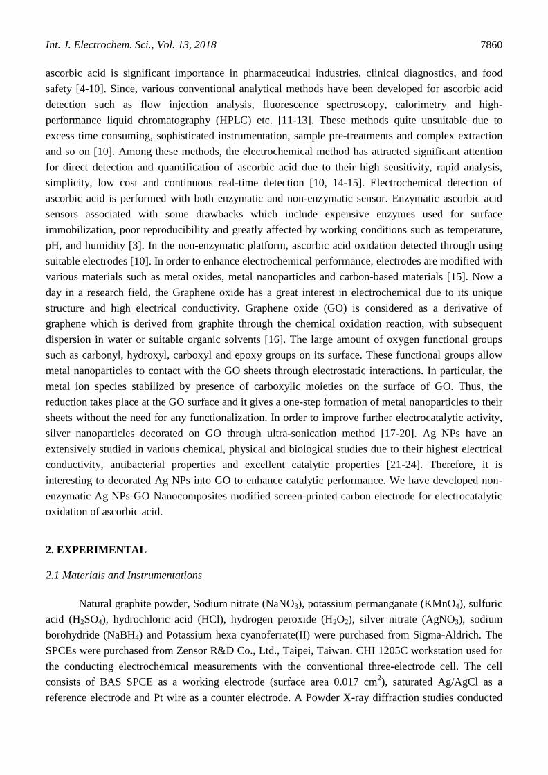
Figure 1. FE-SEM images of GO (A), Ag NPs-GO nanocomposite (B, C) and EDX spectra of
nanocomposite (D).
Surface morphology and elemental composition of Ag NPs-GO nanocomposites were
determined by FE-SEM and EDX. (Figure. 1A) shows smooth and thin sheet-like the structure of
graphene oxide. (Figure. 1B and C) shows a representative SEM images of the Ag NPs-GO
Int. J. Electrochem. Sci., Vol. 13, 2018
7862
nanocomposite. It was clearly seen that a large amount of Ag nanoparticle uniformly distributed at the
GO surface. These Ag nanoparticles in the GO surface are spherical in shape and the sizes ranging
from 50 to 60 nm. The existence of C, O, Ag elements in Ag NPs-GO nanocomposites revealed the
formation of Ag nanoparticles on the surfaces of GO sheets which confirmed through the energy-
dispersive X-ray spectrum results (EDX) (Figure. 1D).
3.2 PXRD and FTIR studies of Ag NPs-GO nanocomposite
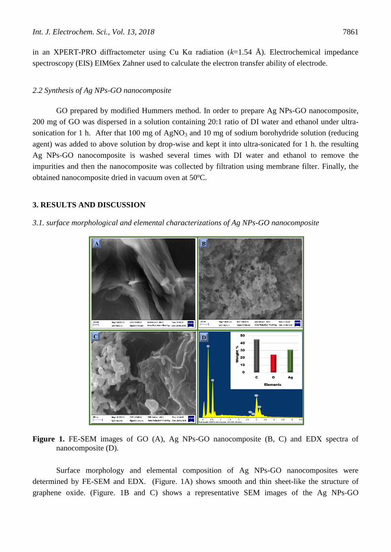
Figure 2. FTIR spectra (A). PXRD spectrum of nanocomposite (B).
(Figure 2A) displays the FT-IR spectra of GO and Ag NPs-GO nanocomposites. GO spectrum
was observed at 3400, 1650 and 1050 cm-1
due to stretching vibration of C-OH (hydroxyl), the
stretching vibration of C=O, and C-O stretch located respectively. Presence of oxygen-containing
groups confirmed that the result of oxidation in the GO and results are also similar to previous studies
[17]. Besides, after decoration with Ag NPs, compare with the spectrum of graphene oxide, the –OH
groups became stretched out while the characteristic peak intensity of GO slightly decreased due to the
interactions between Ag+
ions and oxygen-containing groups on the GO sheets.
The crystallographic structure of synthesized Ag NPs-GO nanocomposites investigated from
XRD pattern (Figure 2B). A sharp diffraction peak observed for GO around 11.7° corresponding to
(001) plane. This result indicated that the successful preparation of GO and matched with references
data. The XRD pattern observed for the Ag NPs-GO nanocomposite around 38.1o, 44.3
o, 64.5
o and
77.5o
corresponding to the crystal lattice planes of (111), (200), (220) and (311) respectively. It
confirmed that the face-centered cubic Ag nanoparticles (JCPDS no 04-0783) formed, respectively
while the peak of GO not observed in the composite due to the metal-nanoparticles strongly interacted
to the GO layers may lead to the sheeting the peak of graphene oxide. Moreover, the sharp peak of
38.1o confirmed that the pure crystalline nature of Ag nanoparticle formed in the Ag NPs-GO
nanocomposites [16-24].
Int. J. Electrochem. Sci., Vol. 13, 2018
7863
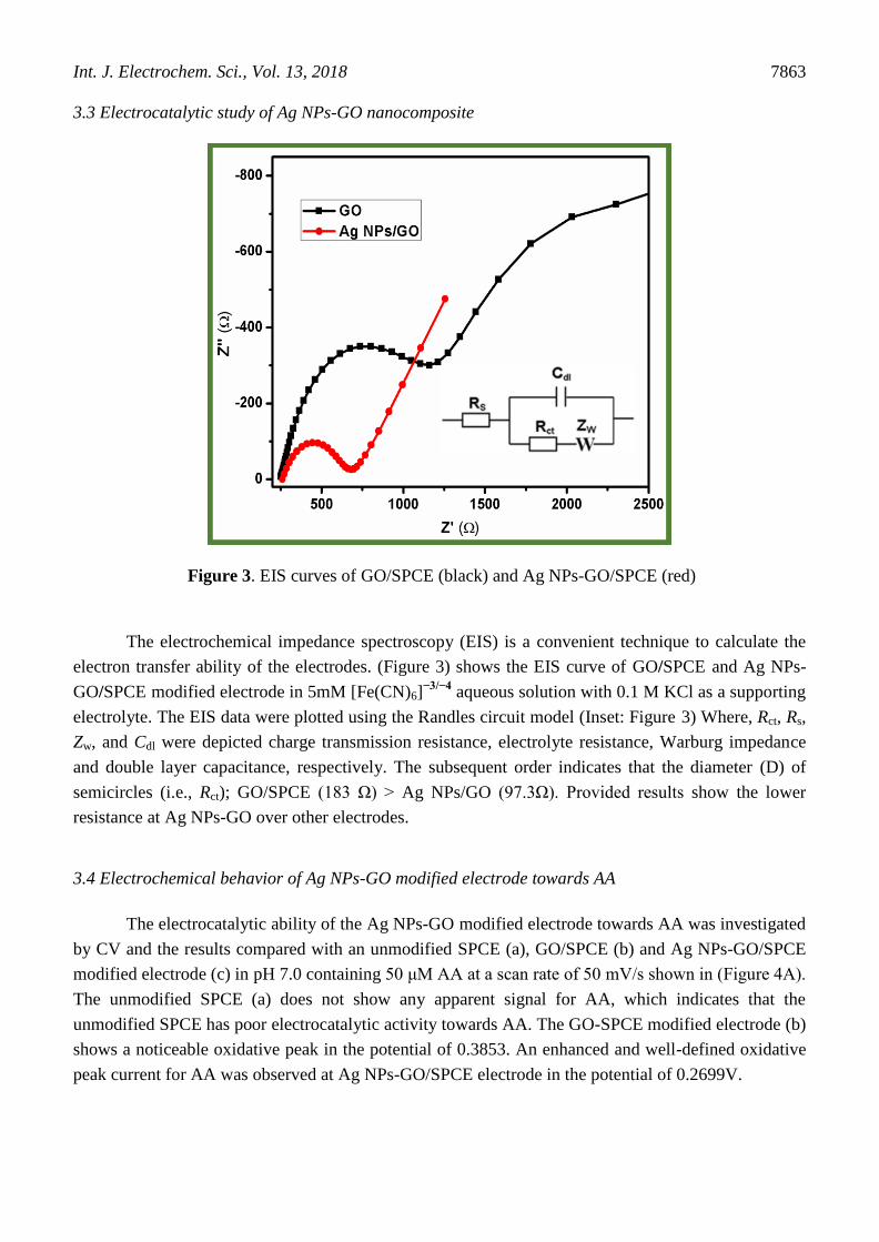
3.3 Electrocatalytic study of Ag NPs-GO nanocomposite
Figure 3. EIS curves of GO/SPCE (black) and Ag NPs-GO/SPCE (red)
The electrochemical impedance spectroscopy (EIS) is a convenient technique to calculate the
electron transfer ability of the electrodes. (Figure 3) shows the EIS curve of GO/SPCE and Ag NPs-
GO/SPCE modified electrode in 5mM [Fe(CN)6]−3/−4
aqueous solution with 0.1 M KCl as a supporting
electrolyte. The EIS data were plotted using the Randles circuit model (Inset: Figure 3) Where, Rct, Rs,
Zw, and Cdl were depicted charge transmission resistance, electrolyte resistance, Warburg impedance
and double layer capacitance, respectively. The subsequent order indicates that the diameter (D) of
semicircles (i.e., Rct); GO/SPCE (183 Ω) > Ag NPs/GO (97.3Ω). Provided results show the lower
resistance at Ag NPs-GO over other electrodes.
3.4 Electrochemical behavior of Ag NPs-GO modified electrode towards AA
The electrocatalytic ability of the Ag NPs-GO modified electrode towards AA was investigated
by CV and the results compared with an unmodified SPCE (a), GO/SPCE (b) and Ag NPs-GO/SPCE
modified electrode (c) in pH 7.0 containing 50 μM AA at a scan rate of 50 mV/s shown in (Figure 4A).
The unmodified SPCE (a) does not show any apparent signal for AA, which indicates that the
unmodified SPCE has poor electrocatalytic activity towards AA. The GO-SPCE modified electrode (b)
shows a noticeable oxidative peak in the potential of 0.3853. An enhanced and well-defined oxidative
peak current for AA was observed at Ag NPs-GO/SPCE electrode in the potential of 0.2699V.
Int. J. Electrochem. Sci., Vol. 13, 2018
7864
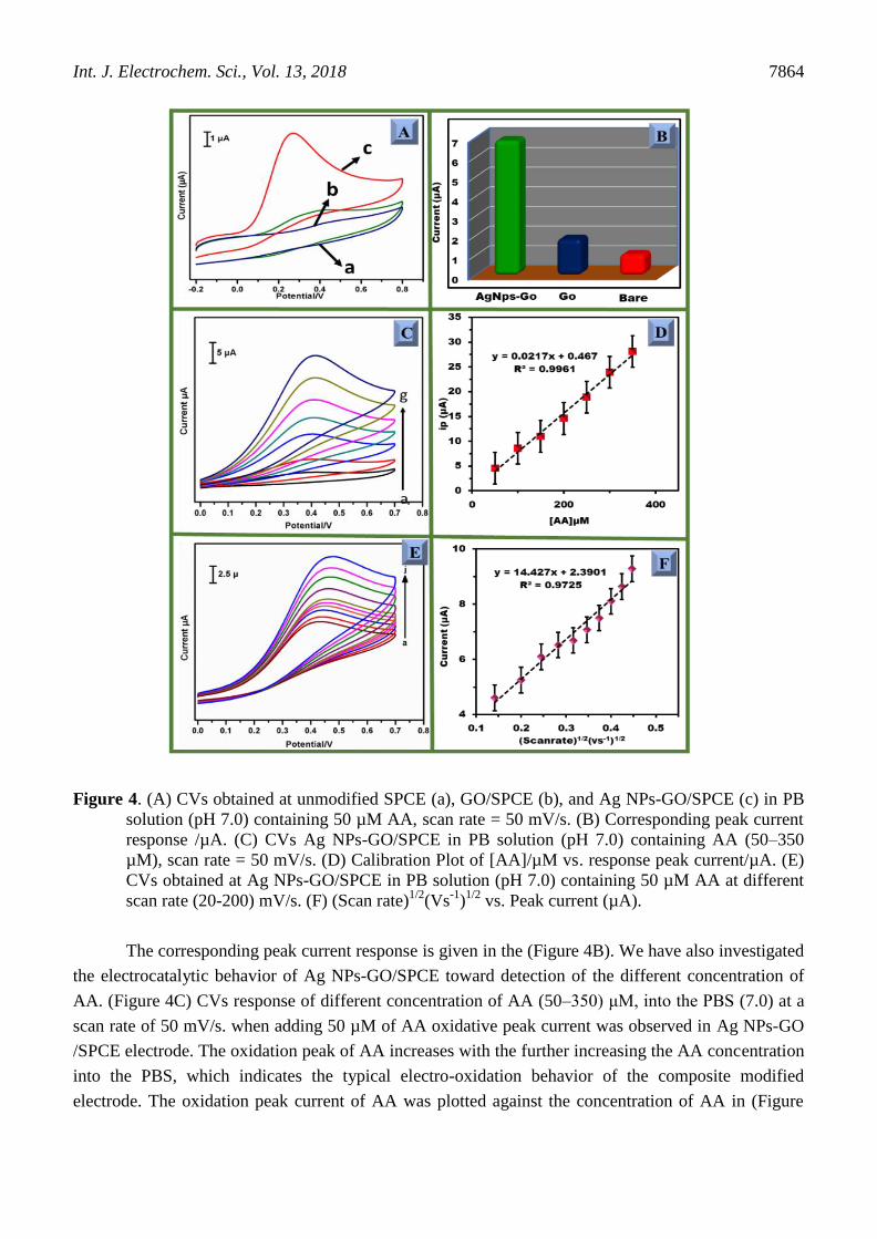
Figure 4. (A) CVs obtained at unmodified SPCE (a), GO/SPCE (b), and Ag NPs-GO/SPCE (c) in PB
solution (pH 7.0) containing 50 µM AA, scan rate = 50 mV/s. (B) Corresponding peak current
response /µA. (C) CVs Ag NPs-GO/SPCE in PB solution (pH 7.0) containing AA (50–350
µM), scan rate = 50 mV/s. (D) Calibration Plot of [AA]/µM vs. response peak current/µA. (E)
CVs obtained at Ag NPs-GO/SPCE in PB solution (pH 7.0) containing 50 µM AA at different
scan rate (20-200) mV/s. (F) (Scan rate)1/2
(Vs-1
)1/2
vs. Peak current (µA).
The corresponding peak current response is given in the (Figure 4B). We have also investigated
the electrocatalytic behavior of Ag NPs-GO/SPCE toward detection of the different concentration of
AA. (Figure 4C) CVs response of different concentration of AA (50–350) μM, into the PBS (7.0) at a
scan rate of 50 mV/s. when adding 50 µM of AA oxidative peak current was observed in Ag NPs-GO
/SPCE electrode. The oxidation peak of AA increases with the further increasing the AA concentration
into the PBS, which indicates the typical electro-oxidation behavior of the composite modified
electrode. The oxidation peak current of AA was plotted against the concentration of AA in (Figure
Int. J. Electrochem. Sci., Vol. 13, 2018
7865
4D). The result also reveals that the Ag NPs-GO composite exhibit excellent electrochemical behavior
towards detection of AA.
(Figure 4E) The effect of scan rate on the electrochemical response of AA at the Ag NPs-GO
modified electrode. It is observed that the oxidation peaks current of AA increases linearly with
increasing the scan rate from 20 to 200 mV s-1
. The oxidation peak current of AA was plotted against
the square root of scan rate shown in (Figure 4F).
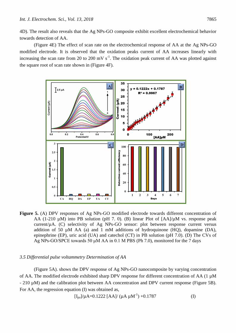
Figure 5. (A) DPV responses of Ag NPs-GO modified electrode towards different concentration of
AA (1-210 µM) into PB solution (pH 7. 0). (B) linear Plot of [AA]/µM vs. response peak
current/µA. (C) selectivity of Ag NPs-GO sensor: plot between response current versus
addition of 50 µM AA (a) and 1 mM additions of hydroquinone (HQ), dopamine (DA),
epinephrine (EP), uric acid (UA) and catechol (CT) in PB solution (pH 7.0). (D) The CVs of
Ag NPs-GO/SPCE towards 50 μM AA in 0.1 M PBS (Ph 7.0), monitored for the 7 days
3.5 Differential pulse voltammetry Determination of AA
(Figure 5A). shows the DPV response of Ag NPs-GO nanocomposite by varying concentration
of AA. The modified electrode exhibited sharp DPV response for different concentration of AA (1 µM
- 210 µM) and the calibration plot between AA concentration and DPV current response (Figure 5B).
For AA, the regression equation (I) was obtained as,
[Ipa]/µA=0.1222 [AA]/ (µA µM-1
) +0.1787 (I)
Int. J. Electrochem. Sci., Vol. 13, 2018
7866
The linear plot revealed good linearity with the slope of 0.997 µA µM-1
.cm-2
. The sensitivity
(1.71 µA µM-1
cm-2
), and limit of detection (25 nM). The linear range, LOD, and sensitivity of Ag
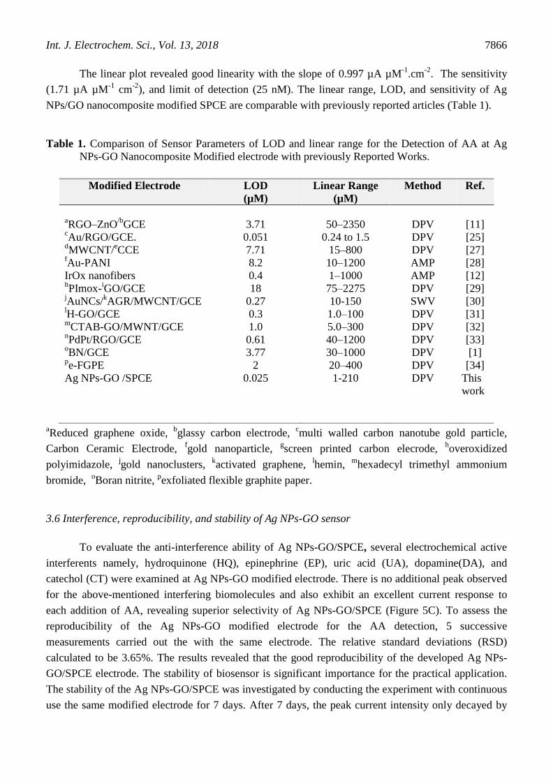
NPs/GO nanocomposite modified SPCE are comparable with previously reported articles (Table 1).
Table 1. Comparison of Sensor Parameters of LOD and linear range for the Detection of AA at Ag
NPs-GO Nanocomposite Modified electrode with previously Reported Works.
Modified Electrode LOD
(µM)
Linear Range
(µM)
Method Ref.
aRGO–ZnO
/bGCE
3.71
50–2350
DPV
[11] cAu/RGO/GCE. 0.051 0.24 to 1.5 DPV [25]
dMWCNT/
eCCE 7.71 15–800 DPV [27]
fAu-PANI 8.2 10–1200 AMP [28]
IrOx nanofibers 0.4 1–1000 AMP [12] hPImox-
iGO/GCE
jAuNCs/
kAGR/MWCNT/GCE
lH-GO/GCE
18
0.27
0.3
75–2275
10-150
1.0–100
DPV
SWV
DPV
[29]
[30]
[31] m
CTAB-GO/MWNT/GCE nPdPt/RGO/GCE
oBN/GCE
pe-FGPE
Ag NPs-GO /SPCE
1.0
0.61
3.77
2
0.025
5.0–300
40–1200
30–1000
20–400
1-210
DPV
DPV
DPV
DPV
DPV
[32]
[33]
[1]
[34]
This
work
aReduced graphene oxide,
bglassy carbon electrode,
cmulti walled carbon nanotube gold particle,
Carbon Ceramic Electrode, fgold nanoparticle,
gscreen printed carbon elecrode,
hoveroxidized
polyimidazole, jgold nanoclusters,
kactivated graphene,
lhemin,
mhexadecyl trimethyl ammonium
bromide, oBoran nitrite,
pexfoliated flexible graphite paper.
3.6 Interference, reproducibility, and stability of Ag NPs-GO sensor
To evaluate the anti-interference ability of Ag NPs-GO/SPCE, several electrochemical active
interferents namely, hydroquinone (HQ), epinephrine (EP), uric acid (UA), dopamine(DA), and
catechol (CT) were examined at Ag NPs-GO modified electrode. There is no additional peak observed
for the above-mentioned interfering biomolecules and also exhibit an excellent current response to
each addition of AA, revealing superior selectivity of Ag NPs-GO/SPCE (Figure 5C). To assess the
reproducibility of the Ag NPs-GO modified electrode for the AA detection, 5 successive
measurements carried out the with the same electrode. The relative standard deviations (RSD)
calculated to be 3.65%. The results revealed that the good reproducibility of the developed Ag NPs-
GO/SPCE electrode. The stability of biosensor is significant importance for the practical application.
The stability of the Ag NPs-GO/SPCE was investigated by conducting the experiment with continuous
use the same modified electrode for 7 days. After 7 days, the peak current intensity only decayed by
Int. J. Electrochem. Sci., Vol. 13, 2018
7867
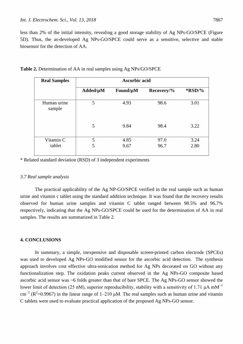
less than 2% of the initial intensity, revealing a good storage stability of Ag NPs-GO/SPCE (Figure
5D). Thus, the as-developed Ag NPs-GO/SPCE could serve as a sensitive, selective and stable
biosensor for the detection of AA.
Table 2. Determination of AA in real samples using Ag NPs/GO/SPCE
Real Samples Ascorbic acid
Added/µM Found/µM Recovery/% *RSD/%
Human urine
sample
5 4.93 98.6 3.01
5 9.84 98.4 3.22
Vitamin C
tablet
5 4.85 97.0 3.24
5 9.67 96.7 2.80
* Related standard deviation (RSD) of 3 independent experiments
3.7 Real sample analysis
The practical applicability of the Ag NP-GO/SPCE verified in the real sample such as human
urine and vitamin c tablet using the standard addition technique. It was found that the recovery results
observed for human urine samples and vitamin C tablet ranged between 98.5% and 96.7%
respectively, indicating that the Ag NPs-GO/SPCE could be used for the determination of AA in real
samples. The results are summarized in Table 2.
4. CONCLUSIONS
In summary, a simple, inexpensive and disposable screen-printed carbon electrode (SPCEs)
was used to developed Ag NPs-GO modified sensor for the ascorbic acid detection. The synthesis
approach involves cost effective ultra-sonication method for Ag NPs decorated on GO without any
functionalization step. The oxidation peaks current observed in the Ag NPs-GO composite based
ascorbic acid sensor was ~6 folds greater than that of bare SPCE. The Ag NPs-GO sensor showed the
lower limit of detection (25 nM), superior reproducibility, stability with a sensitivity of 1.71 μA mM−1
cm−2
(R2=0.9967) in the linear range of 1–210 μM. The real samples such as human urine and vitamin
C tablets were used to evaluate practical application of the proposed Ag NPs-GO sensor.
Int. J. Electrochem. Sci., Vol. 13, 2018
7868
ACKNOWLEDGEMENTS
This work was supported by the National Science Council and the Ministry of Education of Taiwan
(Republic of China) and National Taipei University of Technology, Taipei, Taiwan.
References
1. C.e. Zou, J. Zhong, S. Li, H. Wang, J. Wang, B. Yan, Y. Du, J. Electroanal. Chem., 805 (2017)
110- 119.
2. Y.-G. Lee, B.-X. Liao, Y.-C. Weng, Chemosphere., 173 (2017) 512-519.
3. G. Darabdhara, B. Sharma, M.R. Das, R. Boukherroub, S. Szunerits, Sens. Actuators, B., 238 (2017)
842-851.
4. Q. Li, C. Huo, K. Yi, L. Zhou, L. Su, X. Hou, Sens. Actuators, B., (2018).
5. I. Sebarchievici, A. Lascu, G. Fagadar-Cosma, A. Palade, I. Fringu, M. Birdeanu, B. Taranu, E.
Fagadar-Cosma, C. R. Chim., (2017).
6. M.Y.A. Khan, M. Zahoor, A. Shaheen, N. Jamil, M.I. Arshad, S.Z. Bajwa, N.A. Shad, R. Butt, I.
Ali, M.Z. Iqbal, Mater. Res. Bull., n (2018).
7. L. Zhang, J. Feng, K.-C. Chou, L. Su, X. Hou, J. Electroanal. Chem., 803 (2017) 11-18.
8. L. Fu, A. Wang, G. Lai, W. Su, F. Malherbe, J. Yu, C.-T. Lin, A. Yu, Talanta., 180 (2018) 248-253.
9. P.L. dos Santos, V. Katic, K.C. Toledo, J.A. Bonacin, Sens. Actuators., B 255 (2018) 2437-2447.
10. R. Sha, S. Badhulika, J. Electroanal. Chem., (2018).
11. X. Zhang, Y.-C. Zhang, L.-X. Ma, Sens. Actuators, B 227 (2016) 488-496.
12. S.-j. Kim, Y.L. Kim, A. Yu, J. Lee, S.C. Lee, C. Lee, M.H. Kim, Y. Lee, Sens. Actuators, B., 196
(2014) 480-488.
13. M.A. Alonso-Lomillo, O. Domínguez-Renedo, A. Saldaña-Botín, M.J. Arcos-Martínez, Talanta.,
174 (2017) 733-737.
14. L. Zhang, G. Wang, D. Wu, C. Xiong, L. Zheng, Y. Ding, H. Lu, G. Zhang, L. Qiu, Biosens.
Bioelectron., 100 (2018) 235-241.
15. J.P. Bartolome, A. Fragoso, Inorg. Chim. Acta., 468 (2017) 223-231.
16. Y. Liu, L. Wang, H. Zhang, F. Ran, P. Yang, H. Li, RSC Adv., (64) (2017) 40119-40123.
17. T. Vi, S. Lue, Mater. Sci. Eng., 2016, p. 012033.
18. O. Parkash, C.Y. Yean, R.H. Shueb, Diagnostics., 4(4) (2014) 165-180.
19. K. Hareesh, J. Williams, N. Dhole, K. Kodam, V. Bhoraskar, S. Dhole, Mater. Res. Express., 3 (7)
(2016) 075010.
20. J. Tian, S. Liu, Y. Zhang, H. Li, L. Wang, Y. Luo, A.M. Asiri, A.O. Al-Youbi, X. Sun, Inorg. Chem.,
51(8) (2012) 4742-4746.
21. Y. Haldorai, B.-K. Kim, Y.-L. Jo, J.-J. Shim, Mater. Chem. Phys., 143(3) (2014) 1452-1461.
22. K.R. Reddy, F. Alonso-Marroquin, AIP Publishing., 2017, p. 020003.
23. A. Sudarvizhi, Z.A. Siddiqha, K. Pandian, J Chem Applied Biochem 1(1) (2014) 101.
24. C. Pérez-Ràfols, J. Bastos-Arrieta, N. Serrano, J.M. Díaz-Cruz, C. Ariño, J. De Pablo, M. Esteban,
Sensors., 17(6) (2017) 1458.
25. C. Wang, J. Du, H. Wang, C.e. Zou, F. Jiang, P. Yang, Y. Du, Sens. Actuators, B., 204 (2014) 302-
309.
J. Yan, S. Liu, Z. Zhang, G. He, P. Zhou, H. Liang, L. Tian, X. Zhou, H. Jiang, Colloids Surf B.,
111 (2013) 392-397.
26. B. Habibi, M.H. Pournaghi-Azar, Electrochim. Acta., 55(19) (2010) 5492-5498.
H. Zhang, F. Huang, S. Xu, Y. Xia, W. Huang, Z. Li, Electrochem. Commun., 30 (2013) 46-50.
27. X. Liu, L. Zhang, S. Wei, S. Chen, X. Ou, Q. Lu, Biosens. Bioelectron., 57 (2014) 232-238.
28. Adel A. Abdelwahaba, Yoon-Bo Shimb, Sens. Actuators, B., 221 (2015) 659–665
29. Hao Lin Zou, Bang Lin Li, Hong Qun Luo, Nian Bing Li, Sens. Actuators., B 207 (2015) 535–541
Int. J. Electrochem. Sci., Vol. 13, 2018
7869
30. YuJunYang, WeikunLi, Sens. Actuators., B 56(2014)300–306
31. Jun Yan, Shi Liu, Zhenqin Zhang, Guangwu He, Ping Zhou, Haiying Liang, Lulu Tian,
32. Xuemin Zhou, Huijun Jiang, Colloids Surf B., 111 (2013) 392– 397
33. Weihua Caia, Ting Laia, Haijun Dub, Jianshan Ye, Sens. Actuators, B 193 (2014) 492– 500
© 2018 The Authors. Published by ESG (www.electrochemsci.org). This article is an open access
article distributed under the terms and conditions of the Creative Commons Attribution license
(http://creativecommons.org/licenses/by/4.0/).