simultaneous quantification of 5-fluorouracil and ... · simultaneous quantification of...
TRANSCRIPT

Research Article Open Access
Zafar et al., J Chromatograph Separat Techniq 2014, 5:4http://dx.doi.org/10.4172/2157-7064.1000235
Research Article Open Access
Chromatography Separation Techniques
Volume 5 • Isse 4 • 1000235J Chromat Separation TechniqISSN: 2157-7064 JCGST, an open access journal
Simultaneous Quantification of 5-Fluorouracil and Leucovorin in Pharmaceutical Dosage Form and Human Spiked Plasma by Using RP-HPLC MethodZafar H, Madni MAU*, Arshad S, Altaf H, Khan MI, Mehmood MA and Rehman MDepartment of Pharmacy, The Islamia University of Bahawalpur, Pakistan
*Corresponding author: Madni MAU, Department of pharmacy, The Islamia University of Bahawalpur, Pakistan; E-mail: [email protected]
Received June 28, 2014; Accepted July 11, 2014; Published August 31, 2014
Citation: Zafar H, Madni MAU, Arshad S, Altaf H, Khan MI, et al. (2014) Simultaneous Quantification of 5-Fluorouracil and Leucovorin in Pharmaceutical Dosage Form and Human Spiked Plasma by Using RP-HPLC Method. J Chromatograph Separat Techniq 5: 235. doi:10.4172/2157-7064.1000235
Copyright: © 2014 Zafar H, et al. This is an open-access article distributed under the terms of the Creative Commons Attribution License, which permits unrestricted use, distribution, and reproduction in any medium, provided the original author and source are credited.
AbstractBack ground: 5-Fluorouracil (5-FU) is one of the widely used chemotherapeutic drugs targeting various cancers
including colon cancer. Leucovorin is not a chemotherapy drug, but it is often given as part of chemotherapy. This drug is used to make the chemotherapy more effective and also reduce the risk of side effects. The addition of Leucovorin (LV) to 5-Fuorouracil (5-FU) in advanced colorectal cancer treatment, has shown improved tumor response rates in many trials. Limited data is available for the optimal dosage regimen of both drugs.
Purpose: The purpose of this study was to develop and validate a new method i.e. RP-HPLC for the simultaneous identification and quantification of 5-Fluorouracil (5-FU) and Leucovorin in pharmaceutical dosage form as well as in human spiked plasma.
Method and Results: An HPLC method for the determination of 5-Fluorouracil (5-FU) and Leucovorin in pharmaceutical dosage form in the human spiked plasma, each for 5-Fluorouracil (5-FU) and Leucovorin was developed by using the mobile phase of 20 mM KH2PO4 buffer and methanol at a ratio of (80:20) with pH 5.4 at a flow rate of 1.0 ml min-1. 5-Fluorouracil (5-FU) and Leucovorin were eluted and detected at 242 nm with the retention time of 2.67 and 6.01 min, respectively. The limit of quantification (LOQ) and limit of detection (LOD) values of 5-Fluorouracil (5-FU) were 2.5 and 1.25 ng ml-1, respectively. Similarly, The LOQ and LOD values of Leucovorin were 25 and 12.5 ng ml-1, respectively. Both drugs were eluted through C18 BDS Hypersil column of 150×4.6 mm id with 5 µ particle size. Diclofenac sodium was also co-eluted at 8.61 minutes as an internal standard. The method was found linear for 5-Fluorouracil (5-FU) and Leucovorin in the range of 12.5 to 500 and 25 to 1000 ng ml-1 respectively. Similarly, values of % CV were well within the prescribed limits of ICH guidelines.
Conclusion: The results indicated that the method is sensitive and reliable for the quantification of two drugs simultaneously in pharmaceutical dosage form and in human spiked plasma.
Keywords: 5-Fluorouracil; Leucovorin; Diclofenac sodium; Limit of quantification (LOQ); Limit of detection (LOD); HPLC
IntroductionColorectal cancer is an extremely common type of cancer and has
been considered as the third most common form of the cancer world-wide. It is also known as cancer of the large bowel and includes all cancer originating from the cecum to the anus [1]. Colorectal cancer initially shows no symptom as it breeds slowly and when the symptoms appear large lesions have already developed. Since the late 1980s the role of adjuvant chemotherapy has become increasingly important for the different stages of the cancer and administered according to standard clinical criteria [2]. Five different regimens including, FOLFIRI (5-FU, leucovorin and irinotecan), FOLFOXIRI (leucovorin, 5-FU, oxaliplatin and irinotecan), Cape-Ox (Capecitabine and oxaliplatin), FOLFOX (5-FU, Leucovorin and oxaliplatin) and combination therapy of 5-FU and leucovorin have been used for treating colon cancer. Due to the severe side effects only FOLFOX and combination therapy of 5-FU and leucovorin have been practiced since early 1990s. They are administered intravenously. The only drawback of FOLFOX is because of the oxaliplatin that causes the severe neuropathy along with the other side effects and less tolerated by the elderly patients [3]. So, the adjuvant treatment with 5-FU/LV (Fluorouracil/Leucovorin) has been an international standard of care for stage III colon cancer since the 1990s [4].
Adjuvant chemotherapy with 5-fluorouracil (5-FU) is given to patients with stages II and III tumors under standard schedule and doses [5]. 5-Fluorouracil (5-FU) is an analogue of uracil with a fluorine atom
at C-5 in place of hydrogen and still a widely used anticancer drug [6]. It is available in the monohydrate form, having the structure similarity to that of the pyrimidine molecules of RNA and DNA [7]. It interferes with nucleoside metabolism causing the impairment into RNA and DNA structure, leading to cytotoxicity and cell death [8]. It acts as the most extensively used drug in the treatment of the metastatic colorectal cancer, with stages II and III tumors under standard schedule and doses [9]. The administration of this drug in combination with leucovorin in the adjuvant setting is coupled with a survival benefits for patients with colorectal cancers and gastric cancers and may show considerably high response rate along with no cross resistance [10]. Leucovorin is also known as folinic acid or 5-formyl-5, 6, 7, 8-tetrahydrofolic acid. It is a chemically reduced very potent derivative of folic acid and is useful as an antidote to the drugs which act as folic acid antagonist [11]. Unlike the folic acid, leucovorin does not need di-hydro-folate reductase for the reduction and readily changed to other reduced folic
Citation: Zafar H, Madni MAU, Arshad S, Altaf H, Khan MI, et al. (2014) Simultaneous Quantification of 5-Fluorouracil and Leucovorin in Pharmaceutical Dosage Form and Human Spiked Plasma by Using RP-HPLC Method. J Chromatograph Separat Techniq 5: 235. doi:10.4172/2157-7064.1000235
Page 2 of 8
Volume 5 • Issue 4 • 1000235J Chromat Separation TechniqISSN: 2157-7064 JCGST, an open access journal
acid derivatives i.e. tetra-hydro-folate [12]. This permits purine and thymidine synthesis, and thus RNA, DNA and protein synthesis to occur and is not affected by the blockages of the enzyme by the folic acid antagonists. Leucovorin increases the cytotoxicity properties of fluoropyrimidines such as fluorouracil by their metabolites such as fluorodeoxyuridine monophosphate and methylene tetrahydrofolate. These metabolites form a stable ternary complex with thymidylate synthase, causing the decreased level of the enzyme with in the cell which leads to the cell death [13].
Leucovorin has almost no side effects of its own but when given in combination with 5-FU, along with the beneficial anti-cancer effects it can enhance the severity of side effects of the drug. So, the possible benefit of this combination on overall survival was still doubtful and it became essential, therefore to develop and validate new or innovative analytical methods for 5-FU and LV. Because of the several advantages such as accuracy, precision, rapidity, ease of automation and specificity, HPLC methods can be used for the analysis of the drugs in single/multi component dosage forms. HPLC method eradicates time taking extraction and isolation procedures [14]. The present study is aimed to ensure that cancer patients have an improved quality of life, instead of focusing on the uncommon resources of developing new drugs, with possibly new side-effects. It has been focused our attention towards ensuring the rational and safe use of existing drugs and therapies. Thus, a suitable reverse phase- high performance liquid chromatography (HPLC) method has been developed and validated to quantitatively analyze the concentrations of 5-FU and LV necessary for the therapeutic drug monitoring.
ExperimentalChemicals and Reagents
5-Fluorouracil and leucovorin were donated as a gift sample by Pharmedic laboratories (pvt) Ltd. Lahore, Pakistan. Diclofenac sodium was used as an internal standard (IS). Human plasma was obtained by Blood Bank and Thalassemia Canter, Bahawal Victoria Hospital, Bahawalpur, Pakistan. Potassium di-hydrogen phosphate, Phosphoric acid, HPLC grade methanol, ethanol and acetonitrile were purchased from Merck KGaA, Darmstadt, Germany. De-ionized double distilled water was prepared in Laboratory of Department of Pharmacy IUB.
Chromatographic Equipments and Instruments
For the HPLC analysis Perkin Elmer Series 200 Pump, Perkin Elmer Series 200 UV-Vis Detector, Perkin Elmer Series 200 column Oven and Perkin Elmer NCI 900 system were used and total chrom software version 6.3.1 was used for measurement. Other equipments used included: Filtration Assembly( Sartorius, Germany), Vacuum Pump (ILMVAC, Germany), Sample concentrator (DB.3A, Techne, England), Injection filtration assembly (Sartorius, Germany),
Membrane Filters ( Sartorious Stedim, Germany), Reacti-vialsTM Small Reaction vials (#13223, Thermo Scientific, Rockford, USA), Cellulose acetate 0.45µm filter (Sartorius Stedim Biotech GmbH, Germany). For the determination absorbance and spectrum Plate reader (96-well), Synergy HT, Biotek Instruments USA was used. Gen-5 software was used for measurements. UV-Plates were purchased from Invitrogen. Water distillation apparatus (IM 100 Irmeco GmbH Germany), and all the glass ware was purchased from Pyrex England.
Preparation of phosphate bufferStock solution of 20 mM KH2PO4 was prepared by mixing its 2.738g
in sufficient amount of de-ionized double distilled water to make the final volume of 1000 ml. The pH of this buffer was 4.5.
Preparation of mobile phaseMobile phase was prepared by mixing of 20 mM phosphate
buffer and gradient grade methanol at a ratio of 80:20 (v/v). The pH of this mobile phase was 5.4. By using 0.45 µm membrane filter, the mobile phase was filtered. Later on, just before the use, sonication and degassing of the mobile phase was performed. For every analysis, a freshly prepared mobile phase after filtration, sonication and degassing was used.
Preparation of stock solutions of 5-Fluorouracil and leucovorin
Stock solution of 5-FU (1 mg ml-1) was prepared in the mobile phase. Further dilutions were made up to 1.25 ng ml-1 with the help of mobile phase. In the similar manner, the stock solution of LV (1 mg ml-
1) was prepared in the mobile phase. Further dilutions were made up to 12.5 ng ml-1 with the help of mobile phase. Fresh solutions were made daily, filtered and degassed by sonication and then used.
Selection of suitable wavelength for HPLC analysisThe aim was to develop such method that would allow detection
in terms of concentration and complete separation of 5-FU and LV. Furthermore, the method must also explain and determine any metabolic interactions between these two selected drugs i.e. 5-FU and LV. It was initially decided to test whether ultraviolet (UV) spectrophotometry is a suitable method of analysis and whether it adequately allows for the separation and detection of 5-FU and LV. A Spectra System UV 2020, IRMECO, UV spectrophotometer was used. When subjected to running wavelength scans by UV spectrophotometry, the lambda maximum, λmax, the wavelength at which maximum absorption of the drug occurs, was 258 nm for 5-FU and 240 nm for LV (Figure 1a and b) respectively.
Since their peaks occur at approximately the same wavelength (230-270 nm), it was concluded that UV spectrophotometry is not sensitive enough to allow the separation of the 5-FU and LV (Figure 2a
HN
HNO O
Fluoropyrimidine-2,4(1H,3H)-dione
N
HN
N
HN
O
CsO
HN
HN
H
O
O
H2N
CHO
O
O
cesium(II) 2-(4-((2-amino-5-formyl-4-oxo-1,4,5,6,7,8-hexahydropteridin-6-yl)methylamino)benzamido)pentanedioate
(Leucovorin Calcium)(a) (b)
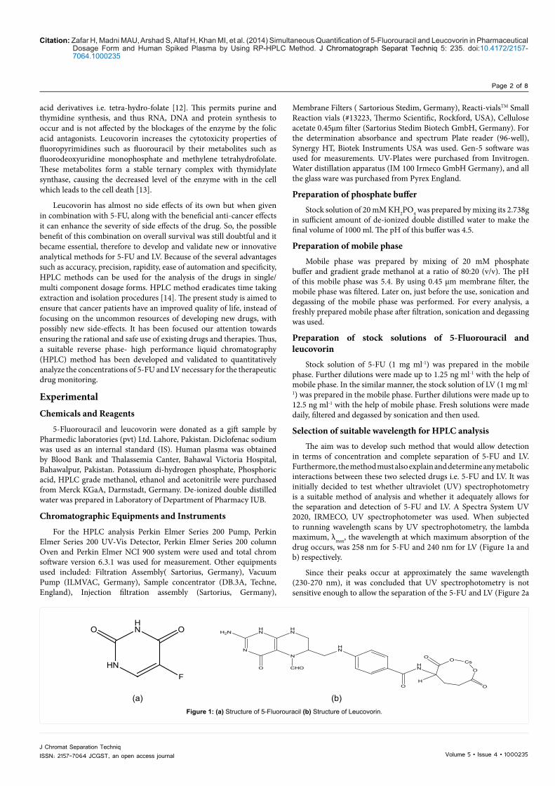
Figure 1: (a) Structure of 5-Fluorouracil (b) Structure of Leucovorin.

Citation: Zafar H, Madni MAU, Arshad S, Altaf H, Khan MI, et al. (2014) Simultaneous Quantification of 5-Fluorouracil and Leucovorin in Pharmaceutical Dosage Form and Human Spiked Plasma by Using RP-HPLC Method. J Chromatograph Separat Techniq 5: 235. doi:10.4172/2157-7064.1000235
Page 3 of 8
Volume 5 • Issue 4 • 1000235J Chromat Separation TechniqISSN: 2157-7064 JCGST, an open access journal
and b). Evidence in the literature showed that high performance liquid chromatography (HPLC) is the best method of quantitatively analyzing 5-FU and LV in plasma, and it was thus decided to develop an HPLC method that would allow for the separation and detection of 5-FU and LV [15].
Chromatographic conditions
This process involves determining the exact analytical conditions, optimal for the quantitative analysis of the particular drugs. RP-HPLC was used for the analysis of 5-Fluorouracil and leucovorin. Both the drugs were eluted through C18 BDS Hypersil column of 150×4.6 mm id, and 5 µm particle size. It was decided to vary the wavelength and HPLC analysis was performed at 239 nm, 242 nm, 248nm, 252 and 258 nm. The optimal wavelength that was selected was 242 nm as it gave sufficiently large peaks for both 5-FU and leucovorin, and fell between their individual λmax values. Both drugs showed adequate absorption of UV radiation at 242 nm. The flow rate was adjusted at 1.0 ml min-1 and run time of this method was adjusted at 15 min. A chromatogram was generated, which showed a peak occurring when the respective compound is eluted.
Peak identification and retention time of 5-Fluorouracil, leucovorin & IS in mobile phase
5-Fluorouracil and Leucovorin peaks were identified by the comparison of retention times of sample and pure drug standard
solutions. The retention times for 5-Fluorouracil and Leucovorin were 2.67 and 6.01 min, respectively. A representative chromatogram is given in Figure 3 a-c. Similarly, the peak of diclofenac sodium was identified by the retention time of pure diclofenac sodium standard solution. The retention time of both the drug was compared with IS which was 8.61 and is shown in Figure 4 a-c.
Blank plasma sample preparation
Human plasma 0.5 ml was taken in a centrifuge tube, vortexed and sonicated for 5 min. Then 1.5 ml of gradient grade methanol was added and vortexed it for 5 min. This sample was centrifuged at 4000 rpm for 5 min. The clear supernanent was taken in the reactivial for drying. This sample was then placed in sample concentrator for 30 min. to evaporate complete methanol. The residues were reconstituted with the filtered mobile phase to make a volume of 0.5 ml. This reconstituted plasma sample was filtered through the 0.45 µm membrane filter paper with the help of injection filtration assembly. Now, 20 µl of sample were injected in the column.
Standard plasma sample preparation contains 5-fluorouracil leucovorin and IS
Human plasma (20 µl) was spiked with known concentrations of 5-FU, LV and IS. Then, vortexed and sonicated this spiked plasma for 5 min each. It was followed by the addition of 60 µl of methanol. This mixture was then again vortexed for 5 min and centrifuged at 4000
(a) (b)
Figure 2: (a) UV spectrum of 5-fluorouracil, showing maximum absorbance at 258 nm. (b) UV spectrum of Leucovorin, showing maximum absorbance at 240 nm. which was 8.61 and is shown in Figure: 4a-c.
(a) (b) (c)
5-fluorouracil 2.67 min
Leucovorin 6.01 min
Leucovorin 6.01 min
5-fluorouracil 2.67 min
Figure 3: Chromatogram of (a) 5-Fluorouracil (b) Leucovorin (c) Simultaneous determination of 5- Fluorouracil and Leucovorin.

Citation: Zafar H, Madni MAU, Arshad S, Altaf H, Khan MI, et al. (2014) Simultaneous Quantification of 5-Fluorouracil and Leucovorin in Pharmaceutical Dosage Form and Human Spiked Plasma by Using RP-HPLC Method. J Chromatograph Separat Techniq 5: 235. doi:10.4172/2157-7064.1000235
Page 4 of 8
Volume 5 • Issue 4 • 1000235J Chromat Separation TechniqISSN: 2157-7064 JCGST, an open access journal
rpm for another 5 min. The supernatant was taken in the reactivial for drying. The spiked and centrifuged human plasma was dried for 30 min and the residue was reconstituted with 20 µl of mobile phase. This reconstituted plasma sample was injected in the column after passing it through 0.45 µm membrane filter.
Peak identification and retention time of 5-Fluorouracil, leucovorin and IS in human spiked plasma
5-Fluorouracil and Leucovorin peaks were identified by the comparison of retention times of sample and pure drug standard solutions, the increase or decrease in size of the peak with a change in the concentration of standard solution was determined. The retention times for 5-Fluorouracil and Leucovorin were 2.67 and 6.01 min, respectively. A representative chromatogram is given in Figure 5a-d. Similarly, the peak of diclofenac sodium was identified by the retention time of pure diclofenac sodium standard solution. The retention time of both the drug was compared with IS which was 8.61 and is shown in Figure 6a-c.
Method validation
The calibration curves of 5-Fluorouracil and Leucovorin are the plots of peak area ratio of the drugs to the IS as a function of concentration of drugs. The unknown concentrations of both drugs were determined from these equations. The precision of the method was based on intra-day variability and determined by triplicate analyses of the calibration standards. The reproducibility was taken as the inter-day variability
and was determined by replicate analyses of the calibration standards. The accuracy of the method was determined by comparing practical amounts recovered from the control samples with actual values present in the samples of both drugs.
Results and DiscussionMethod development and optimization
The RP-HPLC method was based on modifications of methods already available for the determination of 5-FU and LV in Plasma. In the reference method [16], 5 mM KH2PO4 buffer maintained at pH 7.0 was used for 5-FU whereas 10 mM KH2PO4 (pH: 7.0) was applied for LV in combination with acetonitrile in the molar ratio of 70 : 30 (V/V) with a flow rate of 1 ml.min-1. In the present study, a series of experiments have been performed to optimize the ionic concentration buffer and a 20 mM KH2PO4 and methanol in the molar ratio of 80 : 20 (V/V) and final mixture of mobile phase was maintained at pH 5.4. This composition of mobile phase provided better separation and resolution of these drugs with a run time of 10 minutes. Symmetric and better resolved peaks with less tailing were observed with the optimized mobile phase composition. The replacement of acetonitrile by Alsarra and Alarifi, (2004) with methanol also provided better separation and peak symmetry of 5-FU and LV in dosage form and human plasma.
Method Validation
The standardization and validation of newly developed analytic methods was evaluated by the parameters provided in the ICH
(a) (b) (c)
5-Fluorouracil 2.67 min Diclofenac sodium 8.61 min
5-Fluorouracil 2.67 min
Diclofenac sodium 8.61 min Diclofenac sodium 8.61 min Leucovorin 6.01 min
Leucovorin 6.01 min
Figure 4: Chromatogram of (a) 5-Fluorouracil with IS (b) Leucovorin with IS (c) Simultaneous determination of 5- Fluorouracil and Leucovorin with IS.
(a) (b) (c) (d)
5-fluorouracil 2.67 min
5-fluorouracil 2.67 min
Leucovorin 6.01 min
Leucovorin 6.01 min
Figure 5: Chromatogram of (a) Blank plasma (b) plasma with 5-Fluorouracil (c) plasma with Leucovorin (d) Simultaneous determination of 5- Fluorouracil and Leucovorin with plasma.

Citation: Zafar H, Madni MAU, Arshad S, Altaf H, Khan MI, et al. (2014) Simultaneous Quantification of 5-Fluorouracil and Leucovorin in Pharmaceutical Dosage Form and Human Spiked Plasma by Using RP-HPLC Method. J Chromatograph Separat Techniq 5: 235. doi:10.4172/2157-7064.1000235
Page 5 of 8
Volume 5 • Issue 4 • 1000235J Chromat Separation TechniqISSN: 2157-7064 JCGST, an open access journal
guidelines, i.e. linearity, accuracy, precision, stability, sensitivity and robustness [17].
Linearity: In the mobile phase the linearity was accessed by constructing standard curves for both drugs in the linear dynamic range of 12.5 to 500 and 25 to 1000 ng ml-1 for 5-FU and LV (Table 1) respectively. Linear dynamic range was determined by applying straight line fit equation and analysis of correlation coefficient [18]. The values of slope and intercepts for FU and LV were 0.0042, 0.2061 and 0.0091, 0.276, respectively. The correlation coefficient (r2) was found as 0.9990 and 0.9998 for FU and LV, respectively (Figures 7 and 8).
According to the methods described previously the linearity range for the 5-FU was 0.1-10 µg.ml-1 and the results obtained by our method show more linear behavior than that of the previous one. Furthermore the linear behavior of both the drugs in the spiked plasma was accessed by constructing standard curves for both drugs in the linear dynamic range of 100 to 1250 and 125 to 1500 ng.ml-1 for 5-FU and LV (Table 2). This was also described previously by another method where LV showed linear behavior ranging from 50-500 ng.ml-1. The values of slope, intercept for 5-FU and LV were 0.0033, 0.3478 and 0.0096, 0.2956, respectively. The correlation coefficient (r2) was found as 0.9996 and 0.9991. (Figures 9 and 10).
(a) (b) (c)
5-fluorouracil 2.67 min
5-fluorouracil 2.67 min
Leucovorin 6.01 min
Leucovorin 6.01 min
Diclofenac sodium 8.61 min
Diclofenac sodium 8.61 min
Diclofenac sodium 8.61 min
Figure 6: Chromatogram in plasma (a) 5-Fluorouracil with IS (b) Leucovorin with IS (c) Simultaneous determination of 5- Fluorouracil and Leucovorin with IS.
Concentration(5-Fluorouracil) (ng ml-1) Area (mV) Concentration
(Leucovorin) (ng ml-1) Area (mV)
12.5 0.223 25 0.42825.0 0.296 50 0.71850.0 0.429 100 1.285
100.0 0.649 125 1.526125.0 0.765 250 2.683250.0 1.276 500 4.913500.0 2.313 1000 9.214
Table 1: Peak area of different conc. of 5-FU and LV for calibration curve in mobile phase.
Figure 7: Calibration curve of 5-Flurouracil in mobile phase by HPLC method.

Citation: Zafar H, Madni MAU, Arshad S, Altaf H, Khan MI, et al. (2014) Simultaneous Quantification of 5-Fluorouracil and Leucovorin in Pharmaceutical Dosage Form and Human Spiked Plasma by Using RP-HPLC Method. J Chromatograph Separat Techniq 5: 235. doi:10.4172/2157-7064.1000235
Page 6 of 8
Volume 5 • Issue 4 • 1000235J Chromat Separation TechniqISSN: 2157-7064 JCGST, an open access journal
Accuracy and Precision: Accuracy is defined by ISO/IEC as “closeness of agreement between a true quantity value and measured quantity value of an analyte”. Similarly precision expresses a close relationship (degree of scatter) between a series of results obtained from multiple sampling of the same identical sample under the recommended conditions. The inter-batch and intra-batch studies were performed in both cases i.e. mobile phase as well as spiked plasma and the results were compared with the published literature. In this method the mean values of accuracy and % CV values at low, medium and high concentrations were observed for both the drugs and are mentioned in the tables given below (Tables 3 and 4). The results showed good response and accuracy values for both drugs in mobile phase as well as in plasma.
Limit of detection and quantification: The retention times of 5-FU, leucovorin and IS were 2.67, 6.01 and 8.61 min, respectively. The limit of detection (LOD) and limit of quantification (LOQ) for 5-FU were 1.25 and 2.5 ng ml-1, respectively. Similarly, the LOD and LOQ for Leucovorin were 12.5 and 25 ng ml-1, respectively (Table 5). Different studies were carried out by different scientist and according to Alsarra and Alarifi, 2004; the LOQ was 30 ng ml-1 and the limit of detection (LOD) of 5-FU was10 ng.ml-1. Similarly, the values of LOD and LOQ of 5-FU were reported as 10.86 and 32.78 ng.ml-1, respectively by [19].
Stability studies: The freeze–thaw cycle should be repeated two more times, and then analyzed on the third cycle. The % CV of low concentration of 5-FU API and pharmaceutical dosage form dissolved in mobile phase and spiked in plasma were 2.185, 2.385 and 0.308, respectively. Whereas, % CV of low concentration of LV, API and pharmaceutical dosage form dissolved in mobile phase and spiked in plasma were 1.497, 1.539 and 1.537 respectively. The % CV of high concentration of similar dug and pharmaceutical dosage form dissolved in mobile phase and spiked in plasma were 0.129, 0.1601 and 0.188, respectively. Similarly, the % CV of high concentration of similar dug and pharmaceutical dosage form dissolved in mobile phase and spiked in plasma were 0.0861, 0.0874 and 0.113, respectively (Table 6).
Robustness: Robustness processes the ability of an analytical method to persist changes by insignificant but deliberate deviations in method parameters. Parameters that should be considered are percent organic content in the mobile phase, pH of the mobile phase, buffer concentration, and injection volume. By using the standard curves developed earlier, the percentage recovery of 5-FU and LV in human spiked plasma was calculated. The average percentage recovery of 5-FU and LV in the mobile phase was 101.12 and 100.71%, respectively [20-22]. Similarly, the average percentage recovery of 5-FU and LV in spiked plasma was 99.92 and 98.65%, respectively.
ConclusionAn HPLC method for simultaneous determination of 5-Fluorouracil
and Leucovorin for in vitro analysis have not been reported previously. The presented method in addition to its novelty for determination of two ingredients at single wavelength is sufficiently rapid, simple, and sensitive as well as precise and accurate, which complies with ICH guidelines for accuracy, precision, and stability for standards and QC samples. The assay of the both active ingredients was not interfered by the excipients in the pharmaceutical dosage forms and human plasma. The linearity, accuracy, precision, LOD, LOQ, and specificity were established. The method is recommended in the quality control analysis and for bioequivalence and pharmacokinetic studies.
Figure 8: Calibration curve of Leucovorin in mobile phase by HPLC method.
Figure 9: Calibration curve of 5-Flurouracil in spiked plasma by HPLC method.
.
Figure 10: Calibration curve of Leucovorin in spiked plasma by HPLC method.
Concentration(5-Fluorouracil) (ng ml-1) Area (mV) Concentration
(Leucovorin) (ng ml-1) Area (mV)
100 0.623 125 1.593125 0.734 250 2.672250 1.225 500 4.984500 2.038 1000 9.7531000 3.575 1250 12.2361250 4.453 1500 14.624
Table 2: Peak areas of different conc. of 5-FU and LV for calibration curve in spiked plasma.

Citation: Zafar H, Madni MAU, Arshad S, Altaf H, Khan MI, et al. (2014) Simultaneous Quantification of 5-Fluorouracil and Leucovorin in Pharmaceutical Dosage Form and Human Spiked Plasma by Using RP-HPLC Method. J Chromatograph Separat Techniq 5: 235. doi:10.4172/2157-7064.1000235
Page 7 of 8
Volume 5 • Issue 4 • 1000235J Chromat Separation TechniqISSN: 2157-7064 JCGST, an open access journal
Accuracy and precision
5-FU LV
S. D. % CV %Accuracy S. D. % CV % Accuracy
Inter-batch High Conc.
(500 ng ml-1)
Inter-batch High Conc.
(1000 ng ml-1)0.3502 0.0701 99.90 0.6982 0.0699 99.88
Medium Conc.(100 ng ml-1) 0.2189 0.2192 99.89 Medium Conc.
(125 ng ml-1) 0.1256 0.1006 99.87
Low Conc.(12.5 ng ml-1) 0.0515 0.4128 99.80 Low Conc.
(25 ng ml-1) 0.1166 0.4672 99.87
Intra-batch High Conc.
(500 ng ml-1)
Intra-batch High Conc.
(1000 ng ml-1)4.7030 0.9455 99.83 2.4406 0.2441 99.89
Medium Conc.(100 ng ml-1) 0.7865 0.7883 99.78 Medium Conc.
(125 ng ml-1) 1.1012 0.8834 99.72
Low Conc.(12.5 ng ml-1) 0.0821 0.6576 99.48 Low Conc.
(25 ng ml-1) 0.6985 2.8147 99.26
Table 3: Inter-batch & Intra-batch accuracy and precision for 5-FU and LV in Mobile phase.
Accuracy and precision 5-FU LV
S. D. % CV %Accuracy S. D. % CV % Accuracy
Inter-batch High Conc.
(1250 ng ml-1)
Inter-batch High Conc.
(1000 ng ml-1)1.8112 0.1450 99.94 2.0668 0.1380 99.86
Medium Conc.(500 ng ml-1)
1.8718 0.3748 99.88 Medium Conc.(125 ng ml-1)
1.7637 0.1766 99.89
Low Conc.(100 ng ml-1)
1.4562 1.4674 99.23 Low Conc.(25 ng ml-1)
1.2787 1.0246 99.84
Intra-batch High Conc.
(1250 ng ml-1)
Intra-batch High Conc.
(1000 ng ml-1)2.7027 0.2167 99.77 3.6955 0.2469 99.79
Medium Conc.(500 ng ml-1) 1.6034 0.3217 99.70 Medium Conc.
(125 ng ml-1) 2.0857 0.2090 99.80
Low Conc.(100 ng ml-1) 0.8861 0.9017 98.27 Low Conc.
(25 ng ml-1) 1.1694 0.9399 99.53
Table 4: Inter-batch & Intra-batch accuracy and precision for 5-FU and LV in spiked plasma.
Parameter Results (mobile phase) Results (spiked plasma)Rt of 5-Fluro uracil 2.67 ± 0.03 min 2.67 ± 0.03 min
Rt of Leucovorin 6.01 ± 0.03 min 6.01 ± 0.03 min
Rt of diclofenac sodium 8.61 ± 0.03 min 8.61 ± 0.03 min
LOQ of 5-Fluro uracil 2.5 ng ml-1 2.5 ng ml-1
LOD 5-Fluro uracil 1.25 ng ml-1 1.25 ng ml-1
LOQ of Leucovorin 25 ng ml-1 25 ng ml-1
LOD of Leucovorin 12.5 ng ml-1 12.5 ng ml-1
Table 5: Parameters regarding HPLC analysis of 5-Flurouracil and Leucovorin.
5-FU LVS. D. % CV %Accuracy S. D. % CV % Accuracy
5-FU, APIHigh Conc. (500 ng ml-1)
LV, APIHigh Conc. (1000 ng ml-1)0.6438 0.1291 99.72 0.8588 0.0861 99.793
Medium Conc. (12.5 ng ml-1) 0.2692 2.185 98.563 Medium Conc. (25 ng ml-1) 0.3678 1.4974 98.251
Utoral, 250 mginjection
High Conc. (500 ng ml-1)
Kyunrine, 15 mg injectionHigh Conc. (1000 ng ml-1)0.7982 0.1601 99.719 0.8728 0.0874 99.813
Medium Conc. (12.5 ng ml-1) 0.2928 2.385 98.195 Medium Conc. (25 ng ml-1) 0.3750 1.5393 97.444
5-FU in spiked plasmaHigh Conc. (500 ng ml-1)
LV in spiked plasmaHigh Conc. (1000 ng ml-1)0.9383 0.1888 99.401 1.130 0.1133 99.752
Medium Conc. (12.5 ng ml-1) 0.0382 0.308 99.351 Medium Conc. (25 ng ml-1) 0.3784 1.5373 98.462
Table 6: Stability data of 5-FU & LV APIs and pharmaceutical dosage forms in mobile phase and in spiked plasma.

Citation: Zafar H, Madni MAU, Arshad S, Altaf H, Khan MI, et al. (2014) Simultaneous Quantification of 5-Fluorouracil and Leucovorin in Pharmaceutical Dosage Form and Human Spiked Plasma by Using RP-HPLC Method. J Chromatograph Separat Techniq 5: 235. doi:10.4172/2157-7064.1000235
Page 8 of 8
Volume 5 • Issue 4 • 1000235J Chromat Separation TechniqISSN: 2157-7064 JCGST, an open access journal
Acknowledgement
Thanks to Pharmedic laboratories (pvt) Ltd. Lahore, Pakistan who provided pure material of 5-Fluorouracil and leucovorin as a gift.
References
1. Bond J, Tuckey M (2009) An evaluation of current methods of diagnosing colorectal cancer in the United Kingdom. Radiography 15: 106-112.
2. Ribic CM, Sargent DJ, Moore MJ, Thibodeau SN, French AJ, et al. (2003) Tumor Microsatellite-Instability status as a predictor of benefit from Fluorouracil-based adjuvant Chemotherapy for Colon Cancer. The N Engl J Med 349: 247-257.
3. Faithfull S, Deery P (2004) Implementation of capecitabine (Xeloda) into a cancer centre: UK experience. Eur J Oncol Nurs 8 Suppl 1: S54-62.
4. Twelves C, Wong A, Nowacki MP, Abt M, Burris H 3rd, et al. (2005) Capecitabine as Adjuvant Treatment for Stage III Colon Cancer. N Engl J Med 352: 2696-2704.
5. Labianca R, Beretta G, Gatta G, de Braud F, Wils J (2004) Colon cancer. Crit Rev Oncol Hematol 51: 145-170.
6. Rutman RJ, Cantarow A, Paschkis KE (1954) Studies on 2-acetylaminofluorene carcinogenesis: III. The utilization of uracil-2-C14 by pre–neoplastic rat liver. Cancer Research 14: 119-123.
7. Hulme AT, Price SL, Tocher DA (2005) A new polymorph of 5-fluorouracil found following computational crystal structure predictions. J Am Chem Soc 127: 1116-1117.
8. van Kuilenburg AB, van Lenthe H, Maring JG, van Gennip AH (2006) Determination of 5-fluorouracil in plasma with HPLC-tandem mass spectrometry. Nucleosides Nucleotides Nucleic Acids 25: 1257-1260.
9. Maring JG, Schouten L, Greijdanus B, de Vries EG, Uges DR (2005) A simple and sensitive fully validated HPLC-UV method for the determination of 5-fluorouracil and its metabolite 5,6-dihydrofluorouracil in plasma. Ther Drug Monit 27: 25-30.
10. Omura K (2008) Advances in chemotherapy against advanced or metastatic colorectal cancer. Digestion 77 Suppl 1: 13-22.
11. Jardine LF, Ingram LC, Bleyer WA (1996) Intrathecal leucovorin after intrathecal methotrexate overdose. J Pediatr Hematol Oncol 18: 302-309.
12. Bano N, Najam R, Mateen A, Qazi F (2012) High and low dose folinic acid, 5- fluorouracil bolus and continuous infusion for poor-prognosis patients with advanced colorectal carcinoma. Asian Pac J Cancer Prev 13: 3589-3593.
13. Benson AB 3rd, Arnoletti JP, Bekaii-Saab T, Chan E, Chen YJ, et al. (2011) Colon Cancer. J Natl Compr Canc Netw. 9: 1238-1289.
14. Soumaoro LT, Uetake H, Higuchi T, Takagi Y, Enomoto M, et al. (2004) Cyclooxygenase-2 expression: a significant prognostic indicator for patients with colorectal cancer. Clin Cancer Res 10: 8465-8471.
15. Alsarra IA, Alarifi MN (2004) Validated liquid chromatographic determination of 5-fluorouracil in human plasma. J Chromatogr B Analyt Technol Biomed Life Sci 804: 435-439.
16. Vandenbosch C, van Belle S, de Smet M, Taton G, Bruynseels V, et al. (1993) Determination of leucovorin and 5-fluorouracil in plasma by high-performance liquid chromatography. J Chromatogr 612: 77-85.
17. Chitturi SR, Bharathi CH, Reddy AVR, Reddy C, Sharma K, et al. (2008) Impurity profile study of lopinavir and validation of HPLC method for the determination of related substances in lopinavir drug substance. J Pharm Biomed Anal 48: 1430-1440.
18. Belz S, Frickel C, Wolfrom C, Nau H, Henze G (1994) High-performance liquid chromatographic determination of methotrexate, 7-hydroxymethotrexate, 5-methyltetrahydrofolic acid and folinic acid in serum and cerebrospinal fluid. J Chromatogr B Biomed Appl 661: 109-118.
19. Singh R (2013) HPLC method development and validation- an overview. J Pharm Educ Res 4: 27-33.
20. de Mattos AC, Khalil NM, Mainardes RM (2013) Development and validation of an HPLC method for the determination of fluorouracil in polymeric nanoparticles. Braz J Pharm Sci 49: 117-126.
21. Dantus MM, Wells ML (2004) Regulatory issues in chromatographic analysis in the pharmaceutical industry. J Liq Chromatogr Relat Technol 27: 1413-1442.
22. ISO/IEC (2007) Guide 99: International vocabulary of metrology – Basic and general concepts and associated terms (VIM). International Organization for Standardization, Geneva.
Citation: Zafar H, Madni MAU, Arshad S, Altaf H, Khan MI, et al. (2014) Simultaneous Quantification of 5-Fluorouracil and Leucovorin in Pharmaceutical Dosage Form and Human Spiked Plasma by Using RP-HPLC Method. J Chromatograph Separat Techniq 5: 235. doi:10.4172/2157-7064.1000235
Submit your next manuscript and get advantages of OMICS Group submissionsUnique features:
• Userfriendly/feasiblewebsite-translationofyourpaperto50world’sleadinglanguages• AudioVersionofpublishedpaper• Digitalarticlestoshareandexplore
Special features:
• 350OpenAccessJournals• 30,000editorialteam• 21daysrapidreviewprocess• Qualityandquickeditorial,reviewandpublicationprocessing• IndexingatPubMed(partial),Scopus,EBSCO,IndexCopernicusandGoogleScholaretc• SharingOption:SocialNetworkingEnabled• Authors,ReviewersandEditorsrewardedwithonlineScientificCredits• Betterdiscountforyoursubsequentarticles
Submityourmanuscriptat:http://www.editorialmanager.com/biochem