single cell whole genome amplification technique - sigma-aldrich
TRANSCRIPT

1
© The Author 2010. Published by Oxford University Press on behalf of European Society of Human Reproduction and Embryology. This is an Open Access article distributed under the terms of the Creative Commons Attribution Non‐Commercial License (http://creativecommons.org/licenses/by‐nc/2.5), which permits unrestricted non‐commercial use, distribution, and reproduction in any medium, provided the original work is properly cited.
Single Cell Whole Genome Amplification Technique Impacts the Accuracy of SNP Microarray Based Genotyping and Copy Number Analyses
Nathan R. Treff, Ph.D.1,2,3,Jing Su, M.Sc.1,Xin Tao, M.Sc.1,Lesley E. Northrop, Ph.D.1,2,Richard T. Scott
Jr., M.D., H.C.L.D.1,2
1Reproductive Medicine Associates of New Jersey, Morristown, NJ
2Division of Reproductive Endocrinology, Department of Obstetrics Gynecology and Reproductive
Science, UMDNJ‐Robert Wood Johnson Medical School
3Corresponding Author
111 Madison Ave, Suite 100
Morristown, New Jersey 07960
(973) 871‐1254
Mol. Hum. Reprod. Advance Access published December 21, 2010
by guest on January 18, 2011m
olehr.oxfordjournals.orgD
ownloaded from
2
ABSTRACT
Methods of comprehensive microarray based aneuploidy screening in single cells are rapidly emerging.
Whole genome amplification (WGA) remains a critical component for these methods to be successful. A
number of commercially available WGA kits have been independently utilized in previous single cell
microarray studies. However, direct comparison of their performance on single cells has not been
conducted. The present study demonstrates that among previously published methods, a single cell
GenomePlex WGA protocol provides the best combination of speed and accuracy for SNP microarray
based copy number analysis when compared to a REPLI‐g or GenomiPhi based protocol. Alternatively,
for applications that do not have constraints on turn‐around time and that are directed at accurate
genotyping rather than copy number assignments, a REPLI‐g based protocol may provide the best
solution.
KEYWORDS: whole genome amplification; SNP microarray; copy number; single cell genotyping;
aneuploidy screening by guest on January 18, 2011m
olehr.oxfordjournals.orgD
ownloaded from
3
INTRODUCTION
Many groups have developed whole genome microarray based methods to assess chromosome
copy number in order to diagnose aneuploidy in human embryos from a single cell (Treff et al. , 2010a,
Handyside et al. , 2009, Johnson et al. , 2010a, Vanneste et al. , 2009, Gutierrez‐Mateo et al. , 2010).
These developments are in large part due to the failure of fluorescence in situ hybridization (FISH) based
methods to result in the expected clinical benefit of aneuploidy screening for treatment of infertility
(reviewed in Fritz, 2008). Genome wide approaches are certainly more comprehensive than FISH (24
compared to <12 chromosomes, respectively) and some microarray based methods have shown
significantly improved consistency (Treff et al., 2010a, Treff et al. , 2010b) and predictive value for
aneuploidy diagnosis (Northrop et al. , 2010, Scott et al. , 2008).
Some methods of 24 chromosome copy number have also demonstrated accuracy of blinded
predictions in single cells from a variety of cell lines with previously well characterized karyotypic
abnormalities (i.e. Treff et al., 2010a). Unfortunately, other studies have considered a method to be
accurate by only establishing that two different methods of analysis both indicate that an embryo is
abnormal even if the results of the two tests indicate that the abnormalities involved completely
different chromosomes (Gutierrez‐Mateo et al., 2010). This may be inadequate to establish the
accuracy of a test for single cell 24 chromosome aneuploidy diagnosis. Even more troubling is the lack
of any accuracy calculations after analysis of single cells from cell lines with known abnormalities by
technologies such as comparative genomic hybridization (CGH) or array‐CGH. Some microarray based
studies have performed testing of single cells from cell lines (Johnson et al., 2010a, Vanneste et al.,
2009). However, one study suggested that the method was accurate after evaluating only a small
number of single cells (n=7) from cell lines and was unable to obtain an interpretable result in 41% of
blastomeres evaluated, and required analysis by two arrays; BAC and SNP (Vanneste et al., 2009). This
by guest on January 18, 2011m
olehr.oxfordjournals.orgD
ownloaded from
4
may not represent sufficient validation, reliability, or feasibility for routine clinical application. While the
second study involving cell lines (Johnson et al., 2010a) did evaluate a large sample size (n=459), only a
single type of abnormality (trisomy 21) was represented. This may also be inadequate to determine the
accuracy of predicting aneuploidy for all 24 chromosomes.
Unfortunately, these and other preclinical validation considerations have gone overlooked
during the development and implementation of many new technologies for 24 chromosome aneuploidy
screening. Clinical studies have also been limited. For example, case control and observational studies
may not represent sufficient strength of evidence to determine the clinical validity of new technologies.
This is particularly true in light of the experiences with FISH based aneuploidy screening which was
suggested to be clinically beneficial based on case control studies (Gianaroli et al., 1997, Munne et al. ,
2003, Munne et al. , 1999, Munne et al. , 2006, Kahraman et al. , 2000) despite failing to show a
meaningful benefit in all randomized controlled clinical trials (reviewed in Fritz, 2008). While similar
case control and observational clinical studies using comprehensive methods of aneuploidy screening
have been reported (Munne et al., 2010, Wells et al., 2009, Rabinowitz et al., 2010, Schoolcraft et al.,
2010), randomized controlled trials have been limited. Indeed, class I strength of evidence for a
significant improvement in clinical pregnancy and embryo implantation rates (i.e. Scott et al , 2010)
should be made standard for any new aneuploidy screening technology prior to routine implementation.
An equally important clinical trial involves a prospective blinded non‐selection design (i.e. Scott et al.,
2008) in which the negative predictive value of the test is determined. In other words, it is critical to
know if the test produces false positive abnormal diagnoses in embryos that are otherwise capable of
developing into chromosomally normal pregnancies. Such a study is important to confirming whether
the test can be used to safely discard human embryos.
by guest on January 18, 2011m
olehr.oxfordjournals.orgD
ownloaded from
5
WHOLE GENOME AMPLIFICATION
A critical step in every single cell 24 chromosome aneuploidy screening method is whole
genome amplification (WGA). Single cells possess approximately 6‐7 picograms of genomic DNA
(Dolezel et al. , 2003) and microarrays typically require nanogram amounts of DNA to proceed as
recommended. This necessitates amplification of the genome by more than 1000 fold. Moreover, since
these technologies have commonly been applied to quantitatively evaluate chromosomal copy number,
the WGA procedure must result in unbiased amplification to maintain relative quantities of DNA across
the entire genome. Some methods of WGA and microarray based molecular karyotyping rely upon
interpretation of qualitative genotypes rather than quantitative copy number assignments (Handyside et
al., 2009, Johnson et al., 2010a). In these situations and in applications where single gene disorders may
be evaluated directly (Burlet et al., 2006, Lledo et al., 2007, Lledo et al. , 2006, Panelli et al., 2006, Ren et
al., 2007, Renwick et al., 2007, Hellani, 2005, Hellani et al., 2008a) or through microarray based
haplotype inheritance analyses (Handyside et al., 2009), genotyping fidelity is also a critical component
of WGA.
There are a variety of commercially available reagents to perform single cell WGA that have
aided in wide‐spread utilization (Table 1). For example, some groups have used a multiple displacement
amplification (MDA) approach using QIAgen’s “RepliG” technology (Handyside et al., 2009, Handyside et
al , 2004, Sher et al., 2007, Sher et al., 2009) or GE Healthcare’s “GenomiPhi” technology (Vanneste et
al., 2009, Le Caignec et al., 2006, Hellani et al., 2008b). MDA involves the use of a bacteriophage (Φ29)
DNA polymerase that employs rolling circle amplification during incubation at a single temperature
(isothermal)(Dean et al., 2002). Other groups have employed PCR based amplification strategies using
Sigma’s “GenomePlex” technology (Treff et al., 2010a, Gutierrez‐Mateo et al., 2010, Fiegler et al., 2007).
PCR based WGA involves use of a DNA polymerase from the thermophilic bacterium Thermus aquaticus
by guest on January 18, 2011m
olehr.oxfordjournals.orgD
ownloaded from

6
and repeated cycling between temperatures appropriate to sequentially denature and elongate the DNA
(Saiki et al., 1988). Interestingly, comparison studies of commercially available MDA and PCR based
WGA methods have only evaluated performance on input DNA quantities that exceed those found in a
single cell (Park et al., 2005, Barker et al., 2004, Lovmar et al., 2003). The present study performs the
first direct comparison of commercially available single cell WGA methodologies for amplification
reliability, fidelity, and accuracy by SNP microarray analysis.
MATERIALS AND METHODS
Experimental design
This study was designed to evaluate 3 commercially available methods of whole genome
amplification (WGA) on single cells. The evaluation was conducted using a SNP microarray platform with
genomic DNA extracted from large amount of cells serving as a benchmark for genotyping and copy
number accuracy on single cells from the same cell line.
Single cell isolation
Four human fibroblast cell lines were obtained from the Coriell Cell Repository (Camden, NJ).
The karyotype of each cell line was different in the copy number of the X chromosome, and included a
46,XY cell line (GM00323) representing a chromosome X copy number of 1, a 46,XX cell line (GM00321)
representing a chromosome X copy number of 2, a 47,XXX cell line (GM04626) representing a
chromosome X copy number of 3, and a 49,XXXXY cell line (GM00326) representing a chromosome X
copy number of 4. Cells were cultured in Eagle’s Minimum Essential Medium with 15% Fetal Bovine
Serum, 2x Non‐Essential Amino Acid and 1% Penicillin‐Streptomycin‐Glutamine (Invitrogen Corp.,
Carlsbad, CA) at 37°C and 5% CO2. Single cells were isolated following treatment with trypsin/EDTA
(Invitrogen) to detach the adherent fibroblast cultures as recommended. Single cells were then picked
by guest on January 18, 2011m
olehr.oxfordjournals.orgD
ownloaded from
7
up in 1μl of media using a 100μm stripper tip (Midatlantic Diagnostics, Mount Laurel, NJ) under a
dissecting microscope and placed in the bottom of a 0.2ml PCR tube (Ambion Inc,, Austin, TX) holding
WGA method‐specific solutions as described below. Thirty single cells were picked up from each cell
line; 10 single cells for each WGA method. One μl of media was removed to serve as negative controls
for each WGA method. Genomic DNA was also extracted from each cell line immediately after single
cells were obtained using the DNeasy Blood and Tissue Kit (Qiagen Inc., Valencia, CA) as described by the
manufacturer.
Single cell whole genome amplification
The GenomiPhi DNA amplification kit (GE Healthcare, Piscataway, NJ) was used on single cells
according to a previous publication (Le Caignec et al., 2006). One μl of culture media containing a single
cell was loaded into 0.2 ml PCR tubes containing 2.5 μl alkaline lysis buffer (200 mM KOH and 50 mM
DTT (Cui et al. , 1989)). The samples were stored at ‐80°C for at least 30 minutes and then incubated at
65°C for 10 minutes. Two and a half μl of neutralization buffer (0.9 M Tris–HCl, pH 8.3, 0.3 M KCl and 0.2
M HCl (Cui et al., 1989)) was then added to the sample to neutralize the lysis buffer. Nine μl of
GenomiPhi sample buffer containing the random hexamer primers was added to the neutralized cell
lysate, followed by 9 μl of GenomiPhi reaction buffer, and 1 μl of GenomiPhi Enzyme Mix. The
isothermal amplification was performed at 30°C for 3 hours and the reaction was stopped upon
incubation at 65°C for 10 min.
The RepliG Midi Kit (Qiagen) was used on single cells according to a previous publication
(Handyside et al., 2004). Single cells in 1μl of culture media were loaded into 0.2 ml PCR tubes
containing 2.5 μl PBS buffer. Three and a half μl of buffer D2 was added followed by a 10 minute
incubation on ice and a 5 minute incubation at 65°C. Three and a half μl of stop solution was added to
stop the lysis reaction. A WGA master mix containing 10μl nuclease free water, 29μl reaction buffer,
by guest on January 18, 2011m
olehr.oxfordjournals.orgD
ownloaded from
8
and 1μl DNA polymerase was added to the cell lysate followed by the isothermal amplification at 30°C
for 16 h and inactivation at 65°C for 3 min.
The GenomPlex Single Cell Whole Genome Amplification Kit (WGA4; Sigma Aldrich, St. Louis,
MO) was used on single cells as described in a previous publication (Fiegler et al., 2007). Single cells in
1μl of culture media were loaded into 0.2 ml PCR tubes containing 7 μl of nuclease free water. One μl
of alkaline lysis buffer was added followed by incubation at 65°C for 10 min to lyse the cell. One μl of
neutralization buffer was added to neutralize the lysis buffer. Whole genome amplification was
performed following the manufacturer’s instructions (Sigma Aldrich).
WGA DNA from each of the 3 methods described above was purified using the GenElute PCR
Cleanup Kit (Sigma Aldrich) as described in manufacturer’s instructions.
Single cell WGA reliability
The concentration of purified WGA DNA and gDNA was measured using a NanoDrop 8000
spectrophotometer (Thermo Scientific, Wilmington, DE) and DNA yield was calculated. One hundred ng
of WGA DNA and gDNA was loaded to 2% E‐Gel electrophoresis system (Invitrogen) and visualized with a
Kodak Gel Logic 100 system (Kodak, Rochester, NY). Successful WGA was defined as a single cell sample
that yielded more than the required input WGA DNA amount for SNP microarray based analysis (250ng).
For each method, reliability was defined as the percentage of samples that met this definition.
Single cell WGA genotyping fidelity
Three representative WGA DNA samples from each WGA method and each cell line were
evaluated by SNP microarray analysis. Two hundred and fifty ng of WGA DNA or gDNA was processed
with the GeneChip 250K NspI SNP microarray as instructed by the supplier (Affymetrix, Santa Clara, CA).
Genotypes of each SNP were obtained using the Dynamic Model Mapping Algorithm of the GeneChip
by guest on January 18, 2011m
olehr.oxfordjournals.orgD
ownloaded from
9
Genotyping Analysis Software (GTYPE) 4.1 (Affymetrix). Genotyping coverage was defined as the
percentage of SNPs which were successfully assigned a genotype. As such, the SNPs given a “no call”
assignment would contribute to reduced genotyping coverage. Genotyping accuracy was defined as the
percentage of SNPs assigned a genotype that was equivalent to the genotype assigned to purified
genomic DNA from the same cell line. Allele dropout (ADO) was defined as the number of SNPs that
were assigned a homozygous genotype despite being assigned a heterozygous genotype in the purified
genomic DNA profiles from the same cell line.
Single cell WGA copy number accuracy
The same data used to evaluate genotyping accuracy above was also evaluated for copy number
accuracy by using the Copy Number Analysis Tool (CNAT) 4.0.1 (Affymetrix). The copy number (CN)
assignments of each sample were compared with that of the purified genomic DNA from the same cell
line and to the known karyotype of each cell line as reported by the Coriell Cell Repository. Results were
evaluated for accuracy at 3 levels of analysis; each individual SNP, each individual chromosome, and
each individual cell’s 23 chromosome molecular karyotype. The overall copy number assignment for a
single chromosome was determined based on the SNP copy number that represented the majority of
the assignments within that chromosome (Treff et al., 2010a). Diagnostic accuracy was defined as the
percentage of single cells given the correct whole chromosome specific gain, loss, or euploid
assignments.
Statistics and data repository
A Student’s t‐test was used to evaluate significance. Alpha was set at 0.05. Variation was
reported as ± 1 standard error of the mean (S.E.M.). The microarray data discussed in this publication
have been deposited in NCBI's Gene Expression Omnibus and are accessible through GEO Series
accession number GSE24690 (http://www.ncbi.nlm.nih.gov/geo).
by guest on January 18, 2011m
olehr.oxfordjournals.orgD
ownloaded from

10
RESULTS AND DISCUSSION
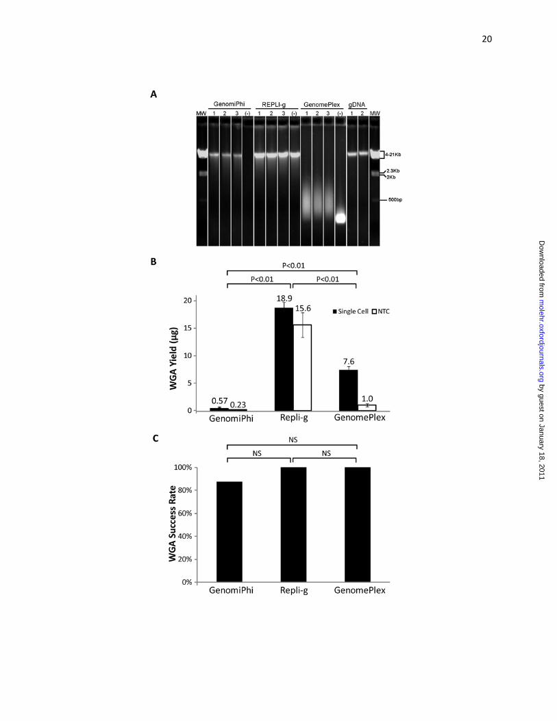
Both GenomiPhi and REPLI‐g methods produced WGA DNA that was equivalent in molecular
weight to that of the genomic DNA (Figure 1A). However, a similar sized DNA smear was detected from
the no template controls amplified with REPLI‐g. As a result, gel electorphoresis of REPLI‐g WGA DNA
alone was insufficient to determine whether amplification was successful. GenomPlex WGA DNA
product size ranged from 100 to 1000 basepairs (Figure 1A). The average WGA DNA yield from the
GenomiPhi protocol was 0.57 ± 0.1 μg, and significantly less (P<0.01) than the 7.63 ± 0.4 μg from
GenomePlex or the 18.93 ± 0.8 μg from REPLI‐g (Figure 1B). Similar quantities of DNA were detected
from the no template controls amplified using the REPLI‐g protocol (15.62 ± 2.3 ug). As a result, DNA
quantitation by UV spectroscopy of REPLI‐g WGA DNA was also insufficient to determine whether
specific amplification was successful. This is consistent with previous studies which have found non‐
specific primer‐directed DNA amplification with no template control MDA reactions (Lage et al., 2003,
Brukner et al., 2005). Eighty‐eight percent (35/40) of the single cells successfully amplified with the
GenomiPhi method by yielding greater than 250 ng of WGA DNA. REPLI‐g and GenomePlex methods
yielded greater than 250ng WGA DNA from 100% of the single cells (Figure 1C). No significant difference
in reliability of obtaining sufficient quantities of DNA for microarray analysis was observed between the
3 methods.
Single cell WGA DNA provided an average of 74% genotyping coverage with the GenomiPhi
protocol and 78% with GenomePlex, which where both significantly lower than the 88% obtained with
REPLI‐g (Figure 2A). Single cell WGA DNA genotypes provided an average of 86% accuracy with the
GenomiPhi protocol, which was significantly less that the 89% accuracy obtained with GenomePlex
(Figure 2B). Both the GenomiPhi and GenomePlex protocols’ genotyping accuracy was significantly
lower that than the 96% obtained with REPLI‐g (Figure 2B). There was an average ADO rate of 14% using
by guest on January 18, 2011m
olehr.oxfordjournals.orgD
ownloaded from
11
GenomiPhi and 11% using GenomePlex, both of which were significantly higher than the 4% obtained
using REPLI‐g (Figure 2C). These results are applicable to performance of methods that require accurate
genotyping and qualitative analysis of aneuploidy, such as those described by Johnson et al. (2010a) and
Handyside et al. (2010), or in situations where one might consider using WGA DNA to genotype specific
genes of interest (i.e. for single gene disorder screening).
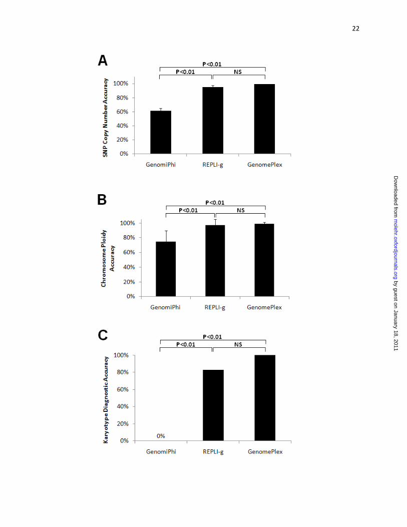
Similarity of single cell copy number assignments with assignments made on genomic DNA and
as expected from the conventional karyotype data for each cell line were evaluated at 3 levels. For
individual SNPs, 62% similarity was obtained using the GenomiPhi protocol, which was significantly less
than the 95% similarity obtained using REPLI‐g or the 99% similarity obtained using GenomePlex (Figure
3A). For individual chromosomes, 75% similarity was obtained using the GenomiPhi protocol, which was
significantly less than the 97% similarity obtained using REPLI‐g or the 99% similarity obtained using
GenomePlex (Figure 3B). For single cell molecular karyotyping diagnosis, 0% accuracy was obtained
using the GenomiPhi protocol, which was significantly less than the 83% similarity obtained using REPLI‐
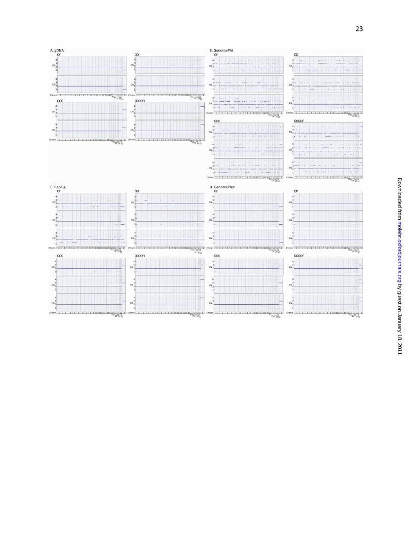
g or the 100% similarity obtained using GenomePlex (Figure 3C). A comprehensive view of the genomic
DNA and single cell copy number assignments are also displayed in Figure 4 and reflects the levels of
accuracy reported above. These results are of particular importance to the performance of methods
that require accurate quantitative analysis of copy number such as those reported by Le Caignec et al.
(2006) and Vanneste et al. (2009), which used GenomiPhi technology, and Feigler et al. (2007), Treff et
al. (2010a), and Gutierrez‐Mateo et al. (2010), which used GenomePlex technology.
Duration of amplification is also important when considering application of single cell WGA
technology to clinical PGD. WGA is only one step necessary to generate a diagnosis for the amplified
sample, which also involves downstream microarray processing and analysis. For example, the most
typical PGD application requires completion of single cell analysis within 24 hours of initiating the
by guest on January 18, 2011m
olehr.oxfordjournals.orgD
ownloaded from
12
procedure in order to avoid embryo cryopreservation. Therefore, while the REPLI‐g protocol may be
suitable for single cell applications that do not have time constraints, the 16 hour turn‐around time may
not allow for its routine use in PGD for aneuploidy screening. A more rapid turn‐around time with
isothermal MDA was represented in this study by the GenomiPhi protocol. Unfortunately, this
shortened MDA protocol performed with the least reliability, fidelity, and accuracy of all methods
tested. In contrast, the GenomePlex protocol provided a more rapid turn‐around time (4 hours) which
could be suitable for application to PGD and produced the highest copy number assignment accuracy of
all methods tested. Therefore, for applications requiring accurate and rapid copy number analysis, such
as PGD for aneuploidy screening, the GenomePlex protocol may be more appropriate than REPLI‐g or
GenomiPhi MDA based protocols. However, for those applications requiring accurate genotyping
analysis without time constraints, the REPLI‐g protocol may be more appropriate than the GenomePlex
or GenomiPhi protocols.
In summary, this study represents the first direct comparison of commercially available single
cell WGA method performance, a necessary step in all 24 chromosome aneuploidy screening
technologies. Clinically relevant measurements of reliability, fidelity and accuracy were evaluated for
each method. In general, a longer MDA protocol was better for genotyping accuracy than PCR, and PCR
was better and faster than MDA for copy number accuracy. Clinicians and laboratory directors should
consider these and other critical pieces of evidence (presented in Table 1 and reviewed in Scott and
Treff, 2010) when evaluating new technologies that intend to predict the chromosomal status and
reproductive potential of human embryos.
AUTHOR CONTRIBUTIONS
N.R.T and R.T.S designed the study, N.R.T., J.S., and L.E.N. wrote the manuscript, J.S., X.T. and
L.E.N. performed the experiments and prepared the microarray data for publication.
by guest on January 18, 2011m
olehr.oxfordjournals.orgD
ownloaded from

13
FUNDING
No extramural funding was used for this study
REFERENCES CITED
Barker DL, Hansen MST, Faruqi AF, Giannola D, Irsula OR, Lasken RS, Latterich M, Makarov V, Oliphant A, Pinter JH et al. Two methods of whole‐genome amplification enable accurate genotyping across a 2320‐SNP linkage panel. Genome Res 2004;14:901‐7.
Brukner, I., B. Paquin, M. Belouchi, D. Labuda, and M. Krajinovic. Self‐priming arrest by modified random oligonucleotides facilitates the quality control of whole genome amplification. Anal. Biochem. 2005;339:345‐347. Burlet P, Frydman N, Gigarel N, Kerbrat V, Tachdijian G, Feyereisen E, Bonnefont J, Frydman R, Munnich A, Steffann J. Multiple displacement amplification improves PGD for fragile X syndrome. Mol Hum Reprod 2006;12:647‐52.
Cui XF, Li HH, Goradia TM, Lange K, Kasasian HH, Jr., Galas D, Arnheim N. Single‐sperm typing: determination of genetic distance between the G gamma‐globin and parathyroid hormone loci by using the polymerase chain reaction and allele‐specific oligomers. Proc Natl Acad Sci U S A 1989;86:9389‐93.
Dean F, Hosono S, Fang L, Wu X, Faruqi A, Bray‐Ward P, Sun Z, Zong Q, Du Y, Du J et al. Comprehensive human genome amplification using multiple displacement amplification. Proc Natl Acad Sci U S A 2002;99:5261‐6.
Dolezel J, Bartos J, Voglmayr H, Greihuber J. Nuclear DNA content and genome size of trout and human. Cytometry A 2003;51:127‐8.
Escudero T, Pere C, Fischer J, Prates R, Tormasi S, Santiago M. Preimplantation genetic diagnosis (PGD) for reciprocal translocations using array comparative genome hybridization (aCGH). Fertil Steril 2010; 94(4). Fiegler H, Geigl JB, Langer S, Rigler D, Porter K, Unger K, Carter NP, Speicher MR. High resolution array‐CGH analysis of single cells. Nucleic Acids Res 2007;35:1‐10.
Fragouli E, Lensi M, Ross R, Katz‐Jaffe M, Schoolcraft WB and Wells D. Comprehensive molecular cytogenetic analysis of the human blastocyst stage. Hum Reprod 2008;23:2596‐2608.
by guest on January 18, 2011m
olehr.oxfordjournals.orgD
ownloaded from
14
Fritz MA. Perspectives on the efficacy and indications for preimplantation genetic screening: where are we now? Hum Reprod 2008;23:2617‐21.
Gianaroli L, Magli MC, Ferraretti A, Fiorentino A, Garrisi J, Munne S. Preimplantation genetic diagnosis increases the implantation rate in human in vitro fertilization by avoiding the transfer of chromosomally abnormal embryos. Fertil Steril 1997;68:1128‐31.
Gutierrez‐Mateo C, Colls P, Sanchez‐Casas Padilla E, Escudero T, Prates R, Ketterson K, Wells D, Munne S. Validation of microarray comparative genomic hybridization for comprehensive chromosome analysis of embryos. Fertil Steril 2010:epub ahead of print.
Handyside AH, Robinson MD, Simpson RJ, Omar MB, Shaw MA, Grudzinskas JG, Rutherford A. Isothermal whole genome amplification from single and small numbers of cells: a new era for preimplantation genetic diagnosis of inherited disease. MolHumReprod 2004;10:767‐72.
Handyside AH, Harton GL, Mariani B, Thornhill AR, Affara NA, Shaw MA, Griffin DK. Karyomapping: a Universal Method for Genome Wide Analysis of Genetic Disease based on Mapping Crossovers between Parental Haplotypes. J Med Genet 2009.
Handyside A.H., Grifo J, Prates R, Tormasi S, Fisher J.M., Munne S. Validation and first clinical application of karyomapping for preimplantation diagnosis (PGD) of Gaucher disease combined with 24 chromosome screening. Fertil Steril 2010;94:S79‐80. Hellani A. Clinical application of multiple displacement amplification in preimplanatation genetic diagnosis. RBM Online 2005;10:376‐80.
Hellani A, Sammour A, Johansson L, El‐Sheikh A. Delivery of a normal baby after preimplantation genetic diagnosis for non‐ketotic hyperglycinaemia. Reprod Biomed Online 2008a;16:893‐7.
Hellani A, Abu‐Amero K, Azouri J, El‐Akoum S. Successful pregnancies after application of array‐comparative genomic hybridization in PGS‐aneuploidy screening. Reprod Biomed Online 2008b;17:841‐7.
Johnson DS, Gemelos G, Baner J, Ryan A, Cinnioglu C, Banjevic M, Ross R, Alper M, Barrett B, Frederick J et al. Preclinical validation of a microarray method for full molecular karyotyping of blastomeres in a 24‐h protocol. Hum Reprod 2010a;25:1066‐75.
Johnson D.S., Hill M, Abae M, Frederick J, Swanson M, Rabinowitz M. First clinical application of DNA microarrays for translocations and inversions. Fertil Steril 2010b;93(5):S13‐S14. Kahraman S, Bahce M, Samli H, Imirzahoglu N, Yakism K, Cengiz G, Donmez E. Healthy births and ongoing pregnancies obtained by preimplantation genetic diagnosis in patients with advanced maternal age and recurrent implantation failure. Hum Reprod 2000;15:2003‐7.
by guest on January 18, 2011m
olehr.oxfordjournals.orgD
ownloaded from
15
Keskintepe L., Sher G., Keskintepe M. Reproductive oocyte/embryo genetic analysis: comparison between fluorescence in‐situ hybridization and comparative genomic hybridization. Reprod. Bio. Online. 2007;15(3):303‐309.
Lage, J.M., J.H. Leamon, T. Pejovic, S. Hamann, M. Lacey, D. Dillon, R. Segraves, B. Vossbrinck, et al. Whole genome analysis of genetic alterations in small DNA samples using hyperbranched strand displacement amplification and array‐CGH. Genome Res. 2003;13:294‐307. Le Caignec C, Spits C, Sermon K, De Rycke M, Thienpont B, Debrock S, Staessen C, Moreau Y, Fryns JP, Van Steirteghem A et al. Single‐cell chromosomal imbalances detection by array CGH. Nucleic Acids Res 2006;34:e68.
Lledo B, Ten J, Galan F, Bernabeu R. Preimplantation genetic diagnosis of Marfan syndrome using multiple displacement amplification. Fertil Steril 2006;86:949‐55.
Lledo B, Bernabeu R, Ten J, Galan F, Cioffi L. Preimplantation genetic diagnosis of X‐linked adrenoleukodystrophy with gender determination using multiple displacement amplification. Fertil Steril 2007;88:1327‐33.
Lovmar L, Fredriksson M, Liljedahl U, Sigurdsson S, Syvanen AC. Quantitative evaluation by minisequencing and microarrays reveals accurate multiplexed SNP genotyping of whole genome amplified DNA. Nucleic Acids Research 2003;31:129‐39.
Munne S, Magli MC, Cohen J, Morton NE, Sadowy S, Gianaroli L, Tucker M, Marquez C, Sable D, Ferraretti A et al. Positive outcome after preimplantation diagnosis of aneuploidy in human embryos. Hum Reprod 1999;14:2191‐9.
Munne S, Sandalinas M, Escudero T, Velilla E, Walmsley R, Sadowy S, Cohen J, Sable D. Improved implantation after preimplantation genetic diagnosis of aneuploidy. Reprod Biomed Online 2003;7:91‐7.
Munne S, Fisher JM, Warner A, Chen S, Zouves C, Cohen J, Group at RCP. Preimplantation genetic diagnosis significantly reduces pregnancy loss in infertile couples: a multicenter study. Fertil Steril 2006;85:S178‐S9.
Munne S, Surrey E.S., Grifo J, Marut E, Opsahl M, Taylor TH. Preimplantation genetic diagnosis using a‐CGH significantly increases ongoing pregnancy rates per transfer. Fertil Steril 2010; 94(4);S81.
Northrop LE, Treff NR, Levy B, Scott RT, Jr. SNP microarray‐based 24 chromosome aneuploidy screening demonstrates that cleavage‐stage FISH poorly predicts aneuploidy in embryos that develop to morphologically normal blastocysts. Mol Hum Reprod 2010;16:590‐600.
by guest on January 18, 2011m
olehr.oxfordjournals.orgD
ownloaded from
16
Panelli S, Damiani G, Espen L, Micheli G, Sgaramella V. Towards the analysis of the genomes of single cells: Further characterisation of the multiple displacement amplification. Gene 2006;372:1‐7.
Park JW, Beaty TH, Boyce P, Scott AF, McIntosh I. Comparing whole‐genome amplification methods and sources of biological samples for single‐nucleotide polymorphism genotyping. Clin Chem 2005;5:1520‐3.
Rabinowitz M, Behr D, Potter D, Ross R, Alper M, Banjevic M. Parental support for single gene PGD and simultaneous 24‐chromosome screening reduces risks of allele misdiagnosis and transfer of aneuploid embryos. Fertil Steril 2009;92:S202. Rabinowitz M, Beltsos A, Potter D, Bush M, Givens C, Smotrich D. Effects of advanced maternal age are abrogated in 122 patients undergoing transfer of embryos with euploid microarray screening results at cleavage stage. Ferti Steril 2010;94(4):S80. Ren Z, Zhou C, Xu Y, Deng J, Zeng H, Zeng Y. Mutation and haplotype analysis of Duchene muscular dystrophy by single cell multiple displacement amplification. Mol Hum Reprod 2007;13:431‐6.
Renwick P, Lewis C, Abbs S, Ogilivie C. Determination of the genetic status of cleavage‐stage human embryos by microsatellite marker analysis following multiple displacement amplification. Prenat Diagn 2007;27:206‐15.
Sher G, Keskintepe L, Keskintepe M, Ginsburg M, Maassarani G, Yakut T, Baltaci V, Kotze D, Unsal E. Oocyte karyotyping by comparative genomic hybrydization provides a highly reliable method for selecting "competent" embryos, markedly improving in vitro fertilization outcome: a multiphase study. Fertil Steril 2007;87:1033‐40.
Sher G, Keskintepe L, Keskintepe M, Maassarani G, Tortoriello D, Brody S. Genetic analysis of human embryos by metaphase comparative genomic hybridization (mCGH) improves efficiency of IVF by increasing embryo implantation rate and reducing multiple pregnancies and spontaneous miscarriages. Fertil Steril 2009;92(6):1886‐1894.
Saiki R, Gelfand D, Stoffel S, Scharf S, Higuchi R, Horn G, Mullis K, Erlich H. Primer directed enzymatic amplification of DNA with a thermostable DNA polymerase. Science 1988;239:487‐91.
Schoolcraft W.B., Treff N.R., Ferry K., Stevens J.M., Katz‐Jaffe M.G., Scott R.T. Clinical application of comprehensive chromosomal screening at the blastocyst stage. Fert Steril 2010a;94:S23.
Schoolcraft W.B., Treff N.R., Ferry K.M., Stevens J.M., Katz‐Jaffe M.G., Scott R.T. First clinical application of SNP microarray based 24 chromosome aneuploidy screening of human blastocysts. Fertil Steril 2010b; 94:S23‐4
by guest on January 18, 2011m
olehr.oxfordjournals.orgD
ownloaded from
17
Scott RT, Jr., Miller KA, Olivares R, Su J, Fratterelli J, Treff NR. Microarray based 24 chromosome preimplantation genetic diagnosis (mPGD) is highly predictive of the reproductive potential of human embryos:a prospective blinded non‐selection trial. Fertil Steril 2008;90:22.
Scott RT, Jr., Treff NR. Assessing the reproductive competence of individual embryos: a proposal for the validation of new "‐omics" technologies. Fertil Steril 2010. Scott RT, Jr., Tao X, Taylor D, Ferry K, Treff N. A prospective randomized controlled trial demonstrating significantly increased clinical pregnancy rates following 24 chromosome aneuploidy screening: biopsy and analysis on day 5 with fresh transfer. Fertil Steril 2010;94:S2.
Sher G, Keskintepe L, Keskintepe M, Ginsburg M, Maassarani G, Yakut T, Baltaci V, Kotze D, Unsal E. Oocyte karyotyping by comparative genomic hybrydization provides a highly reliable method for selecting "competent" embryos, markedly improving in vitro fertilization outcome: a multiphase study. Fertil Steril 2007;87(5):1033‐40. Sher G, Keskintepe L, Keskintepe M, Maassarani G, Tortoriello D, Brody S. Genetic analysis of human embryos by metaphase comparative genomic hybridization (mCGH) improves efficiency of IVF by increasing embryo implantation rate and reducing multiple pregnancies and spontaneous miscarriages. Fertil Steril 2009;92:1886‐94.
Treff N.R., Su J, Tao X, Miller K, Scott R.T. First IVF babies born after rapid 24 chromosome embryo aneuploidy screening and fresh embryo transfer. Fertil Steril 2009a;92(3):S49. Treff N, Tao X, Su J, Northrop L.E., Kamani M, Bergh P, Miller K, Levy B, Scott R. SNP microarray based concurrent screening of 24 chromosome aneuploidy, unbalanced translocations, and single gene disorders in human embryos: first application of comprehensive triple factor PGD. Biol Reprod 2009b; 81:188. Treff N.R., Tao X, Lonczak A, Su J, Taylor D, Scott R. Four hour 24 chromosome aneuploidy screening using high throughput PCR SNP allele ratio analyses. Fertil Steril 2009c;92:S49‐S50. Treff NR, Su J, Tao X, Levy B, Scott RT, Jr. Accurate single cell 24 chromosome aneuploidy screening using whole genome amplification and single nucleotide polymorphism microarrays. Fertil Steril 2010a;94:2017‐21.
Treff NR, Levy B, Su J, Northrop LE, Tao X, Scott RT, Jr. SNP microarray‐based 24 chromosome aneuploidy screening is significantly more consistent than FISH. Mol Hum Reprod 2010b;16:583‐9.
Treff N, Northrop L.E., Kasabwala K, Su J, Levy B, Scott R.T. SNP microarray based concurrent screening of 24 chromosome aneuploidy and unbalanced translocations in preimplantation human embryos. Fertil Steril 2010c;In press.
by guest on January 18, 2011m
olehr.oxfordjournals.orgD
ownloaded from
18
Vanneste E, Voet T, Le Caignec C, Ampe M, Konings P, Melotte C, Debrock S, Amyere M, Vikkula M, Schuit F et al. Chromosome instability is common in human cleavage‐stage embryos. Nat Med 2009;15:577‐83.
Wells, D, Sherlock, JK, Handyside AH, Delhanty JD. Detailed chromosomal and molecular genetic analysis of single cells by whole genome amplification and comparative genomic hybridization. Nucleic Acids Res, 1999;27(4): 1214‐8.
Wells D, Delhanty, JD. Comprehensive chromosomal analysis of human preimplantation embryos using whole genome amplification and single cell comparative genomic hybridization. Mol Hum Reprod 2000;6:1055‐62.
Wells D, Fragouli E, Alfarawaty S, Munne S, Schoolcraft W.B., Katz‐Jaffe M. Highly significant improvement in embryo implantation and increased live birth rate achieved after comprehensive chromosomal screening: implications for single embryo transfer, Fertil Steril 2009; 94(3): S79.
by guest on January 18, 2011m
olehr.oxfordjournals.orgD
ownloaded from
19
FIGURE LEGENDS Figure 1. Reliability of single cell WGA. (A) Gel electrophoresis of purified reaction products of 3
representative samples and 1 no template control (‐) from each of 3 single cell WGA methods
(GenomiPhi, REPLI‐g, and GenomePlex). Representative purified genomic DNA (gDNA) and molecular
weight markers (MW) are included for size references. (B) The mean yield of amplification (± s.e.m.) of
40 single cells (black bars) or 4 no template controls (NTC; white bars) from each of 3 single cell WGA
methods. (C) The rates of successful amplification of 40 single cells from each of 3 single cell WGA
methods.
Figure 2. Genotype fidelity of single cell WGA. (A) The percentage of SNPs evaluated that were
successfully assigned a genotype (genotyping coverage) for each of 3 single cell WGA methods. (B) The
percentage of SNPs assigned a genotype identical to the purified genomic DNA assignments (genotyping
accuracy) for each of 3 single cell WGA methods. (C) The percentage of SNPs assigned a homozygous
genotype in the single cells but also assigned a heterozygous genotype in the purified genomic DNA
samples (Genotyping ADO rate) for each of 3 single cell WGA methods.
Figure 3. Copy number assignment accuracy of single cell WGA. (A) The percentage of SNPs evaluated
that were assigned the expected copy number (SNP copy number accuracy) for each of 3 single cell WGA
methods. (B) The percentage of chromosomes evaluated that were assigned the expected copy number
(chromosome ploidy accuracy) for each of 3 single cell WGA methods. (C) The percentage of cells that
were assigned the expected chromosome loss, gain, or euploidy (karyotype diagnostic accuracy) for
each of 3 single cell WGA methods.
Figure 4. SNP microarray based copy number (CN) graphs of (A) purified genomic DNA, or single cells
amplified with (B) GenomiPhi, (C) REPLI‐g, or (D) GenomePlex protocols. Each panel includes analyses of
each of 4 cell lines possessing one to four X chromosome copies.
by guest on January 18, 2011m
olehr.oxfordjournals.orgD
ownloaded from
20
by guest on January 18, 2011m
olehr.oxfordjournals.orgD
ownloaded from

21
by guest on January 18, 2011m
olehr.oxfordjournals.orgD
ownloaded from
22
by guest on January 18, 2011m
olehr.oxfordjournals.orgD
ownloaded from
23
by guest on January 18, 2011m
olehr.oxfordjournals.orgD
ownloaded from
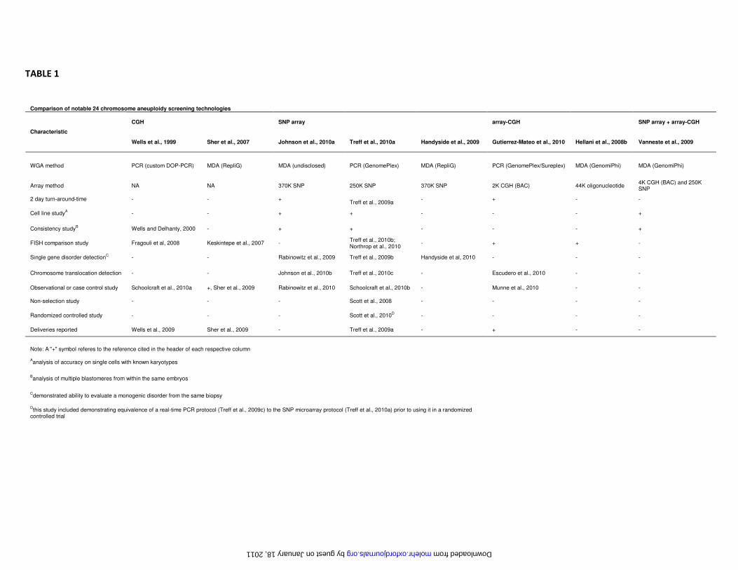
TABLE 1
Comparison of notable 24 chromosome aneuploidy screening technologies
CGH SNP array array-CGH SNP array + array-CGH
Characteristic
Wells et al., 1999 Sher et al., 2007 Johnson et al., 2010a Treff et al., 2010a Handyside et al., 2009 Gutierrez-Mateo et al., 2010 Hellani et al., 2008b Vanneste et al., 2009
WGA method PCR (custom DOP-PCR) MDA (RepliG) MDA (undisclosed) PCR (GenomePlex) MDA (RepliG) PCR (GenomePlex/Sureplex) MDA (GenomiPhi) MDA (GenomiPhi)
Array method NA NA 370K SNP 250K SNP 370K SNP 2K CGH (BAC) 44K oligonucleotide 4K CGH (BAC) and 250K SNP
2 day turn-around-time - - + Treff et al., 2009a
- + - -
Cell line studyA - - + + - - - +
Consistency studyB Wells and Delhanty, 2000 - + + - - - +
FISH comparison study Fragouli et al, 2008 Keskintepe et al., 2007 - Treff et al., 2010b; Northrop et al., 2010
- + + -
Single gene disorder detectionC - - Rabinowitz et al., 2009 Treff et al., 2009b Handyside et al, 2010 - - -
Chromosome translocation detection - - Johnson et al., 2010b Treff et al., 2010c - Escudero et al., 2010 - -
Observational or case control study Schoolcraft et al., 2010a +, Sher et al., 2009 Rabinowitz et al., 2010 Schoolcraft et al., 2010b - Munne et al., 2010 - -
Non-selection study - - - Scott et al., 2008 - - - -
Randomized controlled study - - - Scott et al., 2010D - - - -
Deliveries reported Wells et al., 2009 Sher et al., 2009 - Treff et al., 2009a - + - -
Note: A "+" symbol referes to the reference cited in the header of each respective column
Aanalysis of accuracy on single cells with known karyotypes
Banalysis of multiple blastomeres from within the same embryos
Cdemonstrated ability to evaluate a monogenic disorder from the same biopsy
Dthis study included demonstrating equivalence of a real-time PCR protocol (Treff et al., 2009c) to the SNP microarray protocol (Treff et al., 2010a) prior to using it in a randomized
controlled trial
by guest on January 18, 2011 molehr.oxfordjournals.org Downloaded from