single-incision laparoscopic cholecystectomy: initial
TRANSCRIPT
SIDAM
LdAvpfI
DsoAn
R2FgCWL
©P
ORIGINAL SCIENTIFIC ARTICLES
ingle-Incision Laparoscopic Cholecystectomy:nitial Experience with Critical View of Safetyissection and Routine Intraoperative Cholangiography
rthur Rawlings, MD, Steven E Hodgett, MD, Brent D Matthews, MD, Steven M Strasberg, MD,ary Quasebarth, RN, L Michael Brunt, MD
BACKGROUND: Single-incision laparoscopic cholecystectomy (SILC) is emerging as a potentially less invasivealternative to standard laparoscopic cholecystectomy and natural orifice transluminal endo-scopic surgery cholecystectomy. As this technique is more widely used, it is important tomaintain well-established practices of the critical view of safety (CVS) dissection and intraop-erative cholangiography (IOC). We present our initial experience with SILC using CVS dis-section and routine IOC.
STUDY DESIGN: Fifty-four patients with biliary colic were offered SILC, which was performed through theumbilicus. CVS with photo documentation was attained before clipping and transecting thecystic structures. IOC was done using various needle puncture techniques. Assessment of CVSwas carried out by independent surgeon review of operative still photos or videos using a 3-pointgrading scale: visualization of only 2 ductal structures entering the gallbladder; a clear triangleof Calot; and separation of the base of the gallbladder from the cystic plate.
RESULTS: SILC was performed in 54 patients (15 male and 39 female). Six patients required 1 supple-mentary 3- or 5-mm port. Complete IOC was successful in 50 of 54 patients (92.6%). CVS wasachieved at the time of operation in all 54 patients. Photo documentation review confirmed 3of 3 CVS criteria in 32 (64%) patients, 2 of 3 in 12 patients (24%), 1 of 3 in 3 patients (6%),and 0 in 3 patients (6%).
CONCLUSIONS: As laparoscopic cholecystectomy becomes less invasive, proven safe dissection techniquesshould be maintained. Dissection to obtain the CVS should be the goal of every patient andIOC can be accomplished in a high percentage of patients. This approach places patient safetyconsiderations foremost in the evolution of minimally invasive cholecystectomy. (J Am Coll
Surg 2010;211:1–7. © 2010 by the American College of Surgeons)sMniopbrpsa
coss1
aparoscopic cholecystectomy (LC) has been the gold stan-ard for removal of the gallbladder since the early 1990s.1
s technology has progressed, surgeons have begun to de-elop less invasive methods for this commonly performedrocedure. In 1997, Navarra and colleagues performed theirst single-incision laparoscopic cholecystectomy (SILC).2
n 2007, the first natural orifice transluminal endoscopic
isclosure Information: Drs Matthews and Brunt receive honoraria forpeaking and teaching from Ethicon Endosurgery, Inc, and Covidien. Allther authors have nothing to disclose.bstract presented at the Society of American Gastrointestinal Surgeons An-ual Meeting, Phoenix, AZ, April 2009.
eceived January 26, 2010; Revised February 3, 2010; Accepted February 3,010.rom the Department of Surgery and Institute for Minimally Invasive Sur-ery, Washington University School of Medicine, St Louis, MO.orrespondence address: L Michael Brunt, MD, Department of Surgery,ashington University School of Medicine, 660 S Euclid Ave, Box 8109, St
couis, MO 63110. email: [email protected]
12010 by the American College of Surgeons
ublished by Elsevier Inc.
urgery (NOTES) cholecystectomy was performed byarescaux and colleagues.3 The NOTES approach elimi-
ated an abdominal wall incision, but concern about clos-ng the access route to the abdomen (eg, gastrotomy), lackf standardized equipment, and technical challenges haverecluded the applicability of this investigative approacheyond a small number of centers. Unlike NOTES, lapa-oscopic surgery through a single-incision access site can beerformed with existing instrumentation, although somepecialized equipment has been developed and is readilyvailable.
As more reports of SILC appear in the literature, it isritically important that the same dissection principles andperative goals be maintained that have been in place fortandard LC for many years. The foremost of these is dis-ection to the critical view of safety (CVS) described in995.4 In addition, the ability to carry out intraoperative
holangiography (IOC) should be an integral requirementISSN 1072-7515/10/$36.00doi:10.1016/j.jamcollsurg.2010.02.038
oItpwa
MActdBIts
cEotcciantm
OAs2onattspatd
saifttfutpgpls3fwedada
dicltIumdg
Fi
2 Rawlings et al Single-Incision Laparoscopic Cholecystectomy J Am Coll Surg
f any new approach to surgical removal of the gallbladder.n July 2008, we initiated a program of SILC at our insti-ution, with the goal of obtaining the CVS dissection anderforming routine IOC. We now report our early resultsith this technique and review rates of obtaining the CVS
nd performing an IOC in the first 54 patients.
ETHODSretrospective medical record review was carried out of 54
onsecutive patients undergoing a single-incision approacho LC from July 2008 through October 2009. All proce-ures were performed by 2 attending surgeons (LMB andDM) with assistance by a surgical resident or Minimally
nvasive Surgery Fellow. Study approval was obtained fromhe Institutional Review Board of the Washington Univer-ity School of Medicine and Barnes-Jewish Hospital.
Patients selected for the SILC approach had uncompli-ated symptomatic gallstone disease or biliary dyskinesia.xclusions for the SILC approach were acute cholecystitisr previous periumbilical midline abdominal surgery. Pa-ients with severe morbid obesity were also generally notonsidered candidates for this approach. Data analyzed in-luded patient demographics (ie, age, gender, body massndex, American Society of Anesthesiologists score), oper-tive time, operative approach (ie, 2- versus 3-port tech-ique), results of attempted cholangiography, postopera-ive length of stay, and complications. Data presented areean � SD (range).
perative techniquell laparoscopic cholecystectomies were initiated as a
ingle-site technique. The initial incision was a 1.5- to-cm vertical incision made directly through the umbilicusr a transverse supraumbilical incision. Pneumoperito-eum to 15 mmHg was created either by direct insertion of5-mm optical trocar through the natural fascial defect at
he umbilicus or by a Veress needle insertion at that loca-ion, followed by optical trocar placement. Flaps were dis-ected cephalad for 2 to 3 cm and a second 5-mm low-rofile trocar (Covidien or Apple Medical) was placedbove the initial port. The gallbladder was identified andhe fundus was exposed. For the 2-port technique with a
Abbreviations and Acronyms
CVS � critical view of safetyIOC � intraoperative cholangiographyLC � laparoscopic cholecystectomyNOTES � natural orifice transluminal endoscopic surgerySILC � single-incision laparoscopic cholecystectomy
ouble-suture retraction method, a 2-0 Prolene (Ethicon) a
uture on a Keith needle was then inserted through thebdominal wall in the mid-clavicular line and was retrievedntra-abdominally.This suture was then passed through theundus of the gallbladder and back out the abdominal wallo provide cephalad retraction. A second suture was placedhrough the abdominal wall just to the right of the falci-orm ligament and was then passed through the infundib-lum of the gallbladder and returned outside the body athe right mid-axillary line 2 cm below the rib. Clips werelaced on the suture on each side of where it exited theallbladder neck. This allowed the gallbladder to be “pup-eteered” back and forth in order to provide medial and
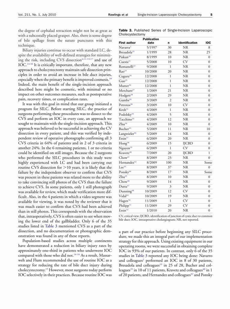
ateral retraction and to expose both the anterior and dor-olateral views of the hepatocystic triangle. Alternatively, a-port technique with a single suture for retraction of theundus was used in which a 3- or 5-mm low-profile trocaras placed above and to the left of the camera port, which
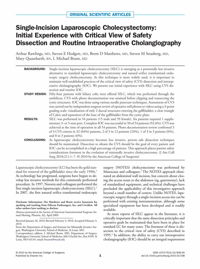
liminated the need for a retraction suture on the gallblad-er infundibulum (Fig. 1). An additional 3- or 5-mm portt an alternate site, usually subcostal, was added at theiscretion of the attending surgeon if exposure with thispproach was inadequate.
In each patient, the primary goal of the dissection was toissect the hepatocystic triangle to the CVS. This entailed
solation of the cystic duct and artery, clearing the hepato-ystic triangle of all extraneous tissue, and separating theower part of the gallbladder from the liver bed (to visualizehe cystic plate) before clipping any ductal structures. AnOC was routinely attempted after the CVS was obtainedsing various needle puncture techniques. Most com-only, a 4F ureteral catheter was introduced into the ab-
omen either through a reusable Veress needle or a 14-auge angiocatheter placed at the mid-clavicular line
igure 1. Operative view of 3-port, single-suture retraction single-ncision laparoscopic cholecystectomy technique.
pproximately 2 cm below the costal margin. The catheter
wcddwwdtttnc
PAidctTpclo
RFaf(�tp
spt
opLtg3ap5
iitfp
an4vC(f(
ptaspiCi
DS
TP
MFTABAP
Al
TO
C
I
C
3Vol. 211, No. 1, July 2010 Rawlings et al Single-Incision Laparoscopic Cholecystectomy
as then secured in a standard fashion with a cholangio-lamp. After completion of the cholangiogram, the cysticuct and cystic artery were doubly clipped with a 5-mmisposable clip applier and then divided. For patients inhom the cystic duct was thickened, it was further securedith a pretied 0-polydioxanone loop suture. The gallblad-er was then dissected off the liver bed using a hook elec-rocautery and removed at the camera port site by enlarginghe fascial opening as needed. All 5-mm fascial defects athe umbilicus were closed using 0-gauge absorbable oronabsorable suture and the single skin incision site waslosed with a 4-0 absorbable subcuticular suture.
hoto documentations a part of the implementation strategy for SILC at our
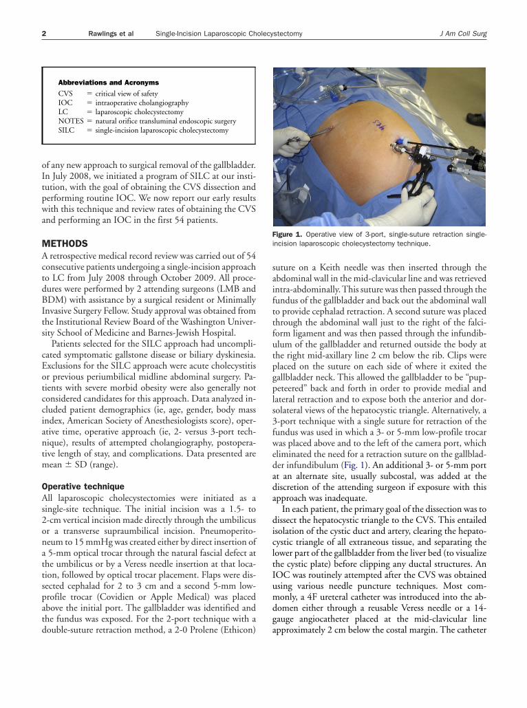
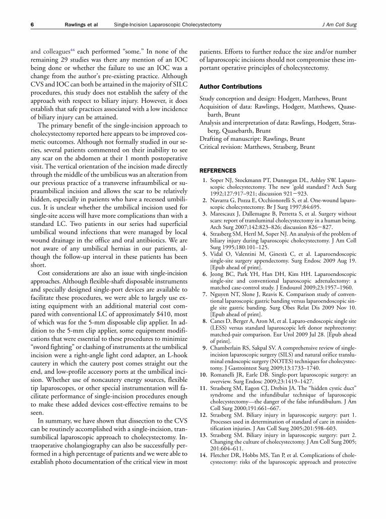
nstitution, an attempt was made to routinely obtain photoocumentation of the CVS before clipping and cutting theystic artery and duct. In some patients, video recording ofhe CVS sequence was obtained in lieu of still photographs.hese images were then independently reviewed by an ex-erienced biliary surgeon (SMS) and were assessed using 3riteria for the critical view: ie, clear hepatocystic triangle;ower gallbladder dissected off the cystic plate; and 2 andnly 2 structures entering the gallbladder.
ESULTSifty-four patients underwent attempted SILC. The meange was 47 � 17.5 (range 20 to 78) years, and 72% wereemale (Table 1). Mean body mass index was 27.7 � 4.7range 17.8 to 40.9). Three patients had a body mass index
35. Five patients had symptomatic biliary dyskinesia andhe rest had uncomplicated symptomatic cholelithiasis. Allrocedures were elective.Operative results are shown in Table 2. A 3-port, single-
ite technique was used in 26 patients. The remaining 28rocedures were initiated as a 2-port, double-suture retrac-
able 1. Patient Demographicsatients
ale, n 15emale, n 39otal, n 54ge (y), mean � SD (range) 47 � 17.5 (20�78)MI, mean � SD (range) 27.7 � 4.7 (17.8�40.9)SA score, mean � SD (range) 2 � 0.5 (1�3)reoperative diagnosis, nSymptomatic cholelithiasis 49Biliary dyskinesia 5
SA, American Society of Anesthesiologists; BMI, body mass index (calcu-ated as kg/m2).
ion technique. Overall, 6 patients required placement of s
ne 3- or 5-mm subcostal or epigastric port to complete therocedure. There were no conversions to a standard 4-portC or to an open procedure. Of the 2 approaches, 5 pa-ients required 1 additional 3- or 5-mm subcostal or epi-astric port in the 2-port approach versus only 1 in the-port technique. One 2-port case also required both theddition of a 5-mm port at the umbilicus and an epigastricort. Mean operating room time was 113.1 � 27.9 (range5 to 206) minutes.An IOC was attempted in all patients and was successful
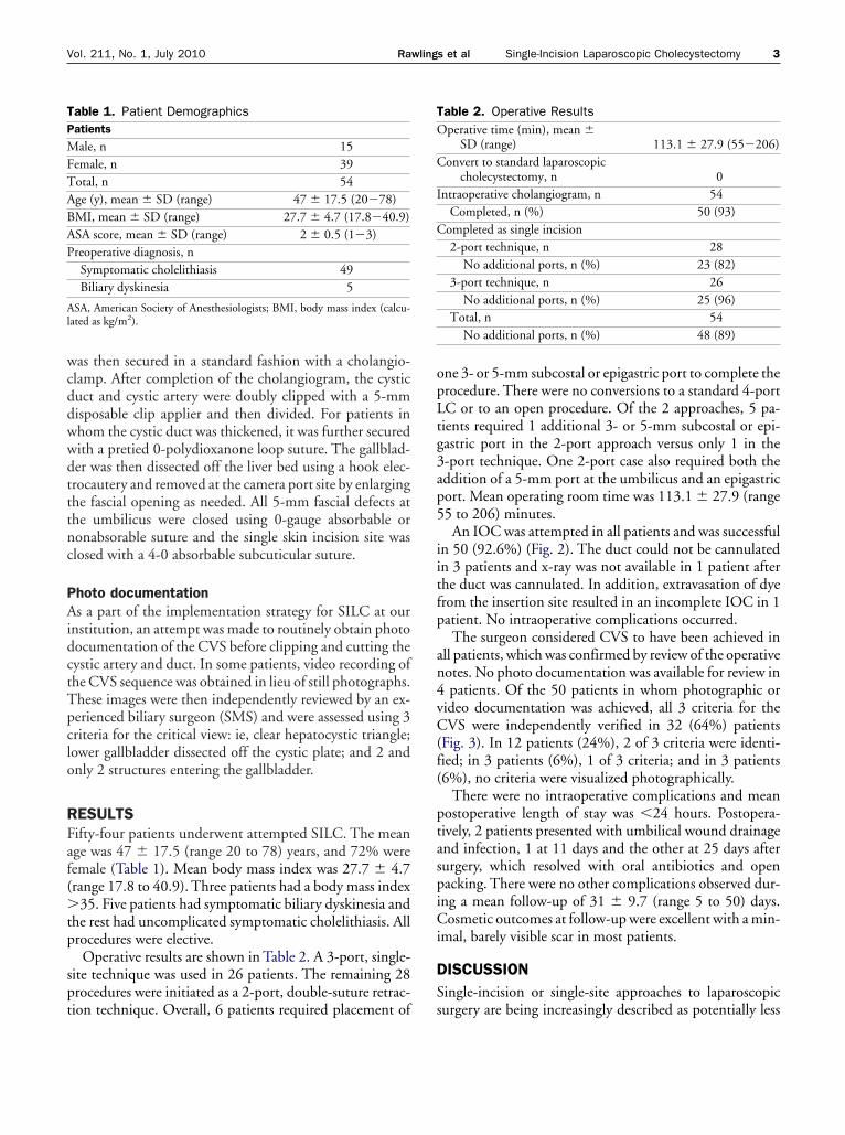
n 50 (92.6%) (Fig. 2). The duct could not be cannulatedn 3 patients and x-ray was not available in 1 patient afterhe duct was cannulated. In addition, extravasation of dyerom the insertion site resulted in an incomplete IOC in 1atient. No intraoperative complications occurred.The surgeon considered CVS to have been achieved in
ll patients, which was confirmed by review of the operativeotes. No photo documentation was available for review inpatients. Of the 50 patients in whom photographic or
ideo documentation was achieved, all 3 criteria for theVS were independently verified in 32 (64%) patients
Fig. 3). In 12 patients (24%), 2 of 3 criteria were identi-ied; in 3 patients (6%), 1 of 3 criteria; and in 3 patients6%), no criteria were visualized photographically.
There were no intraoperative complications and meanostoperative length of stay was �24 hours. Postopera-ively, 2 patients presented with umbilical wound drainagend infection, 1 at 11 days and the other at 25 days afterurgery, which resolved with oral antibiotics and openacking. There were no other complications observed dur-ng a mean follow-up of 31 � 9.7 (range 5 to 50) days.osmetic outcomes at follow-up were excellent with a min-
mal, barely visible scar in most patients.
ISCUSSIONingle-incision or single-site approaches to laparoscopic
able 2. Operative Resultsperative time (min), mean �
SD (range) 113.1 � 27.9 (55�206)onvert to standard laparoscopic
cholecystectomy, n 0ntraoperative cholangiogram, n 54
Completed, n (%) 50 (93)ompleted as single incision2-port technique, n 28
No additional ports, n (%) 23 (82)3-port technique, n 26
No additional ports, n (%) 25 (96)Total, n 54
No additional ports, n (%) 48 (89)
urgery are being increasingly described as potentially less
ifgSlucpip
bPifogmmcuw
1Aatotvdtl“coiit
Fd
Fi
4 Rawlings et al Single-Incision Laparoscopic Cholecystectomy J Am Coll Surg
nvasive, “scarless” procedures, and have been performedor such indications as appendectomy,5 adrenalectomy,6
astric banding,7 and donor nephrectomy.8 Despite thatILC was first reported 13 years ago by Navarro and col-eagues,2 the number of procedures performed was limitedntil the last 2 to 3 years. Development of NOTES chole-ystectomy, which leaves no abdominal scar, might be therimary impetus for the recent surge in interest in single-ncision laparoscopic approaches.9 Numerous educational
igure 2. (A, B) Photo documentation of the critical view of safetyuring single-incision laparoscopic cholecystectomy.
rograms and courses, largely sponsored by industry, have A
een developed to teach surgeons this new approach. AubMed search using various terms10 referring to single-
ncision approaches for cholecystectomy identified 35 dif-erent case or series reports to date, which included a totalf �500 patients. Because single-incision laparoscopic sur-ery can be performed with existing “off the shelf ” equip-ent, it appears to be gaining traction in the surgical com-unity, in contrast to NOTES cholecystectomy. In 1
omprehensive review, there were only 25 patients who hadndergone a NOTES cholecystectomy compared with 142ho had a SILC procedure.9
With the introduction of LC into clinical practice in the990s, a concomitant rise in biliary injuries was seen.4,11-16
lthough SILC does not represent as a major a shift inpproach or technique, such as that which occurred duringhe transition from open to LC, there are technical aspectsf this operation that are departures from some of the basicenets of laparoscopic surgery. First and foremost is that, byirtue of placing both the laparoscopic camera port and allissecting instruments through a single umbilical incision,riangulation between the camera and the working ports isost. This results in a parallel instrument alignment and anin-line” view of the anatomy and field of dissection. Be-ause of the closely placed parallel ports, “sword fighting”f instruments can restrict freedom of movement and view-ng, as well as dissecting angles. Finally, many surgeons,ncluding our group, have used �1 sutures placed directlyhrough the gallbladder to provide retraction and exposure.
igure 3. Intraoperative cholangiogram obtained during single-ncision laparoscopic cholecystectomy.
lthough this does allow manipulation of the gallbladder,
twot
siIaceIdip
psCsadpCacwhrfwttwfawttisdo
hacwscI
adsoIsaBl
TC
F
NBPCRRCGMMHGPKPTZBLEHNKCHLPZCDDVHPE
Cb
5Vol. 211, No. 1, July 2010 Rawlings et al Single-Incision Laparoscopic Cholecystectomy
he degree of cephalad retraction might not be as great asith a subcostally placed grasper. Also, there is some degreef bile spillage from the suture punctures with thisechnique.
Biliary injuries continue to occur with standard LC, de-pite the availability of well-defined strategies for minimiz-ng the risk, including CVS dissection4,11-13,17 and use ofOC.14-16 It is critically important, therefore, that any newpproach to cholecystectomy maintain safe dissection prin-iples in order to avoid an increase in bile duct injuries,specially when the primary benefit is improved cosmesis.18
ndeed, the main benefit of the single-incision approachescribed here might be cosmetic, with minimal or no
mpact on other outcomes measures, such as postoperativeain, recovery times, or complication rates.It was with this goal in mind that our group initiated a
rogram for SILC. Before starting SILC, the practice ofurgeons performing these procedures was to dissect to theVS and perform an IOC in every case, an approach we
ought to maintain with the single-incision approach. Thispproach was believed to be successful in achieving the CVissection in every patient, and this was verified by inde-endent review of operative photographs confirming all 3VS criteria in 64% of patients and in 2 of 3 criteria in
nother 24%. In the 6 remaining patients, 1 or no criteriaould be identified on still images. Because the 2 surgeonsho performed the SILC procedures in this study wereighly experienced with LC and had been carrying outoutine CVS dissection for �10 years, it is likely that theailure by the independent observer to confirm that CVSas present in these patients was related more to the ability
o take convincing still photos of the CVS than the failureo achieve CVS. In some patients, only 1 still photographas available for review, which made verification more dif-
icult. Also, in the 4 patients in which a video segment wasvailable for viewing, it was noted by the reviewer that itas much easier to confirm that CVS had been achieved
han in still photos. This corresponds with the observationhat, intraoperatively, CVS is often easier to see when mov-ng the lower end of the gallbladder. Only 6 of the 35tudies listed in Table 3 mentioned CVS as a part of theissection, and no documentation or photographic dem-nstration was found in any of these reports.
Population-based studies across multiple continentsave demonstrated a reduction in biliary injury rates bypproximately one-third in patients who underwent IOCompared with those who did not.14-16 As a result, Massar-eh and Flum recommended the use of routine IOC as a
trategy for reducing the rate of bile duct injury duringholecystectomy.15 However, most surgeons today perform
OC selectively in their practices. Because routine IOC was opart of our practice before beginning any SILC proce-ure, we made this an integral part of our implementationtrategy for this approach. Using existing equipment in ourperating rooms, we were successful in obtaining completeOC in 93% of our patients. In contrast, only 6 of the 35tudies in Table 3 reported any IOC being done: Navarrond colleagues2 performed an IOC in 8 of 30 patients,resadola and colleagues19 in 25 of 28, Bucher and col-
eagues35 in 10 of 11 patients, Kravetz and colleagues40 in 1
able 3. Published Series of Single-Incision Laparoscopicholecystectomy
irst authorPublication
date n Identification IOC
avarra2 5/1997 30 NR 8resadola19 1/1999 28 NR 25iskun20 8/1999 10 NR 0uesta21 5/2008 10 CV 0omanelli22 9/2008 1 NR 0ao23 10/2008 20 NR 0ugura24 12/2008 1 NR 0uo25 12/2008 1 NR 0utter26 12/2008 1 NR 0erchant27 1/2009 21 NR 0odgett28 2/2009 29 NR 0umbs29 3/2009 2 NR 0etrotos30 3/2009 10 CV 0roh31 4/2009 1 NR 0odolsky32 4/2009 5 NR 0acchino33 4/2009 12 NR 0hu34 4/2009 10 NR 0ucher35 5/2009 11 NR 10angwieler36 5/2009 14 NR 0rsin37 6/2009 20 NR 0ong38 6/2009 15 IJCBD 0guyen39 6/2009 1 CV 0ravetz40 7/2009 20 IJCBD 1how41 8/2009 23 NR 0ernandez42 8/2009 100 NR Some
ee43 8/2009 37 NR 0onsky44 8/2009 17 NR Somehu45 8/2009 10 NR 0how46 9/2009 14 NR 0utta47 9/2009 3 NR 0unning48 10/2009 12 CV 0idal49 10/2009 19 NR 0agen50 11/2009 1 CV 0
hilipp51 11/2009 29 CV 0rsin52 1/2010 20 NR 0
V, critical view; IJCBD, identification of junction of cystic duct to commonile duct; IOC, intraoperative cholangiogram; NR, not reported.
f 20 patients, and Hernandez and colleagues42 and Ponsky
arbcCpaeo
cmravtophcssuwnts
aafipodc“icestcts
cstfe
pop
A
SA
A
DC
R
1
1
1
1
1
6 Rawlings et al Single-Incision Laparoscopic Cholecystectomy J Am Coll Surg
nd colleagues44 each performed “some.” In none of theemaining 29 studies was there any mention of an IOCeing done or whether the failure to use an IOC was ahange from the author’s pre-existing practice. AlthoughVS and IOC can both be attained in the majority of SILCrocedures, this study does not establish the safety of thepproach with respect to biliary injury. However, it doesstablish that safe practices associated with a low incidencef biliary injury can be attained.
The primary benefit of the single-incision approach toholecystectomy reported here appears to be improved cos-etic outcomes. Although not formally studied in our se-
ies, several patients commented on their inability to seeny scar on the abdomen at their 1 month postoperativeisit. The vertical orientation of the incision made directlyhrough the middle of the umbilicus was an alteration fromur previous practice of a transverse infraumbilical or su-raumbilical incision and allows the scar to be relativelyidden, especially in patients who have a recessed umbili-us. It is unclear whether the umbilical incision used foringle-site access will have more complications than with atandard LC. Two patients in our series had superficialmbilical wound infections that were managed by localound drainage in the office and oral antibiotics. We areot aware of any umbilical hernias in our patients, al-hough the follow-up interval in these patients has beenhort.
Cost considerations are also an issue with single-incisionpproaches. Although flexible-shaft disposable instrumentsnd specially designed single-port devices are available toacilitate these procedures, we were able to largely use ex-sting equipment with an additional material cost com-ared with conventional LC of approximately $410, mostf which was for the 5-mm disposable clip applier. In ad-ition to the 5-mm clip applier, some equipment modifi-ations that were essential to these procedures to minimizesword fighting” or clashing of instruments at the umbilicalncision were a right-angle light cord adapter, an L-hookautery in which the cautery post comes straight out thend, and low-profile accessory ports at the umbilical inci-ion. Whether use of noncautery energy sources, flexibleip laparoscopes, or other special instrumentation will fa-ilitate performance of single-incision procedures enougho make these added devices cost-effective remains to beeen.
In summary, we have shown that dissection to the CVSan be routinely accomplished with a single-incision, tran-umbilical laparoscopic approach to cholecystectomy. In-raoperative cholangiography can also be successfully per-ormed in a high percentage of patients and we were able to
stablish photo documentation of the critical view in mostatients. Efforts to further reduce the size and/or numberf laparoscopic incisions should not compromise these im-ortant operative principles of cholecystectomy.
uthor Contributions
tudy conception and design: Hodgett, Matthews, Bruntcquisition of data: Rawlings, Hodgett, Matthews, Quase-
barth, Bruntnalysis and interpretation of data: Rawlings, Hodgett, Stras-
berg, Quasebarth, Bruntrafting of manuscript: Rawlings, Bruntritical revision: Matthews, Strasberg, Brunt
EFERENCES
1. Soper NJ, Stockmann PT, Dunnegan DL, Ashley SW. Laparo-scopic cholecystectomy. The new ‘gold standard’? Arch Surg1992;127:917–921; discussion 921�923.
2. Navarra G, Pozza E, Occhionorelli S, et al. One-wound laparo-scopic cholecystectomy. Br J Surg 1997;84:695.
3. Marescaux J, Dallemagne B, Perretta S, et al. Surgery withoutscars: report of transluminal cholecystectomy in a human being.Arch Surg 2007;142:823–826; discussion 826�827.
4. Strasberg SM, Hertl M, Soper NJ. An analysis of the problem ofbiliary injury during laparoscopic cholecystectomy. J Am CollSurg 1995;180:101–125.
5. Vidal O, Valentini M, Ginestà C, et al. Laparoendoscopicsingle-site surgery appendectomy. Surg Endosc 2009 Aug 19.[Epub ahead of print].
6. Jeong BC, Park YH, Han DH, Kim HH. Laparoendoscopicsingle-site and conventional laparoscopic adrenalectomy: amatched case-control study. J Endourol 2009;23:1957–1960.
7. Nguyen NT, Slone J, Reavis K. Comparison study of conven-tional laparoscopic gastric banding versus laparoendoscopic sin-gle site gastric banding. Surg Obes Relat Dis 2009 Nov 10.[Epub ahead of print].
8. Canes D, Berger A, Aron M, et al. Laparo-endoscopic single site(LESS) versus standard laparoscopic left donor nephrectomy:matched-pair comparison. Eur Urol 2009 Jul 28. [Epub aheadof print].
9. Chamberlain RS, Sakpal SV. A comprehensive review of single-incision laparoscopic surgery (SILS) and natural orifice translu-minal endoscopic surgery (NOTES) techniques for cholecystec-tomy. J Gastrointest Surg 2009;13:1733–1740.
0. Romanelli JR, Earle DB. Single-port laparoscopic surgery: anoverview. Surg Endosc 2009;23:1419–1427.
1. Strasberg SM, Eagon CJ, Drebin JA. The “hidden cystic duct”syndrome and the infundibular technique of laparoscopiccholecystectomy—the danger of the false infundibulum. J AmColl Surg 2000;191:661–667.
2. Strasberg SM. Biliary injury in laparoscopic surgery: part 1.Processes used in determination of standard of care in misiden-tification injuries. J Am Coll Surg 2005;201:598–603.
3. Strasberg SM. Biliary injury in laparoscopic surgery: part 2.Changing the culture of cholecystectomy. J Am Coll Surg 2005;201:604–611.
4. Fletcher DR, Hobbs MS, Tan P, et al. Complications of chole-
cystectomy: risks of the laparoscopic approach and protective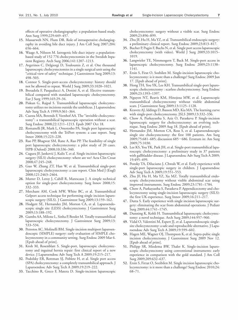
1
1
1
1
1
2
2
2
2
2
2
2
2
2
2
3
3
3
3
3
3
3
3
3
3
4
4
4
4
4
4
4
4
4
4
5
5
5
7Vol. 211, No. 1, July 2010 Rawlings et al Single-Incision Laparoscopic Cholecystectomy
effects of operative cholangiography: a population-based study.Ann Surg 1999;229:449–457.
5. Massarweh NN, Flum DR. Role of intraoperative cholangiog-raphy in avoiding bile duct injury. J Am Coll Surg 2007;204:656–664.
6. Waage A, Nilsson M. Iatrogenic bile duct injury: a population-based study of 152 776 cholecystectomies in the Swedish Inpa-tient Registry. Arch Surg 2006;141:1207–1213.
7. Avgerinos C, Delgiorgi D, Touloumis Z, et al. One thousandlaparoscopic cholecystectomies in a single surgical unit using the“critical view of safety” technique. J Gastrointest Surg 2009;13:498–503.
8. Connor S. Single-port-access cholecystectomy: history shouldnot be allowed to repeat. World J Surg 2009;33:1020–1021.
9. Bresadola F, Pasqualucci A, Donini A, et al. Elective transum-bilical compared with standard laparoscopic cholecystectomy.Eur J Surg 1999;165:29–34.
0. Piskun G, Rajpal S. Transumbilical laparoscopic cholecystec-tomy utilizes no incisions outside the umbilicus. J LaparoendoscAdv Surg Tech A 1999;9:361–364.
1. Cuesta MA, Berends F, Veenhof AA. The “invisible cholecystec-tomy”: a transumbilical laparoscopic operation without a scar.Surg Endosc 2008;22:1211–1213. Epub 2007 Oct 18.
2. Romanelli JR, Mark L, Omotosho PA. Single port laparoscopiccholecystectomy with the TriPort system: a case report. SurgInnov 2008;15:223–228.
3. Rao PP, Bhagwat SM, Rane A, Rao PP. The feasibility of singleport laparoscopic cholecystectomy: a pilot study of 20 cases.HPB (Oxford) 2008;10:336–340.
4. Cugura JF, Jankovic J, Kulis T, et al. Single incision laparoscopicsurgery (SILS) cholecystectomy: where are we? Acta Clin Croat2008;47:245–248.
5. Guo W, Zhang ZT, Han W, et al. Transumbilical single-portlaparoscopic cholecystectomy: a case report. Chin Med J (Engl)2008;121:2463–2464.
6. Mutter D, Leroy J, Cahill R, Marescaux J. A simple technicaloption for single-port cholecystectomy. Surg Innov 2008;15:332–333.
7. Merchant AM, Cook MW, White BC, et al. TransumbilicalGelport access technique for performing single incision laparo-scopic surgery (SILS). J Gastrointest Surg 2009;13:159–162.
8. Hodgett SE, Hernandez JM, Morton CA, et al. Laparoendo-scopic single site (LESS) cholecystectomy. J Gastrointest Surg2009;13:188–192.
9. Gumbs AA, Milone L, Sinha P, Bessler M. Totally transumbilicallaparoscopic cholecystectomy. J Gastrointest Surg 2009;13:533–534.
0. Petrotos AC, Molinelli BM. Single-incision multiport laparoen-doscopic (SIMPLE) surgery: early evaluation of SIMPLE cho-lecystectomy in a community setting. Surg Endosc 2009 Mar 6.[Epub ahead of print].
1. Kroh M, Rosenblatt S. Single-port, laparoscopic cholecystec-tomy and inguinal hernia repair: first clinical report of a newdevice. J Laparoendosc Adv Surg Tech A 2009;19:215–217.
2. Podolsky ER, Rottman SJ, Poblete H, et al. Single port access(SPA) cholecystectomy: a completely transumbilical approach. JLaparoendosc Adv Surg Tech A 2009;19:219–222.
3. Tacchino R, Greco F, Matera D. Single-incision laparoscopic
cholecystectomy: surgery without a visible scar. Surg Endosc2009;23:896–899.
4. Zhu JF, Hu H, Ma YZ, et al. Transumbilical endoscopic surgery:a preliminary clinical report. Surg Endosc 2009;23:813–817.
5. Bucher P, Pugin F, Buchs N, et al. Single port access laparoscopiccholecystectomy (with video). World J Surg 2009;33:1015–1019.
6. Langwieler TE, Nimmesgern T, Back M. Single-port access inlaparoscopic cholecystectomy. Surg Endosc 2009;23:1138–1141.
7. Ersin S, Firat O, Sozbilen M. Single-incision laparoscopic cho-lecystectomy: is it more than a challenge? Surg Endosc 2009 Jun17. [Epub ahead of print].
8. Hong TH, You YK, Lee KH. Transumbilical single-port laparo-scopic cholecystectomy : scarless cholecystectomy. Surg Endosc2009;23:1393–1397.
9. Nguyen NT, Reavis KM, Hinojosa MW, et al. Laparoscopictransumbilical cholecystectomy without visible abdominalscars. J Gastrointest Surg 2009;13:1125–1128.
0. Kravetz AJ, Iddings D, Basson MD, Kia MA. The learning curvewith single-port cholecystectomy. JSLS 2009;13:332–336.
1. Chow A, Purkayastha S, Aziz O, Paraskeva P. Single-incisionlaparoscopic surgery for cholecystectomy: an evolving tech-nique. Surg Endosc 2009 Aug 18. [Epub ahead of print].
2. Hernandez JM, Morton CA, Ross S, et al. Laparoendoscopicsingle site cholecystectomy: the first 100 patients. Am Surg2009;75:681–685; discussion 685�686. Erratum in: Am Surg2009;75:1030.
3. Lee KS, You YK, Park JH, et al. Single-port transumbilical lapa-roscopic cholecystectomy: a preliminary study in 37 patientswith gallbladder disease. J Laparoendosc Adv Surg Tech A 2009;19:495–499.
4. Ponsky TA, Diluciano J, Chwals W, et al. Early experience withsingle-port laparoscopic surgery in children. J LaparoendoscAdv Surg Tech A 2009;19:551–553.
5. Zhu JF, Hu H, Ma YZ, Xu MZ. Totally transumbilical endo-scopic cholecystectomy without visible abdominal scar usingimproved instruments. Surg Endosc 2009;23:1781–1784.
6. Chow A, Purkayastha S, Paraskeva P. Appendicectomy and cho-lecystectomy using single-incision laparoscopic surgery (SILS):the first UK experience. Surg Innov 2009;16:211–217.
7. Dutta S. Early experience with single incision laparoscopic sur-gery: eliminating the scar from abdominal operations. J PediatrSurg 2009;44:1741–1745.
8. Dunning K, Kohli H. Transumbilical laparoscopic cholecystec-tomy: a novel technique. Arch Surg 2009;144:957–960.
9. Vidal O, Valentini M, Espert JJ, et al. Laparoendoscopic single-site cholecystectomy: a safe and reproducible alternative. J Lapa-roendosc Adv Surg Tech A 2009;19:599–602.
0. Hagen ME, Wagner OJ, Thompson K, et al. Supra-pubic singleincision cholecystectomy. J Gastrointest Surg 2009 Nov 12.[Epub ahead of print].
1. Philipp SR, Miedema BW, Thaler K. Single-incision laparo-scopic cholecystectomy using conventional instruments: earlyexperience in comparison with the gold standard. J Am CollSurg 2009;209:632–637.
2. Ersin S, Firzat O, Sozibilen M. Single incision laparoscopic cho-lecystectomy: is it more than a challenge? Surg Endosc 2010;24:
68–71.