sinus lift. rafa mtinez-conde
TRANSCRIPT

8/3/2019 Sinus Lift. Rafa Mtinez-Conde
http://slidepdf.com/reader/full/sinus-lift-rafa-mtinez-conde 1/5
e145
J Clin Exp Dent. 2011;3(2):e145-9. Multiple window antrostomy.
Journal section: Oral Surgery doi:10.4317/j
Publication Types: Review
Multiple window access antrostomy in maxillary sinus grafting.
Presentation of a clinical serie of 10 cases and literature review
Rafael Martínez-Conde 1, Asier Eguia 2, Agurne Uribarri 3, José López-Vicente 3, José Manuel Aguirre 4
1 Professor. Oral and Maxillofacial Surgeon. Oral Medicine. Faculty of Medicine and Dentistry. University of the Basque Coun-
try. Spain.2 Associated Professor. Master of Oral Pathology. Oral Medicine. Faculty of Medicine and Dentistry. University of the Basque
Country. Spain.3 Associated Professor. Oral Medicine. Faculty of Medicine and Dentistry. University of the Basque Country. Spain.4 Head Professor. Director. Master of Oral Pathology. Oral Medicine. Faculty of Medicine and Dentistry. University of the Bas-
que Country. Spain.
Correspondence:
Departamento Estomatología. Universidad del País Vasco.
Barrio Sarriena s/n
48940. Leioa (Vizcaya). Spain.
E-mail: [email protected]
Received : 14/10/2010
Accepted: 17/12/2010
AbstractObjectives: A variation on the usual maxillary sinus grafting technique and its results are presented, using a more
conservative approach that provides better conditions for applying the graft in complicated situations.
Material and Methods: Ten case reports are presented in which the multiple access technique was used due to theexistence of large maxillary sinuses, where a wide surgical approach was needed because several implants were to
be installed or cases in which sinus bone graft was part of a more extensive reconstructive prodedure.
Results: All the implants that were placed after using this technique were correctly integrated and it was possible
to proceed to the prosthesis stage without any problems.
Conclusions: This modied technique of sinus oor bone grafting can have a benecial effect with a lower risk of
perforations, better preservation of vascularisation in the area and improved integration and stability of the implants
and bone graft, specially where wider surgical access is required.
Key words: Antrostomy, maxillary sinus, bone grafting, dental implants.
Martínez-Conde R, Eguia A, Uribarri A, López-Vicente J, Aguirre JM.Multiple window access antrostomy in maxillary sinus grafting. Presenta-
tion of a clinical serie of 10 cases and literature review. J Clin Exp Dent.
2011;3(2):e145-9.
http://www.medicinaoral.com/odo/volumenes/v3i2/jcedv3i2p145.pdf
Article Number: 50417 http://www.medicinaoral.com/odo/indice.htm
© Medicina Oral S. L. C.I.F. B 96689336 - eISSN: 1989-5488
eMail: [email protected]

8/3/2019 Sinus Lift. Rafa Mtinez-Conde
http://slidepdf.com/reader/full/sinus-lift-rafa-mtinez-conde 2/5
e146
J Clin Exp Dent. 2011;3(2):e145-9. Multiple window antrostomy.
IntroductionDue to its versatility and the good results achieved with
its use, oral implantology has become a commonly used
therapeutic tool. Indications of these intraosseous xa-
tion systems have become so widespread that they co-
ver almost any type of edentulism. This widespread use,
sometimes, face situations where there is an insufcientquantity and quality of bone for their installation.
The posterior area of the maxilla is often compromised,
as the presence of the maxillary sinus limits the height
of the remaining amount of available bone. Maxillary
sinus grafting techniques have been developed to solve
this problem. Since it was rst described by Boyne et
al. (1), this method has become a widely-used surgical
technique with a reported success rate of more than 90
% (2). However, despite this, there is still controversy
about certain aspects such as which graft material to use,
whether to perform delayed or simultaneous placement
of dental implants, how to close the antrostomy window
or the type of surgical approach to employ (3-5).
Double window antrostomy approach to the maxillary
sinus has been proposed in the presence of septa (6-9).
Furthermore, this type of surgical access could present
some advantages in another clinical situations where it is
necessary to use a wide surgical approach such as: in hy-
perpneumatised maxillary sinuses, in cases that require a
large surgical access due to the simultaneous installation
of several implants, or in cases where the sinus graft is
part of a more extensive procedure (3,6,7).
Ten cases of multiple antrostomy window surgical ac-
cess for the maxillary sinus grafting procedure, where
the indication was other than the presence of sinus septa,are presented in this paper.
Material and Methods Patient Selection
Betweeen January 2005 and November 2007, ten pa-
tients in which a wide approach to the maxillary sinus
was needed because several implants were to be insta-
lled and the distance between them was very large, or ca-
ses of hyperpneumatised maxillary sinuses, were inclu-
ded in this study (Table1). Patients were fully informed
about the surgical procedure and signed an appropiate
consent form.Surgical Technique
A large buccal mucoperiosteal ap was raised exposing
the lateral wall of the maxilla from its anterior region up
to the area of the tuberosity. In the anterior wall of the
maxilla, an oval-shaped ostectomy was performed using
rotatory instruments. This ostectomy must large enough
to facilitate handling of the sinus lift instruments and
make it possible to detach the Schneirediam membrane
from the anterior portion of sinus oor.
Following extensive detachment in this area, which must
also include the palatal wall, the sinus lift instrument is
moved in a posterior direction, initially along the maxi-
llary sinus oor. At this level it is common to nd some
kind of obstacle due to the presence of irregularities and
septa, especially in the zygomatic-malar complex. It is
not advisable to insist on detaching this area, as the Sch-
neiderian membrane can easily be torn (6,10). However,
it is easy to perform this posterior advance by detachingthe membrane from the external lateral wall of the sinus,
where it is safe to perform an extensive tunnelling in this
buccal area. This tunnelling makes it easy to perform a
second and even a third antrostomy distally in the bony
wall, generally behind the zygomatic-malar complex,
without any risk of tearing the membrane.
This secondary window is used to approach the deta-
chment of the membrane in the posterior region of the
sinus oor and the palatal wall, which is joined with the
anterior area, thus releasing any osseous crests and irre-
gularities that there might be at this level (Fig. 1).
Fig. 1.
Fig. 2.
After raising the sinus membrane it is possible to install
the implant, in the case of immediate implantation (Fig.
2), or to ll the area with the graft material if a dela-
yed technique is to be used. Implantation was delayed
in three cases, either because the lifting procedure for-
med part of a more extensive reconstruction with buccal
8/3/2019 Sinus Lift. Rafa Mtinez-Conde
http://slidepdf.com/reader/full/sinus-lift-rafa-mtinez-conde 3/5
e147
J Clin Exp Dent. 2011;3(2):e145-9. Multiple window antrostomy.
gery was performed to expose the implants in the caseswhere they were left submerged, and placement of the
implants was performed in those where a two-step pro-
cedure had been chosen. All the implants that were pla-
ced immediately, at the same time as the lift procedure,
were correctly integrated and it was possible to proceed
to the prosthesis stage after this period. In the three cases
where placement of the implants was delayed, a suf-
cient quantity of bone was achieved to make their subse-
quent insertion possible without any problems.
DiscussionMaxillary sinus oor bone grafting technique has been
in use for more than thirty years (1) and, since it was rst
described, numerous variations have been reported (5).
Despite all these variations, the most common surgical
access continues to be lateral antrostomy, this involves
the use of the thinnest area of the buccal wall on the
anterior face of the maxillary sinus. From this area, in
a posterior direction, we nd the zygomatic-malar com-
plex, where the bone is thicker. In this buttress region it
is more laborious and difcult to perform the ostectomy,
requiring previous bone reduction, and the lifting of the
“onlay” grafts, or because it was not possible to achievegood primary stability with the implants (Table1).
A mixture of particulate bovine bone (BioOss®, Geist-
lich Pharma AG, Switzerland) with autologous bone har-
vested from the same area and platelet-rich plasma was
used as the graft material (10). In two patients, in whom
the sinus lift formed part of more extensive reconstruc-
tive procedures, cancellous bone chips taken from the
anterior iliac crest mixed with bovine bone (BioOss®,
Geistlich Pharma AG, Switzerland) were utilised. An-
trostomies were closed by placing a collagen membrane
(Bioguide®, Geistlich Pharma AG, Switzerland) to co-
ver osseous defects protecting the graft and preventing
its migration. After this the buccal ap was replaced and
sutured in place.
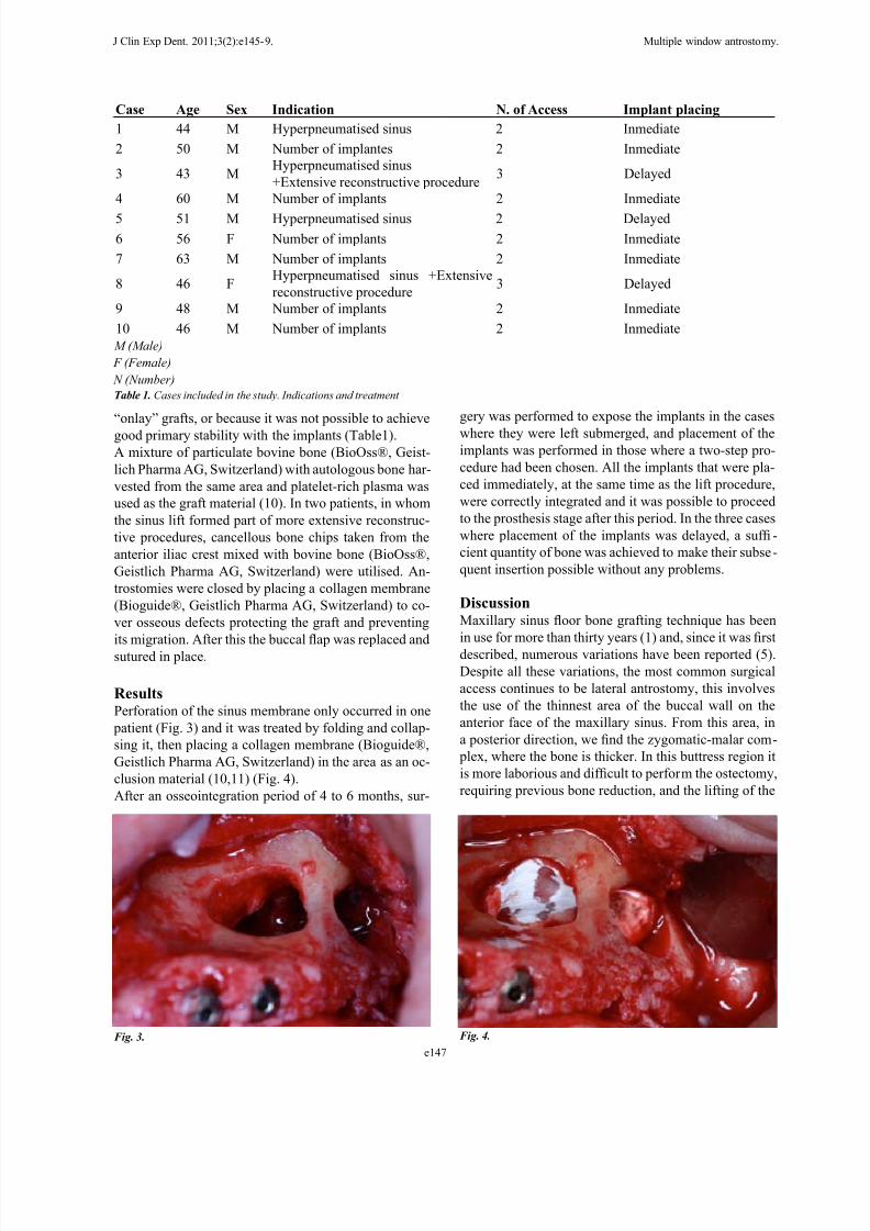
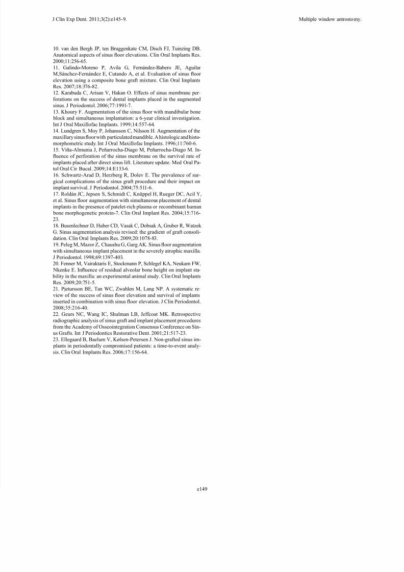
ResultsPerforation of the sinus membrane only occurred in one
patient (Fig. 3) and it was treated by folding and collap-
sing it, then placing a collagen membrane (Bioguide®,
Geistlich Pharma AG, Switzerland) in the area as an oc-
clusion material (10,11) (Fig. 4).
After an osseointegration period of 4 to 6 months, sur-
Case Age Sex Indication N. of Access Implant placing
1 44 M Hyperpneumatised sinus 2 Inmediate
2 50 M Number of implantes 2 Inmediate
3 43 MHyperpneumatised sinus
+Extensive reconstructive procedure3 Delayed
4 60 M Number of implants 2 Inmediate
5 51 M Hyperpneumatised sinus 2 Delayed
6 56 F Number of implants 2 Inmediate
7 63 M Number of implants 2 Inmediate
8 46 FHyperpneumatised sinus +Extensive
reconstructive procedure3 Delayed
9 48 M Number of implants 2 Inmediate
10 46 M Number of implants 2 Inmediate
M (Male)
F (Female)
N (Number)
Table 1. Cases included in the study. Indications and treatment
Fig. 3. Fig. 4.

8/3/2019 Sinus Lift. Rafa Mtinez-Conde
http://slidepdf.com/reader/full/sinus-lift-rafa-mtinez-conde 4/5
e148
J Clin Exp Dent. 2011;3(2):e145-9. Multiple window antrostomy.
membrane is associated with a higher incidence of per-
forations (10). The thickness of the buccal bony wall of
the maxillary sinus decreases again towards the distal
region, in the direction of the tuberosity. By performing
the ostectomy in this area after detaching the membrane
it is easy to avoid accidental perforations.
Perforations or tears of the sinus membrane during thesurgical procedure are the most common complication
of the maxillary sinus lift technique. Although the im-
portance of these perforations for the ultimate success
of the implants is debatable (12-14), when they are very
large they can make it necessary to abort the procedu-
re (13,15,16). According to different authors, the pre-
valence of these perforations varies between 7% and
44% (13,16). Perforation of the sinus membrane only
occurred in one patient in our study (case report No. 6),
which coincided with the presence of a partial septum in
the sinus oor). With multiple antrostomies the approach
to the maxillary sinus is trough the anterior wall, where
the bone is thinner, the use of instruments is simpler and
there is therefore a lower probability of perforating the
membrane.
Masticatory forces are distributed to the cranium by
means of four major buttresses, the outermost of which
is the zygomatic-malar buttress, which forms the joint
between the anterior and posterior walls of the maxillary
sinus. This maxillary pillar is an important bone structu-
re in terms of resistance to masticatory forces, so it is not
advisable to sacrice it during the surgical procedure. If
kept, this pillar or the sections of bone that are preserved
between the antrostomies provide extra support and help
to retain the graft material and its contour, adapting thecollagen membrane that is usually used as a closure for
the ostectomy to that convex area in order to prevent
migration of the graft and the possibility of invasion by
cells of connective origin into the augmented area (4).
Furthermore, these sections of bone that are preserved
between the multiple ostectomies make it possible to
achieve a larger contact area and osteosynthesis screw
xation if buccal bone grafts are used simultaneously.
Once the graft is packed into the maxillary sinus, its in -
tegration and ossication potential is determined by the
presence of osteogenic cells in the area. To a large extent,
these osteogenic cells migrate towards the graft from theremaining exposed bony walls that are in contact with
the graft and from them the new bone progresses towards
the augmented area (17,18). It can therefore be said that
ossication of the graft takes place centripetally, with a
gradient from the exposed maxillary bone inwards into
the graft (18). By trying to conserve the buccal wall we
will therefore improve the integration of the graft due to
the greater contribution of the residual bone.
In the case reports described in this article, none of the
implants were lost during the osseointegration period.
After a healing period of 4 to 6 months all the implants
were successfully integrated. In every case, except whe-
re the sinus lift formed part of a more extensive procedu-
re, we endeavoured to perform immediate implantation,
despite the fact that in many of them there was less than
4 mm of residual alveolar bone remaining (10). It is ne-
cessary to achieve good initial primary stability in order
to perform this simultaneous placement of the implantsand sinus bone graft (19,20). Most studies show a suc-
cess rate of almost 95% for implants after alveolar bone
grafting, with a slight decrease in this rate in cases of im-
mediate implantation (21), especially when there is less
than 4 mm of residual bone (22). The stabilising effect
of the residual buccal wall graft may be benecial for
its integration, isolating it from external lateral forces
that produce micromovements (provisional prostheses,
muscles, mastication, etc.). To achieve better primary
stability Astra Tech ST implants were used, in which the
conical coronal region with microthreads initially help
to anchor them to the bone tissue (23). The preserved
integrity of the buccal bony wall in turn prevents fractu-
res from occurring in the residual alveolar bone during
insertion of the implant, which would destabilise this
anchorage.
Sinus oor bone grafting is a commonly used technique
in oral implantology that has an established method and
indications. However, there are some cases, such as hy-
perpneumatised maxillary sinuses, where wider surgical
access is required due to the need to place several im-
plants or when the sinus lift forms part of more extensive
procedures, in which multiple antrostomies can have a
benecial effect with a lower risk of perforations, better
preservation of vascularisation in the area and improvedintegration and stability of the implants and bone graft.
References1. Boyne PJ, James RA. Grafting of the maxillary sinus oor with
autogenous marrow and bone. J Oral Surg. 1980;38:613-16.
2. Tong DC, Rioux K, Drangsholt M, Beirne OR. A review of survi-
val rates for implants placed in grafted maxillary sinuses using meta-
analysis. Int J Oral Maxillofac Implants. 1998;13:175-82.
3. Wallace SS, Froum SJ. Effect of maxillary sinus augmentation on
the survival of endosseous dental implants. A systematic review. Ann
Periodontol. 2003;8:328-43.
4. Choi KS, Kan JY, Boyne PJ, Goodacre CJ, Lozada JL, Rungcharas-
saeng K. The effects of resorbable membrane on human maxillary sin-
us graft: a pilot study. Int J Oral Maxillofac Implants. 2009;24:73-80.
5. Tofer M. Osteotome-mediated sinus oor elevation: a clinical re-
port. Int J Oral Maxillofac Implants. 2004;19:266-73.
6. Ella B, Noble Rda C, Lauverjat Y, Sédarat C, Zwetyenga N, Siber-
chicot F, et al. Septa within the sinus: effect on elevation of the sinus
oor. Br J Oral Maxillofac Surg. 2008;46:464-7.
7. Maestre-Ferrín L, Galán-Gil S, Rubio-Serano M, Peñarrocha-Diago
M. Maxillary sinus septa: a sytematic review. Med Oral Patol Oral Cir
Bucal. 2010;15:383-6.
8. González-Santana H, Peñarrocha-Diago M, Guarinos-Garbó J,
Sorní-Bröker M. A study of the septa in the maxillary sinuses and
the subantral alveolar processes in 30 patients. J Oral Implantol.
2007;33:340-3.
9. Betts NJ, Miloro M. Modication of the sinus lift procedure for
septa in the maxillay antrum. J Oral Maxillofac Surg. 1994;52:332-3.
8/3/2019 Sinus Lift. Rafa Mtinez-Conde
http://slidepdf.com/reader/full/sinus-lift-rafa-mtinez-conde 5/5
e149
J Clin Exp Dent. 2011;3(2):e145-9. Multiple window antrostomy.
10. van den Bergh JP, ten Bruggenkate CM, Disch FJ, Tuinzing DB.
Anatomical aspects of sinus oor elevations. Clin Oral Implants Res.
2000;11:256-65.
11. Galindo-Moreno P, Avila G, Fernández-Babero JE, Aguilar
M,Sánchez-Fernández E, Cutando A, et al. Evaluation of sinus oor
elevation using a composite bone graft mixture. Clin Oral Implants
Res. 2007;18:376-82.
12. Karabuda C, Arisan V, Hakan O. Effects of sinus membrane per -
forations on the success of dental implants placed in the augmentedsinus. J Periodontol. 2006;77:1991-7.
13. Khoury F. Augmentation of the sinus oor with mandibular bone
block and simultaneous implantation: a 6-year clinical investigation.
Int J Oral Maxillofac Implants. 1999;14:557-64.
14. Lundgren S, Moy P, Johansson C, Nilsson H. Augmentation of the
maxillary sinus oor with particulated mandible. A histologic and histo-
morphometric study. Int J Oral Maxillofac Implants. 1996;11:760-6.
15. Viña-Almunia J, Peñarrocha-Diago M, Peñarrocha-Diago M. In-
uence of perforation of the sinus membrane on the survival rate of
implants placed after direct sinus lift. Literature update. Med Oral Pa-
tol Oral Cir Bucal. 2009;14:E133-6.
16. Schwartz-Arad D, Herzberg R, Dolev E. The prevalence of sur-
gical complications of the sinus graft procedure and their impact on
implant survival. J Periodontol. 2004;75:511-6.
17. Roldán JC, Jepsen S, Schmidt C, Knüppel H, Rueger DC, Acil Y,et al. Sinus oor augmentation with simultaneous placement of dental
implants in the presence of patelet-rich plasma or recombinant human
bone morphogenetic protein-7. Clin Oral Implant Res. 2004;15:716-
23.
18. Busenlechner D, Huber CD, Vasak C, Dobsak A, Gruber R, Watzek
G. Sinus augmentation analysis revised: the gradient of graft consoli-
dation. Clin Oral Implants Res. 2009;20:1078-83.
19. Peleg M, Mazor Z, Chaushu G, Garg AK. Sinus oor augmentation
with simultaneous implant placement in the severely atrophic maxilla.
J Periodontol. 1998;69:1397-403.
20. Fenner M, Vairaktaris E, Stockmann P, Schlegel KA, Neukam FW,
Nkenke E. Inuence of residual alveolar bone height on implant sta-
bility in the maxilla: an experimental animal study. Clin Oral Implants
Res. 2009;20:751-5.
21. Pjetursson BE, Tan WC, Zwahlen M, Lang NP. A systematic re-
view of the success of sinus oor elevation and survival of implantsinserted in combination with sinus oor elevation. J Clin Periodontol.
2008;35:216-40.
22. Geurs NC, Wang IC, Shulman LB, Jeffcoat MK. Retrospective
radiographic analysis of sinus graft and implant placement procedures
from the Academy of Osseointegration Consensus Conference on Sin-
us Grafts. Int J Periodontics Restorative Dent. 2001;21:517-23.
23. Ellegaard B, Baelum V, Kølsen-Petersen J. Non-grafted sinus im-
plants in periodontally compromised patients: a time-to-event analy-
sis. Clin Oral Implants Res. 2006;17:156-64.