sirna lipid nanoparticle potently silences...
TRANSCRIPT
Cancer Therapy: Preclinical
siRNA Lipid Nanoparticle Potently SilencesClusterin andDelaysProgressionWhenCombinedwith Androgen Receptor Cotargeting inEnzalutamide-Resistant Prostate CancerYoshiaki Yamamoto1,2, Paulo J.C. Lin3, Eliana Beraldi1, Fan Zhang1, Yoshihisa Kawai1,2,Jeffrey Leong1, Hidemasa Katsumi3, Ladan Fazli1, Robert Fraser3, Pieter R. Cullis3, andMartin Gleave1
Abstract
Purpose: Lipid nanoparticle (LNP) formulations facilitate tumoruptake and intracellular processing through an enhanced perme-ationand retentioneffect (EPR), and currentlymultipleproducts areundergoing clinical evaluation. Clusterin (CLU) is a cytoprotectivechaperone induced by androgen receptor (AR) pathway inhibitionto facilitate adaptive survival pathway signaling and treatmentresistance. In our study, we investigated the efficacy of siRNA tumordelivery using LNP systems in an enzalutamide-resistant (ENZ-R)castration-resistant prostate cancer (CRPC) model.
Experimental Design: Gene silencing of a luciferase reportergene in the PC-3M-luc stable cell line was first assessed insubcutaneous and metastatic PC-3 xenograft tumors. Upon val-idation, the effect of LNP siRNA targeting CLU in combinationwith AR antisense oligonucleotides (ASO) was assessed in ENZ-RCRPC LNCaP in vitro and in vivo models.
Results: LNP LUC-siRNA silenced luciferase expression in PC-3M-luc subcutaneous xenograft andmetastaticmodels. LNPCLU-siRNA potently suppressed CLU and AR ASO-induced CLU andAKTandERKphosphorylation inENZ-RLNCaP cells in vitro,morepotently inhibiting ENZ-R cell growth rates and increased apo-ptosis when compared with AR-ASO monotherapy. In subcuta-neous ENZ-R LNCaP xenografts, combinatory treatment of LNPCLU-siRNA plus AR-ASO significantly suppressed tumor growthand serum PSA levels compared with LNP LUC-siRNA (control)and AR-ASO.
Conclusions: LNP siRNA can silence target genes in vivo andenable inhibition of traditionally non-druggable genes like CLUand other promising cotargeting approaches in ENZ-R CRPCtherapeutics. Clin Cancer Res; 21(21); 4845–55. �2015 AACR.
IntroductionProstate cancer is the most prevalent cancer in men in the
Western world and the second leading cause of cancer deathsamong males in Western countries (1). Androgen receptor (AR)signaling remains the key driver of castration-resistant prostatecancer (CRPC). Potent AR pathway inhibitors like enzalutamide(ENZ) inhibit AR nuclear translocation and transcriptional activ-
ity (2, 3), but despite significant activity, progression to ENZ-resistant (ENZ-R) CRPC frequently occurs. This progressionoccurs with rising serum PSA levels, thereby implicating ARimportance in disease progression. Activation of adaptive survivalpathways that support AR signaling is an importantmechanismoftreatment resistance. The molecular chaperone clusterin (CLU) isinduced by AR pathway inhibition and highly expressed in ENZ-RCRPC (4), and as a mediator of the stress response conferstreatment resistance when overexpressed (5–7). CLU inhibitsstress-induced apoptosis by suppressing p53-activating stresssignals (8), and conformationally altered Bax (6, 8) in additionto enhancing AKT phosphorylation (9) and transactivation ofNF-kB (10) and autophagy (11). In keeping with these cytoprotectivemechanisms, CLU inhibition potentiates activity of anticancertherapy inmanypreclinicalmodels (12), and is a promising targetfor novel therapeutics.
Although small-interfering RNA (siRNA) offers the promise forpotent and specific gene silencing, poor accumulation at sites ofdisease and intracellular translocation coupled with poor stabil-ity, sensitivity to nucleases, immune stimulation, and rapidclearance have made its therapeutic application difficult. A deliv-ery system is crucial to the efficient delivery to target tissue in orderto overcome these shortcomings. Encapsulation of siRNA using alipid nanoparticle-based delivery system has shown to be crucialin protecting the nucleic acid-based drug fromnucleases as well as
1The Vancouver Prostate Centre and Department of UrologicSciences, University of British Columbia, Vancouver, British Colum-bia, Canada. 2Department of Urology, Graduate School of Medicine,Yamaguchi University, Ube, Japan. 3Department of Biochemistryand Molecular Biology, University of British Columbia, Vancouver,BC, Canada.
Note: Supplementary data for this article are available at Clinical CancerResearch Online (http://clincancerres.aacrjournals.org/).
Y. Yamamoto and P.J.C. Lin, and the Cullis and Gleave Labs contributed equallyto this article.
Corresponding Author: Martin E. Gleave, The Vancouver Prostate Centre andDepartment of Urologic Sciences, University of British Columbia, 2660 OakStreet, Vancouver, British Columbia, Canada V6H 3Z6. Phone: 604-875-4818;Fax: 604-875-5654; E-mail: [email protected]
doi: 10.1158/1078-0432.CCR-15-0866
�2015 American Association for Cancer Research.
ClinicalCancerResearch
www.aacrjournals.org 4845
on June 2, 2018. © 2015 American Association for Cancer Research. clincancerres.aacrjournals.org Downloaded from
Published OnlineFirst June 23, 2015; DOI: 10.1158/1078-0432.CCR-15-0866
prolonging circulation, reducing immune stimulation, andimproving intracellular uptake (13). Themost clinically advanceddelivery of siRNA uses LNP and currently there are six differentsiRNAs encapsulated in LNP drugs undergoing clinical studies.Themost promising study looks at the treatment of transthyretin-induced amyloidosis using a second-generation cationic lipid(DLin-MC3-DMA) that is well tolerated and potently silencestransthyretin (14). Two siRNA-LNP studies focus in the treatmentof hepatic cancers and early indications suggest safe and activetreatments (15). Propensity of LNP systems to accumulate inthe liver has become a major obstacle in the extrahepaticdelivery of siRNA-LNP systems. Although preclinical studiesin prostate cancer (16) and immune cells (17) are promising,distant tumor delivery has not been tested in the clinic. In orderto mitigate this higher Peg-lipid content is used to improve thepharmacodynamic and biodistribution of LNP systems to thetumor site (18–20).
Antisense oligonucleotides (ASO) offer another approach toselectively target genes. Although ASOs are primarily used toinhibit "undruggable" targets (21, 22), they may also be of useagainst drug-resistant targets like that AR in ENZ-resistance(ENZ-R) (23). Although AR extinction approaches using ASOs(24) or shRNA (25) can reduce AR levels and inhibit tumorgrowth in CRPC models, they have neither been studied in thecontext of ENZ-R disease nor in combination with siRNA-mediated cotargeting strategies. In this study, we first investi-gated the efficacy of LNP siRNA tumor delivery in AR-negativePC-3 and AR-positive ENZ-R LNCaP prostate cancer. Genesilencing was first validated using LNP LUC-siRNA to silencePC3 stably expressing firefly luciferace (PC3-M-luc) in vitrofollowed by successful gene silencing in subcutaneous andmetastatic xenograft models. Once in vivo silencing of LUCwas demonstrated, we then evaluated combinatory gene silenc-ing of CLU (using LNP siRNA) and AR (using an AR-ASO) in an
ENZ-R LNCaP model and demonstrated enhanced apoptosisand growth inhibition in vitro and in vivo.
Materials and MethodsCell lines and reagents
LNCaP were kindly provided by Dr. L.W.K. Chung (1992, MDAnderson Cancer Center, Houston, TX) and ENZ-R MR49Fcell lines were generated, and maintained as previously described(4, 26). PC-3M-luc (C6) cells, stably expressing firefly luciferaseprotein, were obtained from Caliper Life Sciences and werecultured in DMEM(Invitrogen) with 5% FBS and 2 mmol/LL-glutamine. Supplementary Table S1 shows source and authen-tication of cell lines. Permanent stocks of cells of authenticated orpurchased were prepared and were stored in liquid nitrogen untiluse. Cells were used for experiments within 6 months. Enzaluta-mide was purchased from Haoyuan Chemexpress Co., Limited.
siRNA and AR antisense oligonucleotideAll siRNAwere purchased from Thermo Scientific or Integrated
DNA Technologies. The lower case letters indicates 2'Omethylmodification, while upper case letter represents unmodified res-idue and "s" indicates phosphorothioate modification.
Clusterin. Sense: 50- AuGAuGAAGACuCuGCuGCdTdT- 30
Antisense: 50 - GCAGCAGAGuCuuCAuCAuGC - 30
Luciferase. Sense: 50 -cuuAcGcuGAGuAcuucGAdTsdT-30
Antisense: 50-UCGAAGuACUcAGCGuAAGdTsdT-30
GFP. Sense: 50 -AcAuGAAGcAGcACGACuUdTsdT-30
Antisense: 50-AAGUCGUGCUGCUUCAUGUdTsdT-30
AR and scrambled (SCRB) antisense was supplied by IsisPharmaceuticals as previously described (4). The AR-ASO tar-geting exon-1 and scrambled (SCRB) control sequences were50-GCGACTACTACAACTT-30 and 50-CAGCGCTGACAACAGT-TTCAT-30, respectively. Prostate cells were treated with theindicated oligonucleotides, using protocols described previous-ly (23, 27, 28).
Lipid nanoparticle encapsulation of siRNAThe ionizable cationic lipids O-(Z,Z,Z,Z-heptatriaconta-
6,9,26,29-tetraen-19-yl)-4-(N,N-dimethylamino)butanoate (DLin-MC3-DMA) and 2,2-dilinoleyl-4-(2-dimethylaminoethyl)-[1,3]-dioxolane (DLin-KC2-DMA), and PEG lipids PEG-DMG andPEG-DSG were purchased from Biofine International Inc. and havebeen previously described (29–31). 1,2-distearoyl-sn-glycero-3-phosphocholine (DSPC)andcholesterolwereobtained fromAvantiand Sigma-Aldrich Co., respectively. The lipid composition of allLNPs containing siRNA (LNP-siRNA) was cationic lipid/DSPC/cholesterol/PEG-DMG (50/10/38.5/1.5; mol%) for in vitro,while cationic lipid/DSPC/cholesterol/PEG-DSG (50/10/37.5/2.5; mol%) or (50/10/35/5; mol%) for in vivo. LNP-siRNAs wereprepared using a microfluidic mixing apparatus as previouslydescribed (30, 32).
Bioluminescence imagingPC-3M-luc cells and tumors in mice were imaged using an
IVIS200 camera (Caliper Life Sciences) as previously described(33). Data were acquired and analyzed using Living Image soft-ware version 3.0 (Caliper Life Sciences).
Translational Relevance
Suppression of androgen receptor (AR) signaling remains atherapeutic goal for castration-resistant prostate cancer(CRPC). Despite newer potent AR-pathway inhibitors, resis-tance frequently occurs. Although cotargeting the AR withadaptive survival pathways is a rational goal, many biologi-cally relevant genes are undruggable with small-moleculeinhibitors. Gene silencing of non-druggable targets usingsmall-interfering RNA (siRNA) is a promising approach butin vivo delivery remains problematic without a delivery system.We developed a lipid nanoparticle (LNP) system demonstrat-ing silencing of luciferase reporter gene using LNP-LUC-siRNAin both subcutaneous andmetatastic PC3-Luc-xenograft mod-els. LNP-CLU-siRNA inhibited AR-antisense–induced upregu-lation of clusterin (CLU) in vitro and in vivo, and significantlysuppressed tumor growth and serum PSA levels in enzaluta-mide-resistant (ENZ-R) LNCaP xenografts compared with AR-antisense monotherapy. These data provide novel proof-of-principle that LNP-siRNA can target genes in vivo enablinginhibition of traditionally non-druggable genes like CLU andother promising cotargeting approaches in ENZ-R CRPCtherapeutics.
Yamamoto et al.
Clin Cancer Res; 21(21) November 1, 2015 Clinical Cancer Research4846
on June 2, 2018. © 2015 American Association for Cancer Research. clincancerres.aacrjournals.org Downloaded from
Published OnlineFirst June 23, 2015; DOI: 10.1158/1078-0432.CCR-15-0866
Cell proliferation assaysPC-3M-luc cells were seeded at a density of 5 � 103 in 96-well
plates, and ENZ-R MR49F and parental LNCaP were seeded at adensity of 1.25� 104 in 48-well plates. Cell viability in PC-3M-luccells and cell growth inMR49F and LNCaP cells weremeasured bycrystal violet assay as previously described (34). Each assay wasdone in triplicate three times.
Western blotting analysisTotal proteins were extracted using RIPA buffer (50 mmol/L
Tris, pH 7.2, 1% NP-40, 0.1% deoxycholate, 0.1% SDS, 100mmol/L NaCl, Roche complete protease inhibitor cocktail) andsubjected to Western blot analysis as described previously (28).Primary antibodies are shown in Supplementary Materials andMethods.
Quantitative reverse transcription-PCRTotal RNA was extracted using TRIzol reagent (Invitrogen Life
Technologies, Inc.) as previously reported (27). Primers(described in Supplementary Table S2) were normalized tob-actin levels as an internal standard, and the comparative cyclethreshold (Ct) method was used to calculate relative quantifica-tion of target mRNAs. Each assay was conducted in triplicate.
In vivo PC-3M-luc subcutaneous tumorMale athymic mice were inoculated subcutaneously with 2 �
106 PC-3M-luc cells. Once tumor bioluminescence signalsreached approximately 1 � 108 photons/second, they were ran-domly assigned for treatment. LNP containing indicated doses ofLUC-siRNA or GFP-siRNA as control was administered intrave-nously (i.v.) through the lateral tail vein. To reduce possibletoxicity due to daily dosing, the dosing regimen was modifiedfrom previous studies performed by Lee and colleagues whererepeat doses administered at 10 mg/kg for 3 consecutive daysfollowed by administration at days 7, 9, and 11 (16) weremodified to daily dosing of 7 mg/kg for 5 days. To evaluate LUCexpression, mice were then imaged on days 0 (before treatment),3, 5, and 8 using an IVIS200 Imaging System and then sacrificedon day 8. LUC expression (photons/second) measured at days 3,5, and 8 was normalized to corresponding animal at day 0 andexpressed as relative increase (%). All animal procedures used inthis manuscript were approved and carried out according to theguidelines of the Canadian Council on Animal Care and appro-priate institutional certification.
In vivo PC-3M-luc metastasis modelPC-3M-luc cells (2 � 106) were injected intravenously into
the tail vein of male athymic mice as described previously (33).Once metastatic bioluminescence signals reached approximately1� 105 photons/second, they were randomly assigned to either 5mg/kg LNP LUC-siRNA or GFP-siRNA as control and i.v. injectedthrough the lateral tail vein daily for 5 days. To evaluate LUCexpression, mice were then imaged on days 0 (before treatment),3, 5, and 8 using an IVIS200 Imaging System and then sacrificedon day 8. LUC expression measured at days 3, 5, and 8 werenormalized to corresponding animal at day 0 and expressed asrelative increase (%).
In vivo ENZ-R MR49F treatmentMale athymic mice were castrated and inoculated subcutane-
ously with 2 � 106 ENZ-R MR49F cells; mice were treated with
ENZ at 10 mg/kg/each orally daily for maintenance of ENZresistance. Once tumors reached 100 mm3, mice were randomlyassigned to 10 mg/kg SCRB or AR-ASO (administered intraper-itoneally, i.p., once daily for 5 days and then three times per weekthereafter) plus either 5 mg/kg LNP siCLU or siLUC siRNA ascontrol with 2.5 or 5% PEG (i.v. through the lateral tail vein oncedaily for 4 days and then 3 times per week thereafter). Tumorvolume and serum PSA was measured as previously described(27). Mice were sacrificed on day 21 and tumors were harvestedfor evaluationbyWestern blot analyses,mRNAexpressionby real-timemonitoring of qPCR, and IHC. Treatment was extended overa 3-week period, with four daily injections in the first week andthree daily injections during the second and third weeks
IHCIHC was performed as previously described previously (27).
All comparisons of staining intensities were made at �200magnifications.
Statistical analysisAll in vitro data were assessed using the Student t test. All in vivo
data were compared using the Kruskal–Wallis test (JMP version8). Levels of statistical significance were set at P < 0.05.
ResultsLNP LUC-siRNA decreases LUC expression in PC-3M-lucsubcutaneous xenografts
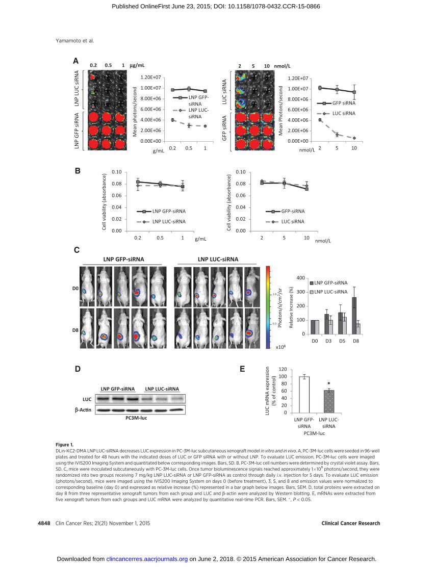
To assess the effects of LNP siRNA tumor delivery in vivo, theexpression of luciferase in PC-3M-luc stably transfected cellstreated with LNP LUC-siRNA containing DLin-KC2-DMA(16, 31) was examined by the IVIS imaging system. The ionizablecationic lipid, DLin-KC2-DMA, is highly active in the liver (31)and also previously shown to silence AR and reduce PSA levelsupon intravenous administration in LNCaP tumor model (16).PC-3M-luc cells showed significant correlation between meanbioluminescence and both the total numbers of these cells invitro (Supplementary Fig. S1) and mean subcutaneous tumorvolume in vivo (Supplementary Fig. S2). LUC siRNA transfectedwith Lipofectamine 2000 or delivered in LNP systems decreasedthe expression of LUC in a dose-dependent manner in PC-3M-lucin vitro (Fig. 1A) without changing the cell numbers, as evaluatedby crystal violet assay (Fig. 1B).
The in vivo activity of LNP LUC-siRNA containing DLin-KC2-DMA was first evaluated using PC-3M-luc subcutaneous xeno-grafts. After tumor bioluminescence signals reached approximate-ly 1 � 108 photons/second, mice were randomly assigned fortreatment with 7 mg/kg LNP LUC-siRNA (5 mice) or LNP GFP-siRNA (5 mice) as control. At baseline, mean bioluminescencesignals were similar in LNP LUC andGFP-siRNA groups (1.22 and1.17�109 photons/second, respectively). LNP LUC-siRNA exhib-ited LUC silencing effects in PC-3M-luc xenografts as early as day 3after initial administration by maintaining LUC expression atbaseline levels, whereas LNP GFP-siRNA treated showed a 143%increase when normalized to day 0 (Fig. 1C). Significant silencingwas achieved at day 8 as the LUC expressionwas below baseline at75%,whereas LNPGFP-siRNA treatment showed a 263% increasein LUC expression. The difference between LNP LUC-siRNA andcontrol LNP GFP-siRNA corresponded to approximately 3-folddecrease in LUC expression (Fig. 1C). As expected, LUC protein intumors collected from representative PC-3M-luc xenografts (n¼ 3
Lipid Nanoparticle siRNA Systems in CRPC
www.aacrjournals.org Clin Cancer Res; 21(21) November 1, 2015 4847
on June 2, 2018. © 2015 American Association for Cancer Research. clincancerres.aacrjournals.org Downloaded from
Published OnlineFirst June 23, 2015; DOI: 10.1158/1078-0432.CCR-15-0866
D E
LNP GFP-siRNA LNP LUC-siRNA
LUC
b-Ac�nPC3M-luc
020406080
100120
LNP GFP-siRNA
LNP LUC-siRNA
LUC
mRN
A ex
pres
sion
(% o
f con
trol
)
*
PC3M-luc
C
0
100
200
300
400
D0 D3 D5 D8
LNP GFP-siRNA
LNP LUC-siRNARe
la�v
e In
crea
se (%
)
LNP GFP-siRNA
D0
D8
LNP LUC-siRNA
x108
Phot
ons/
s/cm
2 /sr
0.2 0.5 1 mg/mL 2 5 10 nmol/L
LUC
siRN
AG
FP si
RNA
µg/mL
Cell
viab
ility
(abs
orba
nce)
0.00
0.02
0.04
0.06
0.08
0.10
0.2 0.5 1
LNP GFP-siRNA
LNP LUC-siRNA
LNP
GFP
siRN
ALN
P LU
C siR
NA
nmol/L
Mea
n Ph
oton
s/se
cond
0.00E+00
2.00E+06
4.00E+06
6.00E+06
8.00E+06
1.00E+07
1.20E+07
2 5 10
GFP siRNA
LUC siRNA
Mea
n ph
oton
s/se
cond
µg/mL
0.00E+00
2.00E+06
4.00E+06
6.00E+06
8.00E+06
1.00E+07
1.20E+07
0.2 0.5 1
LNP GFP-siRNALNP LUC-siRNA
nmol/L
Cell
viab
ility
(abs
orba
nce)
0.00
0.02
0.04
0.06
0.08
0.10
2 5 10
GFP-siRNA
LUC siRNA
B
A
Figure 1.DLin-KC2-DMA LNP LUC-siRNAdecreases LUC expression in PC-3M-luc subcutaneous xenograftmodel in vitro and in vivo. A, PC-3M-luc cellswere seeded in 96-wellplates and treated for 48 hours with the indicated doses of LUC or GFP siRNA with or without LNP. To evaluate LUC emission, PC-3M-luc cells were imagedusing the IVIS200 Imaging System and quantitated below corresponding images. Bars, SD. B, PC-3M-luc cell numbers were determined by crystal violet assay. Bars,SD. C, mice were inoculated subcutaneously with PC-3M-luc cells. Once tumor bioluminescence signals reached approximately 1�108 photons/second, they wererandomized into two groups receiving 7 mg/kg LNP LUC-siRNA or LNP GFP-siRNA as control through daily i.v. injection for 5 days. To evaluate LUC emission(photons/second), mice were imaged using the IVIS200 Imaging System on days 0 (before treatment), 3, 5, and 8 and emission values were normalized tocorresponding baseline (day 0) and expressed as relative increase (%) represented in a bar graph below images. Bars, SEM. D, total proteins were extracted onday 8 from three representative xenograft tumors from each group and LUC and b-actin were analyzed by Western blotting. E, mRNAs were extracted fromfive xenograft tumors from each groups and LUC mRNA were analyzed by quantitative real-time PCR. Bars, SEM. � , P < 0.05.
Yamamoto et al.
Clin Cancer Res; 21(21) November 1, 2015 Clinical Cancer Research4848
on June 2, 2018. © 2015 American Association for Cancer Research. clincancerres.aacrjournals.org Downloaded from
Published OnlineFirst June 23, 2015; DOI: 10.1158/1078-0432.CCR-15-0866
per group) decreased after treatment with LNP LUC-siRNA asopposed to LNP GFP-siRNA treatment (Fig. 1C and D) andtranscript levels decreased by 40% as assessed by quantitativereal-time PCR (Fig. 1E). The reduction of luciferase detection wasnot due to toxicity induced by LNP siRNA treatment as nosignificant body weight loss was observed (Supplementary Fig.S3A), and more importantly, tumor volume increased regardlessof the treatment (LNP GFP or LUC-siRNA; Supplementary Fig.S3B) while showing significant reduction in LUC detection whentreated with LNP LUC-siRNA (Supplementary Fig. S3C–S3E).
LNP LUC siRNA decreases LUC expression in PC-3M-luc in vivometastatic model
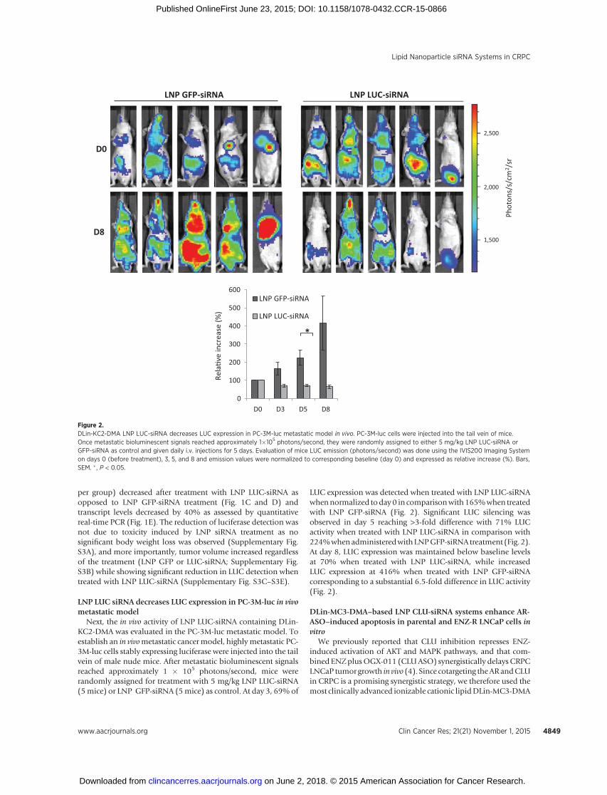
Next, the in vivo activity of LNP LUC-siRNA containing DLin-KC2-DMA was evaluated in the PC-3M-luc metastatic model. Toestablish an in vivometastatic cancer model, highlymetastatic PC-3M-luc cells stably expressing luciferase were injected into the tailvein of male nude mice. After metastatic bioluminescent signalsreached approximately 1 � 105 photons/second, mice wererandomly assigned for treatment with 5 mg/kg LNP LUC-siRNA(5mice) or LNP GFP-siRNA (5mice) as control. At day 3, 69% of
LUC expression was detected when treated with LNP LUC-siRNAwhennormalized today 0 in comparisonwith165%when treatedwith LNP GFP-siRNA (Fig. 2). Significant LUC silencing wasobserved in day 5 reaching >3-fold difference with 71% LUCactivity when treated with LNP LUC-siRNA in comparison with224%whenadministeredwithLNPGFP-siRNA treatment (Fig. 2).At day 8, LUC expression was maintained below baseline levelsat 70% when treated with LNP LUC-siRNA, while increasedLUC expression at 416% when treated with LNP GFP-siRNAcorresponding to a substantial 6.5-fold difference in LUC activity(Fig. 2).
DLin-MC3-DMA–based LNP CLU-siRNA systems enhance AR-ASO–induced apoptosis in parental and ENZ-R LNCaP cells invitro
We previously reported that CLU inhibition represses ENZ-induced activation of AKT and MAPK pathways, and that com-bined ENZplusOGX-011 (CLUASO) synergistically delays CRPCLNCaP tumor growth in vivo (4). Since cotargeting theARandCLUin CRPC is a promising synergistic strategy, we therefore used themost clinically advanced ionizable cationic lipidDLin-MC3-DMA
Rela
�ve
incr
ease
(%)
*
0
100
200
300
400
500
600
D0 D3 D5 D8
LNP GFP-siRNA
LNP LUC-siRNA
LNP GFP-siRNA
D0
D8
LNP LUC-siRNA
Phot
ons/
s/cm
2 /sr
2,500
2,000
1,500
Figure 2.DLin-KC2-DMA LNP LUC-siRNA decreases LUC expression in PC-3M-luc metastatic model in vivo. PC-3M-luc cells were injected into the tail vein of mice.Once metastatic bioluminescent signals reached approximately 1�105 photons/second, they were randomly assigned to either 5 mg/kg LNP LUC-siRNA orGFP-siRNA as control and given daily i.v. injections for 5 days. Evaluation of mice LUC emission (photons/second) was done using the IVIS200 Imaging Systemon days 0 (before treatment), 3, 5, and 8 and emission values were normalized to corresponding baseline (day 0) and expressed as relative increase (%). Bars,SEM. � , P < 0.05.
Lipid Nanoparticle siRNA Systems in CRPC
www.aacrjournals.org Clin Cancer Res; 21(21) November 1, 2015 4849
on June 2, 2018. © 2015 American Association for Cancer Research. clincancerres.aacrjournals.org Downloaded from
Published OnlineFirst June 23, 2015; DOI: 10.1158/1078-0432.CCR-15-0866
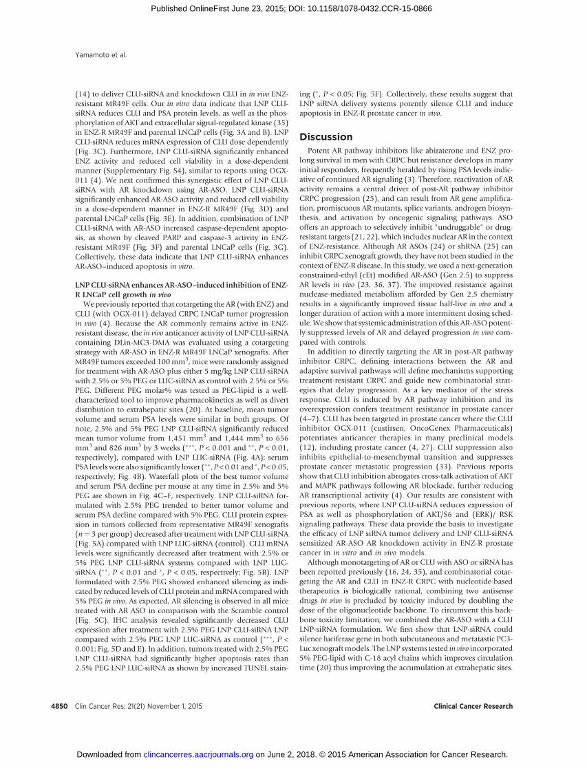
(14) to deliver CLU-siRNA and knockdown CLU in in vivo ENZ-resistant MR49F cells. Our in vitro data indicate that LNP CLU-siRNA reduces CLU and PSA protein levels, as well as the phos-phorylation of AKT and extracellular signal-regulated kinase (35)in ENZ-R MR49F and parental LNCaP cells (Fig. 3A and B). LNPCLU-siRNA reduces mRNA expression of CLU dose dependently(Fig. 3C). Furthermore, LNP CLU-siRNA significantly enhancedENZ activity and reduced cell viability in a dose-dependentmanner (Supplementary Fig. S4), similar to reports using OGX-011 (4). We next confirmed this synergistic effect of LNP CLU-siRNA with AR knockdown using AR-ASO. LNP CLU-siRNAsignificantly enhanced AR-ASO activity and reduced cell viabilityin a dose-dependent manner in ENZ-R MR49F (Fig. 3D) andparental LNCaP cells (Fig. 3E). In addition, combination of LNPCLU-siRNA with AR-ASO increased caspase-dependent apopto-sis, as shown by cleaved PARP and caspase-3 activity in ENZ-resistant MR49F (Fig. 3F) and parental LNCaP cells (Fig. 3G).Collectively, these data indicate that LNP CLU-siRNA enhancesAR-ASO–induced apoptosis in vitro.
LNPCLU-siRNA enhances AR-ASO–induced inhibition of ENZ-R LNCaP cell growth in vivo
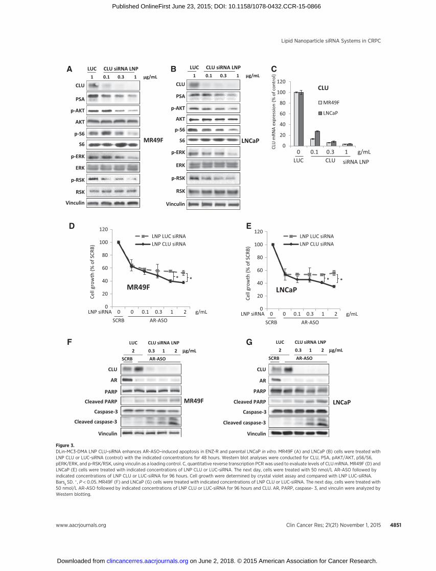
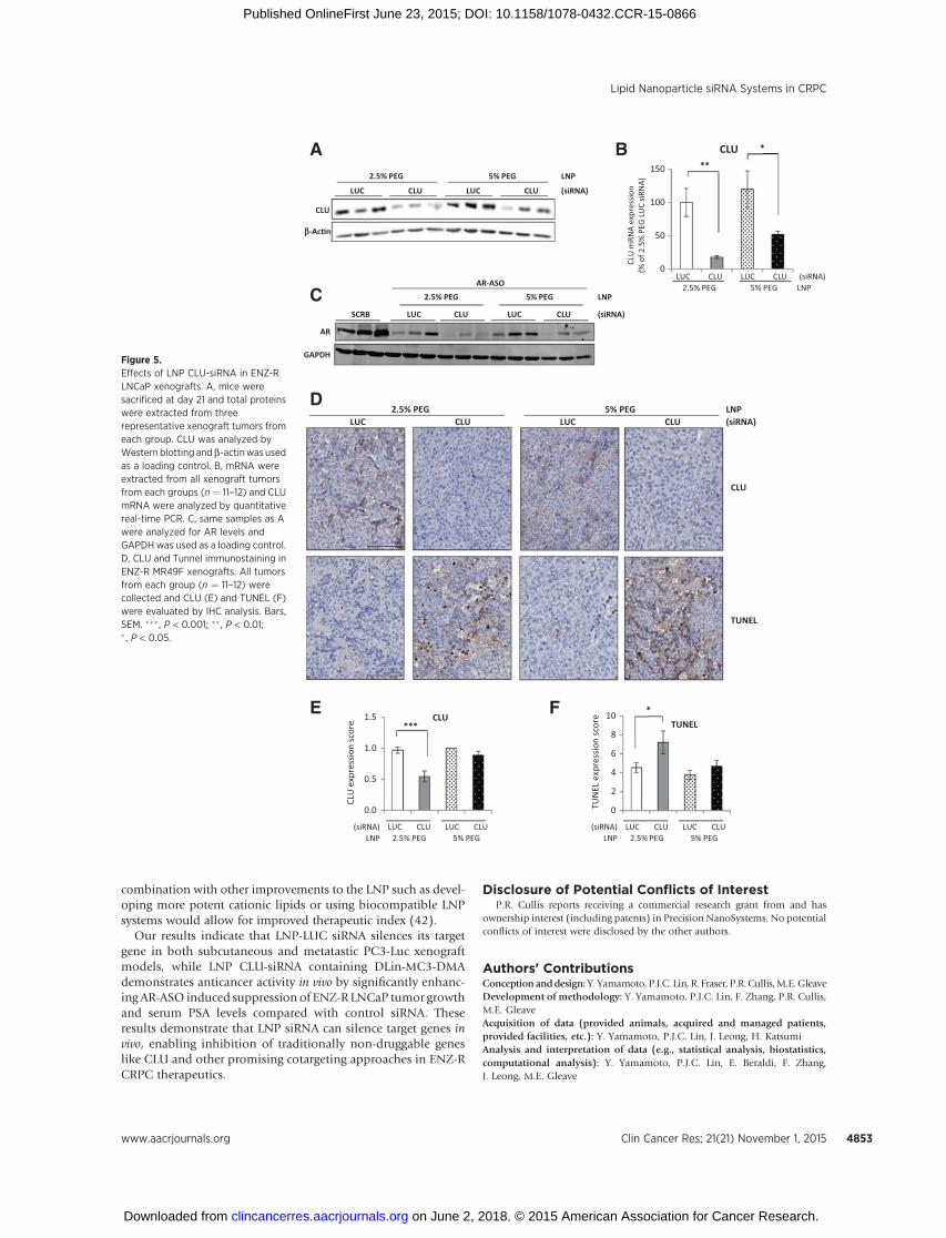
We previously reported that cotargeting the AR (with ENZ) andCLU (with OGX-011) delayed CRPC LNCaP tumor progressionin vivo (4). Because the AR commonly remains active in ENZ-resistant disease, the in vivo anticancer activity of LNP CLU-siRNAcontaining DLin-MC3-DMA was evaluated using a cotargetingstrategy with AR-ASO in ENZ-R MR49F LNCaP xenografts. AfterMR49F tumors exceeded 100mm3,mice were randomly assignedfor treatment with AR-ASO plus either 5 mg/kg LNP CLU-siRNAwith 2.5% or 5% PEG or LUC-siRNA as control with 2.5% or 5%PEG. Different PEG molar% was tested as PEG-lipid is a well-characterized tool to improve pharmacokinetics as well as divertdistribution to extrahepatic sites (20). At baseline, mean tumorvolume and serum PSA levels were similar in both groups. Ofnote, 2.5% and 5% PEG LNP CLU-siRNA significantly reducedmean tumor volume from 1,451 mm3 and 1,444 mm3 to 656mm3 and 826 mm3 by 3 weeks (���, P < 0.001 and ��, P < 0.01,respectively), compared with LNP LUC-siRNA (Fig. 4A); serumPSA levelswere also significantly lower (��,P<0.01 and �,P<0.05,respectively; Fig. 4B). Waterfall plots of the best tumor volumeand serum PSA decline per mouse at any time in 2.5% and 5%PEG are shown in Fig. 4C–F, respectively. LNP CLU-siRNA for-mulated with 2.5% PEG trended to better tumor volume andserum PSA decline compared with 5% PEG. CLU protein expres-sion in tumors collected from representative MR49F xenografts(n¼ 3 per group) decreased after treatment with LNP CLU-siRNA(Fig. 5A) compared with LNP LUC-siRNA (control). CLU mRNAlevels were significantly decreased after treatment with 2.5% or5% PEG LNP CLU-siRNA systems compared with LNP LUC-siRNA (��, P < 0.01 and �, P < 0.05, respectively; Fig. 5B). LNPformulated with 2.5% PEG showed enhanced silencing as indi-cated by reduced levels of CLUprotein andmRNA comparedwith5% PEG in vivo. As expected, AR silencing is observed in all micetreated with AR ASO in comparison with the Scramble control(Fig. 5C). IHC analysis revealed significantly decreased CLUexpression after treatment with 2.5% PEG LNP CLU-siRNA LNPcompared with 2.5% PEG LNP LUC-siRNA as control (���, P <0.001; Fig. 5D and E). In addition, tumors treated with 2.5% PEGLNP CLU-siRNA had significantly higher apoptosis rates than2.5% PEG LNP LUC-siRNA as shown by increased TUNEL stain-
ing (�, P < 0.05; Fig. 5F). Collectively, these results suggest thatLNP siRNA delivery systems potently silence CLU and induceapoptosis in ENZ-R prostate cancer in vivo.
DiscussionPotent AR pathway inhibitors like abiraterone and ENZ pro-
long survival in men with CRPC but resistance develops in manyinitial responders, frequently heralded by rising PSA levels indic-ative of continued AR signaling (3). Therefore, reactivation of ARactivity remains a central driver of post-AR pathway inhibitorCRPC progression (25), and can result from AR gene amplifica-tion, promiscuous AR mutants, splice variants, androgen biosyn-thesis, and activation by oncogenic signaling pathways. ASOoffers an approach to selectively inhibit "undruggable" or drug-resistant targets (21, 22), which includes nuclear AR in the contextof ENZ-resistance. Although AR ASOs (24) or shRNA (25) caninhibit CRPC xenograft growth, they have not been studied in thecontext of ENZ-R disease. In this study, we used a next-generationconstrained-ethyl (cEt) modified AR-ASO (Gen 2.5) to suppressAR levels in vivo (23, 36, 37). The improved resistance againstnuclease-mediated metabolism afforded by Gen 2.5 chemistryresults in a significantly improved tissue half-live in vivo and alonger duration of action with a more intermittent dosing sched-ule.We show that systemic administration of this AR-ASOpotent-ly suppressed levels of AR and delayed progression in vivo com-pared with controls.
In addition to directly targeting the AR in post-AR pathwayinhibitor CRPC, defining interactions between the AR andadaptive survival pathways will define mechanisms supportingtreatment-resistant CRPC and guide new combinatorial strat-egies that delay progression. As a key mediator of the stressresponse, CLU is induced by AR pathway inhibition and itsoverexpression confers treatment resistance in prostate cancer(4–7). CLU has been targeted in prostate cancer where the CLUinhibitor OGX-011 (custirsen, OncoGenex Pharmaceuticals)potentiates anticancer therapies in many preclinical models(12), including prostate cancer (4, 27). CLU suppression alsoinhibits epithelial-to-mesenchymal transition and suppressesprostate cancer metastatic progression (33). Previous reportsshow that CLU inhibition abrogates cross-talk activation of AKTand MAPK pathways following AR blockade, further reducingAR transcriptional activity (4). Our results are consistent withprevious reports, where LNP CLU-siRNA reduces expression ofPSA as well as phosphorylation of AKT/S6 and (ERK)/ RSKsignaling pathways. These data provide the basis to investigatethe efficacy of LNP siRNA tumor delivery and LNP CLU-siRNAsensitized AR-ASO AR knockdown activity in ENZ-R prostatecancer in in vitro and in vivo models.
Although monotargeting of AR or CLU with ASO or siRNA hasbeen reported previously (16, 24, 35), and combinatorial cotar-geting the AR and CLU in ENZ-R CRPC with nucleotide-basedtherapeutics is biologically rational, combining two antisensedrugs in vivo is precluded by toxicity induced by doubling thedose of the oligonucleotide backbone. To circumvent this back-bone toxicity limitation, we combined the AR-ASO with a CLULNP-siRNA formulation. We first show that LNP-siRNA couldsilence luciferase gene in both subcutaneous and metatastic PC3-Luc xenograftmodels. The LNP systems tested in vivo incorporated5% PEG-lipid with C-18 acyl chains which improves circulationtime (20) thus improving the accumulation at extrahepatic sites.
Yamamoto et al.
Clin Cancer Res; 21(21) November 1, 2015 Clinical Cancer Research4850
on June 2, 2018. © 2015 American Association for Cancer Research. clincancerres.aacrjournals.org Downloaded from
Published OnlineFirst June 23, 2015; DOI: 10.1158/1078-0432.CCR-15-0866
F
A
D
LUC CLU siRNA LNP2 0.3 1 2
SCRB AR-ASO
0
20
40
60
80
100
120LNP LUC siRNALNP CLU siRNA
LNCaP
Cell
grow
th (%
of S
CRB)
**
0
20
40
60
80
100
120LNP LUC siRNALNP CLU siRNA
MR49F
Cell
grow
th (%
of S
CRB)
* *
LNP siRNA 0 0 0.1 0.3 1 2 µg/mL
mg/mL mg/mL
µg/mLSCRB AR-ASO
LNP siRNA 0 0 0.1 0.3 1 2SCRB AR-ASO
LUC CLU siRNA LNP1 0.1 0.3 1 μg/mL
LUC CLU siRNA LNP1 0.1 0.3 1 μg/mL
Cleaved caspase-3
CLU
Vinculin
LNCaPCaspase-3
PARP
Cleaved PARP
AR
Cleaved caspase-3
MR49F
CLU
Vinculin
Caspase-3
PARP
Cleaved PARP
AR
LUC CLU siRNA LNP2 0.3 1 2
SCRB AR-ASO
MR49F
CLU
PSA
Vinculin
p-ERK
ERK
p-AKT
AKT
p-RSK
RSK
p-S6
S6p-ERK
LNCaP
p-AKT
AKT
Vinculin
CLU
PSA
ERK
p-RSK
RSK
p-S6
S6
B
E
G
C
0 0.1 0.3 1 µg/mLLUC CLU
0
20
40
60
80
100
120
MR49F
LNCaP
CLU
mRN
A ex
pres
sion
(% o
f con
trol
)
siRNA LNP
CLU
Figure 3.DLin-MC3-DMA LNP CLU-siRNA enhances AR-ASO–induced apoptosis in ENZ-R and parental LNCaP in vitro. MR49F (A) and LNCaP (B) cells were treated withLNP CLU or LUC-siRNA (control) with the indicated concentrations for 48 hours. Western blot analyses were conducted for CLU, PSA, pAKT/AKT, pS6/S6,pERK/ERK, and p-RSK/RSK, using vinculin as a loading control. C, quantitative reverse transcription PCR was used to evaluate levels of CLUmRNA. MR49F (D) andLNCaP (E) cells were treated with indicated concentrations of LNP CLU or LUC-siRNA. The next day, cells were treated with 50 nmol/L AR-ASO followed byindicated concentrations of LNP CLU or LUC-siRNA for 96 hours. Cell growth were determined by crystal violet assay and compared with LNP LUC-siRNA.Bars, SD. � , P < 0.05. MR49F (F) and LNCaP (G) cells were treated with indicated concentrations of LNP CLU or LUC-siRNA. The next day, cells were treated with50 nmol/L AR-ASO followed by indicated concentrations of LNP CLU or LUC-siRNA for 96 hours and CLU. AR, PARP, caspase- 3, and vinculin were analyzed byWestern blotting.
Lipid Nanoparticle siRNA Systems in CRPC
www.aacrjournals.org Clin Cancer Res; 21(21) November 1, 2015 4851
on June 2, 2018. © 2015 American Association for Cancer Research. clincancerres.aacrjournals.org Downloaded from
Published OnlineFirst June 23, 2015; DOI: 10.1158/1078-0432.CCR-15-0866
As shown by Lee and colleagues, the LNP platform using thecationic lipid DLin-KC2-DMA has successfully silenced AR inLNCaP xenograft tumor model (16). This was further improvedwith the use of a more potent cationic lipid DMAP-BLP to whichdoses as low as 1 mg/kg contributed >50% gene silencing (Sup-plementary Fig. S3). Although DMAP-BLP has been shown to bepotent and safe in the brain (30), full toxicity profile of this lipidhas yet to be characterized. However, the most potent lipid in theclinic, DLin-MC3-DMA, has shown to be tolerable and potent(14) and tested. As shown in Figs 4 and 5, combinatory treatmentof AR and CLU silencing decreased PSA levels as well as tumorvolume. Interestingly, LNP formulated with 2.5% PEG was moreeffective compared with 5% PEG in vivo. This can be attributed toPEG interfering with release of siRNA to the cytosol. Currently,less than 3% of siRNA of total siRNA delivered is detected in thecytosol using LNP systems with C14 Peg-lipid (38). With addi-
tional PEG coating on the LNP surface, the PEG can also interferewith intracellular uptake of particles. Although 5% PEG extendscirculation time, the activity of the LNP system would be com-promised, this can be overcome by the incorporation of targetingmoieties (such as DUPA or folate) conjugated to PEG-lipids (39).Targeting siRNA comprising of a targeting moiety conjugateddirectly to the siRNA has been tested in the clinic as Gal-Nacconjugated siRNA accumulates and provide function in the liver(40). However, in order to reach extrahepatic sites, such anapproach requires long circulation time, which can only beachieved by employing a delivery system. Alternative means toimprove treatment also include coencapsulation of AR and CLUsiRNA in the same LNP as this would ensure the same cells wouldhave reagents to silence both AR and CLU. Such approach hasbeen tested showing that up to five different targets silence keyplayers within similar or divergent biologic pathways (41). In
C
A
0
500
1,000
1,500
2,000
2,500
3,000
3,500
−1 0 1 2 3
SCRB
2.5% PEG LNP LUC-siRNA + ARASO
5% PEG LNP LUC-siRNA + ARASO
2.5% PEG LNP CLU-siRNA + ARASO
5% PEG LNP CLU-siRNA + ARASO
Tum
or v
olum
e (m
m3 )
*******
***
Weeks of treatment
0
20
40
60
80
100
120
140
160
−0.5 0.5 1.5 2.5 3
SCRB
2.5% PEG LNP LUC-siRNA + ARASO
5% PEG LNP LUC-siRNA + ARASO
2.5% PEG LNP CLU-siRNA + ARASO
5% PEG LNP CLU-siRNA + ARASO
Seru
m P
SA (n
g/m
L)
* ***
Weeks of treatment
E
PSA
chan
ge (%
)
2.5% PEG
LNP LUC-siRNA+ AR-ASO
LNP CLU-siRNA+ AR-ASO
PSA
chan
ge (%
)
5% PEG
LNP LUC-siRNA+ AR-ASO
LNP CLU-siRNA+ AR-ASO
Tum
or v
olum
e ch
ange
(%)
5% PEG
LNP LUC-siRNA+ AR-ASO
LNP CLU-siRNA+ AR-ASO
2.5% PEG
LNP LUC-siRNA+ AR-ASO
LNP CLU-siRNA+ AR-ASO
Tum
or v
olum
e ch
ange
(%)
B
D
F
150
100
50
0
−50
−100
100
50
0
−50
−100
100
50
0
−50
−100
150
100
50
0
−50
−100
Figure 4.LNP CLU-siRNA enhances AR-ASOactivity in ENZ-R LNCaP xenografts.A, castrated mice were inoculatedsubcutaneously with ENZ-R MR49Fcells. Once tumors reached 100mm3,mice were randomly assigned to 10mg/kg SCRB (12 mice), AR-ASO pluseither 5 mg/kg LNP CLU-siRNA with2.5% PEG (11 mice) or 5% PEG (11mice) or LUC-siRNA as control with2.5% PEG (12 mice) or 5% PEG (11mice). Injections were administeredi.v. once daily for 4 days and thenthree times per week thereafter. Themean tumor volume (A) and theserum PSA level (B) were comparedbetween the five groups � SEM.��� , P < 0.001; �� , P < 0.01; � , P < 0.05.Waterfall plots showed the greatestpercent decline in tumor volume byLNP formulated in 2.5% PEG (C) and5% PEG (D) from baseline at anytime. Waterfall plots of greatestpercent decline in PSA by LNPformulated in 2.5% PEG (E) and 5%PEG (F) from baseline at any time.
Yamamoto et al.
Clin Cancer Res; 21(21) November 1, 2015 Clinical Cancer Research4852
on June 2, 2018. © 2015 American Association for Cancer Research. clincancerres.aacrjournals.org Downloaded from
Published OnlineFirst June 23, 2015; DOI: 10.1158/1078-0432.CCR-15-0866
combination with other improvements to the LNP such as devel-oping more potent cationic lipids or using biocompatible LNPsystems would allow for improved therapeutic index (42).
Our results indicate that LNP-LUC siRNA silences its targetgene in both subcutaneous and metatastic PC3-Luc xenograftmodels, while LNP CLU-siRNA containing DLin-MC3-DMAdemonstrates anticancer activity in vivo by significantly enhanc-ing AR-ASO induced suppression of ENZ-R LNCaP tumor growthand serum PSA levels compared with control siRNA. Theseresults demonstrate that LNP siRNA can silence target genes invivo, enabling inhibition of traditionally non-druggable geneslike CLU and other promising cotargeting approaches in ENZ-RCRPC therapeutics.
Disclosure of Potential Conflicts of InterestP.R. Cullis reports receiving a commercial research grant from and has
ownership interest (including patents) in Precision NanoSystems. No potentialconflicts of interest were disclosed by the other authors.
Authors' ContributionsConception anddesign:Y. Yamamoto, P.J.C. Lin, R. Fraser, P.R. Cullis,M.E.GleaveDevelopment of methodology: Y. Yamamoto, P.J.C. Lin, F. Zhang, P.R. Cullis,M.E. GleaveAcquisition of data (provided animals, acquired and managed patients,provided facilities, etc.): Y. Yamamoto, P.J.C. Lin, J. Leong, H. KatsumiAnalysis and interpretation of data (e.g., statistical analysis, biostatistics,computational analysis): Y. Yamamoto, P.J.C. Lin, E. Beraldi, F. Zhang,J. Leong, M.E. Gleave
A B2.5% PEG 5% PEG LNP
LUC CLU LUC CLU (siRNA)
LUC CLU LUC CLU (siRNA)2.5% PEG 5% PEG LNP
0
50
100
150
CLU
mRN
A ex
pres
sion
(% o
f 2.5
% P
EG L
UC
siRN
A)
**
*
CLU
b-Ac�n
C
AR
GAPDH
SCRB LUC CLU
AR-ASO
LUC CLU
2.5% PEG 5% PEG LNP
(siRNA)
2.5% PEG 5% PEG LNPLUC CLU LUC CLU (siRNA)
D
CLU
TUNEL
(siRNA) LUC CLU LUC CLULNP 2.5% PEG 5% PEG
(siRNA) LUC CLU LUC CLULNP 2.5% PEG 5% PEG
CLU***
0.0
0.5
1.0
1.5
CLU
exp
ress
ion
scor
e
TUN
EL e
xpre
ssio
n sc
ore
TUNEL
0
2
4
6
8
10 *E F
CLU
Figure 5.Effects of LNP CLU-siRNA in ENZ-RLNCaP xenografts. A, mice weresacrificed at day 21 and total proteinswere extracted from threerepresentative xenograft tumors fromeach group. CLU was analyzed byWestern blotting andb-actinwasusedas a loading control. B, mRNA wereextracted from all xenograft tumorsfrom each groups (n ¼ 11–12) and CLUmRNA were analyzed by quantitativereal-time PCR. C, same samples as Awere analyzed for AR levels andGAPDH was used as a loading control.D, CLU and Tunnel immunostaining inENZ-R MR49F xenografts. All tumorsfrom each group (n ¼ 11–12) werecollected and CLU (E) and TUNEL (F)were evaluated by IHC analysis. Bars,SEM. ��� , P < 0.001; �� , P < 0.01;� , P < 0.05.
www.aacrjournals.org Clin Cancer Res; 21(21) November 1, 2015 4853
Lipid Nanoparticle siRNA Systems in CRPC
on June 2, 2018. © 2015 American Association for Cancer Research. clincancerres.aacrjournals.org Downloaded from
Published OnlineFirst June 23, 2015; DOI: 10.1158/1078-0432.CCR-15-0866
Writing, review, and/or revision of the manuscript: Y. Yamamoto, P.J.C. Lin,F. Zhang, J. Leong, P.R. Cullis, M.E. GleaveAdministrative, technical, or material support (i.e., reporting or organizingdata, constructing databases): P.J.C. Lin, E. Beraldi, Y. Kawai, J. Leong,H. Katsumi, R. FraserStudy supervision: R. Fraser, M.E. GleaveOther (pathology): L. Fazli
AcknowledgmentsThe authors thankMary Bowden, Virginia Yago, Darrell Trendall, and Estelle
Li for technical assistance.
Grant SupportThis work was supported by the Canadian Institutes of Health Research
(CIHR) Emerging Team Grant: Personalized siRNA-Based Nanomedicines(FRN:111627).
The costs of publication of this article were defrayed in part by the paymentof page charges. This article must therefore be hereby marked advertisementin accordance with 18 U.S.C. Section 1734 solely to indicate this fact.
Received April 10, 2015; revised June 10, 2015; accepted June 15, 2015;published OnlineFirst June 23, 2015.
References1. Siegel R, Ward E, Brawley O, Jemal A. Cancer statistics, 2011: the impact of
eliminating socioeconomic and racial disparities on premature cancerdeaths. CA Cancer J Clin 2011;61:212–36.
2. TranC,Ouk S, CleggNJ, Chen Y,Watson PA, Arora V, et al. Development ofa second-generation antiandrogen for treatment of advanced prostatecancer. Science 2009;324:787–90.
3. ScherHI, Fizazi K, Saad F, TaplinME, SternbergCN,Miller K, et al. Increasedsurvival with enzalutamide in prostate cancer after chemotherapy. N Engl JMed 2012;367:1187–97.
4. Matsumoto H, Yamamoto Y, Shiota M, Kuruma H, Beraldi E, MatsuyamaH, et al. Cotargeting androgen receptor and clusterin delays castrate-resistant prostate cancer progression by inhibiting adaptive stress responseand AR stability. Cancer Res 2013;73:5206–17.
5. Miyake H, Nelson C, Rennie PS, Gleave ME. Acquisition of chemoresistantphenotype by overexpression of the antiapoptotic gene testosterone-repressed prostate message-2 in prostate cancer xenograft models. CancerRes 2000;60:2547–54.
6. Zhang H, Kim JK, Edwards CA, Xu Z, Taichman R, Wang CY. Clusterininhibits apoptosis by interacting with activated Bax. Nat Cell Biol 2005;7:909–15.
7. Li N, Zoubeidi A, Beraldi E, Gleave ME. GRP78 regulates clusterin stability,retrotranslocation and mitochondrial localization under ER stress in pros-tate cancer. Oncogene 2013;32:1933–42.
8. Trougakos IP, Lourda M, Antonelou MH, Kletsas D, Gorgoulis VG, Papas-sideri IS, et al. Intracellular clusterin inhibits mitochondrial apoptosis bysuppressingp53-activating stress signals and stabilizing the cytosolic Ku70-Bax protein complex. Clin Cancer Res 2009;15:48–59.
9. Ammar H, Closset JL. Clusterin activates survival through the phosphati-dylinositol 3-kinase/Akt pathway. J Biol Chem 2008;283:12851–61.
10. Zoubeidi A, Ettinger S, Beraldi E, Hadaschik B, Zardan A, Klomp LW,et al. Clusterin facilitates COMMD1 and I-kappaB degradation toenhance NF-kappaB activity in prostate cancer cells. Mol Cancer Res2010;8:119–30.
11. Zhang F, Kumano M, Beraldi E, Fazli L, Du C, Moore S, et al. Clusterinfacilitates stress-induced lipidation of LC3 and autophagosome biogenesisto enhance cancer cell survival. Nat Commun 2014;5:5775.
12. Zoubeidi A, Chi K, Gleave M. Targeting the cytoprotective chaperone,clusterin, for treatment of advanced cancer. Clin Cancer Res 2010;16:1088–93.
13. Lin PJ, TamYK, Cullis PR.Development and clinical applications of siRNA-encapsulated lipid nanoparticles in cancer. Clinical Lipidology 2014;9:317–31.
14. Coelho T, Adams D, Silva A, Lozeron P, Hawkins PN, Mant T, et al. Safetyand efficacy of RNAi therapy for transthyretin amyloidosis. N Engl J Med2013;369:819–29.
15. Tabernero J, Shapiro GI, LoRusso PM, Cervantes A, Schwartz GK,Weiss GJ,et al. First-in-humans trial of an RNA interference therapeutic targetingVEGF and KSP in cancer patients with liver involvement. Cancer Discov2013;3:406–17.
16. Lee JB, Zhang K, Tam YY, Tam YK, Belliveau NM, Sung VY, et al. Lipidnanoparticle siRNA systems for silencing the androgen receptor in humanprostate cancer in vivo. Int J Cancer 2012;131:E781–90.
17. Basha G, Novobrantseva TI, Rosin N, Tam YY, Hafez IM, Wong MK, et al.Influence of cationic lipid composition on gene silencing properties oflipid nanoparticle formulations of siRNA in antigen-presenting cells. MolTher 2011;19:2186–200.
18. Li SD, Chono S, Huang L. Efficient gene silencing in metastatic tumor bysiRNA formulated in surface-modified nanoparticles. J Control Release2008;126:77–84.
19. Ambegia E, Ansell S, Cullis P, Heyes J, Palmer L, MacLachlan I. Stabilizedplasmid-lipid particles containing PEG-diacylglycerols exhibit extendedcirculation lifetimes and tumor selective gene expression. BiochimBiophysActa 2005;1669:155–63.
20. Mui BL, Tam YK, JayaramanM, Ansell SM, Du X, TamYY, et al. Influence ofPolyethylene Glycol Lipid Desorption Rates on Pharmacokinetics andPharmacodynamics of siRNA Lipid Nanoparticles. Mol Ther Nucleic Acids2013;2:e139.
21. Zellweger T, Miyake H, Cooper S, Chi K, Conklin BS, Monia BP, et al.Antitumor activity of antisense clusterin oligonucleotides is improved invitro and in vivo by incorporation of 2'-O-(2-methoxy)ethyl chemistry. JPharmacol Exp Ther 2001;298:934–40.
22. Zanardi TA, Han SC, Jeong EJ, Rime S, Yu RZ, Chakravarty K, et al.Pharmacodynamics and subchronic toxicity in mice and monkeys of ISIS388626, a second-generation antisense oligonucleotide that targets humansodium glucose cotransporter 2. J Pharmacol Exp Ther 2012;343:489–96.
23. Yamamoto Y, Loriot Y, Beraldi E, Zhang F, Wyatt AW, Nakouzi NA, et al.Generation 2.5 antisense oligonucleotides targeting the androgen receptorand its splice variants suppress enzalutamide-resistant prostate cancer cellgrowth. Clin Cancer Res 2015;21:1675–87.
24. Zhang Y, Castaneda S,DumbleM,WangM,MileskiM,QuZ, et al. Reducedexpression of the androgen receptor by third generation of antisense showsantitumor activity in models of prostate cancer. Mol Cancer Ther 2011;10:2309–19.
25. Snoek R, Cheng H, Margiotti K, Wafa LA, Wong CA, Wong EC, et al. In vivoknockdown of the androgen receptor results in growth inhibition andregression of well-established, castration-resistant prostate tumors. ClinCancer Res 2009;15:39–47.
26. Kuruma H, Matsumoto H, Shiota M, Bishop J, Lamoureux F, Thomas C,et al. A novel antiandrogen, compound 30, suppresses castration-resistantand MDV3100-resistant prostate cancer growth in vitro and in vivo. MolCancer Ther 2013;12:567–76.
27. Lamoureux F, Thomas C, Yin MJ, Kuruma H, Beraldi E, Fazli L, et al.Clusterin inhibition usingOGX-011 synergistically enhancesHsp90 inhib-itor activity by suppressing the heat shock response in castrate-resistantprostate cancer. Cancer Res 2011;71:5838–49.
28. Shiota M, Zoubeidi A, Kumano M, Beraldi E, Naito S, Nelson CC, et al.Clusterin is a critical downstream mediator of stress-induced YB-1 trans-activation in prostate cancer. Mol Cancer Res 2011;9:1755–66.
29. Jayaraman M, Ansell SM, Mui BL, Tam YK, Chen J, Du X, et al. Maximizingthe potency of siRNA lipid nanoparticles for hepatic gene silencing in vivo.Angew Chem Int Ed Engl 2012;51:8529–33.
30. Rungta RL, Choi HB, Lin PJ, Ko RW, Ashby D, Nair J, et al. LipidNanoparticle Delivery of siRNA to Silence Neuronal Gene Expression inthe Brain. Mol Ther Nucleic Acids 2013;2:e136.
31. Semple SC, Akinc A, Chen J, Sandhu AP, Mui BL, Cho CK, et al. Rationaldesign of cationic lipids for siRNA delivery. Nat Biotechnol 2010;28:172–6.
32. BelliveauNM,Huft J, Lin PJ, Chen S, Leung AK, Leaver TJ, et al.Microfluidicsynthesis of highly potent limit-size lipid nanoparticles for in vivo deliveryof siRNA. Mol Ther Nucleic Acids 2012;1:e37.
33. Shiota M, Zardan A, Takeuchi A, Kumano M, Beraldi E, Naito S, et al.Clusterin mediates TGF-beta-induced epithelial-mesenchymal transition
Yamamoto et al.
Clin Cancer Res; 21(21) November 1, 2015 Clinical Cancer Research4854
on June 2, 2018. © 2015 American Association for Cancer Research. clincancerres.aacrjournals.org Downloaded from
Published OnlineFirst June 23, 2015; DOI: 10.1158/1078-0432.CCR-15-0866
and metastasis via Twist1 in prostate cancer cells. Cancer Res 2012;72:5261–72.
34. Leung SY, Jackson J, Miyake H, Burt H, Gleave ME. Polymeric micellarpaclitaxel phosphorylates Bcl-2 and induces apoptotic regression of andro-gen-independent LNCaP prostate tumors. Prostate 2000;44:156–63.
35. Yang J, Xie SX, Huang Y, Ling M, Liu J, Ran Y, et al. Prostate-targetedbiodegradable nanoparticles loaded with androgen receptor silencingconstructs eradicate xenograft tumors in mice. Nanomedicine 2012;7:1297–309.
36. Burel SA, Han SR, Lee HS, Norris DA, Lee BS, Machemer T, et al. Preclinicalevaluation of the toxicological effects of a novel constrained ethyl modifiedantisense compound targeting signal transducer and activator of transcrip-tion3 inmiceandcynomolgusmonkeys.NucleicAcidTher2013;23:213–27.
37. Seth PP, Siwkowski A, Allerson CR, Vasquez G, Lee S, Prakash TP, et al.Short antisense oligonucleotides with novel 2'-4' conformationaly restrict-ed nucleoside analogues show improved potency without increased tox-icity in animals. J Med Chem 2009;52:10–3.
38. Gilleron J, Querbes W, Zeigerer A, Borodovsky A, Marsico G, Schubert U,et al. Image-based analysis of lipid nanoparticle-mediated siRNA delivery,intracellular trafficking and endosomal escape. Nat Biotechnol 2013;31:638–46.
39. Tam YY, Chen S, Zaifman J, Tam YK, Lin PJ, Ansell S, et al. Small moleculeligands for enhanced intracellular delivery of lipid nanoparticle formula-tions of siRNA. Nanomedicine 2013;9:665–74.
40. Rajeev KG, Nair JK, Jayaraman M, Charisse K, Taneja N, O'Shea J, et al.Hepatocyte-specific delivery of siRNAs conjugated to novel non-nucleo-sidic trivalent N-acetylgalactosamine elicits robust gene silencing in vivo.Chembiochem 2015;16:903–8.
41. LoveKT,MahonKP, LevinsCG,WhiteheadKA,QuerbesW,Dorkin JR, et al.Lipid-like materials for low-dose, in vivo gene silencing. Proc Natl Acad SciU S A 2010;107:1864–9.
42. Maier MA, Jayaraman M, Matsuda S, Liu J, Barros S, Querbes W, et al.Biodegradable lipids enabling rapidly eliminated lipid nanoparticles forsystemic delivery of RNAi therapeutics. Mol Ther 2013;21:1570–8.
www.aacrjournals.org Clin Cancer Res; 21(21) November 1, 2015 4855
Lipid Nanoparticle siRNA Systems in CRPC
on June 2, 2018. © 2015 American Association for Cancer Research. clincancerres.aacrjournals.org Downloaded from
Published OnlineFirst June 23, 2015; DOI: 10.1158/1078-0432.CCR-15-0866
2015;21:4845-4855. Published OnlineFirst June 23, 2015.Clin Cancer Res Yoshiaki Yamamoto, Paulo J.C. Lin, Eliana Beraldi, et al. in Enzalutamide-Resistant Prostate CancerProgression When Combined with Androgen Receptor Cotargeting siRNA Lipid Nanoparticle Potently Silences Clusterin and Delays
Updated version
10.1158/1078-0432.CCR-15-0866doi:
Access the most recent version of this article at:
Material
Supplementary
http://clincancerres.aacrjournals.org/content/suppl/2015/06/24/1078-0432.CCR-15-0866.DC1
Access the most recent supplemental material at:
Cited articles
http://clincancerres.aacrjournals.org/content/21/21/4845.full#ref-list-1
This article cites 42 articles, 18 of which you can access for free at:
Citing articles
http://clincancerres.aacrjournals.org/content/21/21/4845.full#related-urls
This article has been cited by 3 HighWire-hosted articles. Access the articles at:
E-mail alerts related to this article or journal.Sign up to receive free email-alerts
Subscriptions
Reprints and
To order reprints of this article or to subscribe to the journal, contact the AACR Publications Department at
Permissions
Rightslink site. Click on "Request Permissions" which will take you to the Copyright Clearance Center's (CCC)
.http://clincancerres.aacrjournals.org/content/21/21/4845To request permission to re-use all or part of this article, use this link
on June 2, 2018. © 2015 American Association for Cancer Research. clincancerres.aacrjournals.org Downloaded from
Published OnlineFirst June 23, 2015; DOI: 10.1158/1078-0432.CCR-15-0866