site-directed mutagenesis and oxygen isotope incorporation studies of the nucleophilic aspartate of...
TRANSCRIPT
1242 Biochemistry 1994, 33, 1242-1247
Site-Directed Mutagenesis and Oxygen Isotope Incorporation Studies of the Nucleophilic Aspartate of Haloalkane Dehalogenaset
Frens Pries,* Jaap Kingma,* Marjan Pentenga,* Gertie van Pouderoyen,*J C. Margot Jeronimus-Stratingh,ll Andries P. Bruins,ll and Dick B. Janssen'J
Departments of Biochemistry and Pharmacy, University of Groningen. The Netherlands
Received August 4, 1993; Revised Manuscript Received November 12, 1993"
ABSTRACT: Haloalkane dehalogenase catalyzes the hydrolytic cleavage of carbon-halogen bonds in a broad range of halogenated aliphatic compounds. The X-ray structure suggests that Asp124, which is located close to an internal cavity, carries out a nucleophilic attackon the Ca of the substrate, releasing the halogen. To study the mechanism of hydrolysis, this aspartate residue was mutated to alanine, glycine, or glutamate. The mutant enzymes showed no activity toward 1,2-dichloroethane and 1,2-dibromoethane. Incubation of purified wild-type dehalogenase with 1,2-dichloroethane in the presence of H2180 resulted in the incorporation of l 8 0 in 2-chloroethanol and in the carboxylate group of Asp124. This shows that the reaction proceeds by covalent catalysis with the formation of an alkyl-enzyme intermediate that is hydrolyzed by attack of solvent water on the carbonyl carbon of Asp124. On the basis of amino acid sequence similarity between haloalkane dehalogenase and epoxide hydrolases, it is proposed that a conserved aspartate residue is also involved in covalent catalysis by the latter enzymes.
Haloalkane dehalogenase (DhlA) from Xunthobucter au- totrophicus GJlO catalyzes the hydrolysis of a broad range of haloalkanes and related substrates to the corresponding alcohols (Keuning et al., 1985). The sequence of the dhlA gene is known (Janssen et al., 1989) and the three-dimensional structure of the 310 amino acid enzyme was determined by X-ray crystallography (Franken et al., 199 1; Verschueren et al., 1993a). The enzyme is composed of a main domain, formed by a P-sheet and a-helices, and a cap domain, formed mainly by helices and lying on top of the main domain. The topology of the main domain of DhlA is similar to those of several other hydrolytic proteins, which were classified as a/P hydrolase fold enzymes (Ollis et al., 1992).
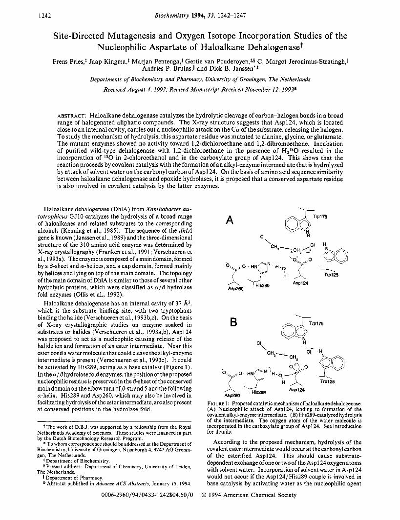
Haloalkane dehalogenase has an internal cavity of 37 A3, which is the substrate binding site, with two tryptophans binding the halide (Verschueren et al., 1993b,c). On the basis of X-ray crystallographic studies on enzyme soaked in substrates or halides (Verschueren et al., 1993a,b), Asp124 was proposed to act as a nucleophile causing release of the halide ion and formation of an ester intermediate. Near this ester bond a water molecule that could cleave the alkyl-enzyme intermediate is present (Verschueren et al., 1993~). It could be activated by His289, acting as a base catalyst (Figure 1). In the a/@ hydrolase fold enzymes, the position of the proposed nucleophilic residue is preserved in the P-sheet of the conserved main domain on the elbow turn of P-strand 5 and the following a-helix. His289 and Asp260, which may also be involved in facilitating hydrolysis of the ester intermediate, are also present at conserved positions in the hydrolase fold.
The work of D.B.J. was supported by a fellowship from the Royal Netherlands Academy of Sciences. These studies were financed in part by the Dutch Biotechnology Research Program.
* To whom correspondence should be addressed at the Department of Biochemistry, University of Groningen, Nijenborgh 4,9747 AG Gronin- gen, The Netherlands.
t Department of Biochemistry.
11 Department of Pharmacy. @ Abstract published in Advance ACS Abstracts, January 15, 1994.
Present address: Department of Chemistry, University of Leiden, The Netherlands.
\
Aspl 24
FIGURE 1 : Proposed catalytic mechanism of haloalkane dehalogenase. (A) Nucleophilic attack of Asp124, leading to formation of the covalent alkyl-enzyme intermediate. (B) His289-catalyzed hydrolysis of the intermediate. The oxygen atom of the water molecule is incorporated in the carboxylate group of Asp124. See introduction for details.
According to the proposed mechanism, hydrolysis of the covalent ester intermediate would occur at the carbonyl carbon of the esterified Asp124. This should cause substrate- dependent exchange of one or two of the Aspl 24 oxygen atoms with solvent water. Incorporation of solvent water in Asp124 would not occur if the Asp124/His289 couple is involved in base catalysis by activating water as the nucleophilic agent
0006-2960/94/0433- 1242$04.50/0 0 1994 American Chemical Society
Nucleophilic Aspartate of Haloalkane Dehalogenase
or if the ester intermediate is hydrolyzed by attack of water at the alkyl Cl carbon, as in several glycosidic enzymes (Sinnott, 1990).
The aim of the work described here was to investigate the role of Asp1 24 by site-directed mutagenesis and by examining the incorporation of l80 from solvent H2180 during substrate hydrolysis.
EXPERIMENTAL PROCEDURES
Materials. All reagents were purchased from Merck or Sigma Chemical Co. Restriction enzymes and other molecular biology enzymes were from Boehringer Mannheim. The expression vector pET3d was from Studier et al. (1990). a-Chymotrypsin and TPCK-treated trypsin were obtained from Worthington Biochemical Corp. The mutagenesis primer with sequence S'GTCGTTCAGG(G,A,T- m T G G G G C G G A T T 3 ' (the codon that was changed for the Asp124 mutations is underlined) was purchased from Eurosequence, Groningen, The Netherlands. Sequencing was done with the T7 sequencing kit from Pharmacia. H2180 (97%) was obtained from Isotec Inc., Miamisburg, OH.
Haloalkane Dehalogenase. Wild-type and mutant D 124G haloalkane dehalogenase were purified from Escherichia coli BL21(DE3) as described by Schanstra et al. (1993). All enzyme used was >99% pure by SDS-PAGE. Stocks of the enzyme were filter-sterilized and stored in TEM (10 mM Tris.SO4, pH 7.5, containing 1 mM P-mercaptoethanol and 1 mM EDTA). For incubations with H2180, the enzyme was concentrated by ultrafiltration over an Amicon PM 10 filter to 10-20 mg/mL. The specific activity of the wild-type enzyme at pH 7.7 was 3.3 units/mg, corresponding to a k,,, of 1.9 s-I.
Construction of the Mutants. Three replacements of Asp 124 in DhlA were made. Mutants D 124G, D 124A, and D124E were made in the DhlA expression vector pPJ123 (Schanstra et al., 1993) with the mutagenesis method of Nelson and Long (1989). Mutant D124G was recloned in the better T7 DhlA expression vector pELA (Schanstra et al., 1993). Sequences were confirmed by T7 DNA polymerase dideoxy sequencing (Sanger et al., 1977).
Growth, Expression, and Purification. Both pPJl23 and pELA constructs were grown at 30 "C in LB medium containing 50 pg/mL ampicillin to an OD600 of about 0.1. Cultivation was continued at 17 OC, and IPTG (0.4 mM) was added after 1 h as an inducer for DhlA expression. Cells were harvested after 16 h (OD600 1-2), washed with TEM buffer, resuspended in TEM buffer, and sonicated for 10 s/mL of cell suspension. Cell debris and other nonsoluble material was removed by centrifugation at 20000g for 15 min.
Assays. Dehalogenase assays were performed as described previously (Janssen et al., 1989). Protein concentrations were determined with Coomassie brilliant blue. Levels of D124G mutant DhlA protein in crude extracts of E . coli BL21 (DE3) were measured by SDS-PAGE on 12.5% acrylamide gels (Laemmli, 1970) and scanning of the dried stained gels with a GeniScan gray-scale scanner using the iPhoto Deluxe program of U-lead Systems Inc. The scanned gels were analyzed with the program Quantiscan, version 1.31, from Microsoft Systems, Biosoft.
Circular Dichroism. Circular dichroism experiments were carried out on a Jobin-Yvon Mark V spectropolarimeter in cuvettes with 0.1-mm optical path length. The enzymes were analyzed at room temperature in 10 mM TriseSO4 buffer, pH 7.5, containing EDTA (1 mM), P-mercaptoethanol (1 mM), sodium azide (0.02% w/v), and 10% glycerol (v/v). The
Biochemistry, Vol. 33, No. 5, 1994 1243
concentrations were 28.9 pM for the wild-type enzyme and 43.2 pM for the D124G mutant. The spectra between 190 and 290 nm were recorded for 30 min, with a band width of 2 nm, and with 0.1-nm step size. The buffer blanks obtained under the same conditions were subtracted from the spectra. The spectra were then converted into mean residue ellipticities [ e ] (deg cm2/dmol) and displayed between 190 and 240 nm.
Incubations with H2l80. Incubations of dehalogenase with H2180 was carried out at 30 OC in 137-pL incubation mixtures containing 22 pL of 50 mM 1,2-dichloroethane, 7 pL of 1 M TrissS04, pH 7.7,40 pL (= 10 nmol) of dehalogenase, and 68 pL of 97% H2180 (48%). The reaction was started by addition of enzyme, and after 2.5 min, 14pL of 2 M ammonium acetate/ ammoniumcarbonate, pH 8.0, and lOpL of 10 mg/mL trypsin that was freshly dissolved in 0.2 M of the same buffer were rapidly added. Digestion was continued for 3 h at 37 "C. Under these conditions more than 95% of the substrate was converted. Controls were performed similarly but with 1,2- dichloroethane omitted.
HPLC Isolation of the Pentapeptide. Separations of proteolytic digests of dehalogenase were carried out on a 4.7- mL Nucleosil 100- 1 OC 18 column. Elution was performed with a linear gradient of 0-66.7% acetonitrile in 0.1% ammonium acetate, pH 6.0. The flow rate was 1 mL/min, and peptides were collected and lyophilized.
Chymotrypsin cleavage was carried out by dissolving the lyophilized peptide in 150 pL of 0.2 M ammonium acetate/ ammonium carbonate buffer, pH 8.0, and incubating with 5 pL of a 10 mg/mL solution of chymotrypsin for 3 h at 35 OC. This was followed by HPLC isolation of the pentapeptide.
Manual Edman degradation was carried out as described by Chang (1983).
Ion-Spray Mass Spectrometry. For mass spectrometry, the lyophilized peptide was dissolved in 250 pL of a solution of 0.02% trifluoroacetic acid in 80% (v/v) methanol in water. The peptide solution was introduced into the mass spectrometer by means of a Jasco Familic lOON syringe pump operating at 5 pL/min. The solution was nebulized and the sample was ionized by pneumatically-assisted electrospray (Bruins et al., 1987). The mass spectrometer was a Nermag R 3010 quadrupole instrument equipped with a custom-built atmo- spheric pressure ionization source (Bruins et al., 1988). Data acquisition and data reduction took place by means of the standard Nermag SIDAR software. Each peptide solution was examined by full-scan mass spectra recorded in 1-amu (atomic mass unit) steps in order to confirm the identity and purity of the peptide. For measurement of the natural isotope distributions and contribution of l 8 0 and (I80)2, a narrow mass range around the M + H+and M + Na+ ions was scanned in 1 / 16th-amu steps in the profile mode of the data system. Sixty scans were averaged for display and calculations. A tabular output from SIDAR was used for calculation of l 8 0 and (l80)2 contributions to themolecular weight of the peptide.
Gas Chromatography-Mass Spectrometry. Incorporation of l 8 0 in 2-chloroethanol formed by the dehalogenase was determined in incubations containing 5 mM 1,2-dichloro- ethane, 40 mM TrissS04, pH 8.2, and 38 nM dehalogenase. The reaction was started by addition of 1,2-dichloroethane and terminated after 5-min incubation at 30 OC by extraction with 1 mL of diethyl ether. The extraction was repeated twice and the combined extracts were concentrated under a stream of nitrogen to 100 pL. Gas chromatography-mass spec- trometry was carried out on a Ribermag R10-10 instrument with electron impact ionization and equipped with a Chrompack CPWax52 CB column that was temperature-

1244 Biochemistry, Vol. 33, No. 5, 1994 Pries et al.
1 2 3 4
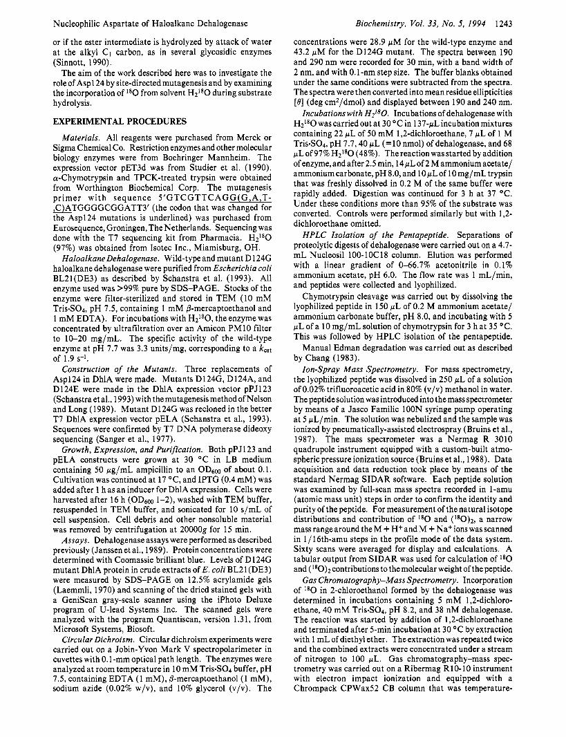
FIGURE 2: SDS-PAGE of crude extract of D124G mutant DhlA in E. coli BL21 (DE3). From left to right: (1) molecular weight markers containing phosphorylase b (94 000), albumin (67 000), ovalbumin (43 OOO),carbonicanhydrase (30 000), and trypsin inhibitor (20 100); (2) 10 pg of crude extract of induced pELA*D 124G; (3) 10 pg of crude extract of induced pET3d, from which pELA is a derivative and that lacks the dhlA gene; (4) 3 pg of purified WT DhlA. Lane 2 contains about 29% DhlA as estimated by densitometric analysis.
programmed from 3 min isothermal at 45 "C to 200 OC at 20 "C/min. Identical incubations were carried out with unlabeled water replacing H2180.
RESULTS
Site-Directed Mutagenesis of Asp124. In order to prove that Aspl 24 is essential for dehalogenase activity, mutants were made by site-directed mutagenesis. Mutant dehalogenase with Asp124 replaced by Gly, Glu, or Ala and expressed in pPJ 123 showed no activity with either 1,2-dichIoroethane or 1,2-dibromoethane, although the wild-type dehalogenase was expressed in pPJ123 at levels of 110 and 115 milliunits/mg of crude extract for these substrates, respectively. The requirement of Aspl 24 was further demonstrated by recloning the D124G mutant in pELA, which gives higher expression (Figure 2). The purified enzyme showed nodetectable activity (less than 10 milliunits/mg) with 1,Zdibromoethane as the substrate.
Correct folding of the purified D124G mutant was tested by circular dichroism and fluorescence spectroscopy. CD spectra of the wild type and mutant were very similar. The ellipticity minima at 208 and 222 nm are similar, indicating that the overall secondary structures are the same for both proteins (Figure 3, top). The fluorescence spectra of the D 124G mutant and wild-type enzyme had the same shape, and the emission maximum was not shifted to higher wavelength (Figure 3, bottom). Fluorescence was strongly quenched by 1,2-dibromoethane. This indicates that the D 124G mutant has the same overall structure as the wild type and still binds 1,2-dibromoethane (Verschueren et al., 1993b) but is not able to convert it.
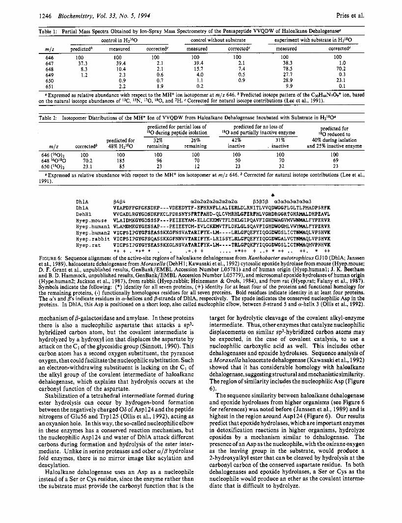
Isolation of a Pentapeptide containing Aspl 24. From the nucleotide sequence of the dehalogenase gene (Janssen et al., 1989) and the specificity of trypsin, Asp124 is predicted to be present in a 24 amino acid tryptic peptide containing the residues Asn 1 17-Argl40. This peptide is expected to elute as one of the later peaks during reversed-phase HPLC (PC/ Gene computer program, Intelligenetics). After separation of trypsin-digested dehalogenase by HPLC, several peaks that eluted from the column were examined by three cycles of manual Edman degradation (Figure 4). The result for peak 1 was Asn-Ile-Thr, which is in agreement with the prediction from the nucleotide sequence.
190 200 210 220 230 240
Wavelength (nm)
wild type I
300 350 400 450 500
Emission wavelength (nm)
FIGURE 3: Circular dichroism (top) and fluorescence (bottom) spectra of wild-type and mutant D124G haloalkane dehalogenase. The CD spectra show no largedifferences at the 208- and 222-nm wavelengths, indicating that the secondary structural elements are the same for both proteins. Fluorescence spectra were recorded at 1 pM enzyme. The different curves represent WT, wild-type haloalkane dehalo- genase; D124G, mutant D124G; 5 p M DBE, mutant D124G with 5 pM 1,2-dibromoethane; and 75 pM DBE, mutant D124G with 75 pM 1,2-dibromoethane.
Chymotrypsin cleavage of the 24 amino acid peptide was expected to yield a pentapeptide with the sequence VVQDW. The presence of tryptophan allowed easy assignment of the strongest UV-absorbing peak (Figure 4) to this pentapeptide.
Ion-Spray Mass Spectrometry. Ion-spray mass spectrom- etry of the presumed Aspl 24-containing peptide indicated the presence of an M + H+ ion at m / z 646, which is in agreement with the mass of the VVQDW pentapeptide predicted from chymotrypsin cleavage behind Lys 120 and Trp125 (Figure 5). A corresponding peak at m / z 688 was always present, which is the M + Na+ ion of the pentapeptide (Figure 5).
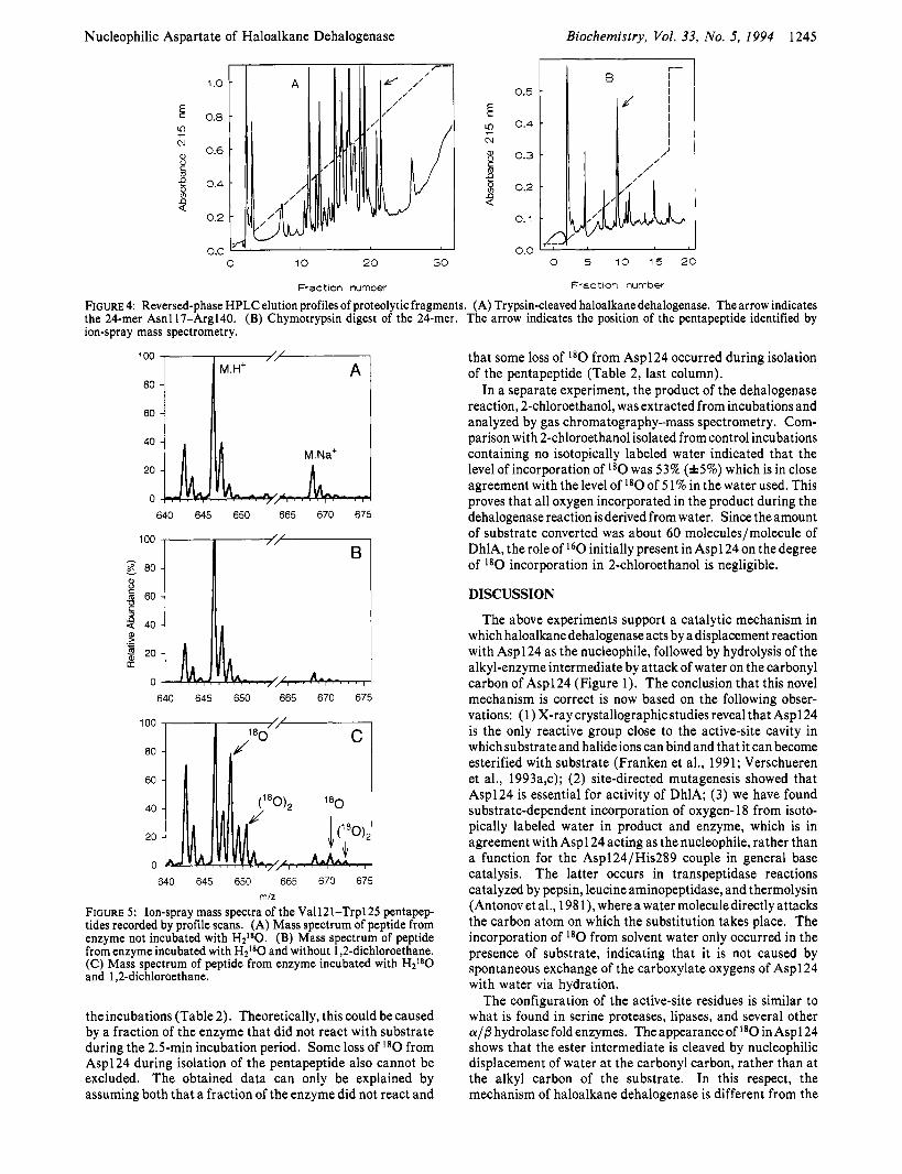
Analysis of the pentapeptide isolated from dehalogenase incubated with H2180 and 1,2-dichloroethane showed that the M + H+ peak at m / z 646 was shifted in part to 648 and 650. Correspondingly, the M + Na+ peak at m/z 688 was shifted to 690 and 692 (Figure 5). This shows that incor- poration of I80 in the pentapeptide had indeed occurred. There was a very limited shift of the mass of the M + H+ and M + Na+ ions of the pentapeptide peaks in control experiments where the peptide was isolated from dehalogenase incubated with H2180 but without substrate (see Table 1).
The normalized peak intensities of the m/z 648 and 650 peaks of the pentapeptide isolated from enzyme incubated with substrate and H2180 were somewhat lower than expected on basis of the level of H2180 in the solvent water used during
Nucleophilic Aspartate of Haloalkane Dehalogenase Biochemistry, Vol. 33, No. 5, 1994 1245
1 .o
0.8 ?
2
9
N a, 0.6
0.4 VI
0.2
0.0 I I 0 10 20 30
Fraction number
I , --. I
0.5
0.4
0.3
0.2
0.1
0.0 0 5 10 15 20
Fraction number
FIGURE 4: Reversed-phase HPLC elution profiles of proteolytic fragments. (A) Trypsin-cleaved haloalkane dehalogenase. The arrow indicates the 24-mer Asnl17-Arg140. (B) Chymotrypsin digest of the 24-mer. The arrow indicates the position of the pentapeptide identified by ion-spray mass spectrometry.
100
80
60
40
20
0
640 645 650 665 670 675
640 645 650 665 670 675
I II II I 40
20
0
640 645 650 665 670 675 miz
FIGURE 5: Ion-spray mass spectra of the Va1121-Trp125 pentapep- tides recorded by profile scans. (A) Mass spectrum of peptide from enzyme not incubated with H2180. (B) Mass spectrum of peptide from enzyme incubated with Hzl*O and without 1,2-dichloroethane. (C) Mass spectrum of peptide from enzyme incubated with H P 0 and 1,2-dichloroethane.
the incubations (Table 2). Theoretically, this could be caused by a fraction of the enzyme that did not react with substrate during the 2.5-min incubation period. Some loss of l 8 0 from Asp124 during isolation of the pentapeptide also cannot be excluded. The obtained data can only be explained by assuming both that a fraction of the enzyme did not react and
that some loss of ‘*O from Asp124 occurred during isolation of the pentapeptide (Table 2, last column).
In a separate experiment, the product of the dehalogenase reaction, 2-chloroethanol, was extracted from incubations and analyzed by gas chromatography-mass spectrometry. Com- parison with 2-chloroethanol isolated from control incubations containing no isotopically labeled water indicated that the level of incorporation of l 8 0 was 53% (f5%) which is in close agreement with the level of l 8 0 of 5 1% in the water used. This proves that all oxygen incorporated in the product during the dehalogenase reaction is derived from water. Since the amount of substrate converted was about 60 molecules/molecule of DhlA, the role of I6O initially present in Aspl 24 on the degree of I8O incorporation in 2-chloroethanol is negligible.
DISCUSSION
The above experiments support a catalytic mechanism in which haloalkane dehalogenase acts by a displacement reaction with Asp124 as the nucleophile, followed by hydrolysis of the alkyl-enzyme intermediate by attackof water on the carbonyl carbon of Asp124 (Figure 1). The conclusion that this novel mechanism is correct is now based on the following obser- vations: (1) X-ray crystallographic studies reveal that Asp124 is the only reactive group close to the active-site cavity in which substrate and halide ions can bind and that it can become esterified with substrate (Franken et al., 1991; Verschueren et al., 1993a,c); (2) site-directed mutagenesis showed that Asp124 is essential for activity of DhlA; (3) we have found substrate-dependent incorporation of oxygen- 18 from isoto- pically labeled water in product and enzyme, which is in agreement with Aspl 24 acting as the nucleophile, rather than a function for the Asp124/His289 couple in general base catalysis. The latter occurs in transpeptidase reactions catalyzed by pepsin, leucine aminopeptidase, and thermolysin (Antonov et al., 198 l ) , where a water moleculedirectly attacks the carbon atom on which the substitution takes place. The incorporation of l 8 0 from solvent water only occurred in the presence of substrate, indicating that it is not caused by spontaneous exchange of the carboxylate oxygens of Aspl 24 with water via hydration.
The configuration of the active-site residues is similar to what is found in serine proteases, lipases, and several other a/P hydrolase fold enzymes. The appearance of l 8 0 in Aspl 24 shows that the ester intermediate is cleaved by nucleophilic displacement of water at the carbonyl carbon, rather than at the alkyl carbon of the substrate. In this respect, the mechanism of haloalkane dehalogenase is different from the
1246 Biochemistry, Vol. 33, NO. 5, 1994 Pries et al.
Table 1: Partial Mass Spectra Obtained by Ion-Spray Mass Spectrometry of the Pentapeptide VVQDW of Haloalkane Dehalogenase’ control in H2160 control without substrate experiment with substrate in HzL80
m J z predictedb measured corrected‘ measured correctedc measured corrected‘ 646 100 100 100 100 100 100 100 647 37.3 39.4 2.1 39.4 2.1 38.3 1 .o 648 8.3 10.4 2.1 15.7 7.4 78.5 70.2 649 1.2 2.3 0.6 4.0 0.5 21.7 0.3 650 0.9 0.7 1 . 1 0.9 28.9 23.1 65 1 2.2 1.9 0.2 9.9 0.1
@ Expressed as relative abundance with respect to the MH+ ion isotopomer at m / z 646. Predicted isotope pattern of the C3oH~N,09+ ion, based on the natural isotope abundances of W, 15N, 170, lSO, and *H. Corrected for natural isotope contributions (Lee et al., 1991).
Table 2: Isotopomer Distributions of the MH+ Ion of VVQDW from Haloalkane Dehalogenase Incubated with Substrate in H2I8O”
predicted for partial loss of I8O during peptide isolation
predicted for no loss of I8O and partially inactive enzyme
predicted for I8O reduced to
predicted for 32% 26% 42% 31% 40% during isolation m l z correctedb 48% H2I80 remaining remaining inactive , inactive and 25% inactive enzyme
646 (I6O)2 100 100 100 100 100 100 648 160180 70.2 185 96 70 50 70 650 (I8O), 23.1 85 23 12 23 32
100 69 23 . I-
@ Expressed as relative abundance with respect to the MH+ ion isotopomer at m / z 646. Corrected for natural isotope contributions (Lee et al., 1991).
D h l A D h l A DehHl Hyep.mouse Hyep.human1 Hyep.human2 Hyep . r abb i t Hyep. r a t
4 P4P4 a2aZaZaZaZaZaZa pSp5p a3a3a3a3a3a3 VIAPDFFGFGKSDKP---VDEEDYTF-EFHRNFLLALIERLDL~ITLVVQD~FLGLTLP~DPSRFK WCADLRGYGDSDKPKCLPDRSNYSFRTFAHD-QLCVMRHLGFERFHLVGHDRGGRTGHRMALDHPEAVL VLAIDMKGYGDSSSP---PEIEEYAM-ELLCKEWVTFLDKLGIPQAVFIGHDUAG~FYPERVR VLAMDMKGYGESSAP---PEIEEYCM-EVLCKEWVTFLDKLGLSQAVFIGHDU~~VWYHALFYPERVR VICPSIPGYGFSEASSKKGFNSVATARIFYK-~----LRLGFQEFYIQGGDWGSLICTNHAQLVPSHVK VICPSIPGYGFSQASSKKGFNNVVTARIFYK-LRISSYLRLGFQEFYIQGGDWGALVCT~QLVPSHVK VICPSIPGYGYSEASSKKGLNSVATARIFYK-~----TRLGFQICFYIQGGDWGSLICT~Q~PNHVK *t t . *t* * . . .t.t t .... t*tt t ..t * tt .. tt. * tt
FIGURE 6: Sequence alignment of the active-site regions of haloalkane dehalogenase from Xanthobacter autotrophicus GJlO (DhlA; Janssen et al., 1989), haloacetate dehalogenase from Moraxella (DehH1; Kawasaki et al., 1992) cytosolic epoxide hydrolase from mouse (Hyep.mouse; D. F. Grant et al., unpublished results, GenBank/EMBL Accession Number L05781) and of human origin (Hyep.human1; J. K. Beetham and B. D. Hammock, unpublished results, GenBank/EMBL Accession Number L05779), and microsomal epoxide hydrolases of human origin (Hype.human2; Jackson et al., 1987), from rabbit (Hyep.rabbit; Heinemann & Ozols, 1984), and from rat (Hyep.rat; Falany et al., 1987). Symbols indicate the following: (*) identity for all seven proteins, (+) identity for at least four of the proteins and functional homology for the remaining proteins, (.) functionally homologous residues for all seven proteins. Bold residues indicate identity in at least four proteins. The a’s and ps indicate residues in a-helices and @-strands of DhlA, respectively. The spade indicates the conserved nucleophilic Asp in the proteins. In DhlA, this Asp is positioned on a short loop, also called nucleophile elbow, between &strand 5 and a-helix 3 (Ollis et al., 1992).
mechanism of @-galactosidase and amylase. In these proteins there is also a nucleophilic aspartate that attacks a sp3- hybridized carbon atom, but the covalent intermediate is hydrolyzed by a hydroxyl ion that displaces the aspartate by attackon the C1 of the glycosidic group (Sinnott, 1990). This carbon atom has a second oxygen substituent, the pyranose oxygen, that could facilitate the nucleophilic substitution. Such an electron-withdrawing substituent is lacking on the C1 of the alkyl group of the covalent intermediate of haloalkane dehalogenase, which explains that hydrolysis occurs at the carbonyl function of the aspartate.
Stabilization of a tetrahedral intermediate formed during ester hydrolysis can occur by hydrogen-bond formation between the negatively charged 06 of Asp124 and the peptide nitrogens of Glu56 and Trp125 (Ollis et al., 1992), acting as an oxyanion hole. In this way, the so-called nucleophilic elbow in these enzymes has a conserved reaction mechanism, but the nucleophilic Asp124 and water of DhlA attack different carbons during formation and hydrolysis of the ester inter- mediate. Unlike in serine proteases and other a/@ hydrolase fold enzymes, there is no mirror image like acylation and deacylation.
Haloalkane dehalogenase uses an Asp as a nucleophile instead of a Ser or Cys residue, since the enzyme rather than the substrate must provide the carbonyl function that is the
target for hydrolytic cleavage of the covalent alkyl-enzyme intermediate. Thus, other enzymes that catalyze nucleophilic displacements on similar sp3-hybridized carbon atoms may be expected, in the case of covalent catalysis, to use a nucleophilic carboxylic acid as well. This includes other dehalogenases and epoxide hydrolases. Sequence analysis of a Moraxella haloacetate dehalogenase (Kawasaki et al., 1992) showed that it has considerable homology with haloalkane dehalogenase, suggesting structural and mechanistic similarity. The region of similarity includes the nucleophilic Asp (Figure 6).
The sequence similarity between haloalkane dehalogenase and epoxide hydrolases from higher organisms (see Figure 6 for references) was noted before (Janssen et al., 1989) and is highest in the region around Asp124 (Figure 6). Our results predict that epoxide hydrolases, which are important enzymes in detoxification reactions in higher organisms, hydrolyze epoxides by a mechanism similar to dehalogenase. The presence of an Asp as the nucleophile, with the oxirane oxygen as the leaving group in the substrate, would produce a 2-hydroxyalkyl ester that can be cleaved by hydrolysis at the carbonyl carbon of the conserved aspartate residue. In both dehalogenases and epoxide hydrolases, a Ser or Cys as the nucleophile would produce an ether as the covalent interme- diate that is difficult to hydrolyze.
Nucleophilic Aspartate of Haloalkane Dehalogenase
ACKNOWLEDGMENT
The authors thank Dr. A. J. W. G. Visser, Biochemistry Department, Agricultural University of Wageningen, for recording the circular dichroism spectra.
REFERENCES Antonov, V. K., Ginodman, L. M., Rumsh, L. D., Kapitannikov,
Y. V., Barshevskaya, T. N., Yavashev, L. P., Gurova, A. G., & Volkova, L. I. (1981) Eur. J. Biochem. 117, 195-200.
Bruins, A. P., & Beaugrand, C. (1988) Proceedings of the 36th ASMS Conference on Mass Spectrometry and Allied Topics, p 1242, American Society for Mass Spectrometry, East Lansing, MI.
Bruins, A. P., Covey, T. R., & Henion, J. D. (1987) Anal. Chem.
Chang, J.-Y. (1983) Methods Enzymol. 91, 455-466. Escobar, W. A., Tan, A. K., & Fink, A. L. (1991) Biochemistry
Falany, C. N., McQuiddy, P., & Kasper, C. B. (1987) J . Biol. Chem. 262, 5924-5930.
Franken, S. M., Rozeboom, H., Kalk, K. H., & Dijkstra, B. W. (1991) EMBO J . 10, 1297.
Gonzalez, J., Takao, T., Hori, H., Besada, V., Rodriguez, R., Padron, G., & Shimonishi, Y. (1992) Anal. Biochem. 205,
Heinemann, F. S., & Ozols, J. (1984) J. Biol. Chem. 259, 797-
Jackson, M. R., Craft, J. A., & Burchell, B. (1987) Nucleic
Janssen, D. B., Pries, F., Van der Ploeg, J., Kazemier, B., Terpstra,
59,2642-2646.
30, 10783-1 0787.
15 1-1 58.
804.
Acids Res. 15, 7188.
P., & Witholt, B. (1989) J . Bacteriol. 171, 6791-6799.
Biochemistry, Vol. 33, No. 5, 1994 1247
Kawasaki, H., Tsuda, K., Matsushita, I., & Tonomura, K. (1992)
Keuning, S., Janssen, D. B., & Witholt, B. (1985) J . Bacteriol.
Laemmli, U. K. (1970) Nature 227, 680-685. Lee, W. N. P., Byerley, L. O., Berger, E. A., & Edmond, J.
(1991) Biol. Muss Spectrom. 20, 451-458. Muderhwa, J. M., Schmid, P. C., & Brockman, H. L. (1992)
Biochemistry 31, 141-148. Nelson, R. M., & Long, G. L. (1989) Anal. Biochem. 180,147-
151. Ollis, D. L., Cheah, E., Cygler, M., Dijkstra, B. W., Frolow, F.,
Franken, S. M., Harel, M., Remington, S. J., Silman, I., Schrag, J., Sussman, J. L., Verschueren, K. H. G., & Goldman, A. (1992) Protein Eng. 5, 197-211.
Sanger, F., Nicklen, S., & Coulsen, A. R. (1977) Proc. Natl. Acad. Sci. U.S.A. 74, 5463-5467.
Schanstra, J. P., Rink, R., Pries, F., & Janssen, D. B. (1993) Protein Expression Pur$ 4, 479-489.
Sinnott, M. L. (1990) Chem. Rev. 90, 1171-1202. Strynadka, N. C., Adachi, H., Jensen, S. E., Johns, K., Sielecki,
A., Betzel, C., Sutoh, K., & James, M. N. G. (1992) Nuture 359, 700-705.
Studier, F. W., Rosenberg, A. H., Dum, J. J., & Dubendorff, J. W. (1990) Methods Enzymol. 185, 60-89.
Verschueren, K. H. G., Franken, S. M., Rozeboom, H. J., Kalk, K. H., & Dijkstra, B. W. (1993a) J . Mol. Biol. 232,856-872.
Verschueren, K. H. G., Kingma, J., Rozeboom, H. J., Kalk, K. H., Janssen, D. B., & Dijkstra, B. W. (1993b) Biochemistry
Verschueren, K. H. G., SeljBe, F., Rozeboom, H. J., Kalk, K. H.,
J . Gen. Microbiol. 138, 1317-1323.
163, 635-639.
32, 9031-9037.
& Dijkstra, B. W. (1993~) Nature 363, 693-698.