skeletal muscle lecture packet 8 reading: chapter 5 copyright 2008 pearson education
TRANSCRIPT

COPYRIGHT 2008 PEARSON EDUCATION
Skeletal MuscleLECTURE PACKET 8
READING: CHAPTER 5

COPYRIGHT 2008 PEARSON EDUCATION
Outline
▪ Overview of the skeletal system▪ Function of bones▪ Bone structure▪ Bone cells▪ Cartilage▪ Tendons and ligaments▪ Joints▪ Bone development▪ Hormonal regulation of bone growth▪ Homeostasis▪ Disorders of the skeletal system

COPYRIGHT 2008 PEARSON EDUCATION
Skeletal System Components
▪ The skeletal system is composed of different types of connective tissues.
- Bones: rigid structure- Cartilage: soft, cushions the joints- Ligaments: attach bone to bone- Tendons: attach muscle to bone
▪ Tendons link the skeletal and the muscular systems together and is not considered part of the skeletal system.

COPYRIGHT 2008 PEARSON EDUCATION
Skeletal System Components
▪ Ligaments link one bone to another.
▪ Tendons link bone to muscle, and it is that dense connective tissue.

COPYRIGHT 2008 PEARSON EDUCATION
Functions of Bone
1. Support and gives shape to the body.2. Protect internal organs. 3. Produce blood cells. 4. Store minerals (particularly calcium and phosphorus).5. Store fat in yellow bone marrow (the soft tissue within some
bones). 6. Along with the muscles, permit flexible body movement.

COPYRIGHT 2008 PEARSON EDUCATION
Bone Structure
▪ There are two types of bone:
▪ Compact bone is very dense, with few internal spaces, which is covered by a glovelike membrane known as the periosteum.
- Periosteum contains blood vessels and nerve cells that provide nourishment to the bone.
▪ Spongy bone is a latticework of tiny beams and thin plates of bone with open areas between.

COPYRIGHT 2008 PEARSON EDUCATION
Bone Structure
▪ Epiphysis is the rounded end of the typical long bone, and it’s composed mainly of red bone marrow where blood cells are made.
▪ Diaphysis is the shaft, or the middle part, of the typical long bone, and has a cavity filled with yellow bone marrow where fat is stored.
▪ Periosteum is the fibrous outer covering of the bone, and it contains nerves, blood vessels, and lymphatic vessels.
- Functions in bone repair and growth.

COPYRIGHT 2008 PEARSON EDUCATION
Bone Structure

COPYRIGHT 2008 PEARSON EDUCATION
Bone Structure
▪ Compact bone contains a repeating structural unit called an osteon.
▪ Each osteon consists of mature bone cells called osteocytes that are arranged in concentric rings around a central canal.

COPYRIGHT 2008 PEARSON EDUCATION
Bone Structure
▪ Each osteocyte lies within a tiny cavity called a lacuna.
▪ Canaliculi are canals that connect the lacunae to the central canal.

COPYRIGHT 2008 PEARSON EDUCATION
Bone Structure

COPYRIGHT 2008 PEARSON EDUCATION
Bone Cells
▪ Osteocytes▪ Osteoblasts: important in bone formation▪ Osteoclasts: important in bone resorption
▪ Osteoblasts and osteoclasts are important in controlling the amount of bone tissue.

COPYRIGHT 2008 PEARSON EDUCATION
Bone Cells: Osteoblasts
▪ Osteoblasts are the bone forming cells.
▪ Osteoblasts produce the matrix of bone by secreting collage (as well as other organic materials) and then depositing calcium salts (hydroxyapatite) on it.

COPYRIGHT 2008 PEARSON EDUCATION
Bone Cells: Osteoclasts
▪ Osteoclasts participate in bone resorption (destruction).
▪ They release acid and collagenase that eat away at the bone, releasing minerals.
▪ These are bone cells on the outer edge of bones.
COPYRIGHT 2008 PEARSON EDUCATION
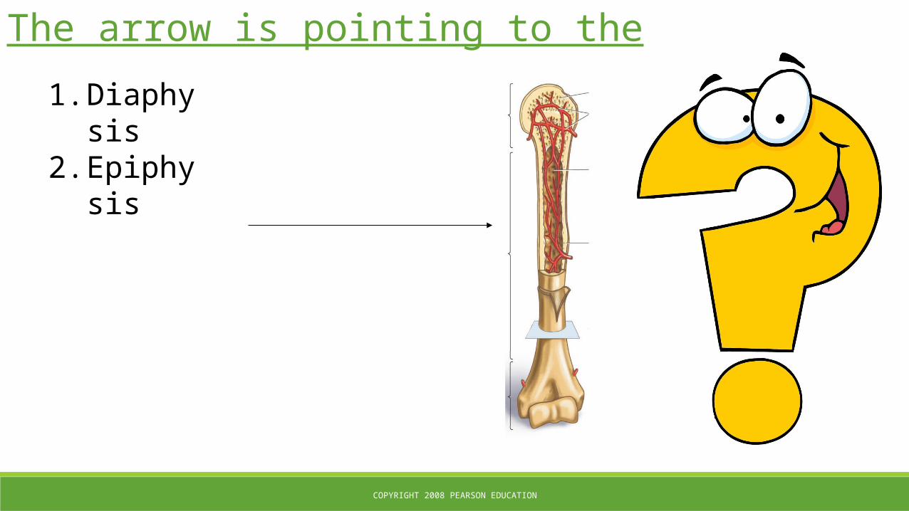
The arrow is pointing to the
1. Diaphysis2. Epiphysis
COPYRIGHT 2008 PEARSON EDUCATION
They secrete calcified material to build bones
1. Osteoclasts2. Osteoblasts
COPYRIGHT 2008 PEARSON EDUCATION
Bone Development
▪ The skeleton begins to form in the fetus at about 6 weeks.▪ Most bones grow through the adolescence but some bones continue to grow until about age 25. ▪ Bones continue to change throughout life.
- Osteoclasts are removing the bone matrix while- Osteoblasts are building the matrix.
▪ Ossification: formation of bones.
COPYRIGHT 2008 PEARSON EDUCATION
Hormonal Regulation of Bone Growth
▪ Growth Hormone (GH) directly stimulates growth of the epiphyseal plate and bone growth during childhood. ▪ Vitamin D causes the intestines to absorb calcium. ▪ Thyroid hormones ensure that the skeleton grow with the proper proportions. ▪ Sex hormones cause adolescents to experience a growth spurt due to an increased level of hormones, but eventually allow for the closing of the growth plate as well.

COPYRIGHT 2008 PEARSON EDUCATION
Hormonal Regulation of Bone Calcium Levels
▪ Parathyroid hormone (PTH) is produced by the parathyroid hormone and causes calcium to be released from bone, thereby increasing blood calcium.
▪ Calcitonin, which is released from the thyroid gland, removes calcium from the blood and causes it to be stored in bone.
▪ Homeostasis is maintaining a balance of calcium levels in blood and in bone.

COPYRIGHT 2008 PEARSON EDUCATION
Hormonal Regulation of Bone Calcium Levels

COPYRIGHT 2008 PEARSON EDUCATION
Healing Broken Bones
▪ Broken bones and fractures are the same thing.
▪ Bone fractures are healed by fibroblasts and osteoblasts.
▪ When a break occurs, there is bleeding followed by a clot.
▪ Fibroblasts secrete collage fibers that form a callus, which links the broken surfaces of bone. Osteoblasts from the periosteum invade the callus and begin to transform the newly deposited cartilage into new bone material, changing it into a bony callus.

COPYRIGHT 2008 PEARSON EDUCATION
Healing Broken Bones

COPYRIGHT 2008 PEARSON EDUCATION
Healing Broken Bones

COPYRIGHT 2008 PEARSON EDUCATION
This hormone is released when blood calcium is low
1. Growth Hormone2. Calcitonin3. Parathyroid Hormone

COPYRIGHT 2008 PEARSON EDUCATION
Calcitonin is produced by the
1. Parathyroid Gland2. Thyroid Gland3. Anterior Pituitary4. Hypothalamus

COPYRIGHT 2008 PEARSON EDUCATION
Cartilage
▪ Cartilage is flexible connective tissue not as strong as bone tissue. It functions to cushion joins and it provides flexibility.
- Found at the ends of long bones, nose, ends of ribs, larynx and trachea, disks between vertebrae and knee, ear flaps, and epiglottis.
▪ It lacks blood vessels, so it’s slow to heal.
▪ Cartilage cells are chondrocytes.
COPYRIGHT 2008 PEARSON EDUCATION
Cartilage in Bones
▪ Two regions of cartilage remain at each end of the long bone.- One is a cap of cartilage over each end of the bone, where
one bone glides over another in a joint. - The second is a plate of cartilage, called an epiphyseal plate,
or growth plate, that separates each end of the bone from its shaft.

COPYRIGHT 2008 PEARSON EDUCATION
Tendons and Ligaments
▪ Ligaments are connective tissue that connects bone to bone.
▪ Tendons are connective tissue that connects muscle to bone.

COPYRIGHT 2008 PEARSON EDUCATION
Joints
▪ Joints are the places where bones meet.
▪ Most of the joints of the body are freely movable synovial joints.

COPYRIGHT 2008 PEARSON EDUCATION
Joints
▪ Joints are the places where bones meet.
▪ Most of the joints of the body are freely movable synovial joints.

COPYRIGHT 2008 PEARSON EDUCATION
Joints

COPYRIGHT 2008 PEARSON EDUCATION
Joints

COPYRIGHT 2008 PEARSON EDUCATION
Integrated

COPYRIGHT 2008 PEARSON EDUCATION
Cartilage cells are
1. Fibrocytes2. Osteocytes3. Chondrocytes

COPYRIGHT 2008 PEARSON EDUCATION
This connective tissue connects bone to bone
1. Ligaments2. Tendons

COPYRIGHT 2008 PEARSON EDUCATION
This connective tissue connects bone to muslce
1. Ligaments2. Tendons