skin cancer diagnosis and surgical management in general ... · further analysis of these data...
TRANSCRIPT

This file is part of the following reference:
Heal, Clare (2010) Skin cancer diagnosis and surgical
management in general practice. PhD thesis, James Cook
University.
Access to this file is available from:
http://researchonline.jcu.edu.au/19038/
The author has certified to JCU that they have made a reasonable effort to gain
permission and acknowledge the owner of any third party copyright material
included in this document. If you believe that this is not the case, please contact
[email protected] and quote
http://researchonline.jcu.edu.au/19038/
ResearchOnline@JCU

DOCTOR OF PHILOSOPHY
SKIN CANCER DIAGNOSIS AND SURGICAL
MANAGEMENT IN GENERAL PRACTICE
Thesis submitted by
Dr Clare Heal MBChB
For the degree of Doctor of Philosophy in
The School of Public Health and Tropical Medicine at James Cook
University
January 2010

C Heal 2
STATEMENT OF ACCESS
I, Clare Heal, author of this work, understand that James Cook University will make this thesis
available for use within the University Library and, via the Australian Digital Thesis Network,
for use elsewhere.
I understand that, as an unpublished work, a thesis has significant protection under the
Copyright Act and I do not wish to place any further restriction on this work.
……………………………….. ……………………………………..
Signature Date

C Heal 3
STATEMENT OF SOURCES
I declare that this thesis is my own work and has not been submitted in any form for another
degree or diploma at any university or other institution of tertiary education. Information
derived from the published or unpublished work of others has been acknowledged in the text
and a list of references is given.
……………………………….. ……………………………………..
Signature Date

C Heal 4
STATEMENT OF THE CONTRIBUTION OF
OTHERS
Some of this research was conducted in collaboration or consultation with other practitioners.
I wish to acknowledge the contribution of my co-authors to a number of manuscripts included
as part of this thesis:
Chapter 2 Risk factors for wound infection after minor surgery
Dr Petra Buttner performed multivariate analysis and edited the paper.
Chapter 3 Does application of chloramphenicol ointment to sutured wounds decrease
the incidence of wound infection?
Dr Petra Buttner contributed to the study design and did statistics. Dr Robert
Cruikshank, Dr David Graham, Dr Sheldon Browning, Ms Jayne Pendergast, Dr
Herwig Drobetz, Mr Robert Gluer and Dr Carl Lisec contributed to the study
design.
Chapter 4 Minor skin excisions in North Queensland
Dr Petra Buttner and Dr Beverly Raasch contributed to study design; performed
statistics and proof read and edited this paper.
Chapter 5 Diagnostic accuracy of excised and biopsied skin lesions by Australian
general practitioners
Dr Petra Buttner performed the statistics, Dr Beverly Raasch, Dr Petra Buttner
and Dr David Weedon contributed to study design and proof read and edited the
manuscript.
Chapter 6 Agreement between histological diagnosis of skin lesions by histopathologists
and a dermatohistopathologist
Dr Petra Buttner performed statistics, Dr Petra Buttner, Dr Beverly Raasch, Dr
David Weedon contributed to the study design and proof read and edited the
manuscript.
Chapter 7 Comparing the case-mix and number needed to treat of GPs and skin cancer
clinic doctors
Dr Beverly Raasch proof read and edited the manuscript.

C Heal 5
ACKNOWLEDGEMENTS
I wish to express my sincere thanks to the many people who contributed to this thesis.
I would like to thank my supervisors, Dr Petra Buttner and Dr Beverly Raasch for their help
over the period of my studies.
I would like to thank the General Practitioners in Mackay who contributed to the design and
helped to collect data for two large randomised controlled trials conducted in Mackay.
I would particularly like to thank the RACGP research foundation and the Chris Silagy
scholarship for the funding of the topical chloramphenicol project.
Finally, I would like to thank my husband Sheldon Browning and daughter Mia Browning for
their tolerance and support.
……………………………….. ……………………………………..
Signature Date

C Heal 6
LIST OF ABBREVIATIONS
AK Actinic keratosis
APC Annual PERCENTAGE CHANGE
BCC Basal cell carcinoma
CM Cutaneous melanoma
CN Common naevus
GP General practitioner
IEC Intra-epithelial carcinoma
JCU James Cook University
KA Keratoacanthoma
MN Melanocytic naevus
NNT Number needed to treat
NPV Negative predictive value
PPV Positive predictive value
RCT Randomised controlled trial
SCC Squamous cell carcinoma
SK Seborrhoeic keratoses
SSI Surgical site infection

C Heal 7
ABSTRACT
Background
Skin cancer is an extremely important health issue in Australia. Squamous cell carcinoma
(SCC) and basal cell carcinoma (BCC) are by far the commonest cancers in Australia with an
incidence of more than four times that of all other registrable cancers combined.(1) Cutaneous
melanoma (CM) is the fifth most common cancer in Australia, with the estimated risk of
developing a melanoma before 75 years of age being one in 26 for Australian men and one in 36
for Australian women.(1) Queensland has the world’s highest recorded incidence of all types of
skin cancer,(2, 3) with incidence rates being even higher in tropical North Queensland.(4)
In North Queensland, the majority of suspicious skin lesions are managed by general
practitioners (GPs),(5) particularly in rural centres such as Mackay where there is no resident
plastic surgeon or dermatologist.(6) It is therefore important that the diagnosis and post surgical
wound management of GPs is optimal.
In 2006 a group of GPs in Mackay, of which I was chief investigator, published a randomised
controlled trial which showed that the wetting of sutures did not increase the incidence of
wound infection but the incidence of infection was nevertheless higher than expected after
minor skin cancer surgery.(7) This trial formed the background to several of the current studies
for this thesis.
The overall aim of the studies presented here was:
1. To improve the management of skin cancer and the conduct of skin cancer surgery by
Australian GPs.
2. To increase patient well-being through appropriate use of post-surgery wound
management.
3. To assess the ability of GPs and pathologists to diagnose skin lesions.
4. To investigate possibilities of research in GP practice settings.

C Heal 8
Methods
The thesis comprises the results of three trials which took part in the Queensland towns of
Mackay and Townsville.
The first study proceeded from the ‘Can sutures get wet’ trial which was a randomised
controlled trial conducted in general practice, in Mackay in 2004-5. Data were recorded from
1247 consecutive patients who attended for minor skin excisions. Further exploration and data
analysis was undertaken from this trial to investigate 857 of these patients for risk factors for
infection, to investigate the numbers needed to treat (by excision) for skin cancers and to
examine the case-mix of skin lesions in regular general practice and a skin cancer clinic.
The second ‘Topical chloramphenicol’ study was also conducted in general practice in Mackay
in 2007. This was a prospective double blinded randomised controlled trial. Nine hundred and
seventy two patients were assessed for infection after receiving either a single topical dose of
chloramphenicol (n=488) or paraffin ointment (n=484; placebo).
The third study had previously been conducted in Townsville and secondary data analysis from
the database of this study formed the ‘Diagnostic accuracy’ and histological agreement studies.
All excised and histologically confirmed skin cancers in Townsville/Thuringowa, from
December 1996 to October 1999 were recorded. Positive predictive values (PPV) and
sensitivities were calculated for the clinical diagnoses and stratified by histological sub-type and
body-site. A stratified sample of 407 of 8,694 skin excisions slides was used to compare the
“standard” histological diagnosis with the diagnosis from an internationally renowned
dermato histo-pathologist.
Results
Secondary data analysis from the ‘Can sutures get wet’ study showed an overall incidence of
infection was 8.6% (95%-confidence interval = [3.5, 13.8]). Excisions from lower legs and feet
(p=0.009) or thighs (p=0.005), excisions of BCC (p=0.006) or SCC (p=0.002) and diabetes
(p<0.001) were found to be independent risk factors for wound infection.
Close to half (46.7%) of lesions excised were skin cancers with more SCCs than BCCs (0.74:1).
Our number needed to treat (NNT) (melanocytic naevi excised per melanoma) was 8.4. Mean
age for excision of melanoma, BCC and SCC was 55, 60.9 and 63.8 yrs respectively. Relative
tumour density was greatest in the face, scalp and neck region for all skin cancers.
C Heal 9
Further analysis of these data comparing mainstream general practitioners with a doctor
working in a designated skin cancer clinic showed that the case-mix of non-melanotic skin
cancers was significantly different for the two groups of doctors (p<0.001). The BCC:SCC ratio
was much higher for skin cancer clinic doctors (4:1) than for GPs (0.6:1). The NNT
(melanocytic naevi excised per melanoma) was 4.7 for the skin cancer doctor and 9.0 for
mainstream GPs.
In the ‘Topical chloramphenicol’ trial, the incidence of infection in the chloramphenicol group
(6.6%; 95%-confidence interval = 95%-CI = (4.9 to 8.8)) was significantly lower compared to
the incidence in the control group (11.0%; 95%-CI = (7.9 to 15.1)) (p=0.010). The absolute
reduction in infection rate was 4.4%, the relative reduction was 40% and the relative risk of
wound infection in the control group was 1.7 times higher (95%-CI = (1.1 to 2.5)) than in the
intervention group.
The third and fourth studies examined a total of 8694 skin excisions reanalysed from the
database of the Townsville study. Positive predictive values (PPV) for the clinical diagnoses
were: BCC 0.727, SCC 0.494 and CM 0.333. Sensitivities for the clinical diagnosis were: BCC
0.639, SCC 0.411, and CM 0.338. For BCC, PPVs and sensitivities were higher for the trunk,
the shoulders and the face and lower for the extremities. The reverse pattern was seen for SCCs.
Further analysis of the data comparing an expert dermato-pathologist with mainstream histo
pathologists, showed positive predictive values for the primary histological diagnosis were
above 90% for BCC, CM and CN. For SCC the positive predictive value was 72.6% (95%-CI =
[65.5, 79.0]).
Overall conclusions
Patients with minor skin excisions in North Queensland have a higher incidence of wound
infection. Groups at high risk of infection after minor surgery were diabetics, those undergoing
excision of a non-melanocytic skin cancer or excision from the lower limb.
Topical chloramphenicol ointment decreases this incidence moderately. GPs in North
Queensland have a high yield of skin cancer from their skin excisions and a low NNT
(melanocytic naevi excised per melanoma). Doctors diagnose skin cancers accurately, but there
are some areas of diagnostic difficulty, in particular in the diagnosis of actinic keratosis (AK)
and SCC. GPs and skin cancer doctors have a different skin cancer case-mix.

C Heal 10
Overall recommendations
1. In view of the level of skin infection associated with skin cancer surgery previously
identified, antibiotic prophylaxis prior to minor surgery in general practice should be
limited to the high risk groups that were identified - the consequences of infection are
often minor and side-effects from antibiotics, such as allergy, can potentially be serious.
2. The use of topical chloramphenicol ointment to prevent infection after minor surgery is
best reserved for high risk groups. The reduction in infection was only 40%, which was
statistically but not clinically relevant. The overutilization of topical antibiotics has
potentially adverse consequences such as antibiotic resistance and allergic contact
dermatitis.
3. The doctors involved in my study in Mackay could consider lowering their threshold for
excision of pigmented lesions. In our sample of Mackay GPs, there was a very high
yield of skin cancers from all excisions, and NNT (melanocytic naevi excised per
melanoma) of 8.4, was lower than published data from comparable cohorts.
4. Educational programs for doctors regarding the diagnosis of skin cancer could focus on
areas of diagnostic weakness which were identified in our study, such as differentiating
melanocytic naevi from malignant melanoma, and differentiating between AK and
SCC.
5. It is important that doctors excising suspicious skin lesions are aware that there is
discordance and lack of agreement between histopathologists regarding the diagnosis of
SCC and AK.

C Heal 11
Table of Contents
List of Figures…………………………………………………………………………………. 14
List of Tables……………………………………………………………………………………14
THESIS INTRODUCTION AND OVERVIEW 17
Chapter 1: LITERATURE REVIEW 22
1.1 Scope and limitations of literature review 22
1.2 Skin cancer incidence in Australia 25
1.3 Management of skin cancer by Australian GPs and Number Needed To Treat. 33
1.4 Surgical site infection 36
1.5 Antibiotic ointment use 42
1.6 Conclusion of literature review 53
CHAPTER 2: RISK FACTORS FOR WOUND INFECTION AFTER MINOR SURGERY55
CHAPTER 3: DOES APPLICATION OF CHLORAMPHENICOL OINTMENT TO
SUTURED WOUNDS DECREASE THE INCIDENCE OF WOUND
INFECTION? 69
CHAPTER 4: MINOR SKIN EXCISIONS IN NORTH QUEENSLAND 84
CHAPTER 5: DIAGNOSTIC ACCURACY OF EXCISED AND BIOPSIED SKIN
LESIONS BY AUSTRALIAN GENERAL PRACTITIONERS 93
CHAPTER 6: AGREEMENT BETWEEN HISTOLOGICAL DIAGNOSIS OF SKIN
LESIONS BY HISTOPATHOLOGISTS AND A
DERMATOPATHOLOGIST 110
CHAPTER 7: COMPARING THE CASE-MIX AND NUMBER NEEDED TO TREAT OF
GPS AND SKIN CANCER CLINIC DOCTORS 115
CHAPTER 8: DISCUSSION CONCLUSIONS AND RECOMMENDATIONS 118
8.1 Update of Literature Review 118
8.2 Overall Conclusions 124
8.3 Overall recommendations 124
8.4 Where to from here – research questions identified and future studies 125

C Heal 12
REFERENCES 127
LIST OF APPENDIXES 135
Appendix 1. Risk factors for wound infection after minor surgery in general practice
.................................................................................................................. 137
Appendix 2. Does single application of topical chloramphenicol to high risk sutured
wounds reduce incidence of wound infection after minor surgery? Prospective
randomised placebo controlled double blind trial........................................ 142
Appendix 3. Minor skin excisions in general practice in North Queensland .................... 149
Appendix 4. Accuracy of clinical diagnosis of skin lesions............................................. 154
Appendix 5. Agreement between histological diagnosis of skin lesions by histopathologists
and a dermato-histopathologist................................................................... 163
Appendix 6. Diagnosing skin cancer in primary care: how do mainstream general
practitioners compare with primary care skin cancer clinic doctors? ........... 168
Appendix 7. Practice based research – Lessons from the field ........................................ 170
Appendix 8. Ethics Approval ......................................................................................... 175
Appendix 9. Consent form and patient information sheet ............................................... 176
Appendix 10. Data collection sheets ................................................................................ 179
Appendix 11. Body site map ............................................................................................ 182
Appendix 12. Excision protocol ....................................................................................... 183
Appendix 13. Study protocol ........................................................................................... 186
Appendix 14. Consort check list ...................................................................................... 197
Appendix 15. Rapid responses to BMJ chloramphenicol article........................................ 200

C Heal 13
Appendix 16. BMJ editorial (Grey et al) .......................................................................... 207
Appendix 17. Rapid responses to BMJ editorial ............................................................... 209
Appendix 18. Antibiotic prophylaxis in dermatologic surgery:
Advisory statement 2008 (Wright et al) ...................................................... 212
Appendix 19. Can sutures get wet? .................................................................................. 222
Appendix 20. How to dissect surgical journals: VII – Comparing outcomes ..................... 226
Appendix 21. How to dissect surgical journals: IX – Sample size ..................................... 234

C Heal 14
List of Figures
Figure 1: Greenmount Station, the first European settlement in Mackay .................................. 19
Figure 2: Malignant melanoma on foot of patient in Mackay, Qld ........................................... 24
Figure 1.1: Trends in age standarised incidence rates (ASR) of melanoma in Australia ........... 33
Figure 2.1: Wound management ............................................................................................. 58
Figure 3.1: Study protocol for the patient ................................................................................ 72
Figure 3.2: Flow-chart of enrolment, randomisation and follow up of patients ......................... 76
List of Tables
Table 1.1: Studies of defined communities measuring incidence of BCC and SCC in Australia
from 1982-2006 ...................................................................................................................... 31
Table 1.2: Australian studies of number of benign lesions excised per melanoma ................... 34
Table 1.3: Classification of surgical wounds .......................................................................... 36
Table 1.4: These categories can be adapted to dermatological surgery .................................... 37
Table 1.5: Incidence and risk factors for surgical site infection following dermatological
surgery prospective observational studies................................................................................ 39
Table 1.6: Guidelines for oral antibiotic prophylaxis of surgical site infection after derma
surgery ................................................................................................................................... 41
Table 1.7: Properties of antibiotic ointments in common use .................................................. 43
Table 1.8: Randomised controlled trials investigating application of antibiotic ointment after
skin surgery ............................................................................................................................ 46
Table 1.9: Randomised placebo controlled double blind studies of antibiotic ointment use on
non-surgical wounds ............................................................................................................... 47
Table 1.10: Randomised controlled studies investigating antibiotic ointment in the treatment of
infected wounds ..................................................................................................................... 50
Table 2.1: Definition of surgical site infection........................................................................ 59
Table 2.2: Reasons for exclusion of 377 patients from study .................................................. 62
Table 2.3: Bivariate correlates between infection after minor surgery and participants’ and
lesions’ characteristics in 857 patients recorded in Mackay, Australia ..................................... 63

C Heal 15
Table 2.4: Correlates of participants’ and lesions’ characteristics with infection after minor
surgery. Results of multivaraible generalised linear modeling based on857 patients from
Mackay, Australia .................................................................................................................. 64
Table 3.1: Reasons for exclusion from study .......................................................................... 75
Table 3.2: Baseline comparisons of intervention (chloramphenicol and control (paraffin) group
............................................................................................................................................... 77
Table 3.3: Incidence of wound infections in intervention (chloramphenical) and control
(paraffin) group ...................................................................................................................... 78
Table 4.1: Histology of 1,247 minor skin excisions recorded in Mackay, North Queensland ... 87
Table 4.2: Age and sex distribution of patients presenting with skin cancers for all skin
excisions in Mackay, North Queensland ................................................................................. 88
Table 4.3: Body site distribution and relative tumour density (RTD) of skin cancers as recorded
in Mackay, North Queensland ................................................................................................. 89
Table 5.1: Positive predictive values (in bold) and 95%-condifence intervals of clinical
diagnosis of skin lesions. Results for pigmented lesions were shaded; rows add up to 100%.... 98
Table 5.2: Sensitivities (in bold) and 95%-confidence interval of clinical diagnosis of skin
lesions. Results for pigmented lesions were shaded; columns add up to 100% ....................... 100
Table 5.3: Positive predictive values of clinical diagnosis of skin lesions stratified by body-
sides. Results for pigmented lesions were shaded .................................................................. 103
Table 5.4: Sensitivities of clinical diagnosis of skin lesions stratified by body-sites. Results for
pigmented lesions were shaded ............................................................................................. 104
Table 6.1: Agreement between diagnosis of Townsville histologies and a dermato-
histopathology specialist (DW). Positive predictive values (in bold) together with 95%-
condifence intervals are given ............................................................................................... 112
Table 7.1: Comparison of lesion excisions in skin cancer clinic and general practice settings 116
Table 8.1: Risk factors for infection, Dixon et al 2006 ......................................................... 119

C Heal 16

C Heal 17
THESIS INTRODUCTION AND OVERVIEW
Introduction and overview
The data for the following thesis were collected from three separate studies conducted in the
rural centre of Mackay and the regional centre Townsville, North Queensland. The author was
involved as principal investigator of the first two studies which were conducted in Mackay in
2004 and 2007. In addition, data was analysed from a separate study conducted by Drs Beverly
Raasch and Petra Buttner in Townsville. Although each study was independent of the other,
they are interrelated in that they examine aspects of the diagnosis and management of skin
cancer in general practice. In this overview, the background and settings for these studies will
be outlined.
Background
The term non-melanocytic skin cancer (NMSC), has previously been used as a collective term
to describe the combination of basal cell carcinoma (BCC) and squamous cell carcinoma (SCC)
This nomenclature has now changed and the terms BCC and SCC will be referred to
individually in this document.
Skin cancer is an extremely important health issue in Australia. SCC and BCC are by far the
commonest cancers in Australia with an incidence of more than four times that of all other
registrable cancers combined.(1) Cutaneous melanoma (CM) is the fifth most common cancer in
Australia, with the estimated risk of developing a melanoma before 75 years of age being one in
26 for Australian men and one in 36 for Australian women.(1) Queensland has the world’s
highest recorded incidence of all types of skin cancer,(2, 3) with incidence rates being even
higher in tropical North Queensland.(4)
In North Queensland, the majority of suspicious skin lesions are managed by general
practitioners (GPs),(5) particularly in rural centres such as Mackay where there is no resident
plastic surgeon or dermatologist. It was recently estimated that, in Australia, 54% of patients
with BCCs and 65% of patients with SCCs are managed in primary care settings, and it could be
projected that these figures would be even higher for rural settings.(6) Skin excisions form a
large proportion of a typical Australian GP’s workload, and this proportion is even greater for
Queensland GPs.(5) Skin excisions are also very costly, with over one million skin excisions
billed to Medicare (Australia’s national health care program) annually(8) and an estimated cost
of $264 million for BCC and SCC in 2000-1 (9% of the total costs for cancer).(9)
C Heal 18
Setting
Mackay is a provincial town in tropical North Queensland. The population of ‘Greater Mackay’,
which includes rural districts belonging to Mackay, (identified as Mackay City Local
Government area in the 2006 census) was 84,890, including 3,298 Aboriginal and Torres Strait
Islander (ATSI) people.(10) The population of Mackay is slightly younger and has a higher(10)
median household income than the Australian population.(10) A large proportion of the
population is of European descent. Traditionally, the main industry was cane farming, and
Mackay is still the largest sugar-producing area in Australia. More recently coal mining has also
become a major industry and the town has experienced an economic boom in the early years of
the new millennium.
The combined ATSI population of 3.9% is higher proportionally than the Queensland average
(2.9%), or the Australian average of 2.3%.(10) Mackay has the largest population of Australian
South Sea Islanders in Australia, which dates back to ‘Blackbirding’ - the recruitment of
indentured labourers to work on cane farms, which took place between 1863 and 1904.(11) The
actual number is estimated to be between 4,000 and 6,000, but there are no accurate figures
available through census information.
Mackay is located on the 21°S latitude, and the climate is hot and humid with the mean daily
maximum temperature ranging between 24 and 30oC during the summer months, and a relative
humidity of 75% to 79%.(12)
One of the first Europeans to travel through the Mackay region was Captain James Cook, who
reached the Mackay coast on June 1, 1770 and named several local landmarks, including Cape
Palmerston, Slade Point and Cape Hillsborough. It was during this trip that the Endeavour’s
botanist, Sir Joseph Banks, briefly recorded seeing Aborigines. The City of Mackay was later
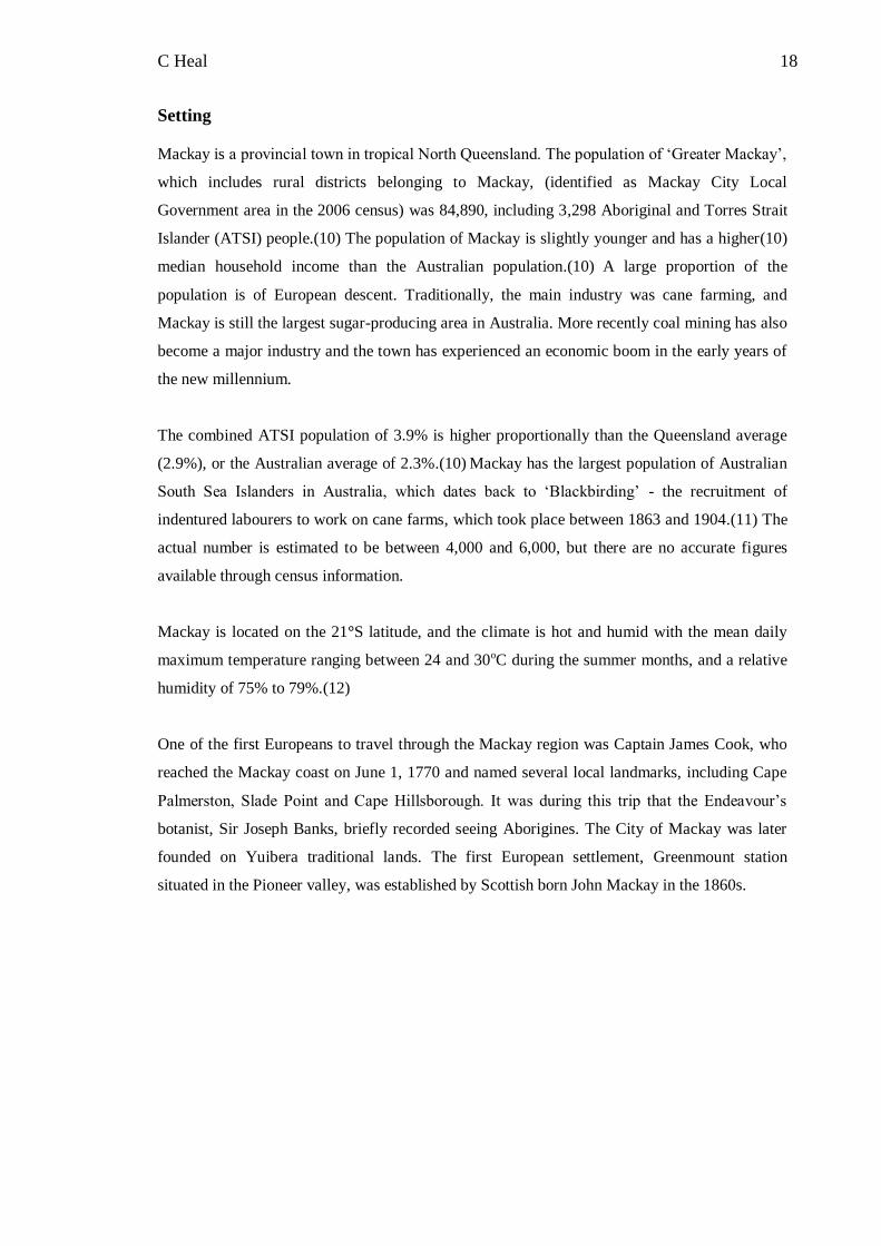
founded on Yuibera traditional lands. The first European settlement, Greenmount station
situated in the Pioneer valley, was established by Scottish born John Mackay in the 1860s.

C Heal 19
Figure 1: Greenmount Station, the first European settlement in Mackay
Townsville is a tropical city in North Queensland. The population of ‘Greater Townsville’ can
be calculated from the 2006 census by combining Townsville Statistical local area (SLA) PtB
(southern outskirts of Townsville), Townsville statistical division (SD) (Central TSV and
Thuringowa) and Thuringowa Statistical local area (SLA) PtB (northern outskirts of
Townsville/Thuringowa). The total population of this district was 154,630, including 8,530
ATSI people.(13)
Townsville’s latitude is 19.160 South and the average daily maximum temperature is
28.80C.(12) Townsville experiences an annual average of 8.4 hours of sunshine per day, and on
average there are 171 days with 10 hours of sunshine per day.(14)
Studies involved in this thesis
In 2006 a group of GPs in Mackay, of which I was chief investigator, published a randomised
controlled trial which showed that the wetting of sutures did not increase the incidence of
wound infection in minor skin cancer surgery.(7) This trial forms the background for this thesis.
Secondary analysis of data from this study is used in the second, fourth and seventh chapter of
this thesis to explore some aspects of skin cancer management by GPs. Chapter 2 describes the
incidence of and risk factors for wound infection after minor surgery.
The ‘Can sutures get wet?’ trial revealed that the incidence of wound infection was high in
comparison with similar cohorts in published studies from other geographical locations. The
proposed thesis further explores these findings and seeks to evaluate an intervention, the use of
topical chloramphenicol ointment, to reduce the infection rate. The broad hypothesis behind this
proposal is that attention to infection control improves outcome and reduces morbidity in skin
cancer surgery. Chapter 3 reports the results of this trial. Chapter 4 describes the demographics
of patients presenting with skin cancers to GPs in Mackay, the sites from which skin cancers are
C Heal 20
removed, and their histology. Chapter 7 compares the case-mix between GPs and a skin cancer
doctor in Mackay.
Between the end of 1996 and October 1999, all excised and histologically confirmed skin
cancers in Townsville, North Queensland, were recorded. Chapter 5 provides a further analysis
of these data, comparing clinical with histological diagnosis of excised and biopsied skin lesions
by Australian GPs. Another re-analysis takes place in Chapter 6 with a comparison of
“standard” histological diagnosis with the diagnosis from an internationally renowned dermato
histo-pathologist.
Chapter 8 provides overall conclusions and recommendations of the thesis.
Overall aims of thesis
1. To improve the management of skin cancer and the conduct of skin cancer surgery by
Australian GPs.
2. To increase patient well-being through the appropriate use of post-surgery wound
management.
3. To assess the ability of GPs and pathologists to diagnose skin lesions.
4. To investigate possibilities of research in GP practice settings.
Specifically, the research questions were:
1. What is the incidence of and risk factors for infection after minor surgery?
2. Does topical chloramphenicol ointment prevent infection after minor surgery in general
practice?
3. Amongst GPs in Mackay, what is the case-mix of lesions that GPs excise in the
management of skin cancer, from which body sites are they excised and what is the age
distribution of patients with skin cancer?
4. Is there a difference in the case-mix of skin cancer managed by mainstream GPs and
skin cancer GPs?
5. How accurately do GPs diagnose different types of skin cancer?
C Heal 21
6. Is there a difference between the diagnostic accuracy of a specialist dermato pathologist
and pathologists in the diagnosis of skin cancer?
Objectives
The objectives specific to each study will be discussed in the respective chapters.
Methods
The methods used will be discussed within the appropriate chapters.
Collaborations and research support
Primary health care research education and development (PHCRED): Collaboration took place
with PHCRED in order to provide research training to doctors and practice nurses. North
Queensland Practice Based Research Network has evolved from this collaboration. Mackay
evidence based medicine group collaborated in the development of the two randomised
controlled trials which were conducted in Mackay.
Professor David Weedon collaborated to the development of chapters 6 and 7.
Available funding
Awarded Chris Silagy scholarship in July 2007 for $20,000.
PHCRED provided a novice research scholarship of $17,000 to Mackay evidence based
medicine group.

C Heal 22
CHAPTER 1: LITERATURE REVIEW
1.1 Scope and limitations of literature review
1.2 Skin cancer incidence in Australia
1.3 Primary care skin cancer management in Australia
1.4 Incidence of and risk factors for infection after skin cancer surgery
1.5 Antibiotic ointments
1.6 Conclusion
1.1 Scope and limitations of literature review
The scope of the literature review with timeframes, limitations and keywords will be discussed
in this section.
1.2.1 Incidence of BCC and SCC
This section discusses the incidence of BCC and SCC in Australia since 1982 until data
collection commenced for the first study of my thesis (2004). With the exception of Tasmania,
this section is limited by lack of registry information since 1999. Comprehensive Medline
search using keywords ‘BCC, SCC, and incidence and Australia’ was used to identify studies
involving BCC and SCC. The search revealed a total of 40 articles, of which 11 were found to
be suitable
1.2.2 Incidence of melanoma in Australia
This section discusses the incidence of melanoma in Australia since 1982 until data collection
commenced for the first study of my thesis (2004). The section is limited by the time lag
between the year of melanoma occurrence, and when the data becomes available, usually
around 4 years in the case of melanoma registries. Therefore information about melanoma after
2000 has been discussed in the section on conclusions.
Comprehensive Medline search using keywords‘melanoma and incidence and Australia’ were
used. In addition, clinical practice guidelines for the management of melanoma in Australia and
New Zealand(15, 16) and Australian Institute of Health and Welfare (AIHW) cancer registry

C Heal 23
information(17) were used to obtain information for the section on melanoma. Information from
new clinical practice guidelines on the management of melanoma in Australia is used in the
section on conclusions.(16)
1.3 Management of skin cancer by Australian GPs and number needed to treat
Comprehensive Medline search was used to identify published literature regarding the number
needed to treat (NNT) by Australian clinicians from 1985 until 2006 when the manuscript in
chapter 2 was published. Keywords ‘number needed to treat’, naevus and melanoma and
Australia were used. A total of 11 articles were identified, 6 of which were found to be suitable
International literature was excluded from the search. Studies published after 2006 are discussed
in the conclusion.
1.4.2 Incidence and risk factors for surgical site infection after dermatological surgery
Comprehensive Medline search using the keywords surgical site infection, and dermatology was
used to identify internationally published material recording the incidence of surgical site
infection after dermatological surgery from 1985 to 2006, when the manuscript in chapter 3 was
published. A total of 66 articles were identified, five of which were found to be suitable.
Material regarding surgical site infection after this date is discussed in the conclusion section.
Micrographic MOHS surgery has been excluded from this section.
1.4.3 Guidelines for oral antibiotic prophylaxis for surgical wounds
Comprehensive Medline search was used to identify existing guidelines for oral antibiotic
prophylaxis for prevention of surgical site infection of dermatological surgery from 1985 to
2006, when the manuscript in chapter 3 was published. Keywords used were antibiotic
prophylaxis and dermatology. A total of 47 articles were identified, five of which were found to
be suitable. Further guidelines developed since this date will be presented in the conclusion
section. Prophylaxis for endocarditis or joint arthroplasty is not included in the scope of this
section.
1.5 Antibiotic ointment use
This sub-chapter reviews the literature available regarding antibiotic ointment and its use in
wound management. Antiseptics such as povidone-iodine, and silver are excluded from the
definition of topical antibiotic for the purposes of this section.

C Heal 24
1.5.3 Infection prevention following surgery
This section discusses the use of antibiotic ointments to prevent infection after minor surgery.
Comprehensive Medline search was used to identify internationally published randomised
controlled trials from 1985 until 2007 when data collection for my randomised controlled trial
(RCT) commenced. Keywords used were antibiotic ointment, prophylaxis and infection. A total
of 22 articles were identified, five of which were found to be suitable.
1.5.4 Infection prevention in non-surgical wounds
A comprehensive Medline search was used to identify internationally published trials
investigating topical antibiotics as infection prophylaxis in minor, non-sutured traumatic
injuries. Studies published between 1985 to 2007, when data collection for the chloramphenicol
trial commenced, was considered to be within the scope of this section. Trials published after
2007 are discussed in the conclusion. Keywords used were antibiotic ointment, prophylaxis and
infection. A total of 22 articles were indentified, two of which were found to be suitable
1.5.5 Treatment of secondary infections
A comprehensive Medline search was used to identify internationally published trials in which
antibiotic ointments were used to treat secondarily infected wounds. Studies published between
1985 and 2007, when data collection commenced for the chloramphenicol trial commenced, was
considered to be within the scope of this section. Trials published after this data are discussed in
the conclusion. Keywords used were antibiotic ointment, prophylaxis and infection. A total of
22 articles were identified, three of which were found to be suitable.
Figure 2: Malignant melanoma on foot of patient in Mackay, Qld

C Heal 25
1.2 Skin cancer incidence in Australia
1.2.1 Incidence of BCC and SCC
With the exception of Tasmania, the presence of BCC and SCC is currently not recorded by
cancer registries in Australia. In Queensland there was compulsory registration of BCC and
SCC until 1999, although no incidence rates were provided. Similarly, in most states there was
compulsory recording of the incidence of SCC and BCC until 1999. Tasmania still records the
incidence of SCC and BCC.
Incidence has been measured either in defined communities at different locations across
Australia, and nationally using a series of household surveys conducted in 1985, 1990, 1995 and
2002.
Methods of measuring incidence rates
Because there is no standardised surveillance system to determine and monitor the incidence of
BCC and SCC, reported incidence rates need to be critically viewed taking into account factors
which can affect the diagnosis of the cases. Incidence is an expression of the number of new
cases of a disease that occur in a defined population over a specific period of time, and by
convention the incidence date is defined as the date of first diagnosis although the disease may
have commenced some time previously. Therefore for BCC and SCC, newly identified skin
cancer may have been previously overlooked and have remained undiagnosed for some time.
The presence of multiple skin cancers may also affect the measurement of incidence rates, as
this may effect the numerator: conventionally only the first lesion of a particular type per patient
per given year should be included.(18) On the other hand, the incidence of all malignant lesions
can better describe the burden of the illness in the population.(19)
In Australian studies, the incidence rate also varies depending on whether, in the process of age
standardisation, world age standardised incidence rates or Australian age standardised incidence
rates are used. If Australian age standardised incidence rates are used, the incidence of skin
cancer appears to be higher as the Australian population is relatively young in comparison with
the world population. The rates are not comparable if different standard populations were used.
In addition, incidence rates vary depending on the age of the population which is used as the
denominator. In some studies(20) the entire population is used as the denominator, thus giving a
conservative estimation of the incidence of skin cancer, as the condition tends to be rare under
the age of 40. However in some studies only certain age groups are used as the denominator,
which may in contrast over-estimate the incidence of skin cancer in the population.
C Heal 26
The validity of skin cancer incidence rates also depends on biopsy rates and standardization of
histological diagnosis. Agreement between histologists regarding the diagnosis of skin cancer is
discussed later in the literature review. In addition, it is difficult to quantify how many BCC and
SCC may be treated by non-surgical procedures in Australia. When excision is not the primary
treatment, no histology is available in order to confirm the diagnosis.(21)
Studies of defined communities
The first of the studies involving defined communities was a longitudinal study conducted in
Maryborough Victoria, which took place between 1982 and 1986.(20) The study took the form
of annual skin examinations. In 1982 a database was established, of 3300 persons aged 40 and
older on the electoral register, and they were invited to participate in the study. A total of 2,669
people aged 40 years and older were recruited (81% of the eligible population). The study was
conducted for one week at the beginning of spring each year for five years (1982-86 inclusive).
Light exposed areas were examined. All participants were asked if they had undergone
treatment for any lesions in the interval in between examinations, and permission was given to
obtain medical records. The age standardised incidence rates for SCC and BCC combined was
estimated to be 873/100,000 per year. The rates for BCC were 672 per 100,000 and SCC 201
per 100,000. Because they had excluded persons under the age of 40 years, but their calculation
used the entire population as the denominator, this estimation is therefore a conservative
estimation (minimal age standardised incidence rate). The age adjusted incidence rate for
combined SCC and BCC in the 40 years or older age group was much higher: 2,152/100,000.
In the Nambour study(22) the incidence and prevalence of BCC and SCC were estimated in a
random sample of the population aged 20 to 69 years. In 1986 a random sample of 3,000
individuals were chosen from the 5,100 persons aged 20 to 69 years listed on the electoral roll
as residents of Nambour. A total of 2,095 adults attended the initial skin cancer survey in
December 1986. Dermatologists examined all participants for prevalent skin cancer – with
histological confirmation – on the head neck and upper limbs. A random sample (10%) received
full body examinations. A cohort was followed up over a 6 year period. In 1987 a postal survey
requested information about skin cancers treated from 1986 to 1987. A further postal survey
was administered in 1990, and subjects were examined for new cancers in 1992.
The incidence of BCC and SCC in 1986 was estimated to be 2,389/100,000 person-years at risk
in men and 1,908/100,000 women. In 1992 at 6 year follow up the incidence rates were 2,528

C Heal 27
/100,000 men and 1679/100,000 in women. In this study 325 patients (16%) were lost to
follow-up, which may possibly have caused selection bias and elevated the incidence rates.
Kricker et al(23) conducted a population-based, longitudinal study in Geraldton, Western
Australia. Initially a population examination study was conducted. In 1987, residents aged 40 to
64 years whose names were on the electoral roll were invited to undergo a whole body skin
examination conducted by a dermatologist.(23) Subjects thought to have skin cancer were asked
to attend their GP for definitive diagnosis, and a report of any pathology was requested.
Subjects who reported diagnosis and treatment of skin cancer in the preceding 2 years were also
asked to provide details of the treating practitioner so medical records could be obtained. The
results were age specific rather than aged standardised. Overall the estimated incident rate for
combined BCC and SCC in this age group was 1,560 per 100,000 person years. Estimated
annual incidence rate for histologically proven BCC was 1,335 per 100,000 person years in men
and 817 per 100,000 person years in females. For SCC the figures were 890 and 289 for men
and women, respectively. In order to measure the rate at which BCC and SCC develops, a
longitudinal follow-up study was then conducted. In 1989 and 1991 subjects were mailed
questionnaires about skin cancers treated in the previous 2 years. The medical records of
subjects who reported treatments were examined.
The cohort was then re-examined by a dermatologist in 1992.(24) For histologically proven
cancers from November 1987 to September 1992 the estimated excision rates per 100,000
person years were for BCC 3,379 in women and 7,067 in men, and for SCC 501 in women and
775 in men. However when only the first skin cancer of each type that occurred during follow
up were measured, the incidence rates for BCC were 2,204 per 100,000 person years in women
and 3,541 in men, and the incidence rates for SCC were 461 in women and 585 in men. As it is
more conventional to record only the first skin cancer of each type as the measure of incidence
rates, this figure has been used in Table 1.1 of comparable incidence rates, where as the higher
figure can be considered to be an excision rate.
The higher incidence rates in the second study may have been caused by close to 30% of
subjects being lost to follow-up. As it is more likely that lower risk subjects were less likely to
participate this could be a cause of selection bias.
Buettner and Raasch(4) conducted a prospective population-based survey to collect
epidemiological information on all excised and histologically confirmed skin cancers in
Townsville. Between December 1996 and December 1997, a total of 3,536 patients with 5,945
histologically confirmed skin cancer lesions were recorded. Age-standardised (world standard

C Heal 28
population) incidence rates of BCC were 2,058.3 for men and 1,194.5 for women, 1,332.3 for
men and 754.8 for women for SCC, and 49.1 for men and 41.7 for women for CM.(4)
A second study was based on data collection between January 1997 and October 1999 including
patients of all ages. A total of 6,708 patients with 13,751 histologically-confirmed NMSC were
recorded, with 38.5% of patients having multiple lesions. Yearly age standardised incidence
rates for BCC were 1,444.8 for men and 942.7 for women. For SCC the rates were 805.0 for
men and 423.6 for women. The occurrence of multiple BCCs and SCCs compromised the
estimation of incidence rates.(18) Incidence rates for excision and yearly average incidence
rates over the 3 years were also calculated.
National Household Surveys
National incidence rates of SCC and BCC have been determined in Australia in a series of
national household surveys of a random sample of the population conducted in 1985, 1990,
1995 and 2002.(2) In the first study, face-to-face interviews were conducted by a market
research company. A total of 30,976 Australians were asked whether they had ever been treated
for skin cancer, and 1,179 responding affirmatively. The treating doctor or hospital was then
approached for confirmation of the diagnosis of people who claimed to have been treated in the
previous 12 months. Respondents who reported being treated for skin cancer were asked for
permission to confirm this diagnosis with the treatment provider. In the first study the estimated
age standardised incidence rates of SCC and BCC combined in the population was 823 per
100,000. For BCC and SCC the rates were 657 and 166 per 100,000, respectively. Rates for
BCC and SCC showed a gradient with respect to latitude.
The second study by a marked research company in 1990 carried out face-to-face interviews
with a stratified random sample of the population. Medical records were checked in those who
answered that they had been treated for skin cancer. A total of 63,450 people were interviewed,
with 3,201 respondents stating that they had been treated for skin cancer and 2,879 giving
sufficient details to be followed up. There was a response from 2,341 (71%) of medical
practitioners. In this study the annual age standardised incidence rate for SCC and BCC
combined was 977 per 100,000, showing a total increase of 19% since 1985. The rates were
1,189 for men and 769 for women.
The third survey was conducted in 1995.(25) During a 12 month period 63,745 people aged
between 14 and 95 years were interviewed, with 10,841 respondents indicating that they had
ever been treated for skin cancer, and 4,671 responding that they had been treated in the

C Heal 29
previous 12 months. This was confirmed by treating physicians in 2,939 subjects. Estimated
age standardised incidence rates for BCC were 788 per 100,000, an increase of 19% since 1985.
SCC rates rose by 93% over the same period, from 166 to 321 per 100,000 inhabitants.
In the fourth and most recent survey in 2002,(26), again face-to- face interviews were used
using a stratified random sample of households. The age standardised incident rate per 100,000
populations for combined BCC and SCC was 1170: for BCC 884 and for SCC 387.
Over the duration of the four surveys, although the rates of BCC and SCC had increased since
1985, the increase was greatest for people aged 60 years and over. Rates for those younger than
60 years had stabilised.

C Heal 30
Author and year
of publication Location and year of trial Population. method SCC and BCC combined/100,000 BCC/100,000 SCC/100,000
Marks et al (20)
1989
Maryborough, Victoria
1982-86 Annual skin examinations 873 age standardised world standard population 672 201
Green and
Battistuta
1990(22)
Nambour, Queensland 1986 20-69, postal surveys and
examination
2,389 men,
1,908 women
Age adjusted incidence rates world standard
population
Green and
Battistuta
1996(22)
Nambour, Queensland 1992 20-69, postal surveys and
examination
2,528/100,000 person years at risk men
1,679/100,000 person years women
Age adjusted incidence rates world standard
population
2,074 men and
1,579 women per
100,000 person
years
1,035 men and
472 women per
100,000
person years
Kricker 1990(23) Geraldton, Western Australia 1987
40-64yrs
Whole body skin
examination
1,560/100,000.
Un-standardised incidence rates
1,335 men
817 women
890 men
289 women
English 1997(24)
Geraldton, Western
Australia
1992
40-64yrs Longitudinal
study. Postal surveys,
whole body skin
examination
2,665 women/100,000
4,126/100,000 men
Un-standardised incidence rates
Can add SCC and BCC rates as only first excision
per person was used
2,204/100,000
person years
women,
3,541/100,000
person years men
461/100,000
person years
women, 585
585/100,000
men
Buettner 1998(4)
Townsville, Australia
1996-1997
All excised skin cancers
3,390.3/100,000 men, 1,948.8/100,000 women
Age standardised incidence rates, world standard
population
Can add SCC and BCC rates as only first excision
per person was used
2,058 men
1,194 women
1,332.3 men
754.8 women
Raasch and Townsville, Australia All excised skin cancers 2,249.8/100,000 men, 1,366.3/100,000 women 1,444.8 for men and 805.0 for men

C Heal 31
Table 1.1: Studies of defined communities measuring incidence of BCC and SCC in Australia from 1982-2006
Buttner 2002(18) 1997-1999 Age standardised incidence rates, world standard population
Can add SCC and BCC rates as only first excision
per person was used
942.7 for women.
and 423.6
Giles 1988(2) National stratified sample
1985 First National survey
823/100,000
Age standardised incidence rates, world standard
population
657/100,000 166/100,000
Marks 1993(27) National stratified sample
1990 Second National survey
977/100,000
1187 men
769 women
Age standardised incidence rates, world standard
population
726 250
(25)Staples1998 National stratified sample
1995 Third National survey
1,109
Age standardised incidence rates, world standard
population
788 321
Staples 2006(26) National stratified sample
2002 Fourth National survey
1,170/100,000
Age standardised incidence rates, world standard
population
884 387

C Heal 32
Registry Information
In Australia, Tasmania has the only Cancer Registry which still records the incidence of BCC
and SCC and also the only registry which has published data from its registry.(28)
The Tasmanian Cancer Registry has published data based on population-based surveillance of
BCC and SCC from 1978 to 1987.(28) Notified cases in Tasmania include all treated BCC and
SCC with a histological diagnosis and those where treatment has been provided at the radiation
oncology clinics. A total of 8,651 NMSC were recorded in 7,160 individuals, representing an
age standardised rate of 161/100,000 per year. Ninety-four per cent of cases were based on
histological diagnosis. Incidence of BCC was higher than the incidence of SCC. The incidence
of NMSC was twice as high in men as in women. Incidence increased substantially with age,
more markedly for SCC than BCC. There was an overall increase of 7% per year in the age
standardised incidence rate of NMSC. The increase was more marked for BCC than for SCC,
and was consistent across age groups and both sexes. A first BCC or SCC during the study
period was associated with a 12-fold increase among men and a 15-fold increase in women of
development of a new BCC or SCC in 5 years. BCC or SCC was 42% of all cancer in the
Tasmanian registry.
Concluding comments
Although less dangerous and less life-threatening than cutaneous melanoma (CM), SCC and
BCC are still invasive cancers, and SCC is capable of metastasing.(29) Because there is
generally no registry style information available in Australia, and because alternate methods of
measuring incidence rates have many influencing factors, it is difficult to accurately quantify
incidence rates.
1.2.2 Incidence of melanoma in Australia
Melanoma is an increasing problem in fair-skinned populations world wide, and Australia as a
country has the world’s highest incidence of melanoma.(30) World age standardised incidence
rates per 100,000 inhabitants for CM were 40.5 for men and 31.8 for women in Australia
between the period of 1983-1999.(31) Previously as a region, Queensland had the world’s
highest incidence rates for melanoma.(32) However it was reported that in 1995, with a world
age standardised incident rate of 56.2/100,000, the Auckland population of New Zealand had
the world’s highest incidence rates of skin cancer.(32)

C Heal 33
Figure 1.1: Trends in age standardised incidence rates (ASR) of melanoma in Australia
Incidence of melanoma in Australia is recorded in population-based state registries. A national
registry exists which is a compilation of state registries.
In Australia, CM is the fourth most common cancer in males, after prostate cancer, bowel
cancer and lung cancer, and the third most common cancer amongst females after breast cancer
and bowel cancer.(33) Between 1980 and 1987 the annual incidence of invasive melanoma in
Queensland was 55.8 per 100,000 for men and 42.9 per 100,000 for women(34) based on world
standard population. The incidence of melanoma in men almost doubled over this period, and
this increase was greatest in men older than 50 years. There were similar incidence rates
documented in New South Wales, where between 1986 and 1988 the annual incidence of
melanoma was 52.5 per 100,000 for men and 42.9 per 100,000 for women.(35) The incidence of
melanoma has been decreasing in young women since the mid 1980’s but has continued to rise
in older age groups.(36)
1.3 Management of skin cancer by Australian GPs and number needed to treat
For the purpose of this section, and in concordance with current NHMRC guidelines,
Hutchinson’s melanocytic freckle (in situ melanoma) is included in the definition of melanoma
and dysplastic naevus is not considered to be malignant or pre-malignant. This is in
concordance with research by Kelly et al(37) which showed that, although patients with
dysplastic naevus syndrome had a higher absolute risk of developing CM, individual dysplastic
naevi did not have a higher risk of malignant change. The term ‘melanocytic naevi’ will be used
as a collective term to describe common naevi and dysplastic naevi.

C Heal 34
In Australia the majority of suspicious skin lesions are managed by GPs(38) and the proportion
of all skin cancers excised by GPs is increasing.(39) It is therefore important that GPs manage
suspicious lesions optimally. The decision to excise is a complex issue which is influenced by
factors such as patient concerns about malignancy, medico legal worries about missing
melanoma, experience and the likelihood from epidemiological data that a pigmented lesion
may be a melanoma.(40)
The number needed to treat (NNT) in skin cancer is often defined as the number of melanocytic
naevi that are excised per melanoma. In other words it is the ratio of pigmented lesions
(melanocytjc naevi and/or seborrhoeic keratosis) to melanoma. It is commonly used as an
indicator of quality of practice in skin cancer management (although its use as an indicator of
quality has many limitations). A 2006 study derived from analysis of billing data from skin
cancer clinics showed a NNT of 28.6.(41) However, in this study, non-pigmented benign lesions
were included in the numerator therefore elevating the NNT. It is difficult to make comparisons
in NNT as there appear to be several different definitions, however the NNT was found to be
between 11 and 29.9 for Australian GPs when based on melanomas and melanocytic naevi only
and up to 36 when seborrhoeic keratoses were included.(35, 40, 42-44)

C Heal 35
Table 1.2: Australian studies of number of benign lesions excised per melanoma
Author, State Setting, sample size Number needed to treat Numerator Comments
Burton et al 1993, Hunter region
NSW(35)
Pathology laboratory reviewed
reports of 1984 and 1988 skin
histopathology data, GPs and
specialists combined
11 naevi per melanoma 1984,
16 naevi per melanoma 1988
Total number of excised
melanocytic naevi
Number of melanoma doubled
(91 to 197), number of naevi
tripled (1,065 to 3,208)
Marks J et al (Victoria)1989-
1994(42)
Analysis of all pigmented
cutaneous lesions excised by
GPs. Histological data, 31943
pigmented lesions
29.9 all pigmented lesions per
melanoma
All pigmented lesions excised
(melanocytic lesions and
seborrhoeic keratosis, excluding
BCCs)
Ratio 11.7: 1 for dermatologists
Del Mar et al 1994 Australia
(43)
Audit 1,896 excised melanocytic
lesions submitted to pathology
service, general practice
19.4 melanocytic naevi per
melanoma, 20.6 melanocytic
naevi and lentigos per
melanoma
Total number of excised
melanocytic naevi and lentigo
NNT decreased with age of
patient;
Secondary data analysis from
available data
Del Mar et al 1995 Australia
(44)
Examination of 5,823
histopathological reports of
melanocytic skin lesions excised
during randomised field trial
17.75 melanoma per
melanocytic naevi control
group, 12.0 melanoma per
melanocytic naevi intervention
group
melanocytic naevi, Secondary data analysis from
available data
English et al 2004 Australia(40)
Retrospective audit 4,741
pigmented lesions, 468 GPs, 223
practices in Perth, Western
Australia
29 pigmented lesions per
melanoma
31 if seborrhoeic keratosis
excluded
Total number of excised
melanocytic naevi and
seborrhoeic keratosis
NNT higher for patients who are
young, female, lower socio-
economic status and recent
graduation of GP
Wilkinson et al 2006
Australia(45)
Analysis of billing data primary
care skin cancer network
28.6 benign lesions per
melanoma
All benign lesions excised from
billing data
C Heal 36
1.4 Surgical site infection
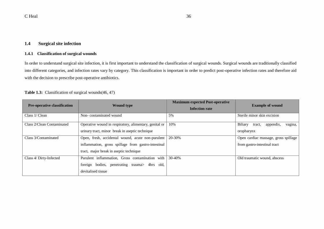
1.4.1 Classification of surgical wounds
In order to understand surgical site infection, it is first important to understand the classification of surgical wounds. Surgical wounds are traditionally classified
into different categories, and infection rates vary by category. This classification is important in order to predict post-operative infection rates and therefore aid
with the decision to prescribe post-operative antibiotics.
Table 1.3: Classification of surgical wounds(46, 47)
Pre-operative classification Wound type Maximum expected Post-operative
Infection rate Example of wound
Class 1/ Clean Non- contaminated wound 5% Sterile minor skin excision
Class 2/Clean Contaminated Operative wound in respiratory, alimentary, genital or
urinary tract, minor break in aseptic technique
10% Biliary tract, appendix, vagina,
oropharynx
Class 3/Contaminated Open, fresh, accidental wound, acute non-purulent
inflammation, gross spillage from gastro-intestinal
tract, major break in aseptic technique
20-30% Open cardiac massage, gross spillage
from gastro-intestinal tract
Class 4/ Dirty-Infected Purulent inflammation, Gross contamination with
foreign bodies, penetrating trauma> 4hrs old,
devitalised tissue
30-40% Old traumatic wound, abscess
C Heal 37
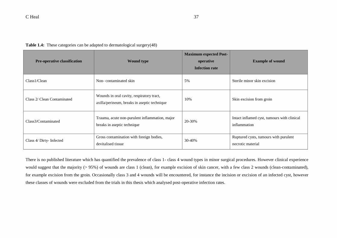
Table 1.4: These categories can be adapted to dermatological surgery(48)
Pre-operative classification Wound type
Maximum expected Post-
operative
Infection rate
Example of wound
Class1/Clean Non- contaminated skin 5% Sterile minor skin excision
Class 2/ Clean Contaminated Wounds in oral cavity, respiratory tract,
axilla/perineum, breaks in aseptic technique 10% Skin excision from groin
Class3/Contaminated Trauma, acute non-purulent inflammation, major
breaks in aseptic technique 20-30%
Intact inflamed cyst, tumours with clinical
inflammation
Class 4/ Dirty- Infected Gross contamination with foreign bodies,
devitalised tissue 30-40%
Ruptured cysts, tumours with purulent
necrotic material
There is no published literature which has quantified the prevalence of class 1- class 4 wound types in minor surgical procedures. However clinical experience
would suggest that the majority (> 95%) of wounds are class 1 (clean), for example excision of skin cancer, with a few class 2 wounds (clean-contaminated),
for example excision from the groin. Occasionally class 3 and 4 wounds will be encountered, for instance the incision or excision of an infected cyst, however
these classes of wounds were excluded from the trials in this thesis which analysed post-operative infection rates.

C Heal 38
1.4.2 Incidence and risk factors for surgical site infection after dermatological surgery
Surgical site infection following minor surgery contributes to patient morbidity and
compromises the cosmetic outcome. Most data regarding incidence and predictors of surgical
site infections are based on studies of general surgical procedures.(47, 49-51)
Of studies looking at infection rates following minor dermatological surgery, most have been
conducted in a hospital specialist setting.(52-55) The infection rates in these studies have been
between 2-3%, with higher infection rates in certain groups. One study was identified which
assessed infections in an out of hospital specialist dermatologist setting. In this study infection
rates were 2%.(52)
As the majority of skin cancer surgery takes place in general practice in Australia,(39) it is
important to study infection in this setting. General practice minor surgery may differ from a
hospital setting, with most procedures taking place in treatment rooms rather than formal
operating theatres.
The quality of evidence with regard to infection rates following minor surgery in a general
practice setting seems to be poor. A comprehensive Medline search revealed only two studies
which adequately recorded the incidence of infection following minor surgery in a general
practice setting.(56) The earliest study, conducted in South Australia, showed an infection rate
of 1.9%.(56) The second study was the ‘Can sutures get wet’ trial, which showed an overall
infection rate of 8%.(7, 57) Secondary analysis of this data to identify risk factors for infection
is the subject of chapter 2.
The acceptable rate of infection following clean minor surgery (class 1) is< 5%.(10, 47, 48, 58,
59) Even within cohorts with a low overall risk of infection, some excisions may be at higher
risk of infection because of body site, pathology or patient factors and environmental
conditions. In these high risk cases infection rate may be greater than 5%. These risk factors
(tabled below) may include excisions from the lower leg, excisions of skin cancers and
excisions from diabetic patients. Excisions from the ear and nose have also been shown to have
a high incidence of infection.(53-56)
C Heal 39
Table 1.5: Incidence and risk factors for surgical site infection following dermatological surgery prospective observational studies
Country, year Setting and sample size Study design
demographics Wound type (All class 1) Incidence of infection Risk factors for infection
Futoryan et al
1995(53)
Boston USA
Hospital department of
dermatological surgery 1,047
procedures
Retrospective audit
patient records
530 Mohs and 517
excisions
24/1047
2.29%
Mohs 13/530 2.45%
excisions 11/517 2.1%
Ear 6/48 12.5%, Ear involving
cartilage 4/14 28.5%
Sylaidis P, 1997(55)
UK
Hospital plastic surgery unit. 351
patients, 464 wounds
Prospective
observational study Clean facial surgery
13/464
2.8%
Nasal area (6.5%), auricular
area (5.2%)
Oncological surgery (12%)
Lathlean 1999(56)
Australia
General practice South Australia, 369
patients
Prospective
observational study Minor skin excisions
7/369
1.9%
4/7 infections lower leg, 6/7
age >80yrs
actual rates not given
Dettenkofer 2003
Germany(54)
German university hospital
dermatology ward, 632 inpatients,
995 dermasurgery procedures
Prospective
observational study
Minor dermatological
surgery
21/995
2.1% Excision BCC 13/172(7.6%)
Amici et al 2005
France(52)
Dermatological outpatients 3,788
procedures
Prospective
observational study,
multicentre
Minor dermatological
surgical procedures
79/3,788
2%
Males, anticoagulants,
Immunosupressants
Skin flaps

C Heal 40
1.4.3 Guidelines for oral antibiotic prophylaxis for surgical wounds
As stated above, in a general surgical setting the acceptable rate of infection following clean
surgery (class 1) is set to less than 5%,(10, 47, 48, 58) and in several studies has been shown to
be less than 2%.(52, 56) In contrast, clean contaminated wounds (class 2) have a risk of
infection of less than 10%. Therefore in a general surgical setting, antibiotic prophylaxis of
surgical wounds is usually considered optional for clean procedures, and reserved for certain at
risk patients or clean procedures that fulfil specific risk criteria, ie where the risk of infection is
greater than 5%.(50, 60)
There is debate about the role of antibiotics prior to skin lesion excision. If guidelines for
prophylaxis after general surgery are to be extrapolated to a dermatological surgery setting, then
most dermatological procedures should not require prophylaxis. Limited guidelines exist
regarding antibiotic prophylaxis of dermatological procedures.(48, 61-63) All of these
guidelines are from the United States of America, and there are currently no guidelines in
Australian practice. Most guidelines advocate the use of prophylaxis in clean-contaminated
(class 2) or contaminated wounds (class 3), but not for clean wounds (class 1). Additionally they
have a role prior to skin surgery in patients at risk of infective endocarditis and in those who
have had recent joint prosthetic surgery.(54, 67, 68, 69)
As stated in the previous sub-chapter, even within cohorts with a low overall risk of infection,
some excisions may be at higher risk because of body site, pathology or patient factors. These
risk factors are not well established and identifying these risk factors is important in order to
develop antibiotic prophylaxis guidelines.

C Heal 41
Table 1.6: Guidelines for oral antibiotic prophylaxis of surgical site infection after derma surgery
Guideline/recommendation Country Antibiotics not
required
Antibiotics indicated
for Type prophylaxis Additional recommendations
Haas et al 1995(48) Department of
dermatology, California Class 1 wounds
Class 2 (clean-
contaminated) wounds
and higher
Single oral dose Endocarditis prophylaxis
Maragh et al 2005 (61) Division dermasurgery
Mayo clinic, Minnesota Class 1 wounds
Class 2 wounds and
higher
Cephalexin 2g orally
30-60mins before
surgery.
Endocarditis prophylaxis, oral,
nasal mucosa, axillary and
anogenital lesions,
immunocompromised patients,
below knee, hand surgery
Messingham S, 2005, (62)
Department of
dermatology, Iowa City,
Iowa
Class 1 and 2
wounds Class 3 or 4 wounds Not specified
Nasal or oral cavity breached,
endocarditis
Nestor M(63) 2005 Centre for cosmetic
enhancement, Florida Class 1 wounds
Class 2 wounds and
higher
Cephalosporin or
dicloxacilliin Endocarditis prophylaxis
C Heal 42
1.5 Antibiotic ointment use
1.5.1 Background/introduction
Some general practitioners may apply topical antibiotic ointment to sutured wounds to prevent
infection after minor surgery. Although there is no data available on the frequency of this
practice amongst GPs in Australia or internationally, a survey of plastic surgeons in the UK
revealed that 66% used chloramphenicol eye ointment in their practice, mainly as prophylaxis
against infection.(64) Other uses for antibiotic ointment in general practice include for the
treatment of secondarily infected wounds,(65) otitis externa, treatment of secondarily infected
eczema(65, 66) and the treatment of impetigo.(66) Antibiotic ointments may also have a role in
accelerating wound healing in both acute and chronic situations.(67, 68)
In contrast to guidelines for the use of oral antibiotics as infection prophylaxis following minor
dermatological surgery, there are no guidelines for the use of topical antibiotics in the same
circumstances. The purpose of this sub-chapter is to review the evidence regarding the
appropriateness and effectiveness of the use of antibiotic ointment as infection prophylaxis and
possible side effects or adverse outcomes.
1.5.2 The ointments
There are several different antibiotic ointments used in clinical practice, some of which are used
more commonly in different countries. The most frequently used of these are Chloromycetin,
Neosporin and Bactroban. Chloromycetin ointment consists of 10mg/g of chloramphenicol, in
plastibase 30W and liquid paraffin.(69) Neosporin ointment is also known as triple antibiotic
ointment (TAO) in the USA. Each gram of Neosporin ointment contains polymixin B sulfate
5,000 units, neomycin sulfate 5mg and bacitracin zinc 400 units in a paraffin ointment base.(69)
Bactroban ointment contains mupirocin and naturally occurring antibiotic.
Neosporin ointment has been available over the counter in the USA since the 1970s, while it has
been confined to a prescription medication in Australia. It ceased to be available in Australia in
October 2006, because of non-availability of an ingredient.(69)
Topical ocular Chloromycetin is widely used in the UK and Australia for the treatment of
conjunctivitis, but is very rarely prescribed for this indication in the US.(70) There is little
evidence for its effectiveness in prophylaxis or treatment of wound infection. Despite this, it is
regularly used in areas outside its main indication. As well as being used for infection
prevention, the ointment has been used as an adhesive for replacement of the nailbed.(71)

C Heal 43
Table 1.7: Properties of antibiotic ointments in common use(66, 72)
Ointment
Trade name,
availability Mode of activity Range of activity Main use
Side effects/
additional
considerations
Mupirocin Bactroban Inhibitor of bacterial protein
synthesis
Gram +ve organisms,
especially staph aureus
Impetigo, elimination of staph
aureus from anterior nares
Anaphylaxis
reported(73)
Bactitracin Ingredient of TAO Interferes with bacterial cell wall
synthesis Gram +ve organisms
Impetigo, furunculosis,
pyodermas
Cross-
sensitisation
with neomycin
Polymixin B
Available singly,
combined with bacitracin
or in TAO
Disrupts bacterial cell membrane
and increases cell permability
Gram-ve organisms
including P aeruginosa,
Enterbacter and E Coli
Limited
spectrum of
activity
Neomycin Available alone, or as
ingredient of TAO
Interferes with bacterial cell wall
sythesis
Aerobic gram +ve and
gram –ve bacilli
Prevention of infection in
superficial abrasions, cuts or
burns
Allergic contact
dermatitis
Polymixin B,
neomycin and
bactitracin
TAO Combination of mechanisms Range of gram +ve and
gram –ve organisms
Prevention of infection in
superficial abrasions, cuts or
burns
Erythromycin Eryacne Inhibitor of bacterial protein
synthesis Gram +ve cocci Acne
Low incidence
of sensitisation
Chloramphenicol Chlormycetin or
Chlorsig Disrupts bacterial cell membrane
Wide range of gram +ve
and gram –ve organisms Bacterial conjunctivitis

C Heal 44
1.5.3 Infection prevention following surgery
Comprehensive Medline search identified a total of 5 randomised trials involving the use of
antibiotic ointment as prophylaxis against wound infection following surgery.
Only one study was found relating to the use of topical chloramphenicol ointment on sutured
wounds.(74) This study investigated the application of Chloromycetin ointment to wounds
following hip replacement. The incidence of superficial wound infection in the intervention
group was reduced (4% v 8%) but results should be viewed with caution as the sample size was
small and the total number of infections analysed even smaller. Therefore although the results
showed statistical significance, clinical significance would be questionable.
Two randomised studies involving Neosporin ointment were identified.(75) (76) One compared
application of Neosporin, bactracin zinc, silver sulfadiazine and paraffin ointment to
uncomplicated clean-contaminated sutured wounds in an emergency department setting.(75).
This was a positive trial which concluded that neosporin did significantly reduce the incidence
of infection (4.5% v 6.6% v 12.1% v 17.6%), however, although the sample size was large, the
number of infections involved was small and no sample size calculation was performed prior to
this study. A second small pilot study found a similar rate of wound infection and adverse
events between uncomplicated sutured soft tissue wounds treated with Neosporin ointment and
bactroban (mupirocin) ointment (4% v 0%).(76)
Bactracin zinc is one of the three antibiotic components of Neosporin ointment. An ointment
containing only Bactracin zinc was compared with white petrolatum ointment for incidence of
infection, incidence of allergic contact dermatitis and wound healing in a large randomised
controlled trial involving wounds following dermatological procedures.(77) The setting was a
general outpatient dermatology clinic and a tertiary referral advanced surgical procedure clinic.
The majority of these wounds were shave biopsies, and therefore non-sutured. Infection was
lower in the Bactracin zinc (intervention) group (0.9% v 2.0%) but, despite a sample size of 922
patients, with a total of only 13 infections, numbers were too small to be statistically significant.
In addition there was no clinical significant difference in wound healing between the two groups
on days 1, 7 and 28. There was no incidence of allergic contact dermatitis in the petroleum
group, and four cases in the bacitracin group (0.9%). The study concluded that the low rate of
infection and absence of allergic contact dermatitis in the white petroleum (control) group
indicated that white petroleum is a safe, effective wound care ointment for ambulatory surgery.

C Heal 45
A large randomised controlled trial conducted in Australia compared the application of
bactroban (mupirocin) ointment to either paraffin ointment or no ointment to post-operative
sutured wounds prior to application of an occlusive dressing.(78) Mupirocin did not
significantly decrease the incidence of wound infection compared to sterile paraffin ointment or
no ointment (2.3% intervention v 1.6% sterile paraffin, 1.4% no ointment.)
We have previously established in our literature that the rate of infection following
dermatological surgery is generally very low (1-5%). In many trials investigating the application
of antibiotic ointment after minor surgery, the total number of infections, which is the outcome
being measured, is very small. Without attention to sample size calculation, type 2 (beta) error
may occur in many of these studies, as the power is simply not sufficient to show a true
difference in infection rate. We note that the greatest total number of infections analysed to
determine the difference between two treatments was 32, and 42 when looking at the difference
between four treatments.

C Heal 46
Table 1.8: Randomised controlled trials investigating application of antibiotic ointment after skin surgery
Author and country Setting and sample
size Wound type Intervention and infection rate
Control and
Infection rate Significance
Dire et al 1995
USA(75)
Emergency department
426 army recruits.
70/30 M:F
Sutured clean
contaminated wounds
Bactitracin zinc 6/109 (5.5%)
neomycin 5/110 (4.5%)
silver sulfadiazine12/99(12.1%)
Petroleum
19/108 (17.6%)
P=0.0034
Smack et al
1996
USA(77)
Dermatology
outpatients army
medical centre
884 patients, 1,207
wounds
Minor dermatological
surgical procedures Bactracin zinc 4/444 (0.9%)
White petroleum
9/440 (2.0%)
P=0.37 (95% CE 0.4-
2.7)
Hood et al 2004
USA(76)
Emergency department
99 patients
Mean age 24.7 M:F
70:30
Sutured clean
contaminated wounds) TAO 0/59 (0%) Mupirocin 2/50 (4%)
P=0.5
Kamath et al 2005
UK(74)
Hospital inpatients
post hip replacement
92 patients, mean age
78.6yrs 72:28 M:F
Sutured (clean) hip
replacement wounds
Chloramphenicol
4/47 (8.5%)
No ointment
8/45(17.8%)
RR 0.430
95% CI 1.62-32.67
Dixon et al 2006
Australia(79)
Skin cancer surgeon
outpatients, 778
patients, 1,801 wounds
Minor dermatological
surgical procedures
Mupirocin
13/562 (2.3%)
Sterile parrafin
12/729 1.6%, No
ointment
7/510 1.4%
P=0.490

C Heal 47
1.5.4 Infection prevention in non-surgical wounds
A comprehensive Medline search identified two randomised controlled trials investigating
topical antibiotics as infection prophylaxis in minor, non-sutured traumatic injuries.
The earlier trial investigated 59 well children age 2-5 years at a day care centre in Alabama, of
which 75% were black.(80) In this randomised controlled double blinded study, triple antibiotic
ointment (TAO) was compared to petroleum control ointment. Ointment was applied three
times a day to insect bites or other minor breaks in skin. A total of 19 children developed
infections: 4/27 in the intervention group and 15/32 in the control group. The antibiotic
ointment was found to be significantly more effective than placebo in preventing impetigo in
these children (15% intervention v 47% placebo).
In the second study, a topical antibiotic gel containing Cetrimide, Bactracin and Polymyxin G
sulfate was compared to Povidone iodine antiseptic cream and a placebo ointment containing a
vehicle gel.(81) A sample group of 177 5-12 year old children attending five primary schools in
Sydney were randomised to receive an assigned topical treatment applied to minor injuries such
as scratches cuts and abrasions. There were only 9 resulting infections, 6/48 patients in the
placebo group, 2/67 patients in the povidone iodine group and 1/62 patients in the antibiotic
ointment group. The incidence of infection was significantly lower in the antibiotic gel
intervention group than the placebo ointment group (12.5% v 1.6%). Incidence of infection was
also lower in the povidone iodine group than the placebo group, (12.5%v 3.0%) but this did not
reach significance. However the number of infections in this trial was very low and the results
should be treated with caution.
Table 1.9: Randomised placebo controlled double blind studies of antibiotic ointment use on
non-surgical wounds
Setting and
sample size
Wound type Intervention and
infection rate
Control and
infection rate
Significance
P value
Maddox et al
1985, (80)
Alabama USA
Rural day care
setting, 59
children
Minor skin
trauma and
abrasions
TAO
(15%)
Petroleum
(47%)
P=0.01
Langord et al
1997(81)
Australia
5 Primary
schools in
Sydney,
Australia, 177
children
Minor
accidental
injuries
Novel gel
containing
cetrimide,
bacitracin and
polymixin B
(1.6%)
1)Placebo
ointment
(12.5%)
2)Povidone
iodine
(3.0%)
P<0.05, 95%
CI,0.011to0.2
07
C Heal 48
1.5.5 Treatment of secondary infections
A comprehensive Medline search identified three trials in which antibiotic ointments were used
to treat secondarily infected wounds (class 4). All of these trials showed topical antibiotic
ointments to be equal or superior to oral antibiotics or antiseptics in the treatment of secondary
infection.
In the earliest of these trials, Villiger et al(82) compared topical mupirocin ointment applied
three times daily with orally administered flucloxacillin or, in patients allergic to penicillin,
erythromycin. All patients were given 4 to 10 days’ treatment. The majority of infections were
impetigo or infected wounds or lacerations. Clinical response in the 101 patients with mupirocin
ointment (86% cured, 13% improved, 1% failed) was significantly better than that seen in the 19
patients treated with erythromycin (47% cured, 26% improved, 26% failed) and similar to that
with the 80 patients treated with flucloxacillin (76% cured, 23% improved, 1% failed). Overall,
the outcome for the 99 oral antibiotic patients was significantly inferior to the 101 mupirocin
patients (p<0.05). When the flucloxacillin and erythromycin data were analysed separately,
there was a significant difference between mupirocin and erythromycin (p<0.05), but not
mupirocin and flucloxacillin (p>0.05). However, numbers of patients in the erythromycin group
were small.
Bactroban cream was found to be as effective as oral cephalexin in the treatment of 706
patients with secondarily infected minor wounds.(65) Neosporin ointment was found to be
superior to antiseptics or a wound protectant in eliminating infection in blister wounds
deliberately infected with staph aureus.(83)
Leyden et al(83) produced a group of six blisters, three on each of the forearms of volunteers by
applying ammonium hydroxide, which caused a dermal blistering reaction. The wounds were
then inoculated with staphylococcus aureus. The groups of three blisters were randomised to be
treated with either TAO, no treatment (control) or a comparative treatment. The comparative
treatments consisted of antiseptics: iodine and benzalkonium chloride, thimerosal, hydrogen
peroxide, camphor and phenol, merbromin: a wound protectant (Johnson and Johnson first aid
cream): and polymixin-B-bacitracin which is an antibiotic ointment containing two of the three
antibiotics in TAO. The blister wounds treated with TAO healed significantly faster than those
treated with benzalkonium, hydrogen peroxide, camphor phenol and iodine and those receiving
no treatment. Interestingly there was no significance difference in healing times between TAO
and the wound protectant, which has no antibacterial activity). The overall clinical appearance

C Heal 49
and healing rates of wounds treated with TAO were ranked to superior to all other treatments
except the polymyxin-B-bacitracin. However it is noted that all wounds ultimately healed.
Bactroban was also found to be significantly more effective than erythromycin and as effective
as flucloxacillin in 200 patients presenting with minor skin infections (mainly impetigo and
infected wounds/lacerations) in general practice.(82)
In the most recent study, Kraus et al(65) reported the combined results of two randomised
controlled trials with a total of 53 participating centres in the US. Patients of any age presenting
with secondarily infected wounds, such as small laceration, abrasion or sutured wound were
eligible to participate. Patients were randomised to either receive topical mupirocin or oral
cephalexin for 10 days. All patients received a placebo in the alternative dosage form. A total of
706 patients were recruited in this trial, 630 completed the trial and 478 were considered
evaluable for clinical efficacy (only if they had a pre therapy pathogen identified). The primary
end point was ‘clinical response’, which was defined as complete resolution or sustained
improvement of signs and symptoms of infection. Clinical recurrence was defined as
reappearance or worsening of signs and symptoms of infection, and unable to determine was
defined as the inability to make a valid assessment of the clinical outcome. Results using
patients evaluable for clinical assessment showed a successful clinical outcome in the mupirocin
group of 233/45 95.1%, and 22/233 95.3% in the cephalexin group. Intention to treat results
were 296/357 (82.9%) in the mupriocin group and 289/349 (82.8%) in the cephalexin group.

C Heal 50
Table 1.10: Randomised controlled studies investigating antibiotic ointment in the treatment of infected wounds
Setting and
sample size Blinding Wound type
Intervention
and outcome Control and outcome Outcome
Villiger et al
1986(82)
Primary care
200 patients Unblinded
Impetigo/ infected
lacerations
Mupirocin
86% cured
1)Flucloxacillin
76% cured 2)erythromycin
47% cured
Response to mupirocin
ointment significantly better
(p<0.05) than erythromycin
and not significantly
different to (p>0.05)
flucoxacillin
Leyden et al
1987(83)
Human –model
study Unblinded
Blisters intentionally
inoculated with staph
aureus
TAO
Mean 9 days to healing
No treatment mean 13.3 days
to healing
Wounds treated with TAO
healed significantly faster
p<0.05
Kraus et al
1998
USA(65)
Primary care
706 patients
between two
identical RCTs
Double blinded
Secondarily infected
lacerations and
abrasions
Mupirocin
95.1% cure rate
Cephalexin
95.3% cure rate
Significantly equivalent
p=0.89, 95%CI-4.0%-3.6%

C Heal 51
1.5.6 Other uses for antibiotic ointment in wound management
It is worth noting that inert petroleum, the vehicle in most antibiotic ointments, may have
wound healing and infection prevention properties.(77, 84) This is likely to be caused by liberal
application as this provides a degree of occlusion, and occlusive dressings have been shown to
shorten wound healing time(85) and provide a barrier against physical infection. Inert petroleum
also prevents adherence to dressings.(77) Studies have also shown antibiotic ointments
accelerate wound healing times.(86)
Neosporin ointment has been found to be safe, effective and significantly better than simple
gauze dressing alone, in minimising the appearance of scars resulting from clean dermabrasion
wounds.(68). Topical antibiotics may also be effective in aiding the healing of chronic wounds
and ulcers.(87)
1.5.7 Antibacterial effects and antibiotic resistance
In one study, antimicrobial treatment including antiseptics and antibiotic ointment were shown
to eradicate bacteria from the skin surface when applied topically.(88) Mupirocin and TAO
ointment were also shown to eradicate bacteria from the multiple keratinised layers of the
stratum corneum and in addition mupirocin prevented repopulation with resident flora.(88)
There is some concern regarding the overuse of topical antibiotics resulting in antibiotic
resistance. Australian guidelines suggest that topical antibiotic use be restricted because of the
capacity of most topical drugs to select resistant micro-organisms and to cause sensitization.(66)
The guidelines also suggest that antimicrobials recommended for topical use are selected from
classes not in use for systemic therapy.(66)
There is a contrary argument that the potential for antimicrobial resistance with topical
antibiotics is actually lower than with systemic antibiotics because of the higher local
concentration achieved by topical delivery.(72) Patterns of antimicrobial activity and resistance
have been examined in vivo for neomycin and mupirocin containing ointments. In one trial,
neomycin plus bactracin was associated with the rapid emergence of drug- resistance organisms
in vitro whereas topical silver nitrate was not.(89) However Neosporin ointment has also been
shown to have a wider spectrum of antibacterial activity compared with mupirocin and to be
usable against mupirocin resistant Gram-positive strains.(90) Chloramphenicol eye drops have
been shown to be effective in the treatment of MRSA ocular surface infections.(91)

C Heal 52
There is no evidence after more than three decades of extensive use worldwide that, with the
exception of mupirocin, topical antibiotics administered on an outpatient basis contribute to any
emerging resistance pattern. Increased use of mupirocin has been associated with the emergence
of resistance, which has been reported both for MRSA (methicillin resistant staphylococcus
aureus) and MSSA (methicillin susceptible staphylococcus aureus). The problem seems to be
associated with prolonged treatment.(92, 93)
1.5.8 Adverse effects
Contact dermatitis
There is some concern about the incidence of allergic contact dermatitis with topical neomycin
use. This has been shown to be as high as 11% in a population referred for diagnostic patch
testing.(94) However there is some evidence that the incidence of this reaction is as low as 1%
when the ointment is used in the general population.(95) The reaction is much more common
amongst subjects previously exposed to neomycin ointment.(95) Contact allergy has been
reported with the use of chloramphenicol ointment but the incidence is thought to be low.(96,
97)
Other adverse outcomes
There is some controversy regarding the link between aplastic anaemia and topical ocular
chloramphenicol.(98, 99) There have been a small number of case reports, most of which
involved prolonged use.(70) However two international case control studies provided no support
to the claim that chloramphenicol eye drops increase the risk of aplastic anaemia.(100) Previous
literature provided only weak support to causality based on a few single case reports. Although
an association between ocular chloramphenicol and aplastic anaemia cannot be excluded, the
risk was estimated to be less than one per million treatment courses.(100) As the baseline
incidence of aplastic anaemia in the population is three per million it is difficult to establish any
causality.(101)
There has been a report of anaphylaxis in one patient following the application of bactroban
ointment.(73)
1.5.9 Conclusion: Should we use topical antibiotics to sutured wounds to prevent
infections?

C Heal 53
Antibiotic prophylaxis is probably prescribed excessively and inappropriately following
dermatological surgery,(49, 62) and is thought best reserved for high risk patients. In contrast to
the existing guidelines for the use of oral antibiotics prior to dermatological surgery discussed in
Chapter 1.5, there are no existing guidelines for the prophylactic use of topical antibiotics.
Despite widespread use, there is very little evidence available regarding the effectiveness of
antibiotic ointment in preventing infection following minor surgery. One reason for the lack of
evidence is, that the low risk of infection after clean surgery means that studies of more than
1,000 procedures (sometimes many more) are required to reliably detect a reduction in
infection.(102) There is some evidence of a 50% reduction in risk from use of prophylactic oral
antibiotics following clean surgery.
Even if more evidence for efficacy became available, the decision to prescribe antibiotic
prophylaxis is complicated and in addition to efficacy, antibiotic costs, adverse effects and
resistance should be taken into account. However, in some circumstances, topical delivery of
antibiotic may be preferably to systemic administration.
If evidence for effectiveness becomes available, it might be justified to consider prophylaxis
with antibiotic ointment in situations where incidence of infection is expected to be greater than
5%.
1.6 Conclusion of literature review
This literature review firstly reviewed the incidence of BCC, SCC and melanoma in Australia,
in order to set the background of this thesis. Although less dangerous and less life-threatening
than cutaneous melanoma, SCC and BCC are still invasive cancers, and SCC is capable of
metastasing.(29) Because there is generally no registry style information available in Australia,
and because alternate methods of measuring incidence rates have many influencing factors, it is
difficult to accurately quantify incidence rates. Australia has one of the world’s highest
incidences of melanoma, although accurate data are limited by time delays in reporting
incidence rates.
A review of number needed to treat (benign and dysplastic naevi excised per melanoma), often
used as a marker of quality of skin cancer management, was found to be between 11 and 29.9
for Australian GPs when based on melanomas and naevi only and up to 36 when seborrhoeic
keratoses were included.(35, 40, 42-44) A review of studies recording incidence of surgical site

C Heal 54
infection after dermatological surgery showed an infection rate of 1.9-2.8%, with higher rates of
infection in certain sub-groups. Finally, most studies of antibiotic ointments used in the
prophylaxis of infection after clean dermatological surgery found no evidence for efficacy,
although a study of clean-contaminated wounds showed some significant difference in infection
rates. It was discussed that several of these studies may have been significantly under-powered.
There appears to be more evidence for use of antibiotic ointment in the treatment of secondary
infections.
This literature review illustrated that there is a high incidence of skin cancer in Australia and in
particular North Queensland. It is therefore important that skin cancer is managed optimally by
Australian GPs. This led to the thesis focusing on surgical management and diagnosis of skin
cancer.
The literature review revealed that there is limited published data which identifies groups at
high risk of infection after minor surgery. It is important to establish these high risk groups in
order to optimize antibiotic prophylactic management.

C Heal 55
CHAPTER 2: RISK FACTORS FOR WOUND INFECTION AFTER
MINOR SURGERY
2.1 Background
This chapter analyses the incidence of infection following minor surgery (class 1) conducted by
general practitioners in Mackay, and compares this incidence with other studies involving
similar patient groups from different geographical locations. Multivariate analysis examines risk
factors for infection. The published article appears in Appendix 1.(57)
In the introduction it has been discussed that there is limited data regarding surgical site
infection after dermatological surgery and limited data regarding risk factors for infection.
There is debate about the role of antibiotics prior to skin lesion excision, and also limited
guidelines about this issue. A better knowledge of risk factors for surgical site infection is
therefore required in order to predict circumstances in which a high incidence of infection is
likely.
It is a secondary analysis of data generated from the ‘Can sutures get wet’ intervention study
described in the introduction.
2.2 Introduction
Surgical site infection following minor surgery contributes to patient morbidity and
compromises the cosmetic outcome. Most data regarding incidence and predictors of surgical
site infection are based on hospital studies.(47, 49, 51) Of studies looking at infection rates
following minor dermatological surgery in an out of the hospital setting, most have been
conducted in specialist dermatology clinics.(52-54) On the other hand, the quality of evidence
with regard to infection rates following minor surgery in a general practice setting seems to be
poor(103) and a comprehensive Medline search revealed only one study which adequately
recorded the incidence of infection following minor surgery in a general practice setting.(56)
In north Queensland, the majority of suspicious skin lesions are managed by general
practitioners (GPs),(38) particularly in rural centres such as Mackay where there is no resident
dermatologist. Skin excisions form a large proportion of a typical Australian GPs workload,
and this proportion is even greater for Queensland GPs.(5) There is evidence that performing
minor surgery in general practice is cost effective compared with a hospital setting.(104) It is

C Heal 56
therefore important that there is adequate data available regarding incidence of and risk factors
for complications such as infection following minor surgery in a general practice setting.
The data from the present study was collected incidentally as part of a randomised controlled
trial which compared the standard management of keeping wounds dry and covered with
allowing wounds to be uncovered and wet in the first 48 hours following minor surgery.(7) As
both arms of the trial showed equivalent infection rates at the 5% significance level (8.4% in the
intervention group and 8.9% in the control group) they have been considered as a single group
for the purpose of this study and analysed together. Explicitly, both arms of the study, both the
control and the intervention group, were considered as a single group and analysed together in
this study. The aims of this study were to determine the incidence of and risk factors for surgical
site infections following minor skin excisions in a primary care setting.
2.3 Methods
Our prospective study of patients presenting for minor skin excisions was conducted initially as
a randomised controlled trial. It is important to describe the methods used for this trial in order
to fully understand the secondary analysis of the data that was generated for this chapter. I will
therefore now provide a detailed description of the methods used.
2.3.1 Study design
We carried out a randomised controlled, multi-centre trial involving patients presenting for
minor skin excisions.
2.3.2 Setting and participants
Nineteen general practitioners (GPs) from four practices in the Mackay area, tropical North
Queensland, Australia (latitude 21°S; inhabitants of Mackay area approximately 75,000)
participated. The GPs were a self-selected group who attended a monthly evidence based
medicine meeting. Data collection took place from October 2004 to May 2005. Consecutive
patients presenting for minor skin excisions were invited to take part in the trial. Practice nurses
were responsible for recruiting patients and collecting data. Demographic information was
collected regarding all patients, as well as clinical information regarding presence or absence of
diabetes, or any other significant medical condition (such as peripheral vascular disease,
anaemia, chronic obstructive airways disease). A body site map was used to define excision site.
At the end of the study practice nurses were asked to re-examine computer records to fill in any

C Heal 57
missing data. The principal researcher visited participating GPs and practice nurses to provide
training and ensure that recording was standardised.
All patients participating were provided with an information sheet, and were asked to give
signed consent. Patients consenting to participate were given written instructions regarding
post-operative wound care.
2.3.3 Eligibility criteria
All patients presenting to a participating GP for ‘minor skin excision’ except for skin excisions
on the face were eligible to participate in the study. Patients who were already taking oral
antibiotics, or who were on immunosuppressive drugs were excluded from the study. Further
exclusion criteria were lacerations, having a flap or 2 layer procedure (tying a ‘bleeder’ did not
count as 2 layer), excision of sebaceous cyst, and skin excision on the face. These exclusions
were made in an attempt to standardise the type of wound being studied, and reduce the number
of confounding factors.
Patients whom oral or topical antibiotics were clinically indicated immediately postoperatively
were also excluded from the study. Specifically, these were patients for whom antibiotics were
indicated because of bacterial endocarditis prophylaxis, joint replacement,(105) or patients for
whom antibiotics were usually prescribed when they had previously undergone a skin excision.
For the purposes of this criteria, ‘usually’ was defined as in 50 >% of previous skin excisions.
At the time of this trial no guidelines existed for the use of antibiotics for infection prophylaxis
after minor surgery, so we therefore had to decide on our own criteria for this purpose. The
results of our trial have now been used to develop criteria for this medical situation.
2.3.4 Surgical wound management protocol
A workshop took place attended by participating GPs to develop guidelines to ensure that
excisions were managed in a standardised manner. The following procedure was agreed upon:
1. Skin preparation – normal saline
2. Usual sterile technique (standard precautions) including sterile gloves
3. Local anaesthetic - type and volume recorded
4. Suture material – nylon – size recorded

C Heal 58
5. No antibiotics, either topical or oral- (if required, or already prescribed exclude from
study). No topical antiseptics eg betadine or alcohol. No antiseptic washes or medicates
soaps.
6. Dressing type – melolin and tape
7. Removal of sutures (ROS) according to site:
o Back – 10 days
o All other sites – 7 days
2.3.5 Intervention
Patients were provided with oral and written instructions regarding post-operative wound
management. The dry group were asked to leave the dressing on and keep dry for the first 48
hours, then bathe and undress as normal until sutures were taken out (Figure 2.1). They were
asked to avoid the use of antiseptic washes or soaps.
The wet group were asked to take the dressing off within the first 12 hours then bathe as normal
until the sutures were taken out. It was felt that patients should leave the GP surgery with a
dressing to absorb immediate bleeding, however it was also felt that defining an exact time to
remove the dressing would be unrealistic, so ‘within 12hrs’ was considered a reasonable
request. They were also asked to avoid the use of antiseptic washes and soaps.
Figure 2.1: Wound management
Wound Management
Bathe as normal until sutures taken out
Avoid use of antiseptics
Take dressing off within first 12 hrs
Wet
After 48hrs take dressing off and bathe as usual
Avoid the use of antiseptics
Keep dressing on for 48hrs
Keep wound dry for 48hrs
Dry
patient
2.3.6 Clinical outcomes
Wounds were assessed for infection by a practice nurse or doctor on the day of removal of
sutures or sooner if the patient re-presented with a perceived infection. Our definition of wound
infection was adapted from standardised surveillance criteria for defining surgical site infections
(SSI) developed by the Centre for Disease Control’s National Nosocomial Infection

C Heal 59
Surveillance System (Table 2.1).(49) All participating doctors and nurses were briefed
regarding the definition of infection by the primary researcher, and were also given written
information.
Table 2.1: Definition of surgical site infection
1) Infection must be within 30 days of excision
2) a) There must be purulent discharge from the wound or
b) The GP must diagnose a wound infection
or
c) The GP commences antibiotics
3) Stitch abscess must not be counted as an infection
2.3.7 Sample size
Sample size was calculated on the basis of a pilot study conducted February to June 2004
involving 543 patients, which showed an overall infection rate of 5.7%. Based on a projected
infection rate of 5%, we decided that an increase in incidence of infection of 5% would be
clinically significant. To come to this conclusion with statistical confidence a power of 80% and
a significance level of 0.05 using a one-sided equivalence test of proportions, a total of 357
patients were required in the intervention group and 357 patients in the control group.
2.3.8 Randomisation
All patients provided written informed consent before enrolling in the study. After agreeing to
participate, patients were randomised by picking a ball out of a hat. This method of
randomisation was chosen by the practice nurses because of ease and acceptability. The practice
nurse enrolled patients and assigned participants to their groups. No blinding took place.
2.3.9 Statistical analysis
We based all analysis on the intention-to-treat principal. Differences between categorical
variables were compared using Chi-square tests. Numerical and categorical variables were
compared using unpaired t-tests. To decide on non inferiority the one-sided 95%-confidence
interval of the difference of infection rates was calculated and compared with the maximum
allowable difference of 5%. P-values less than 0.05 were considered to be statistically
significant.

C Heal 60
Both arms of the study, both the control and the intervention group, were considered as a single
group and analysed together in this study. The statistical analysis takes the cluster sampling
design (four doctors’ practices) into account. Incidence of infection is given together with a
95%-confidence interval (95%-CI). Numerical data was summarized using mean values and
standard deviations ( SD) when approximately normally distributed or median values and
inter-quartile ranges (IQR) when skewed. Bivariate comparisons were conducted using Chi-
square tests (two categorical variables) and unpaired t-tests or non-parametric Mann-Whitney
Wilcoxon tests (categorical and numerical variable). Generalized linear modelling was used to
identify independent risk factors of infection after minor surgery. Relative risks together with
95%-confidence intervals were estimated using binomial distribution and the logarithmic link
function. All variables that were not part of the final model (that is: age, gender, month excision
took place, management of wound, and significant medical condition other than diabetes) were
assessed for potential confounding of the relationships between body-site of skin lesion,
histology of skin lesion, and diabetes with wound infection. Throughout the analysis p-values
less than 0.05 (two-sided hypotheses) were considered to be statistically significant. The
statistical analysis was conducted using SPSS for Windows, release 12 and STATA for
Windows, release 8.
2.3.10 Ethics
Ethics approval was granted by the James Cook University ethics committee - Approval number
H1902.
Having described the methods of the ‘Can sutures get wet’ trial, I will now continue to describe
the statistics used in the secondary data analysis of the current chapter.
2.4 Results
2.4.1 Practice and study characteristics
Participating GPs were younger (median age 44 years) and more predominantly female (64%)
than average for Australian GPs (modal age category 45-54 years; 32% female). Of the total of
1,247 patients who attended for skin excisions during the collection period from October 2004
to May 2005, 377 patients were excluded (294 were ineligible and 83 non-participants) (Table
2.2).
There were no significant differences in the age (participating: 56.3 years of age (± 16.5); not
participating: 58.1 years of age (± 16.2); p=0.208) and gender (participating: 47.6% female; not

C Heal 61
participating: 44.9% female; p=0.407) of participating and excluded patients. A total of 13
participants were eventually lost to follow up. Follow up was completed in 857 (98.5%)
patients.

C Heal 62
Table 2.2: Reasons for exclusion of 377 patients from study
Reason N % Of
excluded
Ineligible Face 256 20.5
Biopsy 10 0.8
Flap 17 1.4
Immunosuppressed 1 0.1
Antibiotics 5 0.4
Not returning for removal of
sutures**
5 0.4
Non-participation Refused 67 5.4
Forgot* 16 1.3
Total 377 30.3
*The practice nurse forgot to invite these patients to participate;
**These five patients knew that they would not be able to return for the removal of sutures.
2.4.2 Infections
Infection occurred in 74 of the 857 excisions (8.6%; 95%-CI = [3.5, 13.8]) (Table 2.3). Infection
rates for the four centres were 2.9%, 7.8%, 10.0%, and 10.2% (p=0.0496). Of all characteristics
recorded, only the presence of diabetes was significantly correlated with the higher incidence of
infection (18.2% compared to 8.4%; p=0.019) (Table 2.3). Participants older than 60 years had a
higher incidence of wound infection (12.4%) compared to younger participants (5.6%), however
this difference was not significant (p=0.056). Squamous cell carcinomas (SCCs) were most
prone to be infected (13.5%), while benign naevi (2.5%) and seborrhoic keratoses (0%) were
least likely to become infected. Of the 74 infections, 25 (33.8%) occurred on the lower leg
(below knee) or foot. There was little variation in incidence of infection with the month of year
in which the excision took place with no evidence of increased infection during the hotter wet
season (p=0.527). There was no significant difference in time to removal of sutures between the
infected and non-infected groups (median time 8 days, IQR = [7, 10] for both groups, p=0.538).

C Heal 63
Table 2.3: Bivariate correlates between infection after minor surgery and participants’ and
lesions’ characteristics in 857 patients recorded in Mackay, Australia
Characteristic Incidence of infection
(74/857)
p-value
Age
≤ 40 years
41 – 50 years
51 – 60 years
61 – 70 years
> 70 years
6.2%
4.8%
5.8%
12.2%
12.7%
p = 0.057
Gender
Male
Female
10.2%
6.9%
p = 0.135
Body site
Scalp and neck
Trunk
Arms and hands
Thighs
Legs and feet
10.6%
5.3%
6.8%
14.0%
15.0%
p = 0.072
Histology of lesion
Melanoma
Basal cell carcinoma
Squamous cell carcinoma
Benign naevus
Dysplastic naevus
Seborrhoic keratosis
Solar keratosis
Other
7.7%
11.4%
13.5%
2.5%
10.5%
0%
9.6%
3.4%
p = 0.118
Date of excision
Wet season (December to February)
Dry season
9.3%
8.3%
p = 0.527
Presence of diabetes
No
Yes
8.4%
18.2%
p = 0.019
Presence of other medical condition* p = 0.324

C Heal 64
No
Yes
8.3%
22.2%
*Medical conditions recorded were COPD(8), anaemia(1), ‘aspirin’(2), ’steroids’(3), ’warfarin’
(2), ischaemic heart disease (1), and peripheral vascular disease (1). Results were adjusted for
the cluster sampling design.
Multivariable generalized linear modeling showed that the body sites legs and feet (p=0.009)
and thighs (p=0.005), the histological subtypes BCC (p=0.006), and SCC (p=0.002), and
prevalence of diabetes (p<0.001) were independently correlated to wound infection (Table 2.4).
Table 2.4: Correlates of participants’ and lesions’ characteristics with infection after minor
surgery. Results of multivariable generalised linear modeling based on 857 patients from
Mackay, Australia
Characteristic Without
infection
(n=783)
With
infection
(n=74)
Relative
risk
95%-CI* p-value
Age
≤ 40 years
41 – 50 years
51 – 60 years
61 - 70 years
> 70 years
151
139
162
166
165
10
7
10
23
24
1
0.67
0.69
1.4
1.2
[0.37, 1.2]
[0.42, 1.1]
[0.66, 2.8]
[0.62, 2.5]
0.190
0.139
0.406
0.534
Gender
Female
Male
380
403
28
46
1
1.3
[0.84, 2.0]
0.232
Histology
All other
BCC**
SCC***
467
124
192
28
16
30
1
2.1
1.8
[1.3, 3.4]
[1.3, 2.6]
0.004
< 0.001
Body site
All other
Legs and feet
Thighs
598
142
43
42
25
7
1
1.9
2.2
[1.1, 3.1]
[1.3, 3.6]
0.019
0.002
Diabetes Mellitus

C Heal 65
No
Yes
765
18
70
4
1
1.7
[1.4, 2.2]
< 0.001
Date of excision
Dry season
Wet season
521
262
47
27
1
1.1
[0.72, 1.6]
0.723
Presence of other
medical condition#
No
Yes
769
14
70
4
1
2.2
[0.43, 10.9]
0.353
Wound management
Dry group
Wet group
378
405
37
37
1
0.95
[0.78, 1.1]
0.577
*95%-CI = 95%-Confidence interval; **BCC = basal cell carcinoma; ***SCC = squamous cell
carcinoma. # Other medical conditions recorded were COPD(8), anaemia(1), ‘aspirin’(2),
’steroids’(3), ’warfarin’ (2), ischaemic heart disease (1), and peripheral vascular disease (1). The
model was adjusted for the cluster sampling design and for the confounding effects of age and
gender of the participants. Date of excision, presence of other medical condition, and wound
management were not part of the model.
2.5 Discussion
The results of the present study suggest that diabetes, excisions from the lower leg and
foot or thighs, and excisions of non-melanocytic skin cancer (SCC and BCC) are
independent risk factors for infection after minor surgery. The latter finding is
consistent with a study conducted in a specialist dermatology clinic setting which
suggested that oncological surgery (excision of skin cancer) is associated with a higher
risk of infection.(55) Body extremities, with reduced blood supply have also previously
been associated with a higher incidence of infection.(55)
The overall incidence of infection in our study of 8.6% was higher than we expected
from published literature looking at a similar general practice cohort (1.9%)(56) and a
similar dermatology clinic cohort (2%).(52) However exclusion of facial excisions from
our study, which may have a lower incidence of infection,(55) may have falsely
elevated our overall incidence of infection. Excluding those for whom antibiotics would

C Heal 66
otherwise have been indicated post-operatively would, however, have lowered the
infection rate. A German study in a university medical centre setting reported a more
comparable infection rate of 8%.(54) It is difficult however to compare the infection
rate between different studies as different variables and methods were used.(53)
The present study did have several limitations. There are various characteristics
influencing the occurrence of infections and although information on as many variables
as possible was recorded, it proved difficult to ensure that all possible predictors of
infection were recorded. There was inadequate data recorded on suture size and
occupation, and consequently, these factors could not be compared. We did not record
smoking, which may be a risk factor for surgical site infections.(49, 106) We also did
not record the size of lesion excised, excision margins or overall wound area, and
therefore are unable to exclude that the increase in incidence of infection recorded for
SCCs and BCCs could be related to the size of the overall wound area rather than the
histology. However there is some evidence that more complicated procedures (flaps or
skin grafts) are associated with an increased infection rates rather than the size of the
excision.(52) Exclusion of facial excisions and more complicated surgery such as flap
or two level procedures prevented analysis of infection rates in these subgroups, and
subsequent comparisons.
Although diabetes was found to be independently correlated to wound infection, the
prevalence of diabetes as well as other medically important conditions was probably
under-recorded, and power was limited to analyse these subgroups.
Surgical training and technique of the GPs involved is a potential confounder which would be
difficult to quantify and was not recorded.
The diagnosis of infection, even when using guidelines, is still subjective and it has been shown
to have inter and intra-observer variation.(107) The definition of infection we used certainly has
limitations, however it is the most widely implemented standard definition of wound
infection(107) and was as close to a gold standard as we could find. We have no evidence to
support intra and inter-practice reproducibility of measurement and recording procedures.

C Heal 67
In addition, there are some limits to generalizing these findings. The GPs involved were not
representative of Australian GPs, being younger and more predominantly female.(108) The
population of Mackay is slightly older and has a lower median household income than the
Australian population.(109) Mackay is a provincial town in tropical north Queensland and has a
hot and humid climate with the mean daily maximum temperature ranging between 24.2 and
30oC during the summer months, and a relative humidity of 75-79%.(110) It is possible
therefore that these findings may not be generalize-able to a temperate climate, although there is
no published evidence that heat and humidity increase infection rates.
Antibiotic prophylaxis is probably prescribed excessively or inappropriately for dermatological
surgery(49, 62) and is thought to be best reserved for high risk patients.(10, 62) There is no data
available on the current prescribing habits of Australian GPs regarding antibiotic prophylaxis
for minor excisions. Although there is also no evidence available regarding what reduction in
the rate of infection we might reasonably expect from the use of prophylactic antibiotics for
minor excisions, there is some evidence of a 50% reduction in risk of infection when
perioperative antibiotic prophylaxis is used following clean surgery.(102) In addition to
efficacy, antibiotic costs, side-effects and resistance must be considered when considering their
use prophylactically. However results of this study could encourage the more judicial use of
prophylactic antibiotics by defining high-risk groups for infection in a general practice setting,
such as diabetics and those undergoing excision of a non-melanocytic skin cancer or excision
from the lower limb. Alternatively, other non-pharmaceutical interventions aiming to reduce
infection rate could be targeted towards these high risk groups.
2.6 Summary
This study found a high overall incidence rate of infection of 8.6%.
Excisions from lower legs and feet or thighs excisions of BCC or SCC and diabetes (p<0.001)
were found to be independent risk factors for wound infection. The results of this study could
encourage the more judicial use of prophylactic antibiotics by defining these high-risk groups in
a general practice setting, such as diabetics and those undergoing excision of a non-melanocytic
skin cancer or excision from the lower limb.
The high incidence of infection in this trial provided stimulus to study an intervention to prevent
surgical site infection, as an effective intervention would be useful to reduce this high rate of
surgical site infection (see chapter 3). Additionally, the large number of infections occurring

C Heal 68
following minor surgery conducted by general practitioners in Mackay would mean that
attaining a meaningful sample size according to a sample size calculation designed to avoid type
2 errors is feasible.

C Heal 69
CHAPTER 3: DOES APPLICATION OF CHLORAMPHENICOL
OINTMENT TO SUTURED WOUNDS DECREASE
THE INCIDENCE OF WOUND INFECTION?
3.1 Background
The literature review demonstrated that there is insufficient evidence for the efficacy of topical
antibiotic ointment in the prophylaxis of wound infection after minor surgery.
This chapter describes a large randomised controlled trial which explores the effectiveness of an
intervention, topical chloramphenicol ointment to reduce infection after minor surgery
conducted in general practice. The published article forms Appendix 2.(111)
The objective of the trial was to establish evidence for the application of antibiotic ointment to
sutured wounds. If the chloramphenicol ointment was found to be effective this could
potentially decrease the incidence of infection following minor surgery in general practice. If
the ointment was found not to be effective this could decrease the over use of topical antibiotic
ointment and consequence adverse outcomes.
It was also hoped that this study would benefit GP researchers by providing more familiarity
with the process of undertaking a practice based clinical trial. It was also hoped that it would
benefit general practice knowledge by establishing evidence for the application of antibiotic
ointment to sutured wounds.
3.2 Introduction
Chloromycetin® ointment consists of 10mg/g of chloramphenicol, in plastibase 30W(112) and
soft white and liquid paraffin.(69) Chloramphenicol has a broad spectrum of activity against
gram positive and gram negative bacteria, rickettsias and chlamydia.(66) Chloramphenicol
ointment is indicated for treatment of bacterial conjunctivitis; however there is little evidence
for its effectiveness in prophylaxis or treatment of wound infection. Despite this, it is regularly
used in areas outside its main indication. Prior to our study, several of the investigating GPs had
applied it to sutured wounds as prophylaxis against wound infection. A survey of UK plastic
surgeons reported that 66% used chloramphenicol eye ointment in their practice, mainly as
prophylaxis against infection.(64) The ointment has been used as an adhesive for replacement of

C Heal 70
the nailbed.(71) A comprehensive Medline search found only one other study relating to the use
of topical chloramphenicol ointment on wounds. This study investigated the application of
chloramphenicol ointment to wounds following hip replacement.(74) The incidence of wound
infection in the intervention group was reduced (4% versus 8%) but the sample size was small
and the results not statistically significant.
Topical ocular chloramphenicol is widely used in the UK and Australia for the treatment of
conjunctivitis, but is very rarely prescribed for this indication in the US.(70) There was
previously some controversy regarding the link between aplastic anaemia and topical ocular
chloramphenicol, based on a small number of single case reports,(70) however two international
case-control studies provided no support for this association. Although the association between
ocular chloramphenicol and aplastic anaemia cannot be excluded, the risk is less than one
million per treatment course.(100) There have been no incidences of aplastic anaemia following
dermatological application reported, despite widespread use.
A prior study of wound infection following minor surgery involving GPs in Mackay,
Queensland, showed an overall incidence of wound infection of 8.6%.(7, 57) This incidence
was higher than expected based on published results of a similar Australian general practice
cohort (1.9%),(56) a skin cancer clinic cohort (1.5%)(113) and of a European dermatology
clinic cohort (2%).(52) The acceptable rate of infection following clean minor surgery is
suggested as less than 5%.(47, 48, 58) A low risk of infection after clean surgery means that
studies of more than 1,000 procedures (sometimes many more) are required, under normal
circumstances, to detect a reduction in infection from an intervention with statistical
confidence.(102) Because of the high incidence of infection in our patient cohort, and high
minor surgery workload,(114) we decided to utilise the increased capacity to investigate a
strategy to reduce infection rate. The present trial sought to establish the effectiveness of topical
chloramphenicol ointment in preventing wound infection after dermatological surgery. We used
the Chloromycetin® brand of chloramphenicol ointment, and this was applied as a single dose
post-operatively, with paraffin ointment used as placebo control.
3.3 Methods
3.3.1 Study design
We carried out a randomised controlled double blind, multi-centre trial involving patients
presenting for minor skin excisions. The study was approved by the James Cook University
ethics committee (approval number H2590).
C Heal 71
3.3.2 Setting and participants
The study was conducted in three private general practices in Mackay, Queensland (latitude
218S; inhabitants 67,000), between June 2007 and March 2008. One of the participating
practices consisted of one general practitioner working in an ‘open access’ designated skin
cancer clinic. Fifteen doctors working in the three practices recruited between one and 200
patients.
The GPs were purposely selected as working at practices that had previously successfully
participated in a wound management project.(7) Consecutive patients presenting for minor skin
excisions were invited to take part in the trial. Practice nurses were responsible for recruiting
patients and collecting data. Demographic information was collected regarding all patients, as
well as clinical information regarding presence of diabetes, or any other predetermined
significant medical conditions. A body site map was used to define excision site. At the end of
the study practice nurses were asked to re-examine computer records to fill in any missing data.
The principal researcher visited participating GPs and practice nurses to provide training and
ensure that recording was standardised.
3.3.3 Eligibility criteria
All patients presenting to a participating GP for ‘minor skin excision’ from all body sites were
eligible to participate in the study. Skin flaps and two layer procedures were recorded and
included. Patients who were already taking oral antibiotics, for whom oral or topical antibiotics
were clinically indicated immediately postoperatively, or who were on immunosuppressive
drugs were excluded from the study. Other exclusion criteria were excision of sebaceous cyst,
history of allergy to any of the ingredients of Chloromycetin® ointment and personal or family
history of aplastic anaemia.
3.3.4 Surgical wound management protocol
We ran a workshop for participating GPs to develop guidelines to ensure that excisions were
managed in a standardised manner. We were unable to reach consensus regarding skin
preparations, so normal saline was used at one centre and chlorhexidine at two centres. The
following procedure was agreed upon:
1. Skin preparation – normal saline or chlorhexidine
2. Usual sterile technique (standard precautions) including sterile gloves
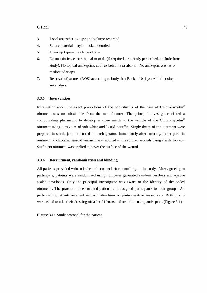
C Heal 72
3. Local anaesthetic - type and volume recorded
4. Suture material – nylon – size recorded
5. Dressing type – melolin and tape
6. No antibiotics, either topical or oral- (if required, or already prescribed, exclude from
study). No topical antiseptics, such as betadine or alcohol. No antiseptic washes or
medicated soaps.
7. Removal of sutures (ROS) according to body site: Back – 10 days; All other sites –
seven days.
3.3.5 Intervention
Information about the exact proportions of the constituents of the base of Chloromycetin®
ointment was not obtainable from the manufacturer. The principal investigator visited a
compounding pharmacist to develop a close match to the vehicle of the Chloromycetin®
ointment using a mixture of soft white and liquid paraffin. Single doses of the ointment were
prepared in sterile jars and stored in a refrigerator. Immediately after suturing, either paraffin
ointment or chloramphenicol ointment was applied to the sutured wounds using sterile forceps.
Sufficient ointment was applied to cover the surface of the wound.
3.3.6 Recruitment, randomisation and blinding
All patients provided written informed consent before enrolling in the study. After agreeing to
participate, patients were randomised using computer generated random numbers and opaque
sealed envelopes. Only the principal investigator was aware of the identity of the coded
ointments. The practice nurse enrolled patients and assigned participants to their groups. All
participating patients received written instructions on post-operative wound care. Both groups
were asked to take their dressing off after 24 hours and avoid the using antiseptics (Figure 3.1).
Figure 3.1: Study protocol for the patient.
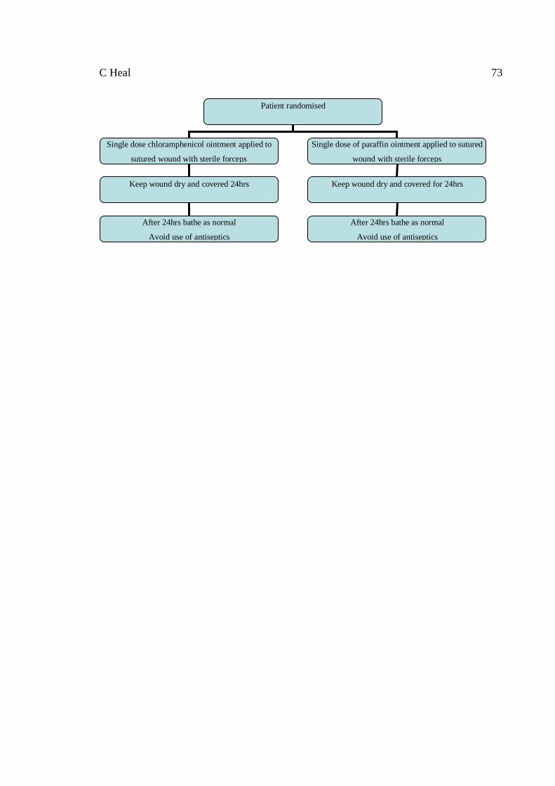
C Heal 73
Patient randomised
Single dose chloramphenicol ointment applied to
sutured wound with sterile forceps
Single dose of paraffin ointment applied to sutured
wound with sterile forceps
Keep wound dry and covered 24hrs
After 24hrs bathe as normal
Avoid use of antiseptics
Keep wound dry and covered for 24hrs
After 24hrs bathe as normal
Avoid use of antiseptics

C Heal 74
3.3.7 Clinical outcomes
Wounds were assessed for infection by the practice nurse or the doctor on the agreed day of
removal of sutures or sooner if the patient re-presented with a perceived infection. Outcome
assessing practice nurses and doctors were blinded to the allocation of intervention and control
groups. Our definition of wound infection was adapted from standardised surveillance criteria
for defining superficial surgical site infections developed by the Centre for Disease Control’s
National Nosocomial Infection Surveillance System (previous Table 2.1).(49) We also
developed our own wound scale, after reviewing several existing scales in the literature,(107) in
order to improve rigour. This wound scale differentiated: no infection or erythema; stitch
abscess; less than 1 cm erythema from the wound margin; greater than 1 cm erythema from the
wound margin; and deep infection or systemic symptoms. All participating doctors and nurses
were briefed regarding the definition of infection by the primary researcher, and were also given
written information. Practice nurses were asked to swab any discharging infections in order to
investigate any pattern of antimicrobial resistance.
3.3.8 Sample size
Sample size was calculated on the basis of our previous study which showed an infection rate of
8.6%.(7) Based on a projected infection rate of 10%, we decided that an absolute decrease in
incidence of infection of 5% would be clinically significant. To come to this conclusion with
statistical confidence a power in excess of 80% and a significance level of 0.05, a total of 473
patients were required in the intervention group and 473 patients in the control group. We did not
adjust for clustering in the sample size calculation at the planning phase of the study. This was
because the participating doctors were considered to be the primary sample units rather than the
practice. As our sample size was large and sufficient for statistical significance we do not feel that
this affected the results of our study.
3.3.9 Statistical analysis
All analysis was based on the intention-to-treat principal. Depending on the distribution, numerical
data was described as mean value and standard deviation (SD) or median value and inter-quartile
range (IQR). Percentages are presented with 95%-confidence intervals (95%-CI). Because the
sample was recruited through 15 different general practitioners and outcome might be more similar
for patients from one medical professional (clustering) compared to another, confidence interval and
the p-value were adjusted for this cluster sampling approach. For the analysis, participating doctors
were considered the primary sampling unit. The “survey” commands of the statistical programme
STATA, release 8 (STATA cooperation, College Station, Texas, USA) were applied to adjust for

C Heal 75
the clustering effects when estimating the standard error. P-values less than 0.05 were considered to
be statistically significant.
3.4 Results
3.4.1 Practice and study characteristics
Of the total of 1,246 patients who attended for skin excisions during the collection period from
June 2007 to March 2008, 232 patients were excluded (Table 3.1).
Table 3.1: Reasons for exclusion from study
Reasons for exclusion from study N=232
Patient declined participation
Patient on oral antibiotics
Doctor did not follow study protocol
History of allergy to Chloromycetin®
Patient did not plan to return for removal of sutures
Shave biopsy conducted
Patient on immune-suppressive drug
Patient with infected sebaceous cyst
139 (59.9%)
39 (16.8%)
20 (8.6%)
2 (0.9%)
12 (5.2%)
6 (2.6%)
4 (1.7%)
10 (4.3%)
Of the remaining 1,014 patients, 509 patients were randomised to the intervention
(chloramphenicol) group, and 505 to the placebo (paraffin) group. A total of 42 patients were
eventually lost to follow up because they had their sutures removed elsewhere. Follow up was
completed in 972 (95.9%) of randomised patients (Figure 3.2).

C Heal 76
Figure 3.2: Flow-chart of enrolment, randomisation and follow up of patients.
3.4.2 Comparisons at baseline
There were large differences between the intervention and the control groups at baseline
(Table 3.2). In the intervention group 71.7% of patients were diagnosed with non-melanoma
skin cancer or solar keratosis compared to 65.1% in the control group.
Patients presenting during study
period n=1246
N=
Randomisation
N=1014
Excluded n=232
(Reasons: Table 1)
Refused to participate n=
Other reasons n= Chloramphenicol n=509
Non complier n=1
Did not receive intervention n=
Placebo (Paraffin) n=505
Non complier n=4
Did not receive intervention n=
Lost to follow up n=21
Lost to follow up n=21
Analysed n =488 Analysed n =484
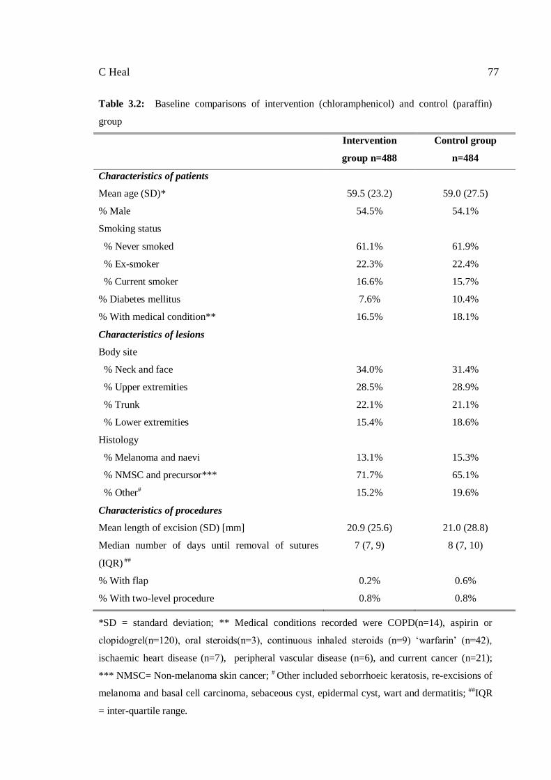
C Heal 77
Table 3.2: Baseline comparisons of intervention (chloramphenicol) and control (paraffin)
group
Intervention
group n=488
Control group
n=484
Characteristics of patients
Mean age (SD)* 59.5 (23.2) 59.0 (27.5)
% Male 54.5% 54.1%
Smoking status
% Never smoked
% Ex-smoker
% Current smoker
61.1%
22.3%
16.6%
61.9%
22.4%
15.7%
% Diabetes mellitus 7.6% 10.4%
% With medical condition** 16.5% 18.1%
Characteristics of lesions
Body site
% Neck and face
% Upper extremities
% Trunk
% Lower extremities
34.0%
28.5%
22.1%
15.4%
31.4%
28.9%
21.1%
18.6%
Histology
% Melanoma and naevi
% NMSC and precursor***
% Other#
13.1%
71.7%
15.2%
15.3%
65.1%
19.6%
Characteristics of procedures
Mean length of excision (SD) [mm] 20.9 (25.6) 21.0 (28.8)
Median number of days until removal of sutures
(IQR) ##
7 (7, 9) 8 (7, 10)
% With flap 0.2% 0.6%
% With two-level procedure 0.8% 0.8%
*SD = standard deviation; ** Medical conditions recorded were COPD(n=14), aspirin or
clopidogrel(n=120), oral steroids(n=3), continuous inhaled steroids (n=9) ‘warfarin’ (n=42),
ischaemic heart disease (n=7), peripheral vascular disease (n=6), and current cancer (n=21);
*** NMSC= Non-melanoma skin cancer; # Other included seborrhoeic keratosis, re-excisions of
melanoma and basal cell carcinoma, sebaceous cyst, epidermal cyst, wart and dermatitis; ##IQR
= inter-quartile range.

C Heal 78
3.4.3 Incidence of infections
Infection occurred in 85 of the 972 excisions (8.7%). The incidence of infection in the
chloramphenicol group (6.6%; 95%-CI = (4.9 to 8.8)) was significantly lower compared to the
incidence in the control group (11.0%; 95%-CI = (7.9 to 15.1)) (p=0.010; adjusted for cluster
sampling). The relative risk of infection was 1.7 times higher in the control compared to the
intervention group (Table 3.3). The number needed to treat (NNT) was 22.8 (number of wounds
treated for each prevented infection, that is 488/21.4). It was calculated that there were 53
infections in 484 control people, hence we expected 53.438 infections in 488 (intervention)
people. Therefore the study prevented 21.438 infections.
There was no significant difference in the wound score between the control and intervention
groups (p=0.253), although 5.5% of patients showed erythema greater than 1cm in the
intervention group compared to 9.1% of patients in the control group (Table 3). Wound swabs
were performed on 24 of the 85 infections. These revealed Staphylococcus aureus infections
which were resistant to benzylpenicillin, but sensitive to all other antibiotics in 22 cases. In one
case, additional resistance to erythromycin but sensitivity to all other antibiotics was noted. In
another case Pseudomonas aeruginosa was cultivated from the wound. These two swabs were
taken from subjects in the control group.
Table 3.3: Incidence of wound infections in intervention (chloramphenicol) and control
(paraffin) group
Intervention group
(n=488)
Control group
(n=484)
Combined results
(n=972)
Number of infections 32 53 85
Incidence of infection 6.6% 11.0% 8.7%
Relative risk of infection
(95%-CI)*
1 (reference category) 1.7; (1.1 to 2.5)
Wound score**
Stitch abscess
< 1cm Erythema
> 1cm Erythema
2.9% (n=14)
13.8% (n=67)
5.5% (n=27)
2.9% (n=14)
12.8% (n=62)
9.1% (n=44)
2.9% (n=28)
13.2% (n=129)
7.3% (n=71)
*95%-CI = 95% confidence interval; **Wound score had two missing values.

C Heal 79
3.5 Discussion
The results of the present study suggest that a single dose of topical chloramphenicol to sutured
wounds can produce a relative reduction in infection rate of about 40%. The absolute reduction
was 4.4% which fell short of our pre-determined reduction for clinical relevance (5%), therefore
this was essentially a negative trial. The incidence of infection in our control group (11%) is
much higher than published literature looking at similar cohorts.(52, 56, 113) The intervention
therefore may not produce a worthwhile absolute reduction in infection in low risk settings
where infection rates are already low, and the number needed to treat in these circumstances,
would be much higher than our figure of 22.8.
3.6 Limitations of study
The present study did have several limitations. There are various characteristics influencing the
occurrence of infections and although information on as many variables as possible was
recorded, it proved difficult to ensure that baseline data was comparable. For example, there
were inadequate data recorded on suture size and occupation, and consequently, these factors
could not be compared. In addition, the prevalence of diabetes and of other medically important
conditions was probably under-recorded, and power was limited to analyse these subgroups.
Surgical training and technique of the GPs involved is a potential confounder which would be
difficult to quantify and was not recorded. However, statistical analysis was adjusted for the
cluster sampling taking the doctor as the primary sampling unit. There was a difference in the
type of skin preparation used by the three participating practices, however there was no
previously published evidence that this makes any difference to infection rates.(115) A total of
42 participants were lost to follow-up. If all 21 participants who were lost to follow up in the
intervention group had developed an infection, then the rates of infection in both groups would
have been similar (10.4% and 11.0%); however we feel this scenario is extremely unlikely.
The diagnosis of infection, although using guidelines, is still subjective and there may be inter-
and intra-observer variation.(107) The definition we used is the most widely implemented
standard definition of wound infection,(49) and we hoped by developing our own wound
assessment scale, to reduce the subjectivity of infection diagnosis. We have no evidence to
support intra- and inter-practice reproducibility of measurement and recording procedures.
The study did not have an arm in which no ointment was applied, therefore we do not know if
the ointment itself had any pro- or anti-infective properties. The ointment base of
Chloromycetin ®consists of a mixture of soft white paraffin, liquid paraffin and plastibase 30W,

C Heal 80
which is a plasticized hydrocarbon gel consisting of 95% mineral oil and 5% polyethylene
glycol. Information about the exact proportions of these constituents was not obtainable from
the manufacturer. Our placebo ointment consisted of 50% soft white paraffin and 50% liquid
paraffin, and was not completely identical to the ointment base of Chloromycetin® as it did not
contain plastibase 30W. Although we think it is unlikely, we are unable to determine if this
substance has an effect on infection. Our trial utilized a single dose of chloramphenicol
ointment only. We have no evidence to surmise that repeated doses might lead to a greater
reduction in infection rate.
3.6.1 Potential of unblinding of intervention
Although there were some large differences observed at baseline, we think it is unlikely that the
intervention was unblended. The placebo ointment was developed using a compound
pharmacist using as close as possible to the ingredients of Chloromycetin®, and the principal
researcher also visited the practices regularly during the trial to ensure that the two ointments
were identical, both at room and refrigerated temperatures. However we did not test with a
formal agreement study to ensure whether the practice nurses remained blinded during the study
period, and this must be acknowledged as a weakness of this trial.
3.6.2 At what level would we consider clinical significance?
When the general practitioner evidence based medicine group initially met to discuss the trial,
we decided that an absolute reduction of infection of 5%, based on a projected infection rate of
10%, would be clinically significant to our practice. Our control group had an infection rate of
11%, therefore an infection rate of 6% in our intervention group would have been considered to
be clinically significant. Our control infection rate was actually 6.6%, so clinical significance
was only missed by a narrow margin, however we felt that it was important and ethical to stick
implicitly to our predetermined levels of significance.
3.6.3 Generalisability
There are some limits to generalizing these findings. The population of Mackay is slightly older
and has a lower median household income than the general Australian population.(109) Mackay
is a provincial town in tropical North Queensland. The climate is hot and humid, with the mean
daily maximum temperature ranging between 24.2 and 30oC during the summer months, and a
relative humidity of 75% to 79%.(110) It is possible that these tropical conditions could increase
sweat production and produce damp dressings, which might reduce the effectiveness of wound
dressings as a potential barrier against exogenous bacteria.(116-118) This would make wounds

C Heal 81
more prone to infection in a tropical environment, and therefore the results may not necessarily
be generalisable to a temperate climate, although there is no published evidence that heat and
humidity increase infection rates. This might also explain why our infection rates were higher
than previous data from temperate climate suggests.(52, 56, 113)
3.6.4 Antibiotic use
There is some concern regarding the overuse of topical antibiotics resulting in antibiotic
resistance. British and Australian guidelines suggest that topical antibiotic use be restricted
because of the capacity of most topical drugs to select resistant micro-organisms and to cause
sensitization. The guidelines also suggest that antimicrobials recommended for topical use are
selected from classes not in use for systemic therapy.(66, 119) There is a contrary argument that
the potential for antimicrobial resistance with topical antibiotics is actually lower than with
systemic antibiotics because of the higher local concentration achieved by topical delivery.(95)
Patterns of antimicrobial activity and resistance have been examined for other antibiotic
ointments.(92, 93) However there is no evidence, over three decades of extensive use
worldwide that, with the exception of mupirocin, topical antibiotics administered on an
outpatient basis contribute to any emerging resistance pattern.(95) Chloramphenicol eye drops
have been shown to be effective in the treatment of methacillin resistant staphylococcus aureus
(MRSA) ocular surface infections.(91)
There is also some concern about the incidence of allergic contact dermatitis with topical
antibiotic use. With topical neomycin, this has been shown to be as high as 11% in a population
referred for diagnostic patch testing.(120) However there is some evidence that the incidence of
this reaction is as low as 1% when the ointment is used in the general population.(95) The
reaction is much more common amongst subjects previously exposed to neomycin
ointment.(120) Contact allergy has been reported with the use of chloramphenicol ointment but
the incidence is thought to be low.(96, 97) Although it is unlikely that there is any connection
between the use of topical chloramphenicol and aplastic anaemia,(70, 100) the study is not large
enough to fully assess the risk in this setting.
Antibiotic prophylaxis is probably prescribed excessively or inappropriately for dermatological
surgery(49, 62) and is thought to be best reserved for high risk patients.(10, 62) There is no data
available on the current prescribing habits of Australian GPs regarding oral or topical antibiotic
prophylaxis for minor excisions. Although there is also no evidence available regarding what
reduction in the rate of infection we might reasonably expect from the use of oral prophylactic
C Heal 82
antibiotics for minor excisions, there is some evidence of a 50% reduction in risk of infection
when perioperative oral antibiotic prophylaxis is used following clean surgery.(102) A similar
reduction in infection rate from a single dose of topical antibiotic, as in this study, may
encourage a reduction in the use of oral antibiotics.
The decision to prescribe antibiotic prophylaxis is complicated and in addition to efficacy,
antibiotic costs, adverse effects and resistance should be taken into account. However, in some
circumstances, topical delivery of antibiotic may be preferably to systemic administration.(66,
119) The results of this study could encourage the judicial use of topical antibiotics following
minor skin surgery. However it must be noted that it is unlikely that topical chloramphenicol
will produce a worthwhile absolute reduction in infection rates in low risk settings in developed
countries. Future research could explore the possibility that important reductions may be seen in
higher risk wounds or in more resource poor settings.
3.6.5 Conclusion
The present study suggests application of a single dose of topical chloramphenicol to high risk
sutured wounds following minor surgery produces a moderate absolute reduction in infection
rate.
3.7 Summary
This trial established that chloramphenicol ointment has limited efficacy in reducing the
incidence of wound infection after minor surgery. The incidence of infection in the
chloramphenicol group (6.6%; 95%-CI = (4.9 to 8.8)) was significantly lower compared to the
incidence in the control group (11.0%; 95%-CI = (7.9 to 15.1)) (p=0.010). The absolute
reduction in infection rate was 4.4%; the relative reduction was 40%. The present study
suggests application of a single dose of topical chloramphenicol to high risk sutured wounds
after minor surgery produces a moderate absolute reduction in infection rate which is
statistically but not clinically significant.
The incidence of infection in the control group in this study (11.0%) was comparable with our
previous study, and again much higher than for similar groups of patients with class 1 wounds
from different geographical locations. One of the risk factors for infection identified from our
previous trial was excision of a skin cancer (see chapter 2), while age was actually found to be a
confounder rather than a risk factor.

C Heal 83
It is therefore important to establish the types of skin lesions that GPs are excising, and the
demographics of the patients who are presenting with skin cancer, in order to understand risk
factors for infection and effective interventions to prevent infection after minor surgery. The
next chapter will examine these factors in more detail.
Diagnosis of skin lesions, C Heal 84
________________________________________________________________________
CHAPTER 4: MINOR SKIN EXCISIONS IN NORTH
QUEENSLAND
4.1 Background
This chapter describes the type of skin surgery that is being conducted by general practitioners
in North Queensland. This is important in order to understand the epidemiology of skin cancer
in this location in order to understand the need for general practitioners to conduct skin surgery.
The number needed to treat (NNT) is the number of melanocytic naevi excised per melanoma,
and can be used to assess performance in skin cancer practice. This chapter is a secondary
analysis of data collected in the ‘Can sutures get wet’ intervention study. The published article
forms Appendix 3.(114)
The objectives of this chapter are to describe the histology of lesions excised by general
practitioners in rural North Queensland and calculate the skin cancer yield and NNT; and to
describe the sites that skin cancers are removed from and the demographics of patients
presenting with skin cancer.
4.2 Introduction
Skin cancer is an extremely important health issue in Australia. Non-melanocytic skin cancer
(NMSC), combining basal cell carcinoma (BCC) and squamous cell carcinoma (SCC), is by far
the commonest cancer in Australia with an incidence of more than four times that of all other
registrable cancers combined.(1) Queensland has the world’s highest recorded incidence of all
types of skin cancer,(2, 3) with incidence rates being even higher in tropical North
Queensland.(4)
In North Queensland, the majority of suspicious skin lesions are managed by general
practitioners (GPs),(38) particularly in rural centres such as Mackay where there is no resident
dermatologist. It was recently estimated that, in Australia, 54% of patients with BCCs and 65%
of patients with SCCs are managed in primary care settings,(6) and it could be projected that
these figures would be even higher for rural settings. Skin excisions form a large proportion of
a typical Australian GP’s workload, and this proportion is even greater for Queensland GPs.(5)
Skin excisions are also very costly, with over one million skin excisions billed to Medicare

Diagnosis of skin lesions, C Heal 85
________________________________________________________________________
annually(8) and an estimated cost of $264 million for NMSC in 2000-1 (9% of the total costs
for cancer).(9)
The data for the present study was collected incidentally as part of a randomised controlled trial
looking at wound management following minor excisions, which has been reported
elsewhere.(7) The purpose of examining the present data was to describe the histology of
lesions that GPs remove during minor excisions in rural North Queensland, the sites that skin
cancers are removed from and the demographics of patients presenting with skin cancer.
4.3 Methods
Sixteen GPs from four practices in the Mackay area, tropical North Queensland, Australia
(latitude 21°S; inhabitants of Mackay area approximately 75,000) participated in the present
study. There are a total of 104 practising GPs in the Mackay area. The participating GPs were a
self-selected group who attended a monthly evidence based medicine meeting. Data collection
for the study took place from October 2004 to May 2005. Data was collected from consecutive
patients presenting for minor skin excisions. This included all patients who had any skin lesion
excised and repaired by direct sutures over the study period. Patients presenting for suturing of
a laceration or incision and drainage of a sebaceous cyst were excluded, as were shave or punch
biopsies. Explicitly, these patients were 1,246 patients who presented for minor skin excisions
during the period of collection for the ‘Can sutures get wet’ trial described in chapter 3. Of the
1,246 patients, 232 were excluded from the ‘Can sutures get wet’ trial. The reasons for
exclusion are detailed in chapter 3. However, written consent was given from all excluded
patients, including the small number that refused to take part in trial, to have data recorded for
research purposes. Demographic data and data on types of skin lesion were recorded on all
1,246 patients who presented for minor skin excisions over the study period.
Practice nurses were responsible for collecting data. A body site map was used to define
excision site, and histology was recorded for all patients. At the end of the study practice nurses
were asked to re-examine computer records to fill in any missing data. The principal researcher
visited participating GPs and practice nurses to provide training and ensure that recording was
standardised.

Diagnosis of skin lesions, C Heal 86
________________________________________________________________________
The data was collated and analysed using SPSS version 12. Standard descriptive statistics and
bivariate statistical tests were applied.
4.3.1 Calculation of relative tumour density
Relative tumour density (RTD) for different body sites was calculated according to Pearl and
Scott.(121) This method has previously been described to compare the density of tumours at
different body sites relative to the density of tumours over the whole body by taking into
account the surface proportion (of the whole body) of a specific body site. Thus the different
body sites become comparable assuming that each skin unit has the same potential for
developing skin cancer. In this way the distribution of tumours over the body surface can be
more readily related to the likely exposure of the surface.
This calculation of relative tumour density (RTD) is performed by taking the proportion of all
lesions at a particular body site and dividing this proportion by the surface proportion of the
body site. A RTD of one corresponds to the density of tumours on the whole body.
4.4 Results
Participating GPs were younger (median age 44 years) and more predominantly female (64%)
than average for Australian GPs (median age category 45-54 years; 32% female).(121) One
doctor who participated worked in a designated skin cancer clinic. Data was recorded from
1,247 consecutive patients who attended for minor skin excisions to the participating doctors
between October 2004 and May 2005.
4.4.1 Histology
SCC was the most frequently excised lesion, accounting for 25.9% of excisions, and all skin
cancers (SCC, BCC or melanoma) together accounted for 46.7% of excisions. This number
increased to 68.9% if solar keratoses were included (Table 4.1). Number of benign or dysplastic
naevi excised per melanoma (NNT) was 9.4 to 1.

Diagnosis of skin lesions, C Heal 87
________________________________________________________________________
Table 4.1: Histology of 1,247 minor skin excisions recorded in Mackay, North Queensland
HISTOLOGY N %
Cutaneous melanoma 20 1.6
Basal cell carcinoma 234 19.2
Squamous cell
carcinoma
316 25.9
Benign naevus 145 11.9
Dysplastic naevus 23 1.9
Seborrhoic keratosis 24 2.0
Solar keratosis 271 22.2
Basal cell papilloma 41 3.4
Re-excision 16 1.3
Other* 131 10.7
Missing 26
Total 1247 100.0
‘Other’included: ‘cyst’ (20), sebaceous cyst (11)**, keratosis unspecified (16), lentigo (11),
scar tissue/keloid (6), cutaneous tag (6), verruca vulgaris (5), dermatofibroma (8), lipoma (5),
keratoacanthoma (9), haemangioma (12), lichen planus (3), dermatis (4) granuloma annulare
(2), pore of winer (1), fibroxanthoma (1), pyogenic granuloma (2), foreign body (1),
neurofibroma (2), xanthogranuloma (1), sebaceous hyperplasia (1), psoriasis (1), and sebaceous
hyperplasia (3).
** Although incision and drainage of sebaceous cyst was excluded, sebaceous cyst was an
incidental finding on pathology report in these 11 patients.
4.4.2 Demographics of patients presenting for excisions
The majority (84.3%) of skin excisions took place in the over 40 years age category, with the
mean age being 56.9 years (SD 16.3). Slightly more (53.2%) of skin excisions took place in
men (Table 4.2).

Diagnosis of skin lesions, C Heal 88
________________________________________________________________________
Table 4.2: Age and sex distribution of patients presenting with skin cancers for all skin excisions
in Mackay, North Queensland.
Melanom
a (n =
20)
BCC
(n = 234)
SCC
(n = 316)
All
excisions
(n = 1247)
Mean age (SD)
[years]
55
(15.0)
60.9
(15.0)
63.8
(12.5)
56.9
(16.3)
Age range [years] 27 - 79 24 – 98 24 - 94 5 - 98
Male to female ratio 0.82 : 1 1.44 : 1 1.56 : 1 1.14 : 1
One BCC was removed from the chest of a 24-year-old male; one SCC was removed from the
leg of a 24-year-old female; and another SCC from the chest of a 29-year-old male. The
majority of SCCs were excised in the 70 to 89 year age group, and the majority of BCCs were
excised from patients 60 to 79 years of age. Eight (40.0%) melanomas were excised from
patients who were younger than 50 years.
4.4.3 Body site distribution of skin cancer
The body site distribution and relative tumour density of skin cancers is documented in
Table 4.3. There was little difference in site distributions between males and females (p=0.7013
for BCC; p=0.8415 for SCC; sample size too small for melanoma).
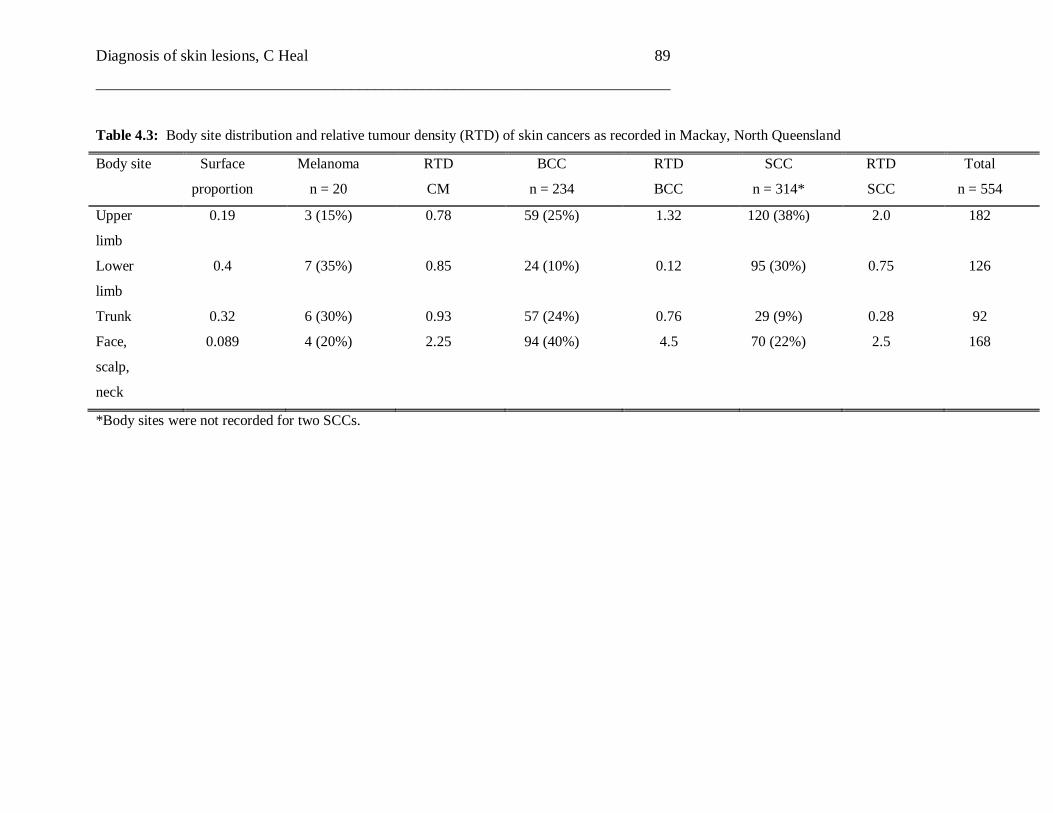
Diagnosis of skin lesions, C Heal 89
________________________________________________________________________
Table 4.3: Body site distribution and relative tumour density (RTD) of skin cancers as recorded in Mackay, North Queensland
Body site
Surface
proportion
Melanoma
n = 20
RTD
CM
BCC
n = 234
RTD
BCC
SCC
n = 314*
RTD
SCC
Total
n = 554
Upper
limb
0.19 3 (15%) 0.78 59 (25%) 1.32 120 (38%) 2.0 182
Lower
limb
0.4 7 (35%) 0.85 24 (10%) 0.12 95 (30%) 0.75 126
Trunk 0.32 6 (30%) 0.93 57 (24%) 0.76 29 (9%) 0.28 92
Face,
scalp,
neck
0.089 4 (20%) 2.25 94 (40%) 4.5 70 (22%) 2.5 168
*Body sites were not recorded for two SCCs.

Diagnosis of skin lesions, C Heal 90
________________________________________________________________________
4.4.4 Comparison of results from designated skin cancer clinic setting to mainstream
general practice setting
The total 1,247 excisions included 76 excisions undertaken in a designated skin cancer clinic.
There was little difference in the demographics of patients seen in the skin clinic setting in
comparison to the patients from GP practices. In the skin clinic, 76% (58/76) of lesions excised
were skin cancers (BCC, SCC or melanoma) compared with 512/1145, 45% in mainstream
general practice setting. The NNT was 5.7 (14+3/3) (mainstream general practice 10.0
134+19+17/17). The BCC: SCC ratio was 4:1 (44/11) (mainstream general practice 190/305
0.6:1).
4.5 Discussion
There are several limitations to analyzing and generalizing our findings. It is known that the age
and gender of general practitioners strongly influences the age and gender of patients who
consult them,(108) thus creating potential selection bias. The participating GPs were not
representative of Mackay GPs, and patients attending general practice are not representative of
the general population.(108) Excisions from the facial region were probably under recorded in
our study, which may have influenced the body site distribution. The sample size from the
designated skin cancer clinic is small, and the comparison of these data with the overall data
must be viewed with caution.
4.5.1 Histology
In 2002, of 374,000 Australians treated for skin cancer it was estimated that 68% were for
BCCs and 32% with SCCs,(6) and previous Australian studies have reflected this ratio of BCCs
to SCC’s.(4, 56) Notably, in our study more SCCs than BCCs were excised. Our ratio of BCC:
SCC was 42.5%:52.5% or 0.74:1. One could reflect that the higher incidence of SCC may be
due to the high rate of cumulative sun exposure of the cane farming population. Hence, one
could speculate that the sun behavior of Mackay residents might differ from other populations.
Another possible explanation of this finding could be that more and more early BCCs are being
treated with cryotherapy or Imiquimod cream by Mackay GPs and hence have no histo-
pathologic confirmation.

Diagnosis of skin lesions, C Heal 91
________________________________________________________________________
The overall NNT for melanoma of 9.4 was much lower than recent data derived from analysis
of billing data from skin cancer clinics (NNT 28.6).(45) However, in this study, non-pigmented
benign lesions were included in the denominator therefore elevating their NNT. In other
studies, the NNT was found to be between 11 and 29 for Australian GPs when based on
melanomas and naevi only (as for our figures) and up to 36 when seborrhoeic keratoses were
included.(35, 42-44) However as there were only 20 melanomas excised the interpretation of
this data is also limited.
4.5.2 Comparison of histology from skin clinic
The doctor working in the skin cancer clinic excised a higher number of skin cancers per
excision and had a lower NNT compared with the overall results. Although with only a total of
76 excisions including 3 melanomas excised in this setting these figures must be interpreted
with caution. Most significantly, there was a much larger BCC:SCC ratio (4:1 v 0.74:1). This
could reflect either an increased pick up of BCC because of the tendency to undertake full-body
skin examination within the skin cancer clinic context and so detect lesions of which the patient
is unaware, or an increased use of excision rather than other management options. The differing
histology in the skin clinic setting could also be influenced by the differing pathology of
patients who self select to attend a skin cancer clinic, the use of epiluminescent microscopy or a
difference in the training and experience of the skin clinic doctor.
4.5.3 Body site distribution
Although only 22% of SCCs were excised from the face, scalp and neck and the most common
site for excision was from the upper limb (38%), when we calculated the RTD, which takes into
account the surface proportion occupied by body sites experiencing different amounts of
ultraviolet radiation, ‘face scalp and neck’ was the most dominant region. Likewise, although
melanomas were more commonly excised from the lower limb, when RTD was calculated,
face, scalp and neck were the most dominant regions. The largest proportion of BCCs was
excised from the head and neck, which was also reflected in the RTD. These findings are
consistent with previous studies.(24, 121-123)
4.5.4 Age
The number of SCCs excised appeared to increase steadily with age, while the majority of
BCCs were excised in the 6th decade, and the most common decade for melanomas was 40-49
Diagnosis of skin lesions, C Heal 92
________________________________________________________________________
years. This is in keeping with the theory that high cumulative sun exposure is regarded as a risk
factor for SCC,(124) while intermittent sun exposure is a consistent risk factor for
melanoma,(125) while both factors appear to contribute to the formation of BCCs.(126)
4.5.5 Conclusion
In this sample of Mackay GPs, close to half (46.7%) of lesions excised were skin cancers. We
excised more SCCs than BCCs (0.74:1), which contrasts with previous studies of Australian
populations. Our NNT is relatively low compared with other studies of Australian GPs, and we
could consider lowering our threshold for excising pigmented lesions.
4.6 Summary
Close to half (46.7%) of lesions excised were skin cancers. We excised more squamous cell
carcinomas than basal cell carcinomas (0.74:1). Mean age for excision of melanoma, BCC and
SCC was 55, 60.9 and 63.8 years respectively.
In this chapter we established that amongst a cohort of GPs in Mackay our skin cancer yield
was comparable to similar cohorts. The number needed to treat (melanocytic naevi excised per
melanoma) was 8.4 and this NNT was much lower than comparable studies. The conclusion of
the study was that the doctors involved in the study could lower our threshold for excising skin
pigmented lesions. However the use of NNT as a marker of quality of skin cancer management
by general practitioners is complex, and many different factors must be taken into
consideration. Firstly, we do not have a measure of the skin cancers that we missed (ie the false
negative diagnosis) so a low NNT may not necessarily be a measure of good quality
management by GPs. Secondly, the measure will be affected by the background incidence and
prevalence of skin cancer in the presenting population. Thirdly the NNT may also be affected
by economics, and the desire to generate income by excising more skin lesions. Fourthly it may
be influenced by medico-legal concerns which may increase the desire to excise, and also the
past clinical experience of a particular doctor. The issue is actually extremely complex, and we
make the recommendation of considering lowering the threshold for excision with extreme
caution.
This chapter also illustrates the importance of GPs and other medical practitioners to be able to
diagnose skin cancer accurately. The next chapter will investigate this in further detail.

Diagnosis of skin lesions, C Heal 93
________________________________________________________________________
CHAPTER 5: DIAGNOSTIC ACCURACY OF EXCISED AND
BIOPSIED SKIN LESIONS BY AUSTRALIAN
GENERAL PRACTITIONERS
5.1 Background
It has been established in the previous chapter that minor skin surgery conducted by general
practitioners in North Queensland produces a high yield of skin cancer. However, there is little
data available regarding the diagnostic accuracy of Australian GPs with regard to excised and
biopsied skin lesions and no published comparison of the diagnostic accuracy of doctors
regarding different subtypes of non-melanotic skin cancer. The published article forms
Appendix 4.(127)
This chapter explores the diagnostic accuracy of Australian GPs regarding lesions that they
have decided to excise. The objective was to analyse historical data of clinical and histological
diagnosis of excised and biopsied non-melanoma skin cancers by Australian GPs.
5.2 Introduction
Skin cancer is an increasing problem in fair-skinned populations world wide, with age
standardised incidence rates per 100,000 for cutaneous melanoma (CM) of 40.5, 13.3 and 6.1
for men, and 31.8, 9.4 and 7.7 for women in Australia, the US and the UK respectively.(31) In
the UK and US, incidence rates for melanoma are rising rapidly. Non-melanocytic skin cancer
(NMSC), combining basal cell carcinoma (BCC) and squamous cell carcinoma (SCC), is the
most common cancer in all three countries.
Australia has the world’s highest recorded incidence of all types of skin cancer,(26, 31, 128)
with incidence rates being even higher in tropical North Queensland.(4) In Australia the
majority of suspicious skin lesions are managed by general practitioners (GPs)(38) and the
proportion of all skin cancers excised by GPs is increasing.(39) Skin excisions form a large
proportion of the typical workload of an Australian GP,(5) with 0.8 per 100 patient encounters
involving excision of a skin lesion.(129) The economic cost of skin cancer to the Australian
health care system is enormous.
Diagnosis of skin lesions, C Heal 94
________________________________________________________________________
In the UK there is some controversy about the ability of general practitioners to recognise skin
cancer and prioritise referrals(130-132) and there is a debate about the appropriateness of both
CM and NMSC being managed in a primary care setting.(130, 131) Similarly in Australia, there
has recently been interest in the diagnostic performance of doctors with respect to skin lesions
which they decide to biopsy or excise.(133, 134) This interest has particularly focused on
assessing the performance of practitioners working in ‘open access’ designated skin cancer
clinics, which are primarily staffed by GPs, and have been a recent rapidly emergent
phenomenon, particularly in the state of Queensland.(135) The Australian studies have shown
that practitioners diagnose a wide range of skin lesions with moderate to high sensitivity.
The main purpose of the present study was to compare the clinical to the histological diagnosis
of excised skin lesions from a large data set which represents close to all skin excisions which
took place over a three year period in a regional centre in North Queensland, at a time prior to
the existence of skin cancer clinics. We aimed to analyse diagnostic accuracy stratified by
histological subtype and body-site. We also aimed to examine the histological nature of false
positive and false negative diagnoses, in order to make inferences about the decisions to excise
and implications of misdiagnoses.
5.3 Methods
The original study was a prospective, population-based survey that aimed to collect information
on all excised and histologically confirmed skin cancers in Townsville. Between the end of
1996 and October 1999, all excised and histologically confirmed skin cancers in
Townsville/Thuringowa, North Queensland, Australia (latitude 19.16ºS; population
approximately 130,000) were recorded. During the study, pathology reports on all excised skin
cancers were collected from the three local pathology laboratories, which routinely collected
reports on NMSC to send to the Queensland Cancer Registry. These laboratories served the
pathology requests from all hospitals, GPs and specialists in Townsville (202 GPs and 42
specialists - including one dermatologist, plastic surgeons and general surgeons, during the
study period). The data collected on excised skin cancers is likely to be almost complete, as
doctors in Townsville routinely seek histology for all excised skin lesions. As Townsville’s
location is relatively isolated (Brisbane is approximately 1,200 km south) it can be assumed that
almost all skin cancers were treated locally. To validate this assumption, 7,370 pathology
reports from the periods February/March and May/June1997, which were sent directly to the
Diagnosis of skin lesions, C Heal 95
________________________________________________________________________
Queensland Cancer Registry from private pathology laboratories and hospitals, were searched
for residents of Townsville treated elsewhere and only one patient was identified. Before the
onset of the study, a histopathology workshop was organized to reach agreement on criteria for
histological diagnosis of skin cancer and other skin lesions. Within the present study BCC and
SCC were differentiated, and all in situ malignancies were excluded. Within the database
multiple skin cancers per patient can be identified. Biopsied lesions were recorded as such and
data was subsequently changed, after the entire lesion was excised. We refer to multiple skin
cancers either if lesions were presented concomitantly for treatment or if subsequent skin
cancers developed and were treated during the study period.(4) Of the 6,708 patients recorded
with NMSC, 38.5% had multiple lesions. Prior to the study all participating pathologists
attended a workshop to reach agreement on criteria for the histological diagnosis of skin lesions
with a consultant dermato-pathologis, and the dermato-pathologist agreed to review a blinded
stratified random sample of 407 histology slides. This part of the study is discussed in more
detail in chapter 6. In 1999, a total of 8,694 patients (mean age 58.6 years; 55.2% male) with up
to 72 excisions per patient were recorded. Incidence rates (per 100,000 inhabitants) of basal cell
carcinoma (BCC) were 1,444.8 for men, 942.7 for women, and of squamous cell carcinoma
(SCC) were 805.0 for men and 423.6 for women. Compared to incidence rates, age standardised
rates of lesions of BCC were 2.1 times higher in men, 1.6 times higher in women, and of SCC
were 1.8 times higher in men and 1.4 times higher in women. Details of these incidence rates,
and comparison with other studies, are discussed in the literature review.
5.3.1 Statistical analysis
In this present study, positive predictive values (PPV) and sensitivity refer to the doctors’
clinical diagnosis of a specific lesion. Histology was considered the “gold standard”. PPV is
defined as the probability that a person with a clinical diagnosis of a particular skin lesion
actually has this skin lesion according to histology. Sensitivity is defined as the probability that
a person with the histological diagnosis of a particular skin lesion will “test positive” for this
skin lesion according to clinical diagnosis.
PPVs and sensitivities together with exact 95%-confidence intervals (95%-CI) were calculated
for the clinical diagnoses of BCC, SCC, intra-epithelial carcinoma (IEC), actinic keratosis
(AK), seborrhoeic keratosis (SK), keratoacanthoma (KA), cutaneous melanoma (CM),
cutaneous melanoma in situ (CM in situ), atypical naevus (AN) and common naevus (CN). For

Diagnosis of skin lesions, C Heal 96
________________________________________________________________________
PPV and sensitivity only the first lesion per patient with the particular clinical diagnosis was
considered; this was to reduce possible bias due to doctors ‘learning’ the clinical diagnosis from
patients presenting for repeated excisions of the same histological type. Sensitivities were
calculated including and excluding cases with missing clinical diagnosis in the denominator. To
infer whether excision was an appropriate option for diagnosis or treatment, we also combined
the squamous cancers and precursors, and similarly the pigmented lesions and determined PPV
for combined clinical diagnoses. Additionally, PPVs and sensitivities were calculated for
clinical diagnoses stratified by body-site.
We defined the number needed to treat (NNT) as the number of all naevi (CN and AN) excised
per CM or CM in situ. This was calculated using histological diagnosis of all excisions during
the three year study period. Comparisons between GPs and specialists were conducted using
Chi-square tests and Fisher’s exact tests. Results were adjusted for multiple testing using the
approach by Bonferroni. Statistical analyses were conducted with SPSS for Windows, release
14.
5.4 Results
Between December 1996 and October 1999 a total of 8,694 patients (mean age 58.6 years;
55.2% male) with up to 72 excisions per patient were recorded. Incidence rates (per 100,000
inhabitants) of BCC were 1,444.8 for men, 942.7 for women, and of SCC were 805.0 for men
and 423.6 for women. Compared to incidence rates, age standardised rates of lesions of BCC
were 2.1 times higher in men, 1.6 times higher in women, and of SCC were 1.8 times higher in
men and 1.4 times higher in women. Details of these incidence rates, and comparison with other
studies, are discussed in the literature review.
5.4.1 Positive predictive values and sensitivities of clinical diagnosis
PPV for BCC was 72.7%, for SCC 49.4, and for CM 33.3 (Table 5.1). Sensitivity for BCC was
63.9%, for SCC 41.1, and for CM 33.8 (Table 5.2). Sensitivities were higher when cases with
missing clinical diagnosis were excluded from the denominator (BCC: 82.0%, SCC: 56.6%,
IEC: 7.8%, AK: 14.4%, SK: 17.0%, KA: 29.9%, CM: 39.5%, CM in situ: 26.3%, AN: 14.3%,
and CN: 59.0%).

Diagnosis of skin lesions, C Heal 97
________________________________________________________________________
Combining the diagnosis of SCC, IEC and AK, the PPV of clinical diagnosis was 66.9% (95%-
CI = [64.7, 69.1]; n=2,708) and sensitivity was 40.2% (95%-CI = [38.7, 41.7]; n=3,933).
Combining CM and CM in situ, the PPV of clinical diagnosis was 38.8% (95%-CI = [31.2,
46.4]; n=160) and sensitivity was 38.1% (95%-CI = [32.0, 44.2]; n=160). Combining all naevi,
the PPV for clinical diagnosis was 51.2% (95%-CI = [46.7, 55.7]; n=477) and sensitivity was
44.4% (95%-CI = [40.3, 48.5]; n=559). These latter sensitivities were calculated including
cases with missing clinical diagnosis in the denominator.

Diagnosis of skin lesions, C Heal 98
________________________________________________________________________
Table 5.1: Positive predictive values (in bold) and 95%-confidence intervals of clinical diagnosis of skin lesions. Results for pigmented lesions
were shaded; rows add up to 100%
Histological diagnosis
Clinical diagnosis BCC SCC IEC Actinic
keratosis
Kerato-
acanthoma
CM CM in
situ
AN CN Seborrhoeic
keratosis
Other*
BCC (n=4555) 72.7%
71.4,74.0
10.9% 4.0% 5.0% 1.0% 0.4% 0%
(N=1)
0% 0.9% 1.3% 3.8%
(N=175)
SCC (n=2306) 20.8% 49.4%
47.3, 51.5
7.9% 10.5% 6.3% 0% 0% 0% 0.1% 1.3% 3.5%
(N=81)
IEC (n=141) 39.0% 3.5% 44.7%
36.3, 53.3
7.8% 0% 0% 0% 0% 0% 0.7% 4.3%
(N=6)
Actinic keratosis
(n=476)
17.0% 26.9% 9.9% 30.9%
26.8, 35.5
4.0% 0% 0% 0% 0.4% 5.0% 5.9%
(n=28)
Keratoacanthoma
(n=357)
3.4% 51.8% 2.0% 3.6% 37.0%
32.0, 42.2
0% 0% 0% 0.3% 0.3% 1.7%
(n=6)
CM (n=138) 16.7% 1.4% 1.4% 2.2% 0% 33.3%
25.5, 41.9
5.1% 0.7% 10.9% 10.1% 18.1%
(N=25)
CM in situ (n=22) 0% 0% 9.1% 4.5% 0% 18.2% 22.7%
7.8, 45.4
4.5% 18.2% 13.6% 9.1%
(n=2)
Atypical naevus
(AN) (n=35)
2.9% 0% 0% 0% 0% 8.6% 0% 5.7%
0.7, 19.2
62.9% 8.6% 11.4%
(n=4)
Common naevus 20.0% 2.3% 1.6% 1.6% 0% 6.3% 0.5% 0.9% 48.9% 9.0% 9.0%

Diagnosis of skin lesions, C Heal 99
________________________________________________________________________
(CN) (n=444) 44.1, 53.6 (n=40)
Seborrhoeic
keratosis (n=111)
9.9% 6.3% 5.4% 9.0% 0.9% 1.8% 0.9% 0% 5.4% 52.3%
42.6, 61.8
8.1%
(n=9)
*Main “other” lesions histologically diagnosed:
BCC: 14.9% lichenoid keratosis (26 of 175) and 8.0% skin, no lesion (14 of 175); SCC: 17.3% lichen simplex chronicus (14 of 81) and 11.1%
dermatofibroma (9 of 81); CM: 44.0% Skin, no lesion (11 of 25) and 16.0% scar (4 of 25); Common naevus: 20.0% lentigo simplex (8 of 40),
17.5% dermatofibroma (7 of 40) and 15.0% haemangioma (6 of 40).

Diagnosis of skin lesions, C Heal 100
________________________________________________________________________
Table 5.2: Sensitivities (in bold) and 95%-confidence interval of clinical diagnosis of skin lesions. Results for pigmented lesions were shaded;
columns add up to 100%
Histological diagnosis
Clinical diagnosis BCC
n=5044
SCC
n=2672
IEC
n=1002
Actinic
keratosis
n=1389
Kerato-
acanthoma
n=544
CM
n=139
CM in
situ n=25
AN
n=23
CN
n=537
Seborrhoeic
keratosis
n=495
BCC 63.9%
62.6, 65.2
20.1% 27.1% 25.9% 12.1% 14.4% 8.0% 4.3% 11.4% 19.8%
SCC 8.0% 41.1%
39.2, 43.0
24.1% 25.0% 35.8% 1.4% 0% 0% 0.4% 9.1%
IEC 0.6% 0.1% 5.0%
3.7, 6.5
0.5% 0% 0.7% 0% 0% 0% 0.2%
Actinic Keratosis 1.3% 3.4% 4.7% 9.4%
8.0, 11.1
3.9% 0% 0% 0% 0.6% 6.5%
Keratoacanthoma 0.2% 5.7% 0.6% 0.9% 23.0%
19.5, 26.8
0% 0% 0% 0.2% 0.2%
CM 0.4% 0.1% 0.2% 0.2% 0% 33.8%
26.0, 42.3
28.0% 8.7% 2.8% 2.6%
CM in situ 0% 0% 0.2% 0.1% 0% 2.9% 24.0%
9.4, 45.1
4.3% 1.3% 0.4%
Atypical naevus
(AN)
0% (n=1) 0% 0% 0% 0% 2.2% 0% 8.7%
1.1, 28.0
4.1% 0.8%
Common naevus 1.5% 0.3% 0.6% 0.5% 0% 20.9% 8.0% 21.7% 41.0% 8.1%
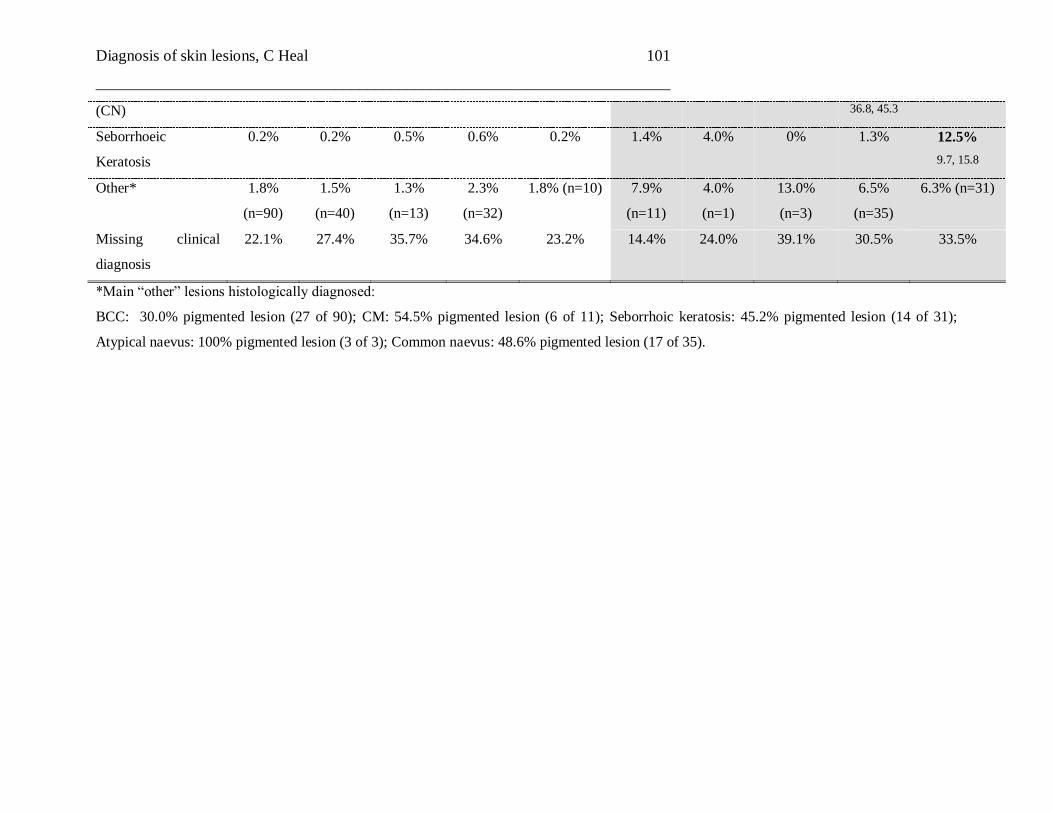
Diagnosis of skin lesions, C Heal 101
________________________________________________________________________
(CN) 36.8, 45.3
Seborrhoeic
Keratosis
0.2% 0.2% 0.5% 0.6% 0.2% 1.4% 4.0% 0% 1.3% 12.5%
9.7, 15.8
Other* 1.8%
(n=90)
1.5%
(n=40)
1.3%
(n=13)
2.3%
(n=32)
1.8% (n=10) 7.9%
(n=11)
4.0%
(n=1)
13.0%
(n=3)
6.5%
(n=35)
6.3% (n=31)
Missing clinical
diagnosis
22.1% 27.4% 35.7% 34.6% 23.2% 14.4% 24.0% 39.1% 30.5% 33.5%
*Main “other” lesions histologically diagnosed:
BCC: 30.0% pigmented lesion (27 of 90); CM: 54.5% pigmented lesion (6 of 11); Seborrhoic keratosis: 45.2% pigmented lesion (14 of 31);
Atypical naevus: 100% pigmented lesion (3 of 3); Common naevus: 48.6% pigmented lesion (17 of 35).

Diagnosis of skin lesions, C Heal 102
________________________________________________________________________
PPV and sensitivities varied with body-site (Tables 5.3 and 5.4). For BCC, PPVs and
sensitivities were higher for the trunk, the shoulders and the face and comparatively lower for
the extremities. For SCC, PPVs were high for the scalp and the extremities and comparatively
lower for shoulders and posterior trunk. PPV for the scalp were above 60% for BCC, SCC, IEC,
CM and naevi.

Diagnosis of skin lesions, C Heal 103
________________________________________________________________________
Table 5.3: Positive predictive values of clinical diagnosis of skin lesions stratified by body-sites. Results for pigmented lesions were shaded
Clinical diagnosis Scalp Face Neck Shoulder Upper
extremities
Anterior
trunk
Posterior
trunk*
Lower
extremities
BCC (n=4517) 67.2% 78.4% 68.4% 82.9% 52.9% 69.6% 82.7% 56.9%
SCC (n=2288) 65.6% 43.4% 44.1% 25.9% 51.8% 43.8% 34.4% 54.2%
IEC (n=139) 66.7% 64.3% 66.7% 28.6% 29.4% 57.1% 16.7% 53.5%
Actinic Keratosis
(n=475) 50.0% 37.8% 13.6% 14.3% 32.5% 12.5% 35.7% 25.6%
Keratoacanthoma
(n=355) 0% 15.8% 15.4% 44.4% 38.0% 33.3% 40.0% 44.6%
CM (n=134) 66.7% 33.3% 14.3% 38.5% 29.4% 9.1% 32.6% 36.8%
CM in situ (n=22) /** 22.2% 0% 100.0% 33.3% 0% 0% 50.0%
Atypical naevus
(n=35) 0% 0% / 0% 0% 0% 14.3% 0%
Common naevus
(n=440) 66.7% 40.3% 34.6% 23.1% 52.6% 57.3% 56.7% 42.0%
Seborrhoeic
Keratosis (n=110) 50.0% 59.4% 87.5% 33.3% 41.7% 55.6% 35.7% 42.1%
*Posterior trunk including buttocks and genitals; **There were no lesions on the scalp clinically diagnosed as CM in situ and there were no lesions
clinically diagnosed as atypical naevus on the neck.

Diagnosis of skin lesions, C Heal 104
________________________________________________________________________
Table 5.4: Sensitivities of clinical diagnosis of skin lesions stratified by body-sites. Results for pigmented lesions were shaded.
Histological
diagnosis
Scalp Face Neck Shoulder Upper
extremities
Anterior
trunk
Posterior
trunk*
Lower
extremities
BCC (n=4988) 64.1% 71.3% 59.7% 68.0% 50.6% 69.5% 64.3% 38.2%
SCC (n=2641) 37.0% 25.7% 29.7% 22.8% 47.8% 30.6% 30.0% 53.7%
IEC (n=991) 11.1% 2.6% 6.7% 6.5% 3.1% 8.0% 12.5% 7.2%
Actinic Keratosis
(n=1388) 5.0% 9.2% 5.9% 0% 12.2% 4.4% 6.8% 8.0%
Keratoacanthoma
(n=536) .0% 16.2% 15.4% 40.0% 24.9% 37.5% 23.5% 21.7%
CM (n=133) 50.0% 42.9% 20.0% 55.6% 31.3% 11.1% 34.8% 23.3%
CM in situ (n=25) /** 50.0% 0% 50.0% 25.0% 0% 0% 25.0%
Atypical naevus
(n=23) / 0% 0% 0% 0% 0% 22.2% 0%
Common naevus
(n=535) 22.2% 25.2% 33.3% 29.2% 49.2% 46.8% 46.8% 46.7%
Seborrhoeic
Keratosis (n=494) 11.8% 11.1% 14.6% 5.6% 7.1% 14.3% 9.1% 12.5%
*Posterior trunk including buttocks and genitals; **There were no lesions on the scalp histologically diagnosed as CM in situ or as atypical
naevus.

C Heal 105
______________________________________________________________________
5.4.2 Comparisons between specialists and GPs
Of the 8,694 first excisions 23.5% did not have a clinical diagnosis and 16.6% of those first
excisions were conducted by specialists. GPs were more likely to give a clinical diagnosis
(77.4%) than specialists (71.6%; p<0.001). For BCC, PPV and sensitivity, when missing values
for clinical diagnosis were excluded, were significantly higher for specialists compared to GPs
(81.2% versus 70.6% and 89.0% versus 80.3%; p<0.001 respectively). All other comparisons
were non-significant when adjusted for multiple testing.
5.5 Discussion
The strengths of our study were a large sample size and close to complete epidemiological data
over a three-year period, which implies reduced selection bias, probably resulting in a
representative sample of skin lesions and doctors. Previous studies(133, 134) have sampled
doctors who may have been biased by having an interest in skin cancer. In the present study
doctors were not aware that their performance was being scrutinised.
5.5.1 Positive predictive values
There are limitations to the use of the terms PPV and sensitivity in this setting as we have no
information about the lesions that doctors decided not to excise. We feel that PPV, which is
calculated from true and false positive clinical diagnoses, is a more accurate representation of
diagnostic performance when examining a skin lesion, hence its use in this study. In the
interpretation of results it is important to remember that PPV is dependent on the prevalence –
the higher the prevalence, the higher the PPV. Therefore the higher prevalence of BCC would
have increased PPV, and the lower prevalence of SCC and CM would have decreased the PPV.
Reasonable weight can be put on the clinical diagnosis of BCC as it was accurate in almost
three quarters of cases. This finding confirms a number of previous studies in general
practice.(133, 134, 136-139) Diagnostic performance was lower for the individual clinical
diagnoses of SCC, IEC, SK, and CN, which were correct in approximately half of cases and
lowest for the individual clinical diagnoses of CM, CM in situ, AK and KA, which were correct
in only around one third of cases. In clinical practice it is more important to identify and treat
appropriately the most serious lesions (SCC, CM and CM in situ) than to misdiagnose but
excise a relatively small number of the less serious (or benign) lesions. Hence from a clinical
perspective the relatively low PPVs for SCC, CM, and CM in situ, indicating an over-diagnosis
of malignancy, are arguably a sign of caution on the part of the doctors.

C Heal 106
______________________________________________________________________
In the majority of cases, when an incorrectly diagnosed lesion had been excised, the excision
was still justifiable – in other words, the result was the excision of a malignancy rather than of a
benign lesion. For example, only 21% of lesions wrongly diagnosed as CM were CN or SK,
while 19.5% were actually NMSC and 5.1% CM in situ. Of the clinical diagnoses of AN, the
majority were CN although 8.6% were finally diagnosed as CM. It was reassuring that only
6.3% of lesions clinically diagnosed as CN proved to be CM. Seemingly, BCC were more
likely to be misdiagnosed as CN than CM, although was not specified if these were pigmented
BCCs. Among the 9% of “other” lesions which were clinically called CM were lesions that
could, with appropriate dermatoscope training(140-142) be easily recognised as non-pigmented
and excision avoided. More than 50% of lesions clinically diagnosed as AK proved to be
NMSC, while less than 10% were actually benign lesions. It is likely that AK which were
excised rather than treated medically or by ablation were either clinically more abnormal or too
thickened to respond to other modalities.(139) Therefore the high proportion of false positive
clinical diagnoses that proved to be malignant was unsurprising.
5.5.2 Sensitivities
The reported sensitivities in this study refer to and represent the diagnostic accuracy of excised
lesions only. The true sensitivity of a clinical diagnosis can be determined only if all relevant
skin lesions are assessed histologically to give the correct number of false negatives. This,
however, would be clinically and ethically unacceptable and most likely unnecessary.
Sensitivity of clinical diagnosis was highest for BCC, with the majority of the misclassification
being SCC again indicating that practitioners usually recognize NMSC. Sensitivity of SCC,
although lower, showed that a high proportion of the misclassified clinical diagnoses were
actually BCC, AK or KA. These latter lesions can be similar in clinical appearance to one
another and may require similar management.
CM were mainly misclassified as CN (20.9%) or BCC (14.4%), which is of clinical
significance. Interestingly, CM in situ were less likely (12%) to be diagnosed as a benign lesion
and therefore seemed to present less of a diagnostic problem than CM (however, numbers were
small). Many excised CN had been correctly diagnosed rather than being excised because they
had been misdiagnosed as CM.

C Heal 107
______________________________________________________________________
For IEC and AK the misclassification was predominantly NMSC, while more than one-third of
these lesions had no clinical diagnosis. Almost 500 SK were excised during the three year study
period. Seborrhoeic keratoses are considered to be completely benign lesions, and there is no
rebate to reward excision in the Australian Medicare system. The misclassification of SK
ranged across pigmented and non-pigmented lesions; close to 30% of the SK that were excised
had been clinically diagnosed as NMSC, but interestingly only 3% had been clinically
diagnosed as melanoma. Seemingly, the majority of SK were excised to exclude NMSC rather
than to exclude melanoma.
5.5.3 Different body-sites
Previous studies have shown that the highest density of NMSC occurs on the face when body
surface is taken into account.(4, 24, 121, 123) However, further analyses of data from a high
sun exposure region have shown that BCC seem to occur more frequently than SCC on the
face, trunk and shoulders and that SCC arise more frequently than BCC on upper and lower
extremities.(114, 122)
PPV and sensitivity for BCC and SCC stratified by body-site appeared to follow opposite
patterns, and were highest in the body-site where these lesions occur more frequently. We may
speculate whether learnt behaviour of doctors leads to over-diagnosis of each respective
malignancy in the body-sites where it more commonly occurs, and to under-diagnosis in body-
sites where it is less common. PPV for the scalp were above 60% for BCC, SCC, IEC, CM and
naevi, seeming to indicate that more weight can be put on these clinical diagnoses in this body-
site. The present stratified analysis of diagnostic accuracy by body-site is to our knowledge the
first of its kind with a large enough sample size to allow detailed analysis. A smaller study in
tertiary referral setting showed a similar pattern for sensitivity of diagnosis of BCC and SCC by
body site.(143) The only other study(144) which stratified analysis by body sites combined
SCC and BCC and so results are difficult to compare.
5.5.4 Comparisons between specialists and GPs
While GPs work in a primary care situation, specialists mostly see referred patients. Because of
these differences in clinical context, it is difficult to present a meaningful comparison between
the diagnostic accuracy of GPs and specialists.(145) In the present analysis, with the exception
of BCCs, there was little difference in the diagnostic performance between the two groups of

C Heal 108
______________________________________________________________________
doctors. Previous studies in this area have produced varying results, but have again been
inadequate in demonstrating any meaningful differences.(145)
5.5.5 Limitations to the study
There are several limitations to the analysis, interpretation and generalisation of these data
which must be acknowledged. Firstly, the clinical diagnosis was obtained from completed
histology request forms as part of a registry style study, rather than being specifically requested
for the purposes of the study. Clinical diagnosis was unrecorded in 23.5% of excisions, and the
reason for this remains unclear. We did not measure the certainty of the clinical diagnosis, or
clarify whether doctors entered the diagnosis which they felt to be correct, or the one they
wished to exclude. We did not measure the reason for the excision. The calculated sensitivities
are likely to be under-estimated because missing clinical diagnoses were included in our
calculations as true negative diagnosis. We also calculated sensitivities which excluded missing
lesions and it is likely that the true figure is somewhere between those values.
Secondly, there were no pre-determined categories for clinical diagnosis, and different
clinicians may categorise their clinical diagnosis in different ways. For example some clinicians
may use the term SCC to incorporate IEC, or ‘changing naevus’ rather than ‘atypical naevus’.
Thirdly, the limitations in the use of PPV and sensitivity have been mentioned before. It must
be acknowledged that this study does not assess how many skin cancers were missed and
therefore failed to be excised. We neither calculated specificity nor negative predictive value as
in the absence of an accurate measurement of the true negative diagnosis we did not feel these
calculations would be meaningful.
5.5.6 Increased training for better diagnosis
There is evidence that training can improve diagnostic accuracy.(44, 146, 147) Identification of
common types of clinical misclassifications based on histological diagnosis, as in the present
study, could help focus and improve skin cancer related educational programs for both
specialists and GPs. Facilitating pathology audits of skin cancer diagnoses and performance is
another practical way of providing feedback to doctors and has been shown to improve the
accuracy of diagnoses.(147, 148) Our present study and others highlight areas where diagnostic
performance is lacking so provides very specific educational needs that should be incorporated
into under- and postgraduate training in the diagnosis of skin cancer.
C Heal 109
______________________________________________________________________
5.5.7 Conclusion
Diagnostic accuracy was highest for BCCs, the most prevalent lesion. Most excisions were
correctly diagnosed or resulted in the removal of malignant lesions. With non-melanocytic
lesions, doctors tended to misclassify benign lesions as malignant, but were less likely to do the
reverse, which can be considered safe practice in a high incidence area. Although it was
reassuring that only a small number of clinically diagnosed common naevi subsequently proved
to be melanoma, a higher proportion of all melanoma had been classified as benign, but the fact
that they had been excised suggests a high index of suspicion. When diagnosing SCC, doctors
were most accurate in the region of the limbs, and when diagnosing BCC, they were more
accurate in the region of the face, trunk and shoulders.
5.6 Summary
In this chapter we explored the ability of medical practitioners to diagnose skin cancer accurately.
A total of 8,694 skin excisions were analysed. PPV for the clinical diagnoses were: Basal cell
carcinoma (BCC) 0.727, squamous cell carcinoma (SCC) 0.494, cutaneous melanoma (CM) 0.333.
Sensitivities for the clinical diagnosis were: BCC 0.639, SCC 0.411, and CM 0.338. For BCC,
PPVs and sensitivities were higher for the trunk, the shoulders and the face and lower for the
extremities. The reverse pattern was seen for SCCs.
Diagnostic accuracy was highest for BCC the most prevalent lesion. Most excisions were correctly
diagnosed or resulted in the removal of malignant lesions. With non-melanocytic lesions, doctors
tended to misclassify benign lesions as malignant, but were less likely to do the reverse. Although a
small number of clinically diagnosed common naevi subsequently proved to be melanoma (6.3%), a
higher proportion of all melanoma had been classified as common naevi (20.9%). Accuracy of
diagnosis was dependant on body-site.
However it is also important for GPs in their diagnosis and management to recognize that there are
difficulties for dermato-pathologists to accurately differentiate different types of malignant and pre
malignant lesions. The next chapter will further explore this idea.
C Heal 110
______________________________________________________________________
CHAPTER 6: AGREEMENT BETWEEN HISTOLOGICAL
DIAGNOSIS OF SKIN LESIONS BY
HISTOPATHOLOGISTS AND A
DERMATOPATHOLOGIST
In this chapter we examine the agreement between the histological diagnosis of skin lesions by
histopathologists and an expert dermatopathologist. This chapter forms a sub-chapter of
chapter 5, as it was developed as a secondary data analysis from the same study from which the
previous chapter was also derived. The objective was to compare “standard” pathology
diagnosis with the diagnosis from an internationally renowned dermatopathologist. The
published article forms Appendix 5.(149)
6.1 Introduction
In Australia the majority of suspicious skin lesions are managed by general practitioners
(GPs)(38) and the proportion of all skin cancers excised by GPs is increasing.(39) It is therefore
important that doctors diagnose skin cancer correctly, and accurate histological diagnosis is
needed to support this. There are several studies in the literature which show inter-observer
variation in the histological diagnosis of melanocytic lesions.(150-152) Literature regarding
keratonocytic lesions is more limited, but diagnostic discordance between pathologists has been
recorded.(153, 154)
The main purpose of the present study was to investigate agreement of the primary histological
diagnosis of excised skin lesions on a stratified sample from a large data set, which represents
close to all skin excisions which took place over a three year period in a regional centre in
North Queensland.
6.2 Methods
As reported in detail in the previous chapter, between the end of 1996 and October 1999, a
study was undertaken which recorded all excised and histologically confirmed skin cancers in
Townsville/Thuringowa, North Queensland, Australia (latitude 19.16ºS; population
approximately 130,000).(4, 122)
To reiterate from the previous chapter, for the duration of the study, pathology reports on
excised skin cancers were collected from the three local pathology laboratories, which routinely
collected reports on NMSC to send to the Queensland Cancer Registry. These laboratories
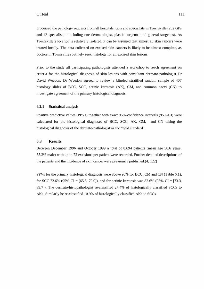
C Heal 111
______________________________________________________________________
processed the pathology requests from all hospitals, GPs and specialists in Townsville (202 GPs
and 42 specialists - including one dermatologist, plastic surgeons and general surgeons). As
Townsville’s location is relatively isolated, it can be assumed that almost all skin cancers were
treated locally. The data collected on excised skin cancers is likely to be almost complete, as
doctors in Townsville routinely seek histology for all excised skin lesions.
Prior to the study all participating pathologists attended a workshop to reach agreement on
criteria for the histological diagnosis of skin lesions with consultant dermato-pathologist Dr
David Weedon. Dr Weedon agreed to review a blinded stratified random sample of 407
histology slides of BCC, SCC, actinic keratosis (AK), CM, and common naevi (CN) to
investigate agreement of the primary histological diagnosis.
6.2.1 Statistical analysis
Positive predictive values (PPVs) together with exact 95%-confidence intervals (95%-CI) were
calculated for the histological diagnoses of BCC, SCC, AK, CM, and CN taking the
histological diagnosis of the dermato-pathologist as the “gold standard”.
6.3 Results
Between December 1996 and October 1999 a total of 8,694 patients (mean age 58.6 years;
55.2% male) with up to 72 excisions per patient were recorded. Further detailed descriptions of
the patients and the incidence of skin cancer were previously published.(4, 122)
PPVs for the primary histological diagnosis were above 90% for BCC, CM and CN (Table 6.1),
for SCC 72.6% (95%-CI = [65.5, 79.0]), and for actinic keratosis was 82.6% (95%-CI = [73.3,
89.7]). The dermato-histopathologist re-classified 27.4% of histologically classified SCCs to
AKs. Similarly he re-classified 10.9% of histologically classified AKs to SCCs.

C Heal 112
______________________________________________________________________
Table 6.1: Agreement between diagnosis of Townsville histologists and a dermato-
histopathology specialist (DW). Positive predictive values (in bold) together with 95%-
confidence intervals are given
Dermato-histopathology specialist
Histological
diagnosis
BCC SCC Actinic
keratosis
Cutaneous
melanoma
Common
naevus
Other
BCC (n=83) 92.8%
84.9,
97.3
2.4% 2.4% 0% 1.2% 1.2%
SCC (n=179) 0% 72.6%
65.5,
79.0
27.4% 0% 0% 0%
Actinic
keratosis
(n=92)
2.2% 10.9% 82.6%
73.3, 89.7
0% 1.1% 3.3%
Cutaneous
melanoma
(n=23)
0% 0% 4.3% 91.3%
72.0, 98.9
4.3% 0%
Common
naevus
(n=30)
0% 0% 6.7% 0% 93.3%
77.9, 99.2
0%
6.4 Discussion
The strengths of our study were a large sample size and close to complete epidemiological data
over a three-year period, which implies reduced selection bias, probably resulting in a
representative sample of skin lesions and doctors. Thus the stratified random sample of slides
that were analysed were likely to be a representative sample of skin cancers removed by doctors
in routine clinical practice.
There are some limitations to the analysis, interpretation and generalisation of these data which
must be acknowledged. Our study looked at agreement between a small group of locally based
histologists and a single dermato-histologist. We have no demographic information regarding
the local histologists, and therefore no information on whether they were representative of
Australian or international histologists. We did not assess the individual histological
characteristics that lead to a diagnosis, and this may have allowed us to define areas to increase

C Heal 113
______________________________________________________________________
reliability. We did not grade whether SCCs were considered to be well or poorly differentiated,
and we did not attempt to measure the degree of agreement.
Agreement between the histologists and the dermato-histopathologist specialist was highest for
BCC, CM and CN. The main area of lack of agreement was between the diagnosis of AK and
SCC. The dermato-histopathologist tended to be more likely to re-classify previously diagnosed
malignant lesions as benign within this sub-group.
Lack of agreement between pathologists regarding the diagnosis of SCC and AK has been
previously recognised in the literature(154, 155) and this is again reflected in our study. This
also illustrates the difficulty involved for doctors in accurately clinically diagnosing lesions for
which a consensus is hard to reach histologically. However this histological diagnosis is of
clinical importance, as the decision on whether to treat a lesion with excision, ablative treatment
or topical medication relies on accurate histological diagnosis.(139) In addition, invasive SCCs
may have the potential to recur and metastasize.(156)
There is a clinical spectrum between invasive SCC, SCC in situ, and AK, however the
boundaries of these conditions are imprecisely defined, and therefore distinction between
diagnosis may be unreliable.(156) To confuse matters further, there is also some controversy in
the literature regarding the proportion of AK which progress to SCC, and indeed whether all
AK should be considered to be SCC.(157)
Given the potential clinical implications of these diagnoses, our results emphasize the need to
improve the reliability of these histological diagnoses. Identification of common types of
histological misclassifications, as in the present study, could help focus pathologists training
and education in order to improve diagnosis.
6.4.1 Conclusion
Agreement between the histologists and the dermato-histopathologist specialist was generally
high. The main area of lack of agreement was between the diagnosis of AK and SCC, with the
dermato-histologist being more likely to classify lesions initially diagnosed as malignant to be
benign.

C Heal 114
______________________________________________________________________
6.5 Summary
Positive predictive values for the primary histological diagnosis were above 90% for basal cell
carcinoma, cutaneous melanoma and common naevus. For squamous cell carcinoma the
positive predictive value was 72.6% (95%-CI = [65.5, 79.0]).
Lack of agreement between histopathologists regarding the diagnosis of squamous cell
carcinoma and actinic keratosis has been previously recognised in the literature and this is again
reflected in our study. The result also illustrates the difficulty involved for doctors in accurately
clinically diagnosing lesions for which a consensus is hard to reach histologically.
In the previous three chapters we explored the case-mix (demographic and tumour
characteristics) of skin cancer excised by GPs in North Queensland, illustrated the importance
of diagnosing skin cancer accuracy, and illustrated some of the areas of diagnostic difficulty.
We established that pathologists may also disagree in their diagnosis of skin cancer. In the next
chapter we use the information from the previous three chapters in order to comment on some of
the limitations in comparing skin cancer doctors with mainstream GPs with regard to diagnostic
accuracy, and examine the difference in case-mix between these two groups.
C Heal 115
______________________________________________________________________
CHAPTER 7: COMPARING THE CASE-MIX AND NUMBER
NEEDED TO TREAT OF GPS AND SKIN CANCER
CLINIC DOCTORS
This chapter presents a letter to the editor in response to a publication comparing the diagnostic
accuracy of skin cancer doctors and general practitioners. The letter also presents some original
research regarding the relative case-mix and number needed to treat (melanocytic naevi excised
per melanoma) using secondary data analysis from the ‘Can sutures get wet?’ project. This
chapter is an extension of both chapter 4 (by discussing case-mix and NNT) and chapter 5 (by
discussing case-mix and diagnostic accuracy) The published article forms Appendix 6.(158)
7.1 Background
There is little data available regarding the diagnostic accuracy of Australian GPs with regard to
excised and biopsied skin lesions. A new recently published study compared the diagnostic
accuracy of GPs and skin cancer clinic doctors.
The objectives of this study are:
(1) To comment on the limitations of comparing skin cancer doctors with mainstream GPs.
(2) To compare the case-mix and number needed to treat between GPs and mainstream skin
cancer doctors using secondary data analysis.
7.2 Letter to the editor
Dear Editor
In a recent article, Youl and colleagues(133) provided information about the ability of doctors to
accurately diagnose skin lesions that they excise or biopsy. We wish to offer some comments
about the comparison between general practitioners (GPs) and skin cancer clinic doctors.
Firstly, the behaviour of GPs and patients in mainstream practice is different from that of
doctors and patients in skin clinics, as is indicated in the comparison of frequency of total skin
examinations (GPs 30.4%, Skin clinic 73.2%). The study does not indicate the circumstances
under which each decision to excise took place. Did patients become aware of a new or
changing skin lesion, and bring it to the attention of the doctor, or did the diagnosis result from

C Heal 116
______________________________________________________________________
a full body skin check which might have resulted in the diagnosis of an earlier, previously
unnoticed and more subtle lesion? It may be useful to separate basal cell carcinoma (BCC) and
squamous cell carcinoma (SCC) into histological subtypes, as early superficial BCC and
intraepidermal SCC may be more difficult to diagnose than other subtypes.(139)
Secondly, the case-mix of non-melanotic skin cancer was quite different between the two
groups (BCC:SCC ratio GPs 1.1:1, skin cancer doctors 2.1:1). This difference in case-mix was
reflected in our own study,(114) where we described the histology of 1,247 lesions excised by
doctors which included 76 lesions undertaken by one doctor in a designated skin cancer clinic.
In an unpublished sub-analysis we divided the results into two settings for comparison (Table
7.1). We found that the skin clinic doctor also had a significantly different (<0.001) non-
melanotic skin cancer case-mix than the GPs, with a much larger BCC:SCC ratio (4:1 to 0.6:1).
We believe this most likely reflects an increased pick up of BCC because of the frequency of
full-body skin examination in the skin clinic context and the consequent detection of lesions of
which the patient is unaware.
Table 7.1: Comparison of lesion excisions in skin cancer clinic and general practice settings
Mean
patient
age
(years)
Proportio
n of
excised
lesions
that were
malignan
t*
BCC :
SCC
ratio
Number
needed to
treat†
Skin cancer clinic 56.5 76% (58/76) 4 : 1 (44/11) 4.7 (14/3)
General practice 56.9 45%
(512/1145)‡
0.6 :
1 (190/305)
9.0 (154/17)
BCC = basal cell carcinoma. SCC = squamous cell carcinoma. * BCC, SCC or melanoma. †
Benign or dysplastic naevi excised per melanoma. ‡ There were 26 cases in which histology
results were missing. All were in the general practice setting
Thirdly, the reported sensitivity and specificity in this study refers only to excised lesions. We
have no information about the lesions that practitioners decided not to excise. The sensitivity
and specificity of a skin examination can only be determined if all relevant skin lesions are
assessed. This would give an accurate representation of the number of true and false negative
diagnosis. However, this would require multiple excisions which would be clinically

C Heal 117
______________________________________________________________________
unacceptable. An important limitation of this study is that it does not assess or compare how
many skin cancers each group of doctors missed.
In conclusion, although this study provides comprehensive information about diagnostic
accuracy, we do not feel a meaningful comparison between the two groups of doctors – based
on the information available – can be made.
7.3 Summary
We found that the case-mix of non-melanocytic skin cancers was significantly different for the
two groups of doctors (P< 0.001) The BCC:SCC ratio was much higher for skin cancer clinic
doctors (4:1) than for GPs (0.6:1). The NNT (melanocytic naevi excised per melanoma) was 4.7
for the skin cancer doctor and 9.0 for mainstream GPs.
There are some limitations in comparing skin cancer doctors with mainstream GPs with regard
to diagnostic accuracy, and the case-mix between these two groups is actually quite
different.We believe the difference in BCC:SCC ratio most likely reflects an increased pick-up
of BCC in skin cancer clinics, owing to the higher frequency of full body skin examinations and
the consequent detection of lesions of which the patient is unaware
The previous chapters of this thesis have presented the results of two large clinical trials
conducted in general practice in North Queensland. The next chapter will discuss the process of
conducting successful practice based research.

C Heal 118
______________________________________________________________________
CHAPTER 8: DISCUSSION, CONCLUSIONS AND
RECOMMENDATIONS
This thesis comprises a literature review and seven original studies. This chapter will integrate
and synthesise the information from the previous chapters. In this chapter, the literature review
will be updated and then the conclusions of the individual chapters with recommendations will
be discussed.
8.1 Update of literature review
New literature (from 2007-2009) which is notable has become available in the following
sections of the literature review.
8.1.1 Incidence of melanoma in Australia
In 2008, new clinical practice guidelines for the management of melanoma in Australia and
New Zealand were published, which provided a summary of the incidence of melanoma in
Australia. In addition, the AIHW cancer registry produces online information regarding
melanoma incidence, although, as stated in the introduction, there is generally a time lag of
around four years before this information becomes available. In the most recent registry
information(17) the age standardised incidence rates in Australia, based on world aged
standardised incidence rates in 2005 were 46.1 for men and 34.6 for women. In 2005 a total of
10,684 melanoma were diagnosed.
A 2002 study used data from the population-based Queensland Cancer Registry to estimate the
annual percentage change in rates across 21 years of incidence data for in situ and invasive
melanomas, stratified by age and sex. In situ melanomas increased by 10.4% per year among
males and 8.4% per year among females. The incidence of invasive lesions has also increased in
males 2.6% per year and females 1.2% per year. Although the shift to more in situ lesions
would tend to reflect improved surveillance, the age standardised incidence is continuing to
increase in all categories. Primary prevention is unlikely to lead to decreases in the overall
incidence of melanoma for at least another 20 years.(159)
A second study analysed data from the Queensland Cancer Registry to investigate geographical
differences and time trends of incidence rate of cutaneous melanoma in Queensland between
1982 and 2002.(160) Yearly age standardised incidence rates world standard population of
cutaneous melanoma averaged over 21 years of observation were highest in the south-eastern

C Heal 119
______________________________________________________________________
part of the state, notably in the statistical division of Moreton for men (54.2) and in Fitzroy for
women (51.4). Inland divisions had on average lower rates than coastal areas. For both men and
women, age standardised incidence rates of invasive and for in situ melanoma increased
between 1982 and 2002 for Queensland as a whole and for almost all its statistical divisions. For
invasive cancer, the increase was strongest for Moreton. For Morton, the annual percentage
change (APC) was men: 5.4%; women: 4.5%; p<0.001, respectively). APCs were higher for in
situ melanoma compared with invasive melanoma for all statistical divisions and both genders.
It was concluded that the behavioural and lifestyle choices might create the observed differences
between statistical divisions.
In conclusion to this updated literature review, the incidence of melanoma in Australia is still
high and increasing, in particular for in-situ melanoma.
8.1.2 Literature update, conclusions, recommendations based on chapter 2: Risk factors
for infection after minor surgery in general practice
The aim of this chapter was to determine the incidence of and risk factors for surgical site
infections (SSI) following minor surgery in a general practice setting. Excisions from lower legs
and feet (p=0.009) or thighs (p=0.005), excisions of BCC (p=0.006) or SCC (p=0.002) and
diabetes (p<0.001) were found to be independent risk factors for wound infection. We felt that
the results of this study could encourage the more judicial use of prophylactic antibiotics by
defining these high-risk groups in a general practice setting, such as diabetics and those
undergoing excision of a non-melanocytic skin cancer or excision from the lower limb.
In 2006, while our ‘Risk factors for infection after minor surgery’ paper was in press, a trial was
published by Dixon et al.(113) It showed the following results (Table 8.1).
Table 8.1: Risk factors for infection, Dixon et al 2006
Setting and
sample size
Study
design
Wound type Incidence
of
infection
Risk factors for
infection
Dixon et al
2006
Australia
Skin cancer
surgery
outpatients,
5,091 lesions,
2,424 patients
Prospective
observational
study
Skin cancer
surgery 1.47%
Below knee
surgery (6.92%)
Groin (10%), Skin
grafts (8.7%)

C Heal 120
______________________________________________________________________
In contrast to our study which had an infection rate of 8.3% this study had a very low infection
rate. This study did not show diabetes to be a risk factor for infection. However both studies
were in agreement that below knee surgery was a risk factor for infection. Dixon et al used
univariate rather than multivariate analysis to determine risk factors for infection.
In 2008 an advisory statement was produced from the Divisions of Dermatological Surgery and
Infectious Diseases at the Mayo Clinic regarding antibiotic prophylaxis in dermatological
surgery (Appendix 11).(105) These guidelines concluded that for the prevention of SSI,
antibiotics may be indicated for procedures on the lower extremities or groin, for wedge
excisions of the lip and ear, skin flaps on the nose, skin grafts and for patients with extensive
inflammatory skin disease.
The guidelines quote ‘An Australian trial involving 857 cutaneous surgical procedures
documented a 8.6% rate of infection, with infections more common in patients with diabetes,
for skin cancer rather than benign lesions, and for surgical sites located on the thighs, legs or
feet’ and ‘ The study by Dixon et al documents increased risk of SSI on sites below the knees.
We have expanded this for patients with prosthetic joints, and to include all sites in the groin
and all areas of the lower extremities based on the findings of the study of Heal, Buettner and
Browning.’
In conclusion, our study, together with the study by Dixon et al, has helped to define risk factors
for infection after dermatological surgery. Even within cohorts with a low overall risk of
infection, some excisions may be at higher risk of infection because of body site, pathology or
patient factors, and infection rate may be greater than 5%.(57, 113) These risk factors may
include excisions from the lower leg, excisions of skin cancers and excisions from diabetic
patients. These risk factors have been adopted to develop a guideline for the use of antibiotic
prophylaxis for minor dermatological surgery.
8.1.3 Literature update, conclusions and recommendations based on chapter 3: Does
single dose of topical chloramphenicol ointment to high risk sutures wound decrease the
incidence of wound infection after minor surgery?
The aim of this study was to compare incidence of infection after minor surgery using a single
application of chloramphenicol ointment versus using paraffin ointment. We concluded that
application of a single dose of topical chloramphenicol to high risk sutured wounds after minor
surgery produces a moderate absolute reduction in infection rate which is statistically but not
clinically significant.

C Heal 121
______________________________________________________________________
The study was published in the BMJ with an accompanying editorial which supported our
findings and recommended that topical antibiotics should not be used for infection prophylaxis
after minor surgery (Appendix 9).(161)
Our article in the BMJ received several rapid responses, some of which questioned our high
infection rate, and criticised the fact that we had not measured skin integrity as a variable.(162)
We responded to these criticisms in ‘rapid responses’. The editorial did suggest that we had
used inflammation as one definition of infection, a matter which we refuted in ‘rapid responses’
(Appendix 10).
Our study analysed a total of 85 infections in order to detect a significant difference between
two treatments. This is the largest number of infections that have been analysed in the published
literature of this type of study. As previously discussed, we feel that previous studies may have
been underpowered to adequately demonstrate the effectiveness of antibiotic ointment in this
setting.
In summary, the main recommendation of this chapter is that prophylactic antibiotics should not
be used routinely after minor surgery. This was supported by an editorial which accompanied
this publication.
8.1.4 Literature update, conclusions and recommendations based on chapter 4: Minor
skin excisions in general practice in North Queensland
In 2007 a study was published in AFP which analysed the billing data in the management of
skin cancer from 1 July 2005 to 30 June 2006 in three Queensland general practices
(metropolitan, provincial, and rural) representing 23,100 patients treated by 23 doctors.(163) As
far as possible, methods were matched to those used in two published studies of skin cancer
clinics. Of the 1,417 skin cancers the number needed to treat (NNT) was 39, compared with 29
in one skin cancer clinic network and 24 in the other. Fifty-four per cent of all excised lesions
were malignant, compared with 60% in one network and 46% in the other, respectively. These
results showed the benign to malignant excision rate was similar in general practice and the skin
cancer clinic networks. This study showed a much higher NNT than our own study, although
skin cancer yield was similar. These results again support our recommendation that GPs in
Mackay could lower their threshold for the excision of pigmented lesions.(163)
A study was published in 2009 which calculated the NNT by skin cancer clinic doctors. It
utilised a large pathology database. NNT was calculated for 10,612 lesions, 6,796 patients, 57

C Heal 122
______________________________________________________________________
doctors and 15 clinics. Overall the NNT was 30 without seborrhoic keratosis and 23 with
seborrhoic keratosis in the denominator. However when four outlying doctors were excluded,
this was decreased to 21 and 15, respectively. There was found to be substantial variability in
individual doctor NNT, with figures varying by individual doctor from 0 to 192 and 117,
respectively.
There are some limitations to the calculation and use of the term NNT in order to measure
performance in the management of skin cancer. The numerator varies in different definitions of
NNT, making results difficult to compare. The measure of NNT may not be indicative of
quality. A high or low NNT does not directly indicate the number of false negative diagnosis of
melanoma, which is the most important factor in the management of melanoma skin cancer.
Case-mix and patient demand may differ in different settings and will influence NNT.
The aim of this chapter was to describe the histology of lesions excised by general practitioners
in rural North Queensland, the sites that skin cancers are removed from and the demographics of
patients presenting with skin cancer. Our findings were that close to half (46.7%) of lesions
excised were skin cancers. We excised more squamous cell carcinomas than basal cell
carcinomas (in a ratio of 1:0.74). Our number needed to treat (melanocytic naevi excised per
melanoma) was 8.4. Mean age for excision of melanoma, basal cell carcinoma and squamous
cell carcinoma was 55, 60.9 and 63.8 yrs respectively. Relative tumour density was greatest in
the face, scalp and neck region for all skin cancers.
My conclusion was that in this sample of Mackay GPs, there was a very high yield of skin
cancers from all excisions. The doctors involved in the study could consider lowering the
threshold for excision of pigmented lesions.
8.1.5 Conclusions and recommendations of chapter 5: Agreement between clinical and
histological diagnosis of skin lesions
The aim of this chapter was to compare the clinical with the histological diagnosis of excised
skin lesions from a set of epidemiological data. We analysed diagnostic accuracy stratified by
histological subtype and body-site and examined the histological nature of misclassified
diagnosis.
My conclusion was that diagnostic accuracy was highest for BCC the most prevalent lesion.
Most excisions were correctly diagnosed or resulted in the removal of malignant lesions. With
non-melanocytic lesions, doctors tended to misclassify benign lesions as malignant, but were

C Heal 123
______________________________________________________________________
less likely to do the reverse. Although a small number of clinically diagnosed common naevi
subsequently proved to be melanoma (6.3%), a higher proportion of all melanoma had been
classified as common naevi (20.9%). Accuracy of diagnosis was dependant on body-site. I
recommended that increased education and training in areas of diagnostic weakness could
improve the management of skin cancers by doctors.
There have been no new papers identified in the literature on the diagnostic accuracy of skin
lesions since this article was published. I am currently developing a manuscript which compares
the diagnostic accuracy of GPs with specialists in the diagnosis of skin cancer.
8.1.6 Conclusions and recommendations of chapter 6: Agreement between histological
diagnosis of skin lesions by pathologists and a histopathologist
The aim of this chapter was to compare agreement for histological diagnosis between local
histopathologists with a dermato-histopathologist from a stratified random sample of excised
skin lesions derived from a set of epidemiological data.
My conclusion was that lack of agreement between histopathologists regarding the diagnosis of
squamous cell carcinoma and actinic keratosis has been previously recognised in the literature
and this is again reflected in our study. The result also illustrates the difficulty involved for
doctors in accurately clinically diagnosing lesions for which a consensus is hard to reach
histologically. There are also implications for the patient - treatment of SCC usually involves
excision, while AK can be treated with ablative procedures. There are also financial
consequences – treatment of SCC generally results in higher Medicare billings than treatment of
AK.
I hope that the results of this study increase awareness between pathologists that there is lack of
agreement in diagnosis, and also that GPs managing skin cancer are aware that diagnoses in
skin cancer are subject to differences in interpretation.
8.1.7 Conclusions and recommendations of chapter 7: Comparing the case-mix and
number needed to treat of GPs and skin cancer clinic doctors
The aims of this chapter were: (1) To comment on the limitations of comparing skin cancer
doctors with mainstream GPs and (2) To compare the case-mix and number needed to treat
between GPs and mainstream skin cancer doctors using secondary data analysis.

C Heal 124
______________________________________________________________________
I found that the case-mix of non-melanocytic skin cancers was significantly different for the two
groups of doctors (p<0.001), and that the BCC:SCC ratio was much higher for skin cancer clinic
doctors (4:1) than for GPs (0.6:1). The NNT (melanocytic naevi excised per melanoma) was 4.7
for the skin cancer doctor and 9.0 for mainstream GPs. I concluded that there are some
limitations in comparing skin cancer doctors with mainstream GPs with regard to diagnostic
accuracy, and the case-mix between these two groups is actually quite different.We believe the
difference in BCC:SCC ratio most likely reflects an increased pick-up of BCC in skin cancer
clinics, owing to the higher frequency of full body skin examinations and the consequent
detection of lesions of which the patient is unaware. I would recommend that these differences
are taken into account in the design and interpretation of any future studies comparing the
diagnostic accuracy or NNT between the two groups.
There have been no further studies which have directly compared the diagnostic accuracy or
case-mix between the two groups of doctors prospectively. A study was published in AFP
which compared the billing data between the two groups and used this to compare the
NNT.(163) I am currently analysing data which compares the diagnostic accuracy between GPs
and specialists, and we will use the conclusions and recommendations of this chapter to help
with the analysis and interpretation of the new data.
8.2 Overall conclusions
Patients with minor skin excisions in North Queensland have a high incidence of wound
infection. Topical chloramphenicol ointment decreases this incidence moderately. GPs in North
Queensland have a high yield of skin cancer from their skin excisions and a low number needed
to treat (melanocytic naevi excised per melanoma). Doctors diagnose skin cancers accurately,
but there are some areas of diagnostic difficulty in particular in the diagnosis of actinic keratosis
and squamous cell carcinoma. GPs and skin cancer doctors have a different skin cancer case-
mix. Enabling factors to conducting research in general practice include ownership of the
project, engaging practice nurses, and adequate funding.
8.3 Overall recommendations
1. I recommend that antibiotic prophylaxis prior to minor surgery in general practice be
limited to the high risk groups that were identified. Antibiotic prophylaxis tends to be
over-utilised in the management of minor surgery, particularly as the consequences of
infection are often minor and side-effects from antibiotics, such as allergy, can
potentially be serious.
C Heal 125
______________________________________________________________________
2. The use of topical chloramphenicol ointment to prevent infection after minor surgery is
best reserved for high risk groups. The reduction in infection was only 40%, which was
statistically but not clinically relevant. The overutilization of topical antibiotics has
potentially adverse consequences such as antibiotic resistance and allergic contact
dermatitis.
3. The doctors involved in my study in Mackay could consider lowering their threshold for
excision of pigmented lesions. In our sample of Mackay GPs, there was a very high
yield of skin cancers from all excisions, and NNT (melanocytic naevi excised per
melanoma) of 8.4, which was lower than published data from comparable cohorts.
4. Educational programs for doctors regarding the diagnosis of skin cancer could focus on
areas of diagnostic weakness which were identified in our study, such as differentiating
melanocytic naevi from malignant melanoma, and differentiating clinically and
histologically between actinic keratosis and squamous cell carcinoma.
5. There is increasing evidence for lack of agreement between histopathologists regarding
the diagnosis of squamous cell carcinoma and actinic keratosis, and it is important that
doctors are aware of this area of discordance.
8.4 Where to from here – research questions identified and future studies
The studies involved in this thesis have illustrated the very high infection rate following minor
surgery in general practice in Mackay. Studies conducted at other locations identified in our
literature review report an infection rate following dermatological surgery of 1-5%. In our first
RCT which was conducted as a background to this thesis, we reported a high overall infection
rates of 8%.(7) The overall infection rate in our antibiotic ointment RCT was 8.7%, with an
infection rate in our placebo group of 11%. Further studies could explore theories for this high
infection rate. This could take the form of a case-control study of people with infections after
minor surgery to identify risk factors for infection, or comparing infection rate in a tropical with
a temperate climate. Other factors could be studied in addition to clinical factors such as
occupation, personal hygiene factors, humidity, smoking, and factors involving practice doctors
and nurses.
The author also plans to combine the results of the two RCTs and analyse the risk factors for
infection in a descriptive study, thus providing stronger evidence for risk factors for infection.

C Heal 126
______________________________________________________________________
There is very little existing evidence in this area, and most are small studies which involve only
a small number of infections. Therefore combining these studies would provide much stronger
statistical and clinical evidence for risk factors for infection. It is very important clinically to
establish risk factors for infection in order to optimise antibiotic prophylaxis and post-operative
wound management.
We have explored diagnostic accuracy of doctors and stratified by body site and histological
sub-type of lesion. Further research which is currently ongoing compares the diagnostic
accuracy of GPs and specialists. Further research could involve the development of educational
intervention to target areas of diagnostic inaccuracy such as the interface between squamous cell
carcinoma and actinic keratosis.
Other future studies identified from the literature review would be a meta-analysis of studies
comparing antibiotic ointments as prophylaxis against wound infection. Again, there are
limiting existing published studies in this area, often with very small numbers of infection, and
combining these studies would provide more evidence for the efficacy of antibiotic ointment.
The thesis has illustrated that large randomised controlled trials may be conducted successfully
in a general practice setting
Following the success of two randomised controlled trials involving minor surgery in general
practice, the author is in the process of developing a new RCT which compares the standard
management of sebaceous cysts with the ‘punch excision technique’.
C Heal 127
______________________________________________________________________
REFERENCES
1. Australian Institute of Health and Welfare, Australasian Association of Cancer
Registeries. Cancer in Australia 2000.Cancer Series No. 23. AIHW Cat. No.
CAN18:AIHW,2003
2. Giles GG, Marks R, Foley P. Incidence of non-melanocytic skin cancer treated in Australia. Br Med J (Clin Res Ed). 1988;296(6614):13-7.
3. Stenbeck KD, Balanda KP, Williams MJ, Ring IT, MacLennan R, Chick JE, et al.
Patterns of treated non-melanoma skin cancer in Queensland--the region with the highest incidence rates in the world. Med J Aust. 1990;153(9):511-5.
4. Buettner PG, Raasch BA. Incidence rates of skin cancer in Townsville, Australia. Int J
Cancer. 1998;78(5):587-93.
5. Del Mar CB, Lowe JB. The skin cancer workload in Australian general practice. Aust Fam Physician. 1997;26 Suppl 1:S24-7.
6. intitiative Ncc. The 2002 national non-melanoma skin cancer survey. Melbourne2003.
7. Heal C, Buettner P, Raasch B, Browning S, Graham D, Bidgood R, et al. Can sutures get wet? Prospective randomised controlled trial of wound management in general practice.
Bmj. 2006;332(7549):1053-6.
8. Health Insurance Commission. Medicare benefits schedule. Item statistics reports, 2004. Available at www.hic.gov.au/statistics/dyn_mbs/forms_tab4.shtml.
9. Australian Institute of Health and Welfare. Health system expenditures on cancer and
other neoplasms in Australia, 2000–1. Health and Welfare Expenditure Series No. 22.
AIHW Cat. No. HWE 29. Canberra: AIHW, 2005. 10. Cho CY, Lo JS. Dressing the part. Dermatol Clin. 1998;16(1):25-47.
11. Hill P, Fa’afoi, E;(1995) The Health and Health Care of Australian South Sea Islanders in
Mackay. Brisbane, University of Queensland. 12. Australian Government – Bureau of Meteorology – climate averages for Townsville MO:
www.bom.gov.au.
13. Australian Bureau of Statistics. 2006 Census of Population and Housing. Canberra: Commonwealth of Australia 2006.
14. Townsville city council website – A social atlas
http://www.townsville.qld.gov.au/about/atlas/intro.asp
15. Australian Cancer Network. Clinical Practice Guidelines for the Management of cutaneous melanoma in Australia. Australian Cancer Network and NHMRC (1999).
16. Australian Cancer Network Melanoma Guidelines Revision Working Party. Clinical
Practice Guidelines for the Management of Melanoma in Australia and New Zealand. Cancer Council Australia and Australian Cancer Network, Sydney and New Zealand
Guidelines Group, Wellington (2008).
17. Australian Institute of Health and Welfare (AIHW), Australasian Association of Cancer
Registries. National Cancer Statistics Clearing House. 2007. http://www.aihw.gov.au/cancer/datacubes/index_2007.cfm> accessed 9th September
2007.
18. Raasch BA, Buettner PG. Multiple nonmelanoma skin cancer in an exposed Australian population. Int J Dermatol. 2002;41(10):652-8.
19. English DR, Armstrong BK, Kricker A, Fleming C. Sunlight and cancer. Cancer Causes
Control. 1997;8(3):271-83. 20. Marks R, Jolley D, Dorevitch AP, Selwood TS. The incidence of non-melanocytic skin
cancers in an Australian population: results of a five-year prospective study. Med J Aust.
1989;150(9):475-8.
21. Raasch B, Woolley T. Management of primary superficial basal cell carcinoma. Aust Fam Physician. 2006;35(6):455-8.
22. Green A, Battistutta D, Hart V, Leslie D, Weedon D. Skin cancer in a subtropical
Australian population: incidence and lack of association with occupation. The Nambour Study Group. Am J Epidemiol. 1996;144(11):1034-40.

C Heal 128
______________________________________________________________________
23. Kricker A, English DR, Randell PL, Heenan PJ, Clay CD, Delaney TA, et al. Skin cancer
in Geraldton, Western Australia: a survey of incidence and prevalence. Med J Aust.
1990;152(8):399-407. 24. English DR, Kricker A, Heenan PJ, Randell PL, Winter MG, Armstrong BK. Incidence of
non-melanocytic skin cancer in Geraldton, Western Australia. Int J Cancer.
1997;73(5):629-33. 25. Staples M, Marks R, Giles G. Trends in the incidence of non-melanocytic skin cancer
(NMSC) treated in Australia 1985-1995: are primary prevention programs starting to have
an effect? Int J Cancer. 1998;78(2):144-8.
26. Staples MP, Elwood M, Burton RC, Williams JL, Marks R, Giles GG. Non-melanoma skin cancer in Australia: the 2002 national survey and trends since 1985. Med J Aust.
2006;184(1):6-10.
27. Marks R, Staples M, Giles GG. Trends in non-melanocytic skin cancer treated in Australia: the second national survey. Int J Cancer. 1993;53(4):585-90.
28. Kaldor J, Shugg D, Young B, Dwyer T, Wang YG. Non-melanoma skin cancer: ten years
of cancer-registry-based surveillance. Int J Cancer. 1993;53(6):886-91. 29. Epstein E, Epstein NN, Bragg K, Linden G. Metastases from squamous cell carcinomas
of the skin. Arch Dermatol. 1968;97(3):245-51.
30. Garbe C, Leiter U. Melanoma epidemiology and trends. Clin Dermatol. 2009;27(1):3-9.
31. Lens MB, Dawes M. Global perspectives of contemporary epidemiological trends of cutaneous malignant melanoma. Br J Dermatol. 2004;150(2):179-85.
32. Jones WO, Harman CR, Ng AK, Shaw JH. Incidence of malignant melanoma in
Auckland, New Zealand: highest rates in the world. World J Surg. 1999;23(7):732-5. 33. AIHW. Australian Health Trends 2001. Canberra: Australian Institute of Health and
Welfare2001.
34. MacLennan R, Green AC, McLeod GR, Martin NG. Increasing incidence of cutaneous melanoma in Queensland, Australia. J Natl Cancer Inst. 1992;84(18):1427-32.
35. Burton RC, Coates MS, Hersey P, Roberts G, Chetty MP, Chen S, et al. An analysis of a
melanoma epidemic. Int J Cancer. 1993;55(5):765-70.
36. Czarnecki D, Meehan CJ. Is the incidence of malignant melanoma decreasing in young Australians? J Am Acad Dermatol. 2000;42(4):672-4.
37. Kelly JW, Yeatman JM, Regalia C, Mason G, Henham AP. A high incidence of
melanoma found in patients with multiple dysplastic naevi by photographic surveillance. Med J Aust. 1997;167(4):191-4.
38. Marks R. Dermatology for the non-dermatologist. In: Marks R, editor. MJA essentials:
dermatology. Sydney: Australasian Medical Publishing, 1997.
39. Askew DA, Wilkinson D, Schluter PJ, Eckert K. Skin cancer surgery in Australia 2001-2005: the changing role of the general practitioner. Med J Aust. 2007;187(4):210-4.
40. English DR, Del Mar C, Burton RC. Factors influencing the number needed to excise:
excision rates of pigmented lesions by general practitioners. Med J Aust. 2004;180(1):16-9.
41. Askew DA, Wilkinson D, Patrick GL. Performance indicators of a primary care skin
cancer clinic network. Med J Aust. 2007;186(3):159. 42. Marks R, Jolley D, McCormack C, Dorevitch AP. Who removes pigmented skin lesions?
J Am Acad Dermatol. 1997;36(5 Pt 1):721-6.
43. Del Mar C, Green A, Cooney T, Cutbush K, Lawrie S, Adkins G. Melanocytic lesions
excised from the skin: what percentage are malignant? Aust J Public Health. 1994;18(2):221-3.
44. Del Mar CB, Green AC. Aid to diagnosis of melanoma in primary medical care. Bmj.
1995;310(6978):492-5. 45. Wilkinson D, Askew DA, Dixon A. Skin cancer clinics in Australia: workload profile and
performance indicators from an analysis of billing data. Med J Aust. 2006;184(4):162-4.
46. Berard F, Gandon J. Postoperative Wound Infections: the Influence of Ultraviolet Irradiation of the Operating Room and of Various Other Factors. Ann Surg.
1964;160(Suppl 2):1-192.
C Heal 129
______________________________________________________________________
47. Cruse PJ, Foord R. The epidemiology of wound infection. A 10-year prospective study of
62,939 wounds. Surg Clin North Am. 1980;60(1):27-40.
48. Haas AF, Grekin RC. Antibiotic prophylaxis in dermatologic surgery. J Am Acad Dermatol. 1995;32(2 Pt 1):155-76; quiz 77-80.
49. Mangram AJ, Horan TC, Pearson ML, Silver LC, Jarvis WR. Guideline for prevention of
surgical site infection, 1999. Hospital Infection Control Practices Advisory Committee. Infect Control Hosp Epidemiol. 1999;20(4):250-78; quiz 79-80.
50. Mangram AJ, Horan TC, Pearson ML, Silver LC, Jarvis WR. Guideline for Prevention of
Surgical Site Infection, 1999. Centers for Disease Control and Prevention (CDC) Hospital
Infection Control Practices Advisory Committee. Am J Infect Control. 1999;27(2):97-132; quiz 3-4; discussion 96.
51. Nguyen D, MacLeod WB, Phung DC, Cong QT, Nguy VH, Van Nguyen H, et al.
Incidence and predictors of surgical-site infections in Vietnam. Infect Control Hosp Epidemiol. 2001;22(8):485-92.
52. Amici JM, Rogues AM, Lasheras A, Gachie JP, Guillot P, Beylot C, et al. A prospective
study of the incidence of complications associated with dermatological surgery. Br J Dermatol. 2005;153(5):967-71.
53. Futoryan T, Grande D. Postoperative wound infection rates in dermatologic surgery.
Dermatol Surg. 1995;21(6):509-14.
54. Dettenkofer M, Wilson C, Ebner W, Norgauer J, Ruden H, Daschner FD. Surveillance of nosocomial infections in dermatology patients in a German university hospital. Br J
Dermatol. 2003;149(3):620-3.
55. Sylaidis P, Wood S, Murray DS. Postoperative infection following clean facial surgery. Ann Plast Surg. 1997;39(4):342-6.
56. Lathlean S. Skin cancer in general practice in South Australia. A five year study. Aust
Fam Physician. 1999;28 Suppl 1:S28-31. 57. Heal C, Buettner P, Browning S. Risk factors for wound infection after minor surgery in
general practice. Med J Aust. 2006;185(5):255-8.
58. Culver DH, Horan TC, Gaynes RP, Martone WJ, Jarvis WR, Emori TG, et al. Surgical
wound infection rates by wound class, operative procedure, and patient risk index. National Nosocomial Infections Surveillance System. Am J Med. 1991;91(3B):152S-7S.
59. Haas AF. Antibiotic prophylaxis. Semin Dermatol. 1994;13(1):27-34.
60. Woods RK, Dellinger EP. Current guidelines for antibiotic prophylaxis of surgical wounds. Am Fam Physician. 1998;57(11):2731-40.
61. Maragh SL, Otley CC, Roenigk RK, Phillips PK. Antibiotic prophylaxis in dermatologic
surgery: updated guidelines. Dermatol Surg. 2005;31(1):83-91.
62. Messingham MJ, Arpey CJ. Update on the use of antibiotics in cutaneous surgery. Dermatol Surg. 2005;31(8 Pt 2):1068-78.
63. Nestor MS. Perioperative use of antibiotics: preventing and treating perioperative
infections. J Drugs Dermatol. 2005;4(6 Suppl):s34-6. 64. Erel E, Platt AJ, Ramakrishnan V. Chloramphenicol use in plastic surgery. Br J Plast
Surg. 1999;52(4):326-7.
65. Kraus SJ, Eron LJ, Bottenfield GW, Drehobl MA, Bushnell WD, Cupo MA. Mupirocin cream is as effective as oral cephalexin in the treatment of secondarily infected wounds. J
Fam Pract. 1998;47(6):429-33.
66. Therapeutic Guidelines. Therapeutic guidelines: antibiotic. Version 13. North Melbourne,
Victoria2006. 67. O'Meara S, Al-Kurdi D, Ovington LG. Antibiotics and antiseptics for venous leg ulcers.
Cochrane Database Syst Rev. 2008;(1):CD003557.
68. Berger RS, Pappert AS, Van Zile PS, Cetnarowski WE. A newly formulated topical triple-antibiotic ointment minimizes scarring. Cutis. 2000;65(6):401-4.
69. Pfizer. Product information: Chloromycetin eye ointment. Pfizer, 2005 (available at
www.pfizer.com.au/ProductInfo.aspx). . 70. Rayner SA, Buckley RJ. Ocular chloramphenicol and aplastic anaemia. Is there a link?
Drug Saf. 1996;14(5):273-6.

C Heal 130
______________________________________________________________________
71. Pasapula C, Strick M. The use of chloramphenicol ointment as an adhesive for
replacement of the nail plate after simple nail bed repairs. J Hand Surg [Br].
2004;29(6):634-5. 72. Thornton Spann C, Taylor SC, Weinberg JM. Topical antimicrobial agents in
dermatology. Clin Dermatol. 2003;21(1):70-7.
73. Saryan JA, Dammin TC, Bouras AE. Anaphylaxis to topical bacitracin zinc ointment. Am J Emerg Med. 1998;16(5):512-3.
74. Kamath S, Sinha S, Shaari E, Young D, Campbell AC. Role of topical antibiotics in hip
surgery. A prospective randomised study. Injury. 2005;36(6):783-7.
75. Dire DJ, Coppola M, Dwyer DA, Lorette JJ, Karr JL. Prospective evaluation of topical antibiotics for preventing infections in uncomplicated soft-tissue wounds repaired in the
ED. Acad Emerg Med. 1995;2(1):4-10.
76. Hood R, Shermock KM, Emerman C. A prospective, randomized pilot evaluation of topical triple antibiotic versus mupirocin for the prevention of uncomplicated soft tissue
wound infection. Am J Emerg Med. 2004;22(1):1-3.
77. Smack DP, Harrington AC, Dunn C, Howard RS, Szkutnik AJ, Krivda SJ, et al. Infection and allergy incidence in ambulatory surgery patients using white petrolatum vs bacitracin
ointment. A randomized controlled trial. Jama. 1996;276(12):972-7.
78. Dixon AJ, Dixon MP, Dixon JB. Prospective study of long-term patient perceptions of
their skin cancer surgery. J Am Acad Dermatol. 2007;57(3):445-53. 79. Dixon AJ, Dixon MP, Dixon JB. Randomized clinical trial of the effect of applying
ointment to surgical wounds before occlusive dressing. Br J Surg. 2006;93(8):937-43.
80. Maddox JS, Ware JC, Dillon HC, Jr. The natural history of streptococcal skin infection: prevention with topical antibiotics. J Am Acad Dermatol. 1985;13(2 Pt 1):207-12.
81. Langford JH, Artemi P, Benrimoj SI. Topical antimicrobial prophylaxis in minor wounds.
Ann Pharmacother. 1997;31(5):559-63. 82. Villiger JW, Robertson WD, Kanji K, Ah Chan M, Fetherston J, Hague IK, et al. A
comparison of the new topical antibiotic mupirocin ('Bactroban') with oral antibiotics in
the treatment of skin infections in general practice. Curr Med Res Opin. 1986;10(5):339-
45. 83. Leyden JJ, Bartelt NM. Comparison of topical antibiotic ointments, a wound protectant,
and antiseptics for the treatment of human blister wounds contaminated with
Staphylococcus aureus. J Fam Pract. 1987;24(6):601-4. 84. Eaglstein WH, Mertz PM. "Inert" vehicles do affect wound healing. J Invest Dermatol.
1980;74(2):90-1.
85. Katz S, McGinley K, Leyden JJ. Semipermeable occlusive dressings. Effects on growth
of pathogenic bacteria and reepithelialization of superficial wounds. Arch Dermatol. 1986;122(1):58-62.
86. Geronemus RG, Mertz PM, Eaglstein WH. Wound healing. The effects of topical
antimicrobial agents. Arch Dermatol. 1979;115(11):1311-4. 87. O'Meara S, Cullum N, Majid M, Sheldon T. Systematic reviews of wound care
management: (3) antimicrobial agents for chronic wounds; (4) diabetic foot ulceration.
Health Technol Assess. 2000;4(21):1-237. 88. Hendley JO, Ashe KM. Effect of topical antimicrobial treatment on aerobic bacteria in the
stratum corneum of human skin. Antimicrob Agents Chemother. 1991;35(4):627-31.
89. Livingston DH, Cryer HG, Miller FB, Malangoni MA, Polk HC, Jr., Weiner LJ. A
randomized prospective study of topical antimicrobial agents on skin grafts after thermal injury. Plast Reconstr Surg. 1990;86(6):1059-64; discussion 65.
90. Jones RN, Li Q, Kohut B, Biedenbach DJ, Bell J, Turnidge JD. Contemporary
antimicrobial activity of triple antibiotic ointment: a multiphased study of recent clinical isolates in the United States and Australia. Diagn Microbiol Infect Dis. 2006;54(1):63-71.
91. Fukuda M, Ohashi H, Matsumoto C, Mishima S, Shimomura Y. Methicillin-resistant
Staphylococcus aureus and methicillin-resistant coagulase-negative Staphylococcus ocular surface infection efficacy of chloramphenicol eye drops. Cornea. 2002;21(7
Suppl):S86-9.

C Heal 131
______________________________________________________________________
92. Bradley SF, Ramsey MA, Morton TM, Kauffman CA. Mupirocin resistance: clinical and
molecular epidemiology. Infect Control Hosp Epidemiol. 1995;16(6):354-8.
93. Miller MA, Dascal A, Portnoy J, Mendelson J. Development of mupirocin resistance among methicillin-resistant Staphylococcus aureus after widespread use of nasal
mupirocin ointment. Infect Control Hosp Epidemiol. 1996;17(12):811-3.
94. Marks JG, Belsito DV, DeLeo VA, Fowler JF, Jr., Fransway AF, Maibach HI, et al. North American Contact Dermatitis Group patch test results for the detection of delayed-type
hypersensitivity to topical allergens. J Am Acad Dermatol. 1998;38(6 Pt 1):911-8.
95. Leyden JJ, Kligman AM. Contact dermatitis to neomycin sulfate. Jama.
1979;242(12):1276-8. 96. Blondeel A, Oleffe J, Achten G. Contact allergy in 330 dermatological patients. Contact
Dermatitis. 1978;4(5):270-6.
97. Buckley SA. Survey of patients taking topical medication at their first presentation to eye casualty. Bmj. 1990;300(6738):1497-8.
98. Doona M, Walsh JB. Use of chloramphenicol as topical eye medication: time to cry halt?
Bmj. 1995;310(6989):1217-8. 99. Doona M, Walsh JB. Topical chloramphenicol is an outmoded treatment. Bmj.
1998;316(7148):1903.
100. Laporte JR, Vidal X, Ballarin E, Ibanez L. Possible association between ocular
chloramphenicol and aplastic anaemia--the absolute risk is very low. Br J Clin Pharmacol. 1998;46(2):181-4.
101. Lancaster T, Swart AM, Jick H. Risk of serious haematological toxicity with use of
chloramphenicol eye drops in a British general practice database. Bmj. 1998;316(7132):667.
102. Platt R. Antibiotic prophylaxis in clean surgery: does it work? Should it be used if it
does? New Horiz. 1998;6(2 Suppl):S53-7. 103. National Primary Care Research and Development Centre and Centre for Public Policy
and Management of the University of Manchester. Outpatient services and primary care.
A scoping review of research into strategies for improving outpatient effectiveness and
efficiency. A report to the NHS Service Delivery and Organisation R & D Programme. Manchester. 2005.
104. O'Cathain A, Brazier JE, Milner PC, Fall M. Cost effectiveness of minor surgery in
general practice: a prospective comparison with hospital practice. Br J Gen Pract. 1992;42(354):13-7.
105. Wright TI, Baddour LM, Berbari EF, Roenigk RK, Phillips PK, Jacobs MA, et al.
Antibiotic prophylaxis in dermatologic surgery: advisory statement 2008. J Am Acad
Dermatol. 2008;59(3):464-73. 106. Nagachinta T, Stephens M, Reitz B, Polk BF. Risk factors for surgical-wound infection
following cardiac surgery. J Infect Dis. 1987;156(6):967-73.
107. Bruce J, Russell EM, Mollison J, Krukowski ZH. The quality of measurement of surgical wound infection as the basis for monitoring: a systematic review. J Hosp Infect.
2001;49(2):99-108.
108. Britt H KS, et al. General Practice activity in Australia 2000-2001. University of Sydney. 2001.
109. CLIB 2001- Basic community profile. 2001 Census of Population and Housing.
Australian Bureau of statistics 2001.
110. Australian Bureua of Meteorology - climate averages for Mackay. [May 2008]; Available from: www.bom.gov.au.
111. Heal CF, Buettner PG, Cruickshank R, Graham D, Browning S, Pendergast J, et al. Does
single application of topical chloramphenicol to high risk sutured wounds reduce incidence of wound infection after minor surgery? Prospective randomised placebo
controlled double blind trial. Bmj. 2009;338:a2812.
112. Allen L. Compounding Gels. Secundum Artem. 2007;Vol 4(No. 5).

C Heal 132
______________________________________________________________________
113. Dixon AJ, Dixon MP, Askew DA, Wilkinson D. Prospective study of wound infections in
dermatologic surgery in the absence of prophylactic antibiotics. Dermatol Surg.
2006;32(6):819-26; discussion 26-7. 114. Heal C, Buettner P, Raasch B, Browning S. Minor skin excisions in general practice in
North Queensland. Aust Fam Physician. 2006;35(10):825-8.
115. Edwards PS, Lipp A, Holmes A. Preoperative skin antiseptics for preventing surgical wound infections after clean surgery. Cochrane Database Syst Rev. 2004;(3):CD003949.
116. Edlich R. Biochemical and clinical aspects. IK Cohen RD, WJ Linblad, editor. Sydney:
WB Saunders; 1992.
117. Thomas S. Wound management and dressings. London: The Pharmaceutical Press; 1990. 118. Colebrook L, Hood AM. Infection through soaked dressings. Lancet. 1948;2(6531):682.
119. British National Formulary2006.
120. JG Marks DB, VA Delco, JF Fowler, AF Fransway, HI Mailbach, et al. North American contact dermatitis group patch test results for the detection of delayed-type
hypersensitivity topical allergerns. J Am Acad Dermatol. 1998;38:911-8.
121. Pearl DK, Scott EL. The anatomical distribution of skin cancers. Int J Epidemiol. 1986;15(4):502-6.
122. Raasch B, Maclennan R, Wronski I, Robertson I. Body site specific incidence of basal
and squamous cell carcinoma in an exposed population, Townsville, Australia. Mutat Res.
1998;422(1):101-6. 123. Green A, MacLennan R, Youl P, Martin N. Site distribution of cutaneous melanoma in
Queensland. Int J Cancer. 1993;53(2):232-6.
124. Kricker A, Armstrong BK, English DR. Sun exposure and non-melanocytic skin cancer. Cancer Causes Control. 1994;5(4):367-92.
125. Armstrong BK. Epidemiology of malignant melanoma: intermittent or total accumulated
exposure to the sun? J Dermatol Surg Oncol. 1988;5(4):367-92. 126. Kricker A, Armstrong BK, English DR, Heenan PJ. A dose-response curve for sun
exposure and basal cell carcinoma. Int J Cancer. 1995;60(4):482-8.
127. Heal CF, Raasch BA, Buettner PG, Weedon D. Accuracy of clinical diagnosis of skin
lesions. Br J Dermatol. 2008;159(3):661-8. 128. Diepgen TL MV. The epidemiology of skin cancer. Brit J Dermatol. 2002;146(61):1-6.
129. Charles J, Knox S, Britt H. Skin excisions in Australian general practice. Aust Fam
Physician. 2006;35(7):474-5. 130. McKenna DB, Marioni JC, Lee RJ, Prescott RJ, Doherty VR. A comparison of
dermatologists', surgeons' and general practitioners' surgical management of cutaneous
melanoma. Br J Dermatol. 2004;151(3):636-44.
131. Khorshid SM, Pinney E, Bishop JA. Melanoma excision by general practitioners in north-east Thames region, England. Br J Dermatol. 1998;138(3):412-7.
132. Thomson MA, Loffeld A, Marsden JR. More skin cancer detected from nonurgent
referrals. Br J Dermatol. 2005;153(2):453-4. 133. Youl PH, Baade PD, Janda M, Del Mar CB, Whiteman DC, Aitken JF. Diagnosing skin
cancer in primary care: how do mainstream general practitioners compare with primary
care skin cancer clinic doctors? Med J Aust. 2007;187(4):215-20. 134. Moffatt CR, Green AC, Whiteman DC. Diagnostic accuracy in skin cancer clinics: the
Australian experience. Int J Dermatol. 2006;45(6):656-60.
135. Wilkinson D, Bourne P, Dixon A, Kitchener S. Skin cancer medicine in primary care:
towards an agenda for quality health outcomes. Med J Aust. 2006;184(1):11-2. 136. Nixon RL, Dorevitch AP, Marks R. Squamous cell carcinoma of the skin. Accuracy of
clinical diagnosis and outcome of follow-up in Australia. Med J Aust. 1986;144(5):235-9.
137. Jonna BP, Delfino RJ, Newman WG, Tope WD. Positive predictive value for presumptive diagnoses of skin cancer and compliance with follow-up among patients
attending a community screening program. Prev Med. 1998;27(4):611-6.
138. Green A, Leslie D, Weedon D. Diagnosis of skin cancer in the general population: clinical accuracy in the Nambour survey. Med J Aust. 1988;148(9):447-50.
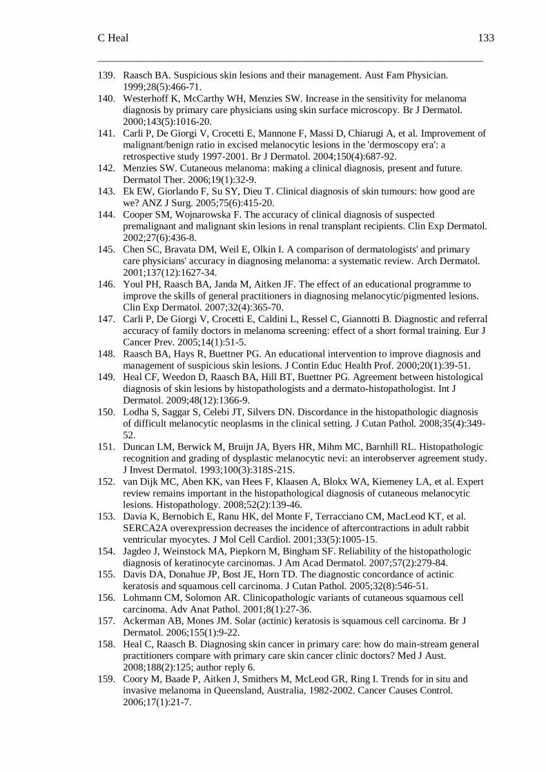
C Heal 133
______________________________________________________________________
139. Raasch BA. Suspicious skin lesions and their management. Aust Fam Physician.
1999;28(5):466-71.
140. Westerhoff K, McCarthy WH, Menzies SW. Increase in the sensitivity for melanoma diagnosis by primary care physicians using skin surface microscopy. Br J Dermatol.
2000;143(5):1016-20.
141. Carli P, De Giorgi V, Crocetti E, Mannone F, Massi D, Chiarugi A, et al. Improvement of malignant/benign ratio in excised melanocytic lesions in the 'dermoscopy era': a
retrospective study 1997-2001. Br J Dermatol. 2004;150(4):687-92.
142. Menzies SW. Cutaneous melanoma: making a clinical diagnosis, present and future.
Dermatol Ther. 2006;19(1):32-9. 143. Ek EW, Giorlando F, Su SY, Dieu T. Clinical diagnosis of skin tumours: how good are
we? ANZ J Surg. 2005;75(6):415-20.
144. Cooper SM, Wojnarowska F. The accuracy of clinical diagnosis of suspected premalignant and malignant skin lesions in renal transplant recipients. Clin Exp Dermatol.
2002;27(6):436-8.
145. Chen SC, Bravata DM, Weil E, Olkin I. A comparison of dermatologists' and primary care physicians' accuracy in diagnosing melanoma: a systematic review. Arch Dermatol.
2001;137(12):1627-34.
146. Youl PH, Raasch BA, Janda M, Aitken JF. The effect of an educational programme to
improve the skills of general practitioners in diagnosing melanocytic/pigmented lesions. Clin Exp Dermatol. 2007;32(4):365-70.
147. Carli P, De Giorgi V, Crocetti E, Caldini L, Ressel C, Giannotti B. Diagnostic and referral
accuracy of family doctors in melanoma screening: effect of a short formal training. Eur J Cancer Prev. 2005;14(1):51-5.
148. Raasch BA, Hays R, Buettner PG. An educational intervention to improve diagnosis and
management of suspicious skin lesions. J Contin Educ Health Prof. 2000;20(1):39-51. 149. Heal CF, Weedon D, Raasch BA, Hill BT, Buettner PG. Agreement between histological
diagnosis of skin lesions by histopathologists and a dermato-histopathologist. Int J
Dermatol. 2009;48(12):1366-9.
150. Lodha S, Saggar S, Celebi JT, Silvers DN. Discordance in the histopathologic diagnosis of difficult melanocytic neoplasms in the clinical setting. J Cutan Pathol. 2008;35(4):349-
52.
151. Duncan LM, Berwick M, Bruijn JA, Byers HR, Mihm MC, Barnhill RL. Histopathologic recognition and grading of dysplastic melanocytic nevi: an interobserver agreement study.
J Invest Dermatol. 1993;100(3):318S-21S.
152. van Dijk MC, Aben KK, van Hees F, Klaasen A, Blokx WA, Kiemeney LA, et al. Expert
review remains important in the histopathological diagnosis of cutaneous melanocytic lesions. Histopathology. 2008;52(2):139-46.
153. Davia K, Bernobich E, Ranu HK, del Monte F, Terracciano CM, MacLeod KT, et al.
SERCA2A overexpression decreases the incidence of aftercontractions in adult rabbit ventricular myocytes. J Mol Cell Cardiol. 2001;33(5):1005-15.
154. Jagdeo J, Weinstock MA, Piepkorn M, Bingham SF. Reliability of the histopathologic
diagnosis of keratinocyte carcinomas. J Am Acad Dermatol. 2007;57(2):279-84. 155. Davis DA, Donahue JP, Bost JE, Horn TD. The diagnostic concordance of actinic
keratosis and squamous cell carcinoma. J Cutan Pathol. 2005;32(8):546-51.
156. Lohmann CM, Solomon AR. Clinicopathologic variants of cutaneous squamous cell
carcinoma. Adv Anat Pathol. 2001;8(1):27-36. 157. Ackerman AB, Mones JM. Solar (actinic) keratosis is squamous cell carcinoma. Br J
Dermatol. 2006;155(1):9-22.
158. Heal C, Raasch B. Diagnosing skin cancer in primary care: how do main-stream general practitioners compare with primary care skin cancer clinic doctors? Med J Aust.
2008;188(2):125; author reply 6.
159. Coory M, Baade P, Aitken J, Smithers M, McLeod GR, Ring I. Trends for in situ and invasive melanoma in Queensland, Australia, 1982-2002. Cancer Causes Control.
2006;17(1):21-7.
C Heal 134
______________________________________________________________________
160. Buettner PG, MacLennan R. Geographical variation of incidence of cutaneous melanoma
in Queensland. Aust J Rural Health. 2008;16(5):269-77.
161. Grey JE, Healy B, Harding K. Antibiotic prophylaxis for minor dermatological surgery in primary care. Bmj. 2009;338:a2749.
162. Weatherhead SC, Lawrence CM. Antibiotics for skin surgery. Preoperative integrity of
skin surface predicts infection risk. Bmj. 2009;338:b516. 163. Byrnes P, Ackermann E, Williams ID, Mitchell GK, Askew D. Management of skin
cancer in Australia--a comparison of general practice and skin cancer clinics. Aust Fam
Physician. 2007;36(12):1073-5.

C Heal 135
______________________________________________________________________
LIST OF APPENDIXES
Appendix 1. Risk factors for wound infection after minor surgery in general practice
Appendix 2. Does single application of topical chloramphenicol to high risk sutured
wounds reduce incidence of wound infection after minor surgery?
Prospective randomised placebo controlled double blind trial
Appendix 3. Minor skin excisions in general practice in North Queensland
Appendix 4. Accuracy of clinical diagnosis of skin lesions
Appendix 5. Agreement between histological diagnosis of skin lesions by
histopathologists and a dermato-histopathologist
Appendix 6. Diagnosing skin cancer in primary care: how do mainstream general
practitioners compare with primary care skin cancer clinic doctors?
Appendix 7. Practice based research – Lessons from the field
Appendix 8. Ethics Approval
Appendix 9. Consent form and patient information sheet
Appendix 10. Data collection sheets
Appendix 11. Body site map
Appendix 12. Excision protocol
Appendix 13. Study protocol
Appendix 14. Consort check list
Appendix 15. Rapid responses to BMJ chloramphenicol article
Appendix 16. BMJ editorial (Grey et al)

C Heal 136
______________________________________________________________________
Appendix 17. Rapid responses to BMJ editorial
Appendix 18. Antibiotic prophylaxis in dermatologic surgery:
Advisory statement 2008 (Wright et al)
Appendix 19. Can sutures get wet?
Appendix 20. How to dissect surgical journals: VII – Comparing outcomes
Appendix 21. How to dissect surgical journals: IX – Sample size

Diagnosis of skin lesions, C Heal 137
______________________________________________________________________
APPENDIX 1.
RISK FACTORS FOR WOUND INFECTION AFTER
MINOR SURGERY IN GENERAL PRACTICE

Diagnosis of skin lesions, C Heal 138
______________________________________________________________________

Diagnosis of skin lesions, C Heal 139
______________________________________________________________________

Diagnosis of skin lesions, C Heal 140
______________________________________________________________________

Diagnosis of skin lesions, C Heal 141
______________________________________________________________________

Diagnosis of skin lesions, C Heal 142
______________________________________________________________________
APPENDIX 2.
DOES SINGLE APPLICATION OF TOPICAL
CHLORAMPHENICOL TO HIGH RISK SUTURED
WOUNDS REDUCE INCIDENCE OF WOUND
INFECTION AFTER MINOR SURGERY?
PROSPECTIVE RANDOMISED PLACEBO
CONTROLLED DOUBLE BLIND TRIAL

Diagnosis of skin lesions, C Heal 143
______________________________________________________________________

Diagnosis of skin lesions, C Heal 144
______________________________________________________________________

Diagnosis of skin lesions, C Heal 145
______________________________________________________________________

Diagnosis of skin lesions, C Heal 146
______________________________________________________________________
Diagnosis of skin lesions, C Heal 147
______________________________________________________________________
Diagnosis of skin lesions, C Heal 148
______________________________________________________________________

Diagnosis of skin lesions, C Heal 149
______________________________________________________________________
APPENDIX 3.
MINOR SKIN EXCISIONS IN GENERAL
PRACTICE IN NORTH QUEENSLAND

Diagnosis of skin lesions, C Heal 150
______________________________________________________________________

Diagnosis of skin lesions, C Heal 151
______________________________________________________________________

Diagnosis of skin lesions, C Heal 152
______________________________________________________________________

Diagnosis of skin lesions, C Heal 153
______________________________________________________________________

Diagnosis of skin lesions, C Heal 154
______________________________________________________________________
APPENDIX 4.
ACCURACY OF CLINICAL DIAGNOSIS OF SKIN
LESIONS

Diagnosis of skin lesions, C Heal 155
______________________________________________________________________

Diagnosis of skin lesions, C Heal 156
______________________________________________________________________

Diagnosis of skin lesions, C Heal 157
______________________________________________________________________

Diagnosis of skin lesions, C Heal 158
______________________________________________________________________

Diagnosis of skin lesions, C Heal 159
______________________________________________________________________

Diagnosis of skin lesions, C Heal 160
______________________________________________________________________

Diagnosis of skin lesions, C Heal 161
______________________________________________________________________

Diagnosis of skin lesions, C Heal 162
______________________________________________________________________

Diagnosis of skin lesions, C Heal 163
______________________________________________________________________
APPENDIX 5.
AGREEMENT BETWEEN HISTOLOGICAL
DIAGNOSIS OF SKIN LESIONS BY
HISTOPATHOLOGISTS AND A
DERMATOPATHOLOGIST

Diagnosis of skin lesions, C Heal 164
______________________________________________________________________

Diagnosis of skin lesions, C Heal 165
______________________________________________________________________

Diagnosis of skin lesions, C Heal 166
______________________________________________________________________

Diagnosis of skin lesions, C Heal 167
______________________________________________________________________

Diagnosis of skin lesions, C Heal 168
______________________________________________________________________
APPENDIX 6.
DIAGNOSING SKIN CANCER IN PRIMARY
CARE: HOW DO MAINSTREAM GENERAL
PRACTITIONERS COMPARE WITH PRIMARY
CARE SKIN CANCER CLINIC DOCTORS?

Diagnosis of skin lesions, C Heal 169
______________________________________________________________________

Diagnosis of skin lesions, C Heal 170
______________________________________________________________________
APPENDIX 7.
PRACTICE BASED RESEARCH – LESSONS FROM
THE FIELD

Diagnosis of skin lesions, C Heal 171
______________________________________________________________________

Diagnosis of skin lesions, C Heal 172
______________________________________________________________________

Diagnosis of skin lesions, C Heal 173
______________________________________________________________________

Diagnosis of skin lesions, C Heal 174
______________________________________________________________________

Diagnosis of skin lesions, C Heal 175
______________________________________________________________________
APPENDIX 8.
ETHICS APROVAL

Diagnosis of skin lesions, C Heal 176
______________________________________________________________________
APPENDIX 9.
CONSENT FORM AND
PATIENT INFORMATION SHEET
School: James Cook University School of Medicine
Project: Topical Chloromycetin ointment to sutured wounds
Chief Investigator: Dr Clare Heal
Contact Detail: James Cook University, Mackay Base Hospital, Q4740
Tel 49686630
Ethics Officer Contact detail: Tina Langford Tel 47814342
PATIENT INFORMATION SHEET
We would like to invite you to take part in a research study being conducted by your
General Practitioner under the supervision of the James Cook University.
DO YOU NEED TO TAKE PART?
Before telling you anything about the study we need to make it clear that you do not need to take part unless
you want to. Whether you decide to participate or not will make no difference to the care you receive as a patient at the practice.
If you do not wish to take part simply inform your doctor or nurse.
There will be no additional cost to you if you are happy to participate
GENERAL INFORMATION ABOUT THE STUDY
The aim of the study is to determine whether applying antibiotic ointment makes any
difference to the chance of getting an infection.
IF YOU TAKE PART
If you decide to take part in the study you will be randomly assigned to one of two
groups.
TOPICAL ANTIBIOTIC GROUP
You will receive a single application of an antibiotic ointment after your wound
has been sutured. Please keep your wound dry and covered for 24hrs. After
24hrs take your dressing off and shower or bathe as normal. If the wound
becomes hot, swollen or red, return to your doctor to have the wound checked.
CONTROL GROUP
You will receive a single application of Vaseline ointment after your wound has

Diagnosis of skin lesions, C Heal 177
______________________________________________________________________
been sutured. Please keep your dressing covered and dry for the first 24hrs.
After 24hrs you may take your dressing off and shower or bathe as normal. If the
wound becomes hot, swollen or red, return to your doctor to have your wound
checked
If you have any concerns you can discuss these with your doctor. Your doctor will also
ask you to sign the consent form.
WILL THE INFORMATION BE CONFIDENTIAL?
All the information collected during the study will be confidential. Only your GP and the
study investigators will know that the information is related to you.
WHAT IF I GET A WOUND INFECTION?
Wound infections occur in around 5-20% of skin excisions, so there is a chance that
you will get an infection, whether you have topical antibiotics or not. If you notice that
your wound has become infected, we would like you to see your GP straight away. You
will also be asked to attend your GP after a suitable period of time to have your sutures
removed. If at this point the wound is infected, it will be treated like any other infection,
possibly with antibiotic tablets.
POSSIBLE RISKS, DISCOMFORTS, BENEFITS
There is a very small possibility of having a local allergy to the antibiotic ointment. This
would not be serious and could be treated with a different ointment.
There may be a tiny risk of developing life-threatening anaemia when Chloromycetin
ointment is applied to the eye, however if is thought to completely safe in a single dose
applied to the skin.
Hopefully the results of this study will show if antibiotic ointment can help to prevent
infection after minor skin surgery.
INFORMED CONSENT FORM
School: James Cook University School of Medicine
Project: Topical Chloromycetin to sutured wounds
Chief Investigator: Dr Clare Heal
Contact Detail: James Cook University, Mackay Base Hospital, Q4740
Tel 49686630
Description:
This study involves being randomly assigned to a group where either antibiotic
ointment will be applied to your wound, or no ointment will be applied (our
Diagnosis of skin lesions, C Heal 178
______________________________________________________________________
current normal management) The wound will be reassessed when you come
back for removal of sutures, so there is no extra time commitment involved. The
outcomes of this study will be used to improve the way wounds are managed by
GPs in Mackay. The results of the study may be used for a publication, but all
information will be kept confidential.
CONSENT
The aims of this study have been clearly explained to me and I understand what is wanted of me. I
know that taking part in this study is voluntary and I am aware that I can stop taking part in it at any
time and may refuse to answer any questions.
I understand that any information I give will be kept strictly confidential and that no names
will be used to identify me with this study without my approval.
Name: (printed)
Signature:
Date:

Diagnosis of skin lesions, C Heal 179
______________________________________________________________________
APPENDIX 10.
DATA COLLECTION SHEETS

Diagnosis of skin lesions, C Heal 180
______________________________________________________________________
Wound Care Project- Data Collection Sheet. Please record patients included in trial
ID Date Dr Patient name DOB Sex
Flap
2
level
Site
(see
body
map)
Size
length
Tea
Cup/
Yrs
DM
Y/N
Other
Eg PVD
Anaemia,
cancer
histology
ROS
Date
Wound
score
0-4
Infec
Y/N
Abs
Y/N
Compliant
Y/N

Diagnosis of skin lesions, C Heal 181
______________________________________________________________________
TOTAL

Diagnosis of skin lesions, C Heal 182
______________________________________________________________________
APPENDIX 11.
BODY SITE MAP BODY SITE MAPBODY SITES
1) Anterior arm 7) Anterior thigh 13) Torso posterior
2) Posterior arm 8) Posterior thigh 14) Neck
3) Anterior forearm 9) Anterior leg 15) Scalp
4) Posterior forearm 10) Posterior leg 16) Face
5) Back hand 11) Foot 17) Nose
6) Palm hand 12)Torso anterior 18) Ear
19) Lip

Diagnosis of skin lesions, C Heal 183
______________________________________________________________________
APPENDIX 12.
EXCISION PROTOCOL
EXCISION PROTOCOL
1) Skin prep – N Saline
2) Usual aseptic technique, including sterile gloves
3) Nylon sutures
4) Local anaesthetic – 1% lignocaine with adrenaline
5) Dressing – Melolin and tape, Primapore, Cutiplast ® or Cosmopor, (not tegaderm,
hydrofilm or waterproof dressing)
6) No other topical antiseptic or antibiotic
INCLUSION CRITERIA
All sutured wounds:
Patients presenting for excision of a minor skin lesion
All body sites including face
Diabetes, Anaemia, PVD, malignancy (but record chronic disease)
Flap/2 level procedure
Multiple excisions on same date (give each excision separate number)
Excisions on same patient on different dates
Lacerations
Sutured punch or shave biopsies
Excision of sebaceous cyst
EXCLUSION CRITERIA
Incision and drainage of infected sebaceous cyst
Immunosuppressive drugs
Already taking oral antibiotics
Oral/topical abs clinically indicated postoperatatively
Personal/Family History of Aplastic anaemia
Non-sutured biopsies, steristripped skin tears
Allergy to any ingredients of Chloromycetin ointment

Diagnosis of skin lesions, C Heal 184
______________________________________________________________________
DIAGOSIS OF INFECTION
1) Within 30 days of excision
2) a) Purulent discharge from wound or
b) Doctor diagnosis infection or commences antibiotics
3) Stitch abscess does not count as infection
AND
0- no infection 3- Erythema >2cm
1- Stitch abscess 4- Deep infection/ Systemic symptoms 2- Erythema <2cm
BODY SITE CATEGORIES (see body map)
1)Anterior arm 7)Anterior thigh 12)Anterior torso
2)Posterior arm 8)Posterior thigh 13Posterior torso
3)Anterior forearm 9)Anterior leg 14)Neck
4)Posterior forearm 10)Posterior leg 15)Scalp
5)Back hand 11)Foot 16)Face
6)Palm hand
TEA
Tea cup years
average number of cups per day/ number of years consumed for
eg 5/10 = 5 cups of tea per day for 10 years
HISTOLOGY
1)MM- Malignant melanoma
2)BCC- Basal Cell Carcinoma
3)SCC- Squamous Cell Carcinoma
4)BN- Benign naevus
5)DN- Dysplastic naevus
6)SK- Solar keratosis
7)SW- Seborrhoic keratosis

Diagnosis of skin lesions, C Heal 185
______________________________________________________________________
8)Other (write in)
11)RMM- re-excision malignant melanoma
12)RBCC- re-excision Basal Cell Carcinoma
13)RSCC- re-excision Squamous Cell Carcinoma
MEDICAL CONDITIONS TO RECORD
Anaemia
Peripheral vascular disease
Cancer (current)
Oral steroids
Continuous inhaled steroids
Oral warfarin
Aspirin or clopidogrel
Ischaemic Heart Disease
COPD
SMOKING
Yes/No/Ex

Diagnosis of skin lesions, C Heal 186
______________________________________________________________________
APPENDIX 13.
STUDY PROTOCOL
Dr Clare Heal – Study Protocol
1. PROJECT INFORMATION
1.1 Project Outline
Aim
To determine whether the application of topical chloramphenicol ointment
(Chloromycetin) to sutured wounds reduces the incidence of wound infection following
minor surgery.
Background / Literature review:
A previous study of wound infection involving GPs in Mackay, Queensland showed an
overall incidence of wound infection of 8.6%, with infection rates at four general
practice centres of 2.9%, 7.8%, 10.0% and 10.2%.1,2 This was higher than we expected
from published results in a similar Australian general practice cohort (1.9%)3 and a
similar European dermatology clinic cohort (2%)4 . The acceptable rate of infection
following clean minor surgery is 5%5,6,7 . We have consequently decided to investigate a
strategy to reduce this infection rate.
Although some practitioners apply topical antibiotic ointment for infection prophylaxis
following minor excisions, there is little data available regarding the effectiveness of
this treatment. There are several different antibiotic ointments used in clinical practice
including Chloromycetin, neosporin and mupirocin.
Chloromycetin ointment consists of 10mg/g of chloramphenicol, in plastibase 30W and
liquid paraffin.8 It is indicated for treatment of bacterial conjunctivitis and there is little
evidence for its effectiveness in prophylaxis or treatment of wound infection. Despite
this, it is regularly used in areas outside its main indication. Several of the investigating
GPs had applied it to sutured wounds as prophylaxis against wound infection. A survey
of UK plastic surgeons reported that 66% used chloramphenicol eye ointment in their

Diagnosis of skin lesions, C Heal 187
______________________________________________________________________
practice, mainly as prophylaxis against infection.9 The ointment has been used as an
adhesive for replacement of the nailbed.10 Comprehensive medline search found only
one study relating to the use of topical Chloromycetin ointment on wounds. This study
investigated the application of Chloromycetin ointment to wounds following hip
replacement.11 The incidence of wound infection in the intervention group was reduced
(4% v 8%) but the sample size was small and the results not statistically significant.
Neosporin ointment is also known as triple antibiotic ointment (TAO) in the USA. Each
gram of Neosporin ointment contains polymixin B sulfate 5,000 units, neomycin sulfate
5mg and bacitracin zinc 400units in a paraffin ointment base12. Comprehensive medline
search revealed two randomised studies involving Neosporin ointment. One compared
application of Neosporin, bactracin zinc, silver sulfadiazine and paraffin ointment to
uncomplicated sutured wounds. This concluded that neosporin did reduce the incidence
of infection (4.5%v 6.6%v12.1%v17.6%); however the sample size was too small for
the results to be statistically significant.13 A second small pilot study found a similar
rate of wound infection and adverse events between uncomplicated sutured soft tissue
wound treated with Neosporin ointment or mupirocin ointment (4%v 0%).14 Neosporin
ointment has been found to be safe, effective and significantly better than simple gauze
dressing alone, in minimising the appearance of scars resulting from clean dermabrasion
wounds.15
Bactroban ointment contains mupirocin. Interestingly, a recent large Australian study
showed that application of mupirocin ointment prior to application of an occlusive
dressing did not decrease the incidence of wound infection (2.3% intervention v 1.4%
control).16
There is some concern regarding the overuse of topical antibiotics resulting in antibiotic
resistance. Australian guidelines suggest that topical antibiotic use be restricted because
of the capacity of most topical drugs to select resistant micro-organisms and to cause
sensitization. The guidelines also suggest that antimicrobials recommended for topical
use are selected from classes not in use for systemic therapy.17 Patterns of antimicrobial
activity and resistance have been examined for Neosporin and mupirocin ointment In
one trial, neomycin plus bactracin was associated with the rapid emergence of drug-
resistance organisms whereas topical silver nitrate was not.18 However Neosporin

Diagnosis of skin lesions, C Heal 188
______________________________________________________________________
ointment has also been shown to have a wider spectrum of antibacterial activity
compared with mupirocin and to be usable against mupirocin resistant Gram-positive
strains. 19Chloramphenicol eye drops have been shown to be effective in the treatment
of MRSA ocular surface infections.20
In one study, antimicrobial treatment including antiseptics and antibiotic ointment were
shown to eradicate bacteria from the skin surface when applied topically. Mupirocin and
Neosporin ointment were also been shown to eradicate bacteria from the multiple
keratinised layers of the stratum corneum and in addition neosporin prevented
repopulation with resident flora.21 There are no similar medline listed studies involving
Chloromycetin ointment.
There is some concern about the incidence of allergic contact dermatitis with topical
neomycin use. This has been shown to be as high as 11% in a population referred for
diagnostic patch testing.22 However there is some evidence that the incidence of this
reaction is as low as 1% when the ointment is used in the general population.23 The
reaction is much more common amongst subjects previously exposed to neomycin
ointment.22Contact allergy has been reported with the use of chloramphenicol ointment
but the incidence is thought to be low.24, 25
Neosporin ointment has been available over the counter in the USA since the 1970s,
while it has been confined to a prescription medication in Australia. It ceased to be
available in Australia in Oct 2006, because of non-availability of an ingredient.26.
Topical ocular chloramphenicol is widely used in the UK and Australia for the
treatment of conjunctivitis, but is very rarely prescribed for this indication in the US.27
There was previously some controversy regarding the link between aplastic anaemia
and topical ocular chloramphenicol, based on a small number of single case reports,27
however two international case-control studies provided no support to this association.
Although the association between ocular chloramphenicol and aplastic anaemia cannot
be excluded, the risk is less than one million per treatment courses28. There have been
no incidences of aplastic anamia following dermatological application, despite
widespread use. Because of current accessibility in Australia and lack published data we
decided to study the use of Chloromycetin ointment.

Diagnosis of skin lesions, C Heal 189
______________________________________________________________________
Our trial seeks to establish the effectiveness of Chloromycetin ointment in preventing
wound infection after dermatological surgery. We intend to apply it as a single dose
post operatively, with sterile paraffin ointment (lacrilube) used as a control. Wound
infections will be swabbed in order to investigate patterns of antimicrobial resistance.
1. Heal C, Buettner P, Raasch B et al. ‘Can sutures get wet?’ A randomised
controlled trial of wound management in general practice. BMJ 2006;332:1053-
4
2. Heal C, Buttner P, Browning S. Risk factors for infection after minor surgery in
general practice. MJA 2006 185;(5):255-9
3. Lathlean S. Skin cancer in general practice in South Australia. Aust fam
physician 1999; 28 (Supp 1).
4. Amici J, Rogues A, Lasheras A et al. A prospective study of the incidence and
complications associated with dermatological surgery. Br Journ Derm
2005;153:967-971
5. Culver DH, Horan TC, Gaynes RP et al. Surgical wound infection rates by
wound class, operative procedure, and patient risk index. National Nosocomial
Infections Surveillance Sytem.Am J Med. 1991;91:152S-7S
6. Haas AF, Grekin RC. Antibiotic prophylaxis in dermatological surgery. J Am
Acad Dermatol 32:15-176 1995
7. Cruse JE, Foord R. The Epidemiology of wound infection. Surgical clinics of
North America. 1980;60(1):27-39
8. Chloromycetin product information. Pfizer. 1st March 2005.
9. Pasapula C, Strick M. The use of chloramphenicol ointment as an adhesive for
replacement of the nail bed after simple nail bed repairs. J Hand Surg (Br). 2004
Dec;29(6):634-5
10. Erel E, Platt A, Ramakrishnan V. Chloramphenicol use in plastic surgery. Br J
Plast Surg 52(1999)326-327.
11. Kamath S, Sinha S, Shaari E, Young D, Campbell AC. Role of topical
antibiotics in hip surgery. A prospective randomised study.
Injury. 2005 Jun;36(6):783-7. Epub 2005 Mar 29.
12. Neosporin product information. Pfizer.
13. Dire DJ, Coppola M, Dwyer DA, Lorette JJ, Karr JL. Prospective evaluation of
topical antibiotics for preventing infections in uncomplicated soft-tissue wounds
repaired in the ED. Acad Emerg Med. 1995 Jan ;2(1)4-10
Diagnosis of skin lesions, C Heal 190
______________________________________________________________________
14. Hood R, Shermock KM, Emerman C. A prospective, randomised pilot
evaluation of topical triple antibiotic versus mupriocin for prevention of
uncomplicated soft tissue wound infection. Am J Emerg Med. 2004 Jan;(1):1-3
15. Berger RS, Pappert AS, Van Zile PS, Cetnarowski WE. A newly formulated
topical triple-antibiotic ointment minimizes scarring. Cutis. 2000 Jun;65(6):401-
4
16. Dixon AJ, Dixon MP, Dixon JB. Randomized clinical trial of the effect of
applying ointment to surgical wounds before occlusive dressings. British Journal
of Surgery 2006;93:937-943
17. Therapeutic Guidelines: Antibiotic Version 12, 2003.
18. Livingston DH, Cryer HG, Miller FB et al. A randomised prospective study of
topical antibiotics on skin grafts after thermal surgery. Plast reconstr surg. 1990
Dec;86(6):1059-64
19. Jones RN, Li Q, Kohut B, Biedenbach DJ, Bell J, Turnidge JD. Contemporary
antimicrobial activity of triple antibiotic ointment: a multiphased study of recent
clinical isolates in the United States and Australia. Diagn Microbiol Infect Dis.
2006 Jan;54(1):63-71.
20. Fukuda M et al. MRSA. Cornea
21. Hendley JO, Ashe KM. Effect of topical antimicrobial treatment on aerobic
bacteria in the stratum corneum of the human skin. Antimicrob Agents
Chemother. 1991 Apr;35(4):627-31
22. Marks JG, Belsiion DV, DeLeo VA et al. North American contact dermatitis
group patch test results for the detection of delayed-type hypersensitivity to
topical allergens. J Am Acad Dermatol. 1998;38:911-918.
23. Leyden JJ, Kligman AM. Contact dermatitis to neomycin sulfate. JAMA.
1979;242:1276-78
24. Blondeel A, Oleffe J, Achten G. Contact allergy in 330 dermatological patients.
Contact Dermatitis. 1978 Oct;4(5):270-6
25. Buckley S. Survey of patients taking topical medication at their first presentation
to eye casualty. Br Med J 1999;300:1497-8
26. Pfizer customer advice line. Accessed 3/2/2007
27. Rayner SA, Buckley RJ. Ocular chloramphenicol and aplastic anaemia. Is there
a link? Drug.Saf 1996;14(5):273-6
28. Laporte J-R, Vidal X, Ballarin E, Ibanez L. Possible association between ocular
chloramphenicol and aplastic anaemia- the absolute risk is low. Br J Clin
Pharmacol 1998;46:181-4

Diagnosis of skin lesions, C Heal 191
______________________________________________________________________
1.2 Detailed Methodology
.
Design
Randomised Control Trial involving 1200 patients presenting for excision of minor skin
lesions.
Methods
Our previous study showed an infection rate of 8.6% following minor skin excisions.
Sample size calculations based on a baseline infection rate of 8%, with a 50% reduction
in incidence of infection to 4% being considered to be clinically significant, indicate
that a sample size of 601 will be needed in each of the control and experimental groups.
Practice and study characteristics
It is proposed that 4-6 GPs from around 1-2 practices in the Mackay area participate.
The GPs are to be purposively selected from a larger evidence based medicine group.
Workshops will be held to agree on the design and methods of the trial.
Recruitment, randomisation and blinding
Patients are to be randomised within each participating practice using computer
generated random numbers and opaque sealed envelopes. All patients participating are
to be provided with an information sheet, and will be asked to give signed consent.
They will also be given written instructions regarding post-operative wound care.
Ointment will be placed in numbered sterile vials. The practice nurses will be
responsible for the application of ointment. Only the principle researcher will be aware
of the identity of the ointments.
Inclusion and Exclusion criteria
Inclusion criteria:
All sutured wounds:
Patients presenting to a participating GP for excision of a minor skin lesion.

Diagnosis of skin lesions, C Heal 192
______________________________________________________________________
All body sites
Lacerations
Sutured biopsies
More than one excision in same patient
Patient presenting for excisions on more than one occasion
Exclusion criteria:
Already taking oral antibiotics
Oral or topical antibiotics clinically indicated immediately postoperatively
Excision of sebaceous cyst
History of allergy to any of ingredients of Chloromycetin ointment.
Personal or family history of aplastic anaemia
Non-sutured biopsies
Steristripped skin tears
Surgical Wound Management Protocol
Skin preparation –Normal saline
Usual sterile technique
1% lignocaine with adrenaline
Suture material – nylon
No antibiotics, either topical or oral- (if required or already prescribed exclude from
study)
Dressing type – melolin and tape
Ointment will be applied with sterile green spatula
Actual advice to patients

Diagnosis of skin lesions, C Heal 193
______________________________________________________________________
Wound Management
After 24hrs bathe as normal
Avoid use of antiseptics
Practice nurse will apply first does of ointment
Keep wound dry and covered for 24hrs
Take dressing off after 24hrs
Ointment
After 24hrs bathe as normal
Avoid the use of antiseptics
Keep wound dry and covered for 24hrs
Take dressing off after 24hrs
No ointment
patient
Removal of sutures according to site
Back – 10 days
All other sites – 7 days
Practice nurse or doctor will assess wound for infection on day of ROS, or sooner if
patient re-presents with perceived infection.
Wound infection will be defined according to Surgical Site Infection definition defined
by National Nosocomial Infection Surveillance System (NNIS) from CDC Atlanta,
USA.
A wound swab will be taken from all infected wounds and sent for microscopy, culture
and sensitivity testing.
Or
0= No infection
1= Stitch abscess
2=Redness<1cm
3=Redness>1cm
4=Deep infection/systemic symptoms
Diagnosis of skin lesions, C Heal 194
______________________________________________________________________
Ethics
The trial has been given ethical approval through the JCU ethics committee. The trial
has been registered.
Data Collection
Data will be collected for 6-9 months from June 2007- March 2008
Data Analysis
Data will be analysed using SPSS.
Data will be analysed by intention-to-treat.
Outcomes and significance
A large GP study giving statistically significant results would be extremely useful,
relevant and practical to everyday General Practice. If antibiotic ointment is found to
significantly reduce infection rate after minor surgery, then the outcome is likely to be
beneficial to the general public and result in reduced infection rates. If application in
antibiotic ointment is found to result in no significantly reduced infection rates then
reduction in the use of antibiotic ointment will reduce antibiotic resistance and potential
allergic reactions.
1.3 Participant welfare particulars
The project would be considered to be experimental category 2 as a basic clinical
assessment of the wound site will be involved
Participants will be selected as detailed above.
Date Patient
ID
DOB Sex Site
(use
body
map)
DM
Y/N
Other
Eg PVD
Anaemia
Oint ROS
Date
Histology Inf
Y/N
Compliant?
Y/N

Diagnosis of skin lesions, C Heal 195
______________________________________________________________________
Informed consent will be obtained from the participant prior to testing following
discussion with the General Practitioner.
Results of the project will be available to participants who wish to discuss them with
their GP and this is to be advertised in participating practices when available.
Data will be kept in a locked cabinet in researchers office and destroyed after 10 years.
There will be no payment for participation in the project.
Participants will be able to withdraw from the project at any time.
1.4 Confidentiality
Confidentiality will be ensured by using codes as patient identifiers on data collection
sheets
1.5 Data Retention and Storage
Data will be held for a sufficient length of time to allow reference (10 years)
1.6 Comments
There have been no other ethics applications for this project.
2 Information page
Attached
3 Informed consent form
Attached
4 Letters of approval/support/permission
The project will take place in General Practices in the Mackay region. Each individual
practitioner is responsible for their own workplace and therefore giving permission for
the project to take place by participating.

Diagnosis of skin lesions, C Heal 196
______________________________________________________________________
5 Questionnaire
No questionnaire will be involved
6 Current First Aid Certificate
Not required
7 Suitability Cards
This research will not involve working with children
8 Health Service District Patients
The patients involved will be private patients of the individual GPs involved, and the
research will not involve the relevant Health Service District.

Diagnosis of skin lesions, C Heal 197
______________________________________________________________________
APPENDIX 14.
CONSORT CHECK LIST
CONSORT Checklist of items to include when reporting a randomised trial
PAPER SECTION
And topic
Item Description Reported on
Page #
TITLE & ABSTRACT 1 How participants were allocated to interventions (e.g., "random
allocation", "randomised", or "randomly assigned").
3
INTRODUCTION
Background
2 Scientific background and explanation of rationale. 4
METHODS
Participants
3 Eligibility criteria for participants and the settings and locations where the
data were collected.
6
Interventions 4 Precise details of the interventions intended for each group and how and
when they were actually administered.
6,7
Objectives 5 Specific objectives and hypotheses. 5
Outcomes 6 Clearly defined primary and secondary outcome measures and, when
applicable, any methods used to enhance the quality of measurements
(e.g., multiple observations, training of assessors).
7,8
Sample size 7 How sample size was determined and, when applicable, explanation of
any interim analyses and stopping rules.
8

Diagnosis of skin lesions, C Heal 198
______________________________________________________________________
Randomization --
Sequence generation
8 Method used to generate the random allocation sequence, including
details of any restrictions (e.g., blocking, stratification)
7
Randomization --
Allocation concealment
9 Method used to implement the random allocation sequence (e.g.,
numbered containers or central telephone), clarifying whether the
sequence was concealed until interventions were assigned.
7
Randomization --
Implementation
10 Who generated the allocation sequence, who enrolled participants, and
who assigned participants to their groups.
7
Blinding (masking) 11 Whether or not participants, those administering the interventions, and
those assessing the outcomes were blinded to group assignment. When
relevant, how the success of blinding was evaluated.
7
Statistical methods 12 Statistical methods used to compare groups for primary outcome(s);
Methods for additional analyses, such as subgroup analyses and
adjusted analyses.
8,9
RESULTS
Participant flow
13 Flow of participants through each stage (a diagram is strongly
recommended). Specifically, for each group report the numbers of
participants randomly assigned, receiving intended treatment,
completing the study protocol, and analyzed for the primary outcome.
Describe protocol deviations from study as planned, together with
reasons.
10(fig2)
Recruitment 14 Dates defining the periods of recruitment and follow-up. 5
Baseline data 15 Baseline demographic and clinical characteristics of each group. 11

Diagnosis of skin lesions, C Heal 199
______________________________________________________________________
Numbers analyzed 16 Number of participants (denominator) in each group included in each
analysis and whether the analysis was by "intention-to-treat". State the
results in absolute numbers when feasible (e.g., 10/20, not 50%).
10
Outcomes and estimation 17 For each primary and secondary outcome, a summary of results for each
group, and the estimated effect size and its precision (e.g., 95%
confidence interval).
12
Ancillary analyses 18 Address multiplicity by reporting any other analyses performed, including
subgroup analyses and adjusted analyses, indicating those pre-specified
and those exploratory.
N/A
Adverse events 19 All important adverse events or side effects in each intervention group. N/A
DISCUSSION
Interpretation
20 Interpretation of the results, taking into account study hypotheses,
sources of potential bias or imprecision and the dangers associated with
multiplicity of analyses and outcomes.
12, 13,14,
15
Generalizability 21 Generalizability (external validity) of the trial findings. 14
Overall evidence 22 General interpretation of the results in the context of current evidence. 14,15

Diagnosis of skin lesions, C Heal 200
______________________________________________________________________
APPENDIX 15.
RAPID RESPONSES TO BMJ
CHLORAMPHENICOL ARTICLE

Diagnosis of skin lesions, C Heal 201
______________________________________________________________________

Diagnosis of skin lesions, C Heal 202
______________________________________________________________________

Diagnosis of skin lesions, C Heal 203
______________________________________________________________________

Diagnosis of skin lesions, C Heal 204
______________________________________________________________________
Diagnosis of skin lesions, C Heal 205
______________________________________________________________________

Diagnosis of skin lesions, C Heal 206
______________________________________________________________________

Diagnosis of skin lesions, C Heal 207
______________________________________________________________________
APPENDIX 16.
BMJ EDITORIAL (GREY ET AL)

Diagnosis of skin lesions, C Heal 208
______________________________________________________________________

Diagnosis of skin lesions, C Heal 209
______________________________________________________________________
APPENDIX 17.
RAPID RESPONSES TO BMJ EDITORIAL
Diagnosis of skin lesions, C Heal 210
______________________________________________________________________
Diagnosis of skin lesions, C Heal 211
______________________________________________________________________

Diagnosis of skin lesions, C Heal 212
______________________________________________________________________
APPENDIX 18.
ANTIBIOTIC PROPHYLAXIS IN
DERMATOLOGIC SURGERY:
ADVISORY STATEMENT 2008 (WRIGHT ET AL)

Diagnosis of skin lesions, C Heal 213
______________________________________________________________________

Diagnosis of skin lesions, C Heal 214
______________________________________________________________________

Diagnosis of skin lesions, C Heal 215
______________________________________________________________________

Diagnosis of skin lesions, C Heal 216
______________________________________________________________________

Diagnosis of skin lesions, C Heal 217
______________________________________________________________________

Diagnosis of skin lesions, C Heal 218
______________________________________________________________________
Diagnosis of skin lesions, C Heal 219
______________________________________________________________________

Diagnosis of skin lesions, C Heal 220
______________________________________________________________________

Diagnosis of skin lesions, C Heal 221
______________________________________________________________________

Diagnosis of skin lesions, C Heal 222
______________________________________________________________________
APPENDIX 19.
CAN SUTURES GET WET?

Diagnosis of skin lesions, C Heal 223
______________________________________________________________________

Diagnosis of skin lesions, C Heal 224
______________________________________________________________________

Diagnosis of skin lesions, C Heal 225
______________________________________________________________________

Diagnosis of skin lesions, C Heal 226
______________________________________________________________________
APPENDIX 20.
HOW TO DISSECT SURGICAL JOURNALS: VII –
COMPARING OUTCOMES

Diagnosis of skin lesions, C Heal 227
______________________________________________________________________

Diagnosis of skin lesions, C Heal 228
______________________________________________________________________

Diagnosis of skin lesions, C Heal 229
______________________________________________________________________

Diagnosis of skin lesions, C Heal 230
______________________________________________________________________

Diagnosis of skin lesions, C Heal 231
______________________________________________________________________

Diagnosis of skin lesions, C Heal 232
______________________________________________________________________

Diagnosis of skin lesions, C Heal 233
______________________________________________________________________
Diagnosis of skin lesions, C Heal 234
______________________________________________________________________
APPENDIX 21.
HOW TO DISSECT SURGICAL JOURNALS: IX –
SAMPLE SIZE

Diagnosis of skin lesions, C Heal 235
______________________________________________________________________

Diagnosis of skin lesions, C Heal 236
______________________________________________________________________

Diagnosis of skin lesions, C Heal 237
______________________________________________________________________