skin peptides in xenopus laevis: morphological ...jcb.rupress.org/content/103/6/2299.full.pdf ·...
TRANSCRIPT
Skin Peptides in Xenopus laevis: Morphological Requirements for Precursor Processing in Developing and Regenerating Granular Skin Glands Bernhard E. Flucher,* Christine Lenglachner-Bachinger, * Kurt Pohlhammer,* Hans Adam,* and Christa Mollay¢ • Institute of Zoology, University of Salzburg, and * Institute of Molecular Biology, Austrian Academy of Sciences, A-5020 Salzburg, Austria
Abstract. The biosynthesis of the peptides caerulein and PGL a in granular skin glands of Xenopus laevis proceeds through a pathway that involves discrete mor- phological rearrangements of the entire secretory com- partment. Immunocytochemical localization of these peptides during gland development indicates that bio- synthetic precursors are synthesized in intact secretory cells, whereas posttranslational processing requires morphological reorganization to a vacuolated stage. The bulk of the processed secretory material is then stored in vacuolae-derived storage granules. In the mature gland, storage granules are still formed at a low level. However, in this case processing takes place in a distinct cytoplasmic structure, the multicored body, which we suggest to be functionally equivalent
to vacuolae. When granular glands regenerate after having lost
all their storage granules upon strong stimuli, another morphological pathway is used. 2 wk after gland de- pletion, secretory cells become arranged in a mono- layer that covers the luminal surface of the gland. Storage granules are formed continuously within these intact secretory cells. Here, precursor processing does not require a vacuolated stage as in newly generated glands but occurs in multicored bodies. Most storage granules seem to be formed in the third week of re- generation. The high biosynthetic activity is also reflected by the high activity of the putative processing enzyme dipeptidyl aminopeptidase during this period of regeneration.
S KIN glands of amphibians, in particular those of anu- rans, contain large quantities of biologically active pep- tides and of biogenic amines (11). These compounds are
either identical or highly homologous to hormones or neuro- transmitters of the diffuse endocrine system of higher verte- brates (12). Some peptides that have originally been isolated from amphibian skin were subsequently found in the mam- malian central nervous system and/or the gastrointestinal tract, and vice versa (13, 14). The active peptides thus far iso- lated from Xenopus laevis skin secretion are caerulein, thy- rotropin-releasing hormone, xenopsin, PGL a (2-4), and several others (16).
The decapeptide caerulein was first isolated from the skin of Hyla caerulea (1). This peptide bears a close resemblance to gastrin and cholecystokinin in both its primary structure and in its biological activity (7). Hoffmann et al. (18) found cDNA clones that code for a hitherto unknown peptide. As- suming that all established processing reactions had oc- curred, it was suggested that this new peptide would consist of 24 residues with an amino-terminal tyrosine and a carboxy-terminal leucine amide. It was therefore designated PYL a, according to the nomenclature of Tatemoto and Mutt (37). However, another proteolytic cleavage, in addition to
the predicted processing events, seems to occur in vivo. Cleavage at a single arginine residue yields a peptide that is shorter than PYL a by three amino acids from the amino ter- minus. This peptide, PGL a, has been isolated from the skin secretion and was characterized by HPLC and mass spec- troscopic analysis (3, 16).
Secretory peptides from frog skin are formed in dermatous granular glands (6, 10). Mature granular glands in X. laevis skin consist of a syncytial secretory compartment filled with spheroidal "granules: These granules have been referred to in the literature as "secretory granules" However, this term implies a direct descent from the Golgi apparatus and the liberation of secretory products by exocytosis. This though does not reflect the situation in X. laevis skin glands where the mature granules have exclusively an extracellular storage function. To distinguish these granules from membrane- bounded intracellular secretory granules we will be using the term "storage granules: The fine structure of the mature granular gland has been described (10, 19). Initial formation of granular glands takes place during metamorphosis (39); however, a steady de novo formation of new glands occurs in adult animals as well. During the development of imma- ture granular glands, a stage is reached where cell mem-
© The Rockefeller University Press, 0021-9525/86112/2299/11 $1.00 The Journal of Cell Biology, Volume 103 (No. 6, Pt. 1 ), Dec. 1986 2299-2309 2299
on June 19, 2018jcb.rupress.org Downloaded from http://doi.org/10.1083/jcb.103.6.2299Published Online: 1 December, 1986 | Supp Info:
branes of secretory cells disintegrate but where no mature granules have as yet been formed. Based on the foamy ap- pearance of such glands in light microscopic preparations, Spannhof (35) named this stage "Schaumstadium;" here it will be referred to as "vacuolated stage"
Considering these different morphological stages of the developing gland we were interested to see if these reflect distinct biochemical processes in the biosynthesis of secre- tory peptides. However, granular glands formed by de novo synthesis are unsuitable for the study of specific biochemical parameters, such as processing enzymes. Therefore, we also examined the granular gland during regeneration.
After experimentally stimulated discharge of the secretory products (10) glands require a period of several weeks before comparable quantities of secretion can again be released (5). We were interested to see if regenerating glands undergo similar morphological changes as newly forming glands in the process of their maturation and on what time scale this process occurs. Furthermore, by using a biochemical marker we hoped that in the regenerating gland we could more pre- cisely identify the morphological stage in which precursor processing occurs. For this purpose, the putative processing enzyme dipeptidyl aminopeptidase (DAP) ~ has been cho- sen. This enzyme can easily be identified and isolated from skin secretion (25). The morphology of the regenerating granular gland, the immunoreactivity towards the secretory skin peptide caerulein, and the enzymatic activity of DAP were then studied.
The data presented here show that de novo formation and regeneration of granular glands proceed through two differ- ent pathways. Whereas biosynthesis, processing, and storage are confined to distinct, consecutive developmental stages in the newly forming gland, these processes occur simultane- ously during regeneration within intact secretory cells and do not require the formation of a syncytially organized sec- retory compartment. Thus skin glands of X. laevis offer intriguing possibilities to study the relationship between bio- chemical processes and morphological structures in the de- velopment of a secretory system.
Materials and Methods
Production of Antisera
Antigens. Caerulein-8-conjugate was generously supplied by K. Richter (Institute for Molecular Biology, Salzburg, Austria); PYL ~ was synthe- sized by Dr. Leban (Biogen, Basel, Switzerland). PYL a was adsorbed on KAI(SO4)2 according to the method of Bradley et al. (8).
Immunization. A group of four rabbits (mixed breed) was immunized with either antigen. Caerulein-8-conjugate (g00 p.g/animal) was diluted in 0.5 ml 0.01 M phosphate-buffered 0.15 M saline (PBS), pH 7.5 and sus- pended in an equal volume of complete Freund's adjuvant and then ad- ministered by subeutanous injection at multiple sites along the spine. PYL ~ (50 ~g/animal) was adsorbed to KAI(SO4)2, suspended with incomplete Freund's adjuvant, and injected in the axillar lymph node (32). Booster im- munizations were performed at intervals of "~1 mo by intravenous and sub- cutanous injections. The rabbits were bled from the lateral ear vein 6-12 d after every booster immunization. All sera were lyophilized and screened for anti-caerulein or anti-PYL a antibodies, respectively, with the peroxi- dase-anti-peroxidase (PAP) method and the dot-blot test (36, 38).
Antisera. For the experiments reported here, anti-caerulein antiserum, anti-CL, and the anti-PYL ~ antiserum, pYLa-V/2, were used. Optimal di-
1. Abbreviations used in this paper: Ala-Pro-pNA, alanyl-prolyl-p-nitro- anilide; DAP, dipeptidyl aminopeptidase; IGS, immunogold staining; PAP, peroxidase-anti peroxidase.
lutions for both antisera were found to be 1:1,000 for the PAP method, and between 1:500 and 1:1,000 for the immunogold staining (IGS) technique.
Specificity Test
To establish method specificity of the applied immunocytochemical meth- ods, various controls were performed. These included using increasing dilu- tions of the primary antiserum, omitting it, or replacing it by preimmune serum or unrelated antisera (15, 40). To show serum specificity the following tests were carried out.
Absorption Test. The specific antisera were incubated with increasing concentrations of the respective antigen as well as unrelated antigens such as other Xenopus laevis skin peptides (29).
Peptides used in specificity tests were: caeruletid (Montedison Far- maceutica, Freiburg, FRG), h-gastrin 1-17 (UCB-Bioproducts S. A., Brux- elles, Belgium), penta-gastrin (generously supplied by K. Richter; Institute for Molecular Biology, Salzburg, Austria), PGL a (isolated from frog skin secretion), xenopsin (Peninsula Laboratories Inc., Merseyside, England), vasopressin (Sigma Chemical Co., St. Louis, MO) and BSA (Serva Fein- biochemica GmbH & Co., Heidelberg, FRG).
Tissue Preparation
Adult Xenopus laevis were anesthetized with MS 222 (Sandoz Ltd., Basel, Switzerland). Samples of dorsal skin were fixed in Bouin's solution, de- hydrated, and embedded in paraplast for histological staining (hematoxylin- eosin, azocarmin-aniline blue)(31) and for immunocytochemistry at the light microscopic level. Alternatively, small pieces of skin were quick- frozen in liquid propane and freeze-dried at -40°C before fixation in form- aldehyde vapors (33). For immunocytochemistry at the electron micro- scopic level, fixation in Karnovsky's solution or, alternatively, in buffered 4% paraformaldehyde and 0.25% glutaraldehyde was chosen. After three washes in Millonig's buffer (pH 7.3; containing 3% sucrose) the samples were usually postfixed in 1% osmium tetroxide and then embedded in Epon 812 (Serva Feinbiochemica GmbH & Co.) or LR-White (London Resin Comp., Basingstoke, England). Alternatively, some specimens, not treated with osmium tetroxide, were subjected to 0.5 mg/ml potassium borohydride in PBS for three times 15 rain and embedded in 20% polyvinyl-alcohol, 10,000 tool wt (Janssen Pharmaceutica, Beerse, Belgium) according to the method of Small (34).
Isolation of Skin Secretion
Discharge of secretory granules from the granular skin glands of Xenopus laevis was stimulated either by adrenaline injection (100 p.g/kg) into the dor- sal lymph sac (10) or by mild electric shock (15 V). After this initial stimula- tion frogs were killed at weekly intervals with MS 222 (Sandoz), and sam- ples of dorsal skin were then processed for light or electron microscopic studies. Alternatively, frogs were again stimulated 1, 2, 3, 4, or 8 wk after initial treatment and their secretion collected in 0.9% NaCI for enzymatic studies.
Protein-Chemical Procedures
Storage granules were isolated from the secretion by centrifugation at 6,000 rpm for 30 min. The granules were then lysed by adding 0.05 M ammonium acetate, pH 7.0. Subsequent precipitation of proteins between 65 and 90% ammonium sulfate yields a partially purified DAP. Alanyl-prolyl-p-nitro- anilide (Ala-Pro-pNA) (Bachem, Bubendorf, Switzerland) was used as sub- strate and the liberation of free pNA measured at 410 nm. Proteins were separated on 10% SDS PAGE (23).
PAP Method
Tissue sections (6 I-tm) which were mounted on glass slides were submerged in xylene to remove paraffin, rehydrated, and washed in PBS (pH 7.5) and then treated with pepsin (4 mg/ml 0.2 N HCI) for 20 min at 37°C (30). The proteolytic reaction was stopped by washing thoroughly in ice-cold PBS. Before the primary antiserum, 5% normal goat serum was applied to the sections. The sections were then incubated with anti-CL or anti-PYLa-V/2 for 40-44 h at 4°C in a humid chamber. After washing the samples with PBS they were exposed to goat anti-rabbit gamma globulin (Behring GmbH, Frankfurt, FRG) at a dilution of 1:50, washed, and incubated in PAP comp]ex (DAKO, Kopenhagen, Denmark) at a dilution of 1:100 at room tem- perature and washed again. Subsequently, the sections were transferred to a solution of 75 [.tg diaminobenzidine and 10 [.tl H20~ in 100 ml PBS for
The Journal of Cell Biology, Volume 103, 1986 2300

Figure 1. Light micrographs of the skin of adult Xenopus laevis. (a) Overall view of Bouin-fixed and azocarmin-aniline blue stained sections show mature gran- ular glands (G), mucous glands (M), and the vacuolated stage (V) of prema- ture granular glands. The glands lie in the dennis and open towards the surface via ducts. (b) Early stage in granular gland development where the cell mem- branes of the secretory cells are still in- tact. (c) Detail of the vacuolated stage. Cell membranes have now disappeared and the nuclei are situated at the periph- ery of the acinus, however, storage gran- ules are still absent at this stage. (a) Bar, 100 lam. (b) Bar, 10 ~tm. (c) Bar, 10 I.tm.
Figure 2. Electron micrographs of the vacuolated stage of a premature granular gland. The tissue was fixed in Karnovsky's solution and 1% OSO4. (a) Residual cell organelles are confined to areas close to the nucleus (N) at the periphery of the acinus. Near the Golgi appara- tus (GA) vesicular structures can be observed (arrowheads). (b) The interior of the gland is filled with vacuolated material. The vacuoles (V) contain droplets and electron-opaque cores (arrows), the sites where immunoreactive material first becomes aggregated during granular gland development. (a) Bar, 1 ~m. (b) Bar, 1 lain.
Flucher et al. Skin Peptides in Xenopus laevis 2301
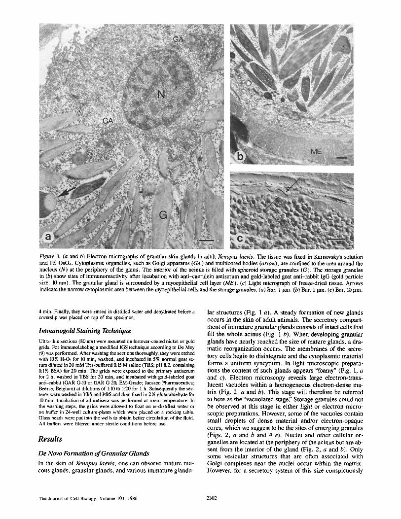
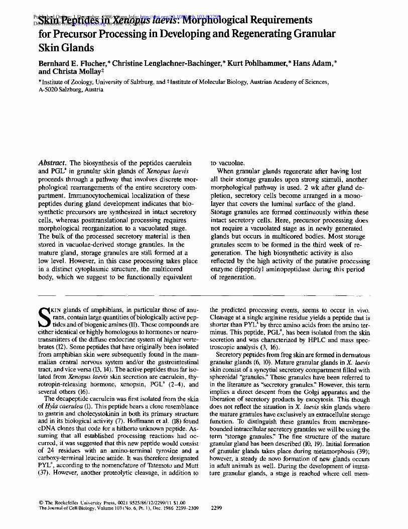
Figure 3. (a and b) Electron micrographs of granular skin glands in adult Xenopus laevis. The tissue was fixed in Karnovsky's solution and l% OSO4. Cytoplasmic organelles, such as Golgi apparatus (GA) and multicored bodies (arrow), are confined to the area around the nucleus (N) at the periphery of the gland. The interior of the acinus is filled with spheroid storage granules (G). The storage granules in (b) show sites of immunoreactivity after incubation with anti-caerulein antiserum and gold-labeled goat anti-rabbit IgG (gold particle size, 10 nrn). The granular gland is surrounded by a myoepithelial cell layer (ME). (c) Light micrograph of freeze-dried tissue. Arrows indicate the narrow cytoplasmic area between the myoepithelial cells and the storage granules. (a) Bar, 1/am. (b) Bar, 1 ~,n. (c) Bar, 10 p.m.
4 min. Finally, they were rinsed in distilled water and dehydrated before a coverslip was placed on top of the specimen.
lmmunogold Staining Technique
Ultra-thin sections (80 nm) were mounted on formvar-coated nickel or gold grids. For immunolabeling a modified IGS technique according to De Mey (9) was performed. After washing the sections thoroughly, they were etched with 10% H202 for 10 min, washed, and incubated in 5% normal goat se- rum diluted in 20 mM Tris-buffered 0.15 M saline (TBS; pH 8.2, containing 0.1% BSA) for 20 min. The grids were exposed to the primary antiserum for 2 h, washed in TBS for 20 rain, and incubated with gold-labeled goat anti-rabbit (GAR G-10 or GAR G 20, EM-Grade; Janssen Pharmaceutica; Beerse, Belgium) at dilutions of 1:10 to 1:20 for 1 h. Subsequently the sec- tions were washed in TBS and PBS and then fixed in 2% glutaraldehyde for 10 rain. Incubation of all antisera was performed at room temperature. In the washing steps, the grids were allowed to float on re-distilled water or on buffer in 24-well culture-plates which were placed on a rocking table. Glass beads were put into the wells to obtain better circulation of the fluid. All buffers were filtered under sterile conditions before use.
Resu l t s
De Novo Formation of Granular Glands
In the skin of Xenopus laevis, one can observe mature mu- cous glands, granular glands, and various immature glandu-
lar structures (Fig. 1 a). A steady formation of new glands occurs in the skin of adult animals. The secretory compart- ment of immature granular glands consists of intact cells that fill the whole acinus (Fig. 1 b). When developing granular glands have nearly reached the size of mature glands, a dra- matic reorganization occurs. The membranes of the secre- tory cells begin to disintegrate and the cytoplasmic material forms a uniform syncytium. In light microscopic prepara- tions the content of such glands appears "foamy" (Fig. 1, a and c). Electron microscopy reveals large electron-trans- lucent vacuoles within a homogeneous electron-dense ma- trix (Fig. 2, a and b). This stage will therefore be referred to here as the "vacuolated stage: Storage granules could not be observed at this stage in either light or electron micro- scopic preparations. However, some of the vacuoles contain small droplets of dense material and/or electron-opaque cores, which we suggest to be the sites of emerging granules (Figs. 2, a and b and 4 e). Nuclei and other cellular or- ganelles are located at the periphery of the acinus but are ab- sent from the interior of the gland (Fig. 2, a and b). Only some vesicular structures that are often associated with Golgi complexes near the nuclei occur within the matrix. However, for a secretory system of this size conspicuously
The Journal of Cell Biology, Volume 103, 1986 2302

Figure 4. Caerulein and PYL~-immunocytochemistry. (a and b) Colocalization of caerulein- and PYL~-immunoreactive material in the granular gland. 6-~tm sections of Bouin-fixed tissue were treated with anti-caerulein (a) or anti-PYL ~ antisera (b), followed by a routine PAP staining method (for details see Materials and Methods). Caerulein- and PYL~-immunoreactive material could be demonstrated within the same granular gland (G) in serial sections, whereas the vacuolated stage (V) and the mucous gland (M) showed no immunoreac- tivity. (c-e) Electron micrographs of immunolabeled storage granules. Ultrathin sections of 4% paraformaldehyde/0.25 % glutaraldehyde- (c and d) or Karnovsky-fixed (e) tissue were subsequently incubated with anti-caerulein (c) or anti-PYL a antisera (d and e) and gold- labeled goat anti-rabbit IgG (gold particle size, 10 nm). Embedding media: (c) LR-White; (d) polyvinyl-alcohol. (c and d) Both, caerulein- and PYLa-immunoreactive material is located within the storage granules. (e) In the vacuolated stage immunoreactive material is localized in the electron-opaque cores within the vacuoles (arrows). Here, the secretory peptides can be demonstrated first during granular gland development. (a and b) Bar, 100 ~tm. (c) Bar, 0.5 tim. (d) Bar, 0.5 ~tm. (e) Bar, 0.5 ~tm.
Flucher et al. Skin Peptides in Xenopus laevis 2303
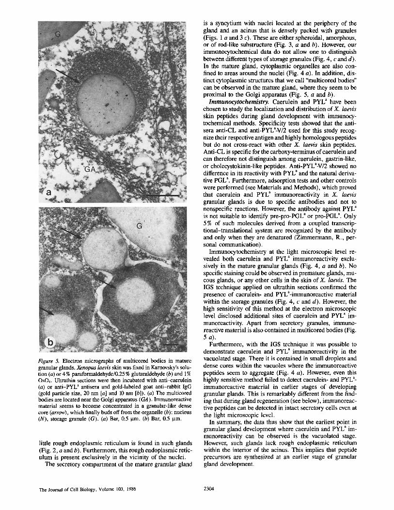
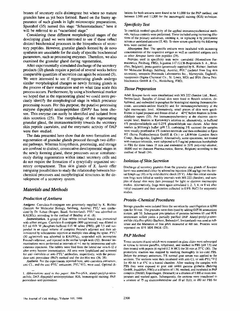
Figure 5. Electron micrographs of multicored bodies in mature granular glands. Xenopus laevis skin was fixed in Karnovsky's solu- tion (a) or 4 % paraformaldehyde/0.25 % glutaraldehyde (b) and 1% OsO4. Ultrathin sections were then incubated with anti-caerulein (a) or anti-PYL" antisera and gold-labeled goat anti-rabbit IgG (gold particle size, 20 nm [a] and 10 nm [b]). (a) The multicored bodies are located near the Golgi apparatus (GA). Immunoreactive material seems to become concentrated in a granular-like dense core (arrow), which finally buds off from the organelle (b); nucleus (N), storage granule (G). (a) Bar, 0.5 Ixm. (b) Bar, 0.5 ~tm.
little rough endoplasmic reticulum is found in such glands (Fig. 2, a and b). Furthermore, this rough endoplasmic retic- ulum is present exclusively in the vicinity of the nuclei.
The secretory compartment of the mature granular gland
is a syncytium with nuclei located at the periphery of the gland and an acinus that is densely packed with granules (Figs. 1 a and 3 c). These are either spheroidal, amorphous, or of rod-like substructure (Fig. 3, a and b). However, our immunocytochemical data do not allow one to distinguish between different types of storage granules (Fig. 4, c and d). In the mature gland, cytoplasmic organelles are also con- fined to areas around the nuclei (Fig. 4 a). In addition, dis- tinct cytoplasmic structures that we call "multicored bodies" can be observed in the mature gland, where they seem to be proximal to the Golgi apparatus (Fig. 5, a and b).
Immunocytochemistry. Caerulein and PYL a have been chosen to study the localization and distribution of X. laevis skin peptides during gland development with immunocy- tochemical methods. Specificity tests showed that the anti- sera anti-CL and anti-PYLa-V/2 used for this study recog- nize their respective antigen and highly homologous peptides but do not cross-react with other X. laevis skin peptides. Anti-CL is specific for the carboxy-terminus of caerulein and can therefore not distinguish among caerulein, gastrin-like, or cholecystokinin-like peptides. Anti-PYLa-V/2 showed no difference in its reactivity with PYL ~ and the natural deriva- tive PGL a. Furthermore, adsorption tests and other controls were performed (see Materials and Methods), which proved that caerulein and PYL" immunoreactivity in X. laevis granular glands is due to specific antibodies and not to nonspecific reactions. However, the antibody against PYL a is not suitable to identify pre-pro-PGL ~ or pro-PGL a. Only 5 % of such molecules derived from a coupled transcrip- tional-translational system are recognized by the antibody and only when they are denatured (Zimmermann, R., per- sonal communication).
Immunocytochemistry at the light microscopic level re- vealed both caerulein and PYL a immunoreactivity exclu- sively in the mature granular glands (Fig. 4, a and b). No specific staining could be observed in premature glands, mu- cous glands, or any other cells in the skin of X. laevis. The IGS technique applied on ultrathin sections confirmed the presence of caerulein- and pYLa-immunoreactive material within the storage granules (Fig. 4, c and d). However, the high sensitivity of this method at the electron microscopic level disclosed additional sites of caerulein and PYL" im- munoreactivity. Apart from secretory granules, immuno- reactive material is also contained in multicored bodies (Fig. 5 a).
Furthermore, with the IGS technique it was possible to demonstrate caerulein and PYL" immunoreactivity in the vacuolated stage. There it is contained in small droplets and dense cores within the vacuoles where the immunoreactive peptides seem to aggregate (Fig. 4 a). However, even this highly sensitive method failed to detect caerulein- and PYL a- immunoreactive material in earlier stages of developing granular glands. This is remarkably different from the find- ing that during gland regeneration (see below), immunoreac- tive peptides can be detected in intact secretory cells even at the light microscopic level.
In summary, the data thus show that the earliest point in granular gland development where caerulein and PYL a im- munoreactivity can be observed is the vacuolated stage. However, such glands lack rough endoplasmic reticulum within the interior of the acinus. This implies that peptide precursors are synthesized at an earlier stage of granular gland development.
The Journal of Cell Biology, Volume 103, 1986 2304
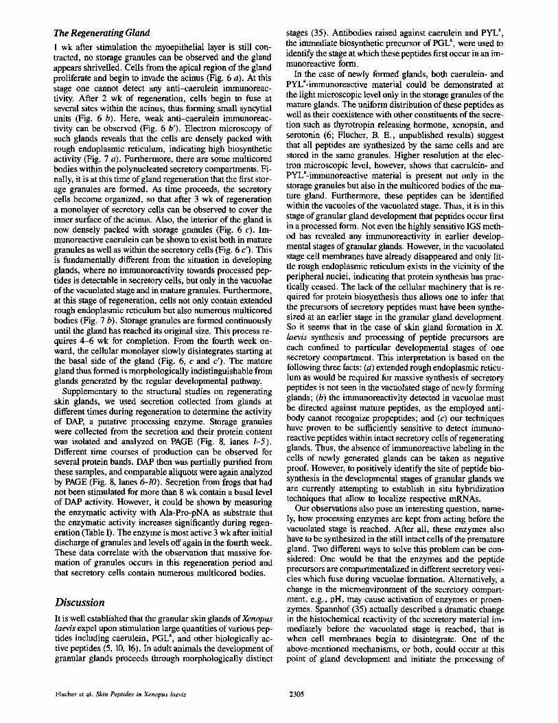
The Regenerating Gland 1 wk after stimulation the myoepithelial layer is still con- tracted, no storage granules can be observed and the gland appears shrivelled. Cells from the apical region of the gland proliferate and begin to invade the acinus (Fig. 6 a). At this stage one cannot detect any anti-caerulein immunoreac- tivity. After 2 wk of regeneration, cells begin to fuse at several sites within the acinus, thus forming small syncytial units (Fig. 6 b). Here, weak anti-caerulein immunoreac- tivity can be observed (Fig. 6 b'). Electron microscopy of such glands reveals that the cells are densely packed with rough endoplasmic reticulum, indicating high biosynthetic activity (Fig. 7 a). Furthermore, there are some multicored bodies within the polynucleated secretory compartments. Fi- nally, it is at this time of gland regeneration that the first stor- age granules are formed. As time proceeds, the secretory cells become organized, so that after 3 wk of regeneration a monolayer of secretory cells can be observed to cover the inner surface of the acinus. Also, the interior of the gland is now densely packed with storage granules (Fig. 6 c). Im- munoreactive caerulein can be shown to exist both in mature granules as well as within the secretory cells (Fig. 6 c'). This is fundamentally different from the situation in developing glands, where no immunoreactivity towards processed pep- tides is detectable in secretory cells, but only in the vacuolae of the vacuolated stage and in mature granules. Furthermore, at this stage of regeneration, cells not only contain extended rough endoplasmic reticulum but also numerous multicored bodies (Fig. 7 b). Storage granules are formed continuously until the gland has reached its original size. This process re- quires 4-6 wk for completion. From the fourth week on- ward, the cellular monolayer slowly disintegrates starting at the basal side of the gland (Fig. 6, c and c'). The mature gland thus formed is morphologically indistinguishable from glands generated by the regular developmental pathway.
Supplementary to the structural studies on regenerating skin glands, we used secretion collected from glands at different times during regeneration to determine the activity of DAP, a putative processing enzyme. Storage granules were collected from the secretion and their protein content was isolated and analyzed on PAGE (Fig. 8, lanes 1-5). Different time courses of production can be observed for several protein bands. DAP then was partially purified from these samples, and comparable aliquots were again analyzed by PAGE (Fig. 8, lanes 6-10). Secretion from frogs that had not been stimulated for more than 8 wk contain a basal level of DAP activity. However, it could be shown by measuring the enzymatic activity with Ala-Pro-pNA as substrate that the enzymatic activity increases significantly during regen- eration (Table I). The enzyme is most active 3 wk after initial discharge of granules and levels off again in the fourth week. These data correlate with the observation that massive for- mation of granules occurs in this regeneration period and that secretory cells contain numerous multicored bodies.
Discussion
It is-well established that the granular skin glands of Xenopus laevis expel upon stimulation large quantities of various pep- tides including caerulein, PGL a, and other biologically ac- tive peptides (5, 10, 16). In adult animals the development of granular glands proceeds through morphologically distinct
stages (35). Antibodies raised against caerulein and PYL a, the immediate biosynthetic precursor of PGL a, were used to identify the stage at which these peptides first occur in an im- munoreactive form.
In the case of newly formed glands, both caerulein- and PYL~-immunoreactive material could be demonstrated at the light microscopic level only in the storage granules of the mature glands. The uniform distribution of these peptides as well as their coexistence with other constituents of the secre- tion such as thyrotropin releasing hormone, xenopsin, and serotonin (6; Flucher, B. E., unpublished results) suggest that all peptides are synthesized by the same cells and are stored in the same granules. Higher resolution at the elec- tron microscopic level, however, shows that caerulein- and PYLa-immunoreactive material is present not only in the storage granules but also in the multicored bodies of the ma- ture gland. Furthermore, these peptides can be identified within the vacuoles of the vacuolated stage. Thus, it is in this stage of granular gland development that peptides occur first in a processed form. Not even the highly sensitive IGS meth- od has revealed any immunoreactivity in earlier develop- mental stages of granular glands. However, in the vacuolated stage cell membranes have already disappeared and only lit- tle rough endoplasmic reticulum exists in the vicinity of the peripheral nuclei, indicating that protein synthesis has prac- tically ceased. The lack of the cellular machinery that is re- quired for protein biosynthesis thus allows one to infer that the precursors of secretory peptides must have been synthe- sized at an earlier stage in the granular gland development. So it seems that in the case of skin gland formation in X. /ae~'s synthesis and processing of peptide precursors are each confined to particular developmental stages of one secretory compartment. This interpretation is based on the following three facts: (a) extended rough endoplasmic reticu- lum as would be required for massive synthesis of secretory peptides is not seen in the vacuolated stage of newly forming glands; (b) the immunoreactivity detected in vacuolae must be directed against mature peptides, as the employed anti- body cannot recognize propeptides; and (c) our techniques have proven to be sufficiently sensitive to detect immuno- reactive peptides within intact secretory ceils of regenerating glands. Thus, the absence of immunoreactive labeling in the cells of newly generated glands can be taken as negative proof. However, to positively identify the site of peptide bio- synthesis in the developmental stages of granular glands we are currently attempting to establish in situ hybridization techniques that allow to localize respective mRNAs.
Our observations also pose an interesting question, name- ly, how processing enzymes are kept from acting before the vacuolated stage is reached. After all, these enzymes also have to be synthesized in the still intact cells of the premature gland. Two different ways to solve this problem can be con- sidered: One would be that the enzymes and the peptide precursors are compartmentalized in different secretory vesi- cles which fuse during vacuolae formation. Alternatively, a change in the microenvironment of the secretory compart- ment, e.g., pH, may cause activation of enzymes or proen- zymes. Spannhof (35) actually described a dramatic change in the histochemical reactivity of the secretory material im- mediately before the vacuolated stage is reached, that is when cell membranes begin to disintegrate. One of the above-mentioned mechanisms, or both, could occur at this point of gland development and initiate the processing of
Flucher et al. Skin Peptides in Xenopus laevis 2305
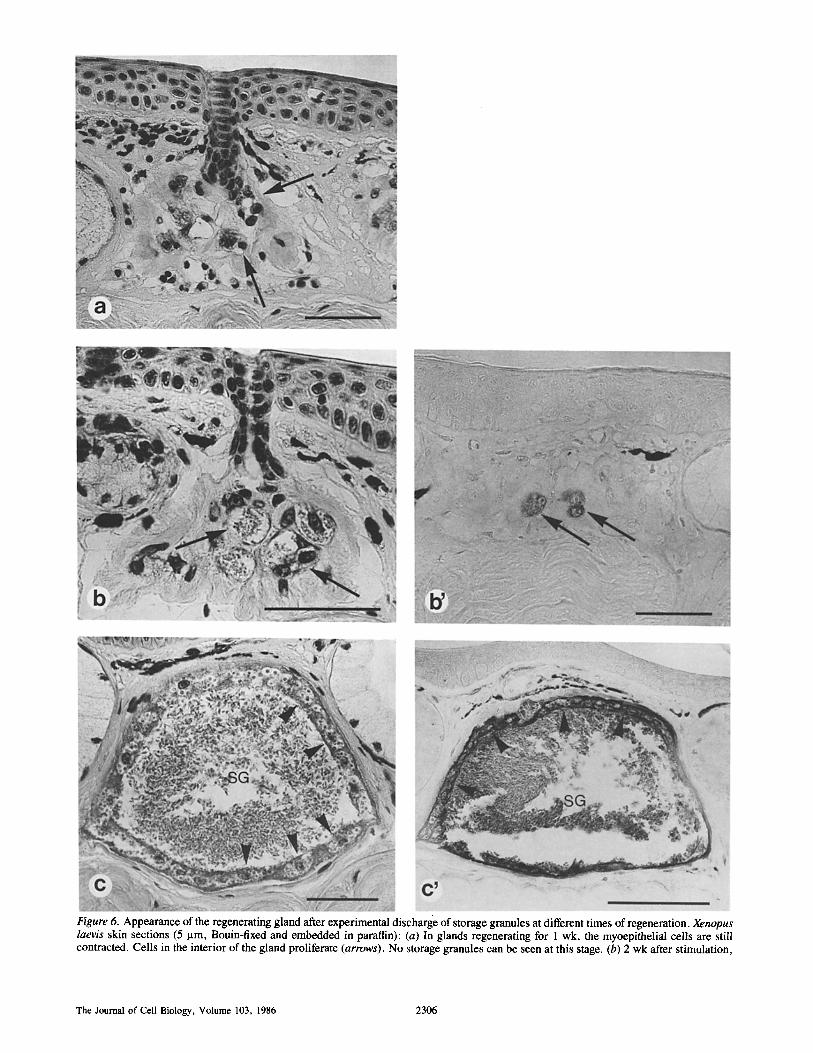
Figure 6. Appearance of the regenerating gland after experimental discharge of storage granules at different times of regeneration. Xenopus laevis skin sections (5 lam, Bouin-fixed and embedded in paraffin): (a) In glands regenerating for 1 wk, the myoepithelial cells are still contracted. Cells in the interior of the gland proliferate (arrows). No storage granules can be seen at this stage. (b) 2 wk after stimulation,
The Journal of Cell Biology, Volume 103, 1986 2306
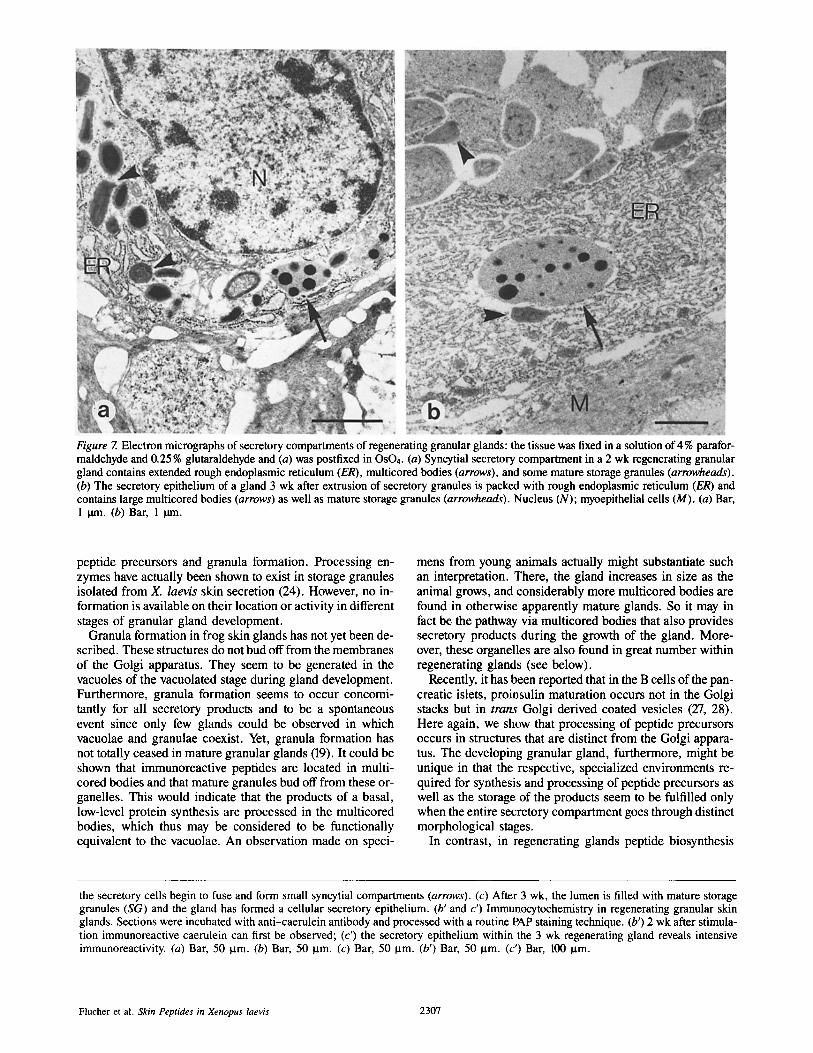
Figure 7. Electron micrographs of secretory compartments of regenerating granular glands: the tissue was fixed in a solution of 4 % parafor- maldehyde and 0.25 % glutaraldehyde and (a) was postfixed in OsO4. (a) Syncytial secretory compartment in a 2 wk regenerating granular gland contains extended rough endoplasmic reticulum (ER), multicored bodies (arrows), and some mature storage granules (arrowheads). (b) The secretory epithelium of a gland 3 wk after extrusion of secretory granules is packed with rough endoplasmic reticulum (ER) and contains large multicored bodies (arrows) as well as mature storage granules (arrowheads). Nucleus (N); myoepithelial cells (M). (a) Bar, 1 ~rn. (b) Bar, 1 p.m.
peptide precursors and granula formation. Processing en- zymes have actually been shown to exist in storage granules isolated from X. laevis skin secretion (24). However, no in- formation is available on their location or activity in different stages of granular gland development.
Granula formation in frog skin glands has not yet been de- scribed. These structures do not bud off from the membranes of the Golgi apparatus. They seem to be generated in the vacuoles of the vacuolated stage during gland development. Furthermore, granula formation seems to occur concomi- tantly for all secretory products and to be a spontaneous event since only few glands could be observed in which vacuolae and granulae coexist. Yet, granula formation has not totally ceased in mature granular glands (19). It could be shown that immunoreactive peptides are located in multi- cored bodies and that mature granules bud off from these or- ganelles. This would indicate that the products of a basal, low-level protein synthesis are processed in the multicored bodies, which thus may be considered to be functionally equivalent to the vacuolae. An observation made on speci-
mens from young animals actually might substantiate such an interpretation. There, the gland increases in size as the animal grows, and considerably more multicored bodies are found in otherwise apparently mature glands. So it may in fact be the pathway via multicored bodies that also provides secretory products during the growth of the gland. More- over, these organelles are also found in great number within regenerating glands (see below).
Recently, it has been reported that in the B cells of the pan- creatic islets, proinsulin maturation occurs not in the Golgi stacks but in trans Golgi derived coated vesicles (27, 28). Here again, we show that processing of peptide precursors occurs in structures that are distinct from the Golgi appara- tus. The developing granular gland, furthermore, might be unique in that the respective, specialized environments re- quired for synthesis and processing of peptide precursors as well as the storage of the products seem to be fulfilled only when the entire secretory compartment goes through distinct morphological stages.
In contrast, in regenerating glands peptide biosynthesis
the secretory cells begin to fuse and form small syncytial compartments (arrows). (c) After 3 wk, the lumen is filled with mature storage granules (SG) and the gland has formed a cellular secretory epithelium. (b' and c') Immunocytochemistry in regenerating granular skin glands. Sections were incubated with anti-caerulein antibody and processed with a routine PAP staining technique. (b') 2 wk after stimula- tion immunoreactive caerulein can first be observed; (c') the secretory epithelium within the 3 wk regenerating gland reveals intensive immunoreactivity. (a) Bar, 50 p.m. (b) Bar, 50 lam. (c) Bar, 50 I.tm. (b') Bar, 50 p.m. (c') Bar, 100 p.m.
Flucher et al. Skin Peptides in Xenopus laevis 2307
Figure 8. SDS PAGE of total pro- tein content and partially purified DAP isolated from granules de- rived from regenerating glands. Lanes 1-5, proteins in storage granules expelled by glands that were allowed to regenerate for (1) 8, (2) 1, (3) 2, (4) 3, and (5) 4 wk. Lanes 6--10, DAP partially purified from secretory granules isolated from glands aged (6) 8, (7) 1, (8) 2, (9) 3, and (10) 4 wk. The band previously identified as DAP (reference 9) is indicated by a dot.
requires a different structural organization. During regen- eration, secretory cells become organized in a monolayer that covers the inner surface of the glandular compartment. These cells contain extended rough endoplasmic reticulum and large multicored bodies. Furthermore, it could be shown by immunocytochemical techniques that the secretory epi- thelium contains immunoreactive caerulein. Thus, both bio- synthesis and processing of peptide precursors seem to occur simultaneously in these cells. This finding is basically differ- ent from the development of newly generated glands. Also, as opposed to the situation in the developing gland, granules are formed continuously during regeneration as soon as the secretory cells become biosynthetically active in the second week of regeneration. Finally, the mature gland is again densely packed with these storage granules within a period of 4-6 wk.
Granules from X. laevis skin glands are highly unstable structures without membranes that can therefore not be iso- lated quantitatively. Nevertheless, it could be demonstrated that granules contain biologically active peptides (5, 10) and enzymes that are required to generate those peptides from their respective biosynthetic precursors (26). However, we have noted previously that frogs that have not been stimu- lated for a considerable time expel very low concentrations of processing enzymes. This indicated that protein synthesis and processing do not occur continuously at high rates and that processing enzymes are likely to be degraded in mature glands. Since we are currently studying such enzymes, it was of practical interest to see at what time after stimulation skin glands would contain high levels of processing activity.
DAPs are known to be involved in the processing of promelittin to melittin in bee venom glands (21) and in the maturation of alpha-factor in yeast (20). The characteristic feature of this type of enzyme is that it recognizes only every
Table I. Specific Activity of DAP at Different Times of Regeneration aider Initial Discharge of Secretory Granules*
Regeneration period Specific activity
wk #mol/rng per min
1 1.5 2 1.6 3 2.0 4 1.4 8 0.4
* Specific activity of DAP is expressed in micromoles Ala-Pro-pNA cleaved per milligram protein per minute.
other amino acid from the amino-terminal side of a protein, whereby a particular DAP can cleave exclusively after a few, specific residues (22). The caerulein sequence is contained in a precursor sequence in which it is flanked by pairs of ba- sic amino acids (17). After excision at these sites, the peptide still has an additional amino-terminal fragment. To remove this extra sequence a DAP is suitable and, elsewhere, we have reported the purification of such an enzyme of appropri- ate specificity from skin secretion ofXenopus laevis (25). We now have found that high levels of this enzyme are present in storage granules when the morphological appearance of the regenerating gland suggests high processing activity. These data strengthen the role of DAP as processing enzyme in X. laevis and suggest, furthermore, that granular glands must be depleted every 3-4 wk if one wants to isolate pro- cessing enzymes.
In summary then, the data show that at least final process- ing of precursor proteins is blocked in intact secretory cells of developing glands, whereas this process is completed in
The Journal of Cell Biology, Volume 103, 1986 2308
the secretory cells of the regenerating skin glands. This ob- servation thus seems to indicate that the granular gland in the skin ofX. laevis might represent a system in which a particu- lar cell type can be subjected to two distinctly different de- velopmental programs. It remains to be elucidated which factors commit the cell to either pathway.
We thank Dr. G. Szelky and Dr. J. Thalhamer for their excellent advice on antibody production and Dr. V. Small and Dr. J. DeMey for their help to establish the IGS protocol. Furthermore, we thank Dr. H. C. Bauer for stimulating discussions.
This study was supported by Grant No. P5948B from the Fonds zur Frr- derung der wissenschaftlichen Forschung, Austria.
Received for publication 12 August 1986.
References
1. Anastasi, A., V. Erspamer, and R~ Endan. 1968. Isolation and amino acid sequence of caerulein, the active decapeptide of the skin ofHyla caerulea. Arch. Biochem. Biophys. 125:57-68.
2. Anastasi, A., G. Bertaccini, J. M. Cei, G. Decaro, V. Erspamer, M. Im- picciatore, and M. Roseghini. 1970. Presence of caerulein in extracts of the skin of Leptodactylus pentadactylus labyrinthicus and of Xenopus laevis. Brit. J. Pharmacol. 38:221-228.
3. Andrea, D., H. Aschauer, G. Kreil, and R. B. Merrifield. 1985. Solid- phase synthesis of PYL a and isolation of its natural counterpart, PGL ~ (PYL a- (4-24)) from skin secretion of Xenopus laevis. Eur. J. Biochem. 149:531-535.
4. Araki, K., S. Tachibana, M. Uchyama, T. Nakajima, and T. Yasuhara. 1973. Isolation and structure of a new active peptide "xenopsin" on the smooth muscle, especially on a strip of fundus from rat stomach, from the skin of Xenopus laevis. Chem. Pharm. Bull. 21:2801-2804.
5. Balls, M., G. W. Bennett, R. H. Clothier, C. A. Mardsen, and A. D. Waters. 1981. Adrenaline-induced discharge of thyrotropin releasing hormone and 5-hydroxytryptamine from the granular glands in amphibian skin. J. Phys- iol (Lond.). 316:42-43.
6. Bennett, G. W., M. Balls, R. H. Clothier, C. A. Mardsen, G. Robinson, and G. D. Wemyss-Holden. 1981. Location and release of TRH and 5-HT from amphibian skin. Biol. Int. Rep. 5:151-158.
7. Bertaccini, G. 1976. Active peptides of non-mammalian origin. Phar- macol. Rev. 28:127-177.
8. Bradley, L. M., S. M. Shigi, andJ. Nort. 1980. Secondary immunization to nitrophenyl haptens. In Selected Methods in Cellular Immunology. B. B. Mishell and S. N. Shiigi, editors. Freeman, Cooper & Co., San Francisco. 45-54.
9. De Mey, J. 1983. Colloidal gold probes in immunocytochemistry. In Im- munocytochemistry. J. M. Polak and S. Van Noorden, editors. Wright-PSG, London. 82-112.
10. Dockray, G. J., and C. R. Hopkins. 1975. Caerulein secretion by dermal glands in Xenopus laevis. J. Cell Biol. 64:724-733.
11. Erspamer, V. 1971. Biogenic amines and active polypeptides of the am- phibian skin. Annu. Rev. Pharmacol. 11:327-347.
12. Erspamer, V. 1978. Correlations between active peptides of the amphib- ian skin and peptides of avian and mammalian gut and brain. Abstracts of the 19th Congress of the Italian Pharmacological Society. 109-156.
13. Erspamer, V. 1983. Amphibian skin peptides in mammals-looking ahead. Trends Neurosci. 6:200-201.
14. Feurle, G. E., R. E. Carraway, E. Rix, and W. Knauf. 1985. Evidence for the presence of Xenopsin-related peptide(s) in the gastric mucosa of mam- mals. J. Clin. Invest. 76:156-162.
15. Flucher, B. E. 1984. Detecting the cause of non-specific staining in im- munocytochemistry (PAP-method). Mikroskopie. 41:219-226.
16. Gibson, B., L. Poulter, and D. H. Williams. 1986. Mass spectrometric evidence for additional processing of the caerulein, PGL a and Xenopsin pre- cursors from Xenopus laevis. J. Biol. Chem. In press.
17. Hoffmann, W., T. C. Bach, H. Seliger, and G. Kreil. 1983. Biosynthesis of caerulein in the skin of Xenopus laevis: partial sequences of precursors as deduced from cDNA clones. EMBO (Eur. Mol. Biol. Organ.) Z 2:111-114.
18. Hoffmann, W., K. Richter, and G. Kreil. 1983. A novel peptide desig- nated PYL' and its precursor as predicted from cloned mRNA of Xenopus laevis skin. EMBO (Fur. Idol. Biol. Organ.) J. 2:711-714.
19. Hopkins, C. R. 1971. The fine structure of the cutaneous poison glands in Xenopus laevis. J. Physiol. (Lond.). 219:9P-10P.
20. Julius, D., L. Blair, A. Brake, G. Sprague, and J. Thorner. 1983. Yeast alpha-factor is processed from a larger precursor polypeptide: the essential role of a membrane-bound dipeptidyl aminopeptidase. Cell. 32:839-852.
21. Kreil, G., L. Haiml, and G. Suchanek. 1980. Stepwise cleavage of the pro-part of promelittin by dipeptidyl aminopeptidase IV. Eur. J. Biochem. 111:49-58.
22. Kiillertz, G., P. Oehme, and A. Barth. 1981. Dipeptidyl-Peptidase IV - Chemie, Biochemie und physiologische Aspekte. Beitr~ige zur Wirkstoff- chemie, Berlin. 11.
23. Laemmli, U. K. 1970. Cleavage of structural proteins during the assem- bly of the head of bacteriophage P4. Nature (Lond.). 227:680-685.
24. MoUay, C., W. Hoffmann, A. Hutticher, K. Richter, U. Vilas, R. Vla- sak, and G. Kreil. 1984. Multiple processing steps in the generation of peptides from bee venom glands and frog skin. In Protein Transport and Secretion. D. L. Oxender, editor. Alan R. Liss Inc., New York. 345-353.
25. Mollay, C., U. Vilas, A. Hutticher, and G. Kreil. 1986. Isolation of a dipeptidyl aminopeptidase, a putative processing enzyme, from skin secretion of Xenopus laevis. Eur. J. Biochem. In press.
26. Mollay, C., J. Wichta, and G. Kreil. 1986. Partial characterization of an amidating enzyme present in skin secretion ofXenopus laevis. FEBS (Fed. Eur. Biochem. Soc.) Lett. 202:251-254.
27. Orci, L., P. Halban, M. Amherdt, M. Ravazzola, J-D. Vassalli, and A. Perrelet. 1984. Nonconverted, amino acid analog-modified proinsulin stays in a Golgi-derived clathrin-coated membrane compartment. J. Cell Biol. 99:2187- 2192.
28. Orci, L., M. Ravazzola, M. Amherdt, O. Mardsen, J-D. Vassalli, and A. Perrelet. 1985. Direct identification of prohormone conversion site in in- sulin-secreting cells. Cell. 42:671-681.
29. Petrusz, P., P. Dimeo, P. Ordronneau, C. Weaver, and D. A. Keefer. 1975. Improved immunoglobulin-enzyme bridge method for light microscopic demonstration of hormone-containing cells of the rat adenohypophysis. Histo- chemistry. 46:9-26.
30. Reading, M. 1977. A digestion technique for the reduction of background staining in the immunoperoxidase method. J. Clin. Pathol. 33:88-90,
31. Romeis, B. 1968. Mikroskopische Technik. R. Oldenburg Verlag, Miinchen-Wien. 174-175.
32. Sigel, M. B., Y. N. Sinha, and W. P. Vanderlaan. 1983. Production of antibodies by inoculation into lymph nodes. Methods Enzymol. 93:3-12.
33. Simonsberger, P. 1984. A freeze-drying apparatus to aid specimen prepa- ration for light and electron microscopy. Mikroskopie. 41:208-218.
34. Small, J. V., D. O. Fiirst, and L De Mey. 1986. Localization of filamin in smooth muscle. J. Cell Biol. 102:210-220.
35. Spannhof, L. 1953. Zur Genese, Morphologie und Physiologic der Haut- druesen bei Xenopus laevis. Wissenschafiliche Zeitschrift der Humboldt Uni- versitaet, Mathematisch, naturwissenschaftliche Reihe. 3:295-305.
36. Sternberger, L. A., P. H. Jr. Hardy, J. J. Cuculis, and H. G. Meyer. 1970. The unlabeled antibody method of immunohistochemistry. Preparation and properties of soluble antigen-antibody complex (horseradish peroxidase- anti horseradish peroxidase) and its use in identification of spirochetes. J. Histochem. Cytochem. 18:315.
37. Tatemoto, K., and V. Mutt. 1980. Isolation of two novel candidate hor- mones using a chemical method for finding naturally occurring polypeptides. Nature (Lond.). 285:417-418.
38. Towbin, H., T. Staekelin, and J. Gordon. 1979. Electrophoretic transfer of proteins from polyacrylamide gels to nitrocellulose sheets: procedure and some applications. Proc. Natl. Acad. Sci. USA. 76:4350-4354.
39. Vanable, J. W., Jr. 1964. Granular gland development during Xenopus laevis metamorphosis. Dev. Biol. 10:331-357.
40. Van Leeuven, F. 1982. Specific immunocytochemical localization of neuropeptides: a utopian goal? In Techniques in Immunohistochemistry. Vol. 1. G. R. Bullock and P. Petrusz, editors. Academic Press, London. 283-299.
Flucher et al. Skin Peptides in Xenopus taevis 2309