small bowel stromal tumors: approach by capsule endoscopy
TRANSCRIPT
9
Small Bowel Stromal Tumors:
Approach by Capsule Endoscopy
Anca Trifan, Ana Maria Singeap and Carol Stanciu “Gr. T. Popa“ University of Medicine and Pharmacy Iaşi,
Institute of Gastroenterology and Hepatology Iaşi Romania
1. Introduction
Small bowel tumors (SBTs) are rare, accounting for 3-6% of all digestive neoplasms (Gay Delvaux, 2008) which is strikingly low when one considers that the small bowel represents 75% of the length and 90% of the mucosal surface area of the alimentary tract. However, the accuracy of this estimate is uncertain because traditional small bowel examining methodologies have proved inadequate. Gastrointestinal stromal tumors (GISTs) are the most common mesenchimal tumors of the gastrointestinal tract (Nowain et al., 2005). Recognized as a distinctive entity in early 1980s
(Mazur Clark, 1983), GISTs have been a very active area over the last decade with remarkable progress in diagnostic modalities, pathophysiologic understanding, and new treatments. Until a decade ago, most of the small bowel was out of the range of endoscopic examination. The advent of capsule endoscopy (CE) was a major breakthrough for endoscopic diagnosis of small bowel diseases (Pennazio, 2005). CE is a safe, painless and sensitive endoscopic imaging of the entire small bowel, and several studies have revealed its diagnostic superiority over other methods such as push enteroscopy (Mylonaki et al., 2003), small bowel follow-through (Costamagna et al., 2002), angiography (Saperas et al., 2007), erythrocyte scintigraphy, CT–enterography and magnetic resonance-enterography (Eliakim et al., 2004; Golder et al., 2006; Jensen et al., 2011). Several published studies (Arakawa et al., 2009; Fukumoto et al., 2009; Pasha et al., 2008) showed that CE and double-balloon enteroscopy (DBE) are nearly equal in their ability to detect lesions when the entire small bowel is examined. Following the introduction of CE in clinical practice it was shown that the frequency of SBTs is higher than previously published (Cobrin et al., 2006; Estevez et al., 2007), and GISTs are the most frequent tumors identified by CE (Rondonotti et al., 2008). Nevertheless, CE lacks the ability to obtain biopsy specimens and performed therapeutic procedures, and therefore the role of CE in the diagnostic work-up of SBTs, including GISTs is still debated. The rising incidence and importance of GISTs represent an interesting challenge for CE considering the particular characteristics of these neoplasms. This review will discuss the main features of GISTs –one of the most advancing fields of gastrointestinal oncology- with particular emphasize on the role of capsule endoscopy in their management.
www.intechopen.com

New Techniques in Gastrointestinal Endoscopy
150
2. Definition
Gastrointestinal stromal tumors (GISTs) are defined as specific mesenchymal tumors of the gastrointestinal tract arising from the gastrointestinal wall, omentum, mesentery or retroperitoneum, that express the KIT (CD117, stem cell factor receptor) protein, a cell
membrane receptor with tyrosine kinase activity (Miettinen Lasota, 2001). Previously, GISTs were described as smooth muscle tumors and gastrointestinal autonomic nerve tumors (GANTs). The above definition excludes gastrointestinal true smooth muscle tumors (leiomyomas, leiomyosarcomas, leiomyoblastomas), neurofibromas and schwannomas.
3. Etiology
The etiology of GISTs is not known. Familial GISTs with inheritable KIT or PDGFRA (platelet-derived growth factor receptor alpha ) mutations have been identified (Nishida et al., 1998). GISTs can also be a component of Carney triad (gastric stromal sarcoma, extra-adrenal paraganglioma, pulmonary chondroma) (Carney, 1999). A relationship between neurofibromatosis 1 and GISTs typically occurring in the small bowel has also been postulated (Miettinen et al., 2002).
4. Epidemiology
GISTs are the most common mesenchymal tumors of the gastrointestinal tract, with an estimated incidence of 10-20 cases per million per year (Nilsson et al., 2005). However, the true epidemiology of GISTs has been difficult to determine. GISTs typically occur in older adults (age 55 to 65 years), with a mild male predominance in some series (Tran et al., 2005). Nevertheless, GISTs have been reported in all ages, including children. GISTs may occur in the entire length of the gastrointestinal tract, the most frequent location being the stomach (60-70%), followed by small bowel (20-30%), colon and rectum (5%) and esophagus (1%) (Miettinen et al., 2006). Within the small bowel, half of the stromal tumors are located in the jejunum, 25% in the duodenum and 25% in the ileum. GISTs are the most frequent
histological type of primary small bowel tumors (Miettinen Lasota, 2006a). Occasional, GISTs primary in the mesentery and omentum have also been reported (Miettinen et al, 1999).
5. Pathogenesis
The current theory is that an oncogenic mutation (acquired as a result of unidentified factors) on the KIT gene plays the central role in GISTs tumorigenesis. In the gastrointestinal tract, KIT (originally also called CD117) is normally expressed by the interstitial cells of Cajal, and therefore it has been proposed that GISTs originate from these cells (Blay et al., 2005). In the normal cell, the KIT receptor ligand is stem cell factor and, when bound, it leads to activation of the receptor and subsequent controlled downstream cellular cascades including several of cellular proliferative mechanisms and antiapoptotic pathways. In GISTs cells, mutations in the KIT genes lead to activation of tyrosine kinase receptor independent of the receptor ligand. These gain-of-function mutations allow uncontrolled cell proliferation and inhibition of normal apoptosis - well-known steps in carcinogenesis.
www.intechopen.com
Small Bowel Stromal Tumors: Approach by Capsule Endoscopy
151
Mutations of the KIT genes occur in approximately 90% of GISTs. Less than 5% gain-of-function mutation occur in PDGFRA gene (Heinrich et al., 2003). Nearly 10% of GISTs do not have detectable mutation in either KIT or PDGFRA genes.
6. Pathology
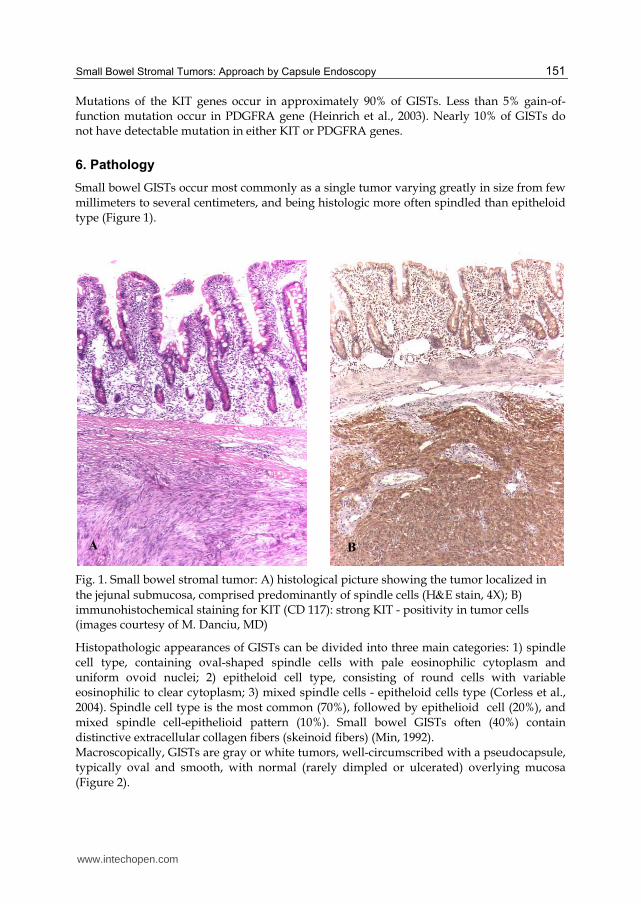
Small bowel GISTs occur most commonly as a single tumor varying greatly in size from few millimeters to several centimeters, and being histologic more often spindled than epitheloid type (Figure 1).
Fig. 1. Small bowel stromal tumor: A) histological picture showing the tumor localized in
the jejunal submucosa, comprised predominantly of spindle cells (HE stain, 4X); B) immunohistochemical staining for KIT (CD 117): strong KIT - positivity in tumor cells (images courtesy of M. Danciu, MD)
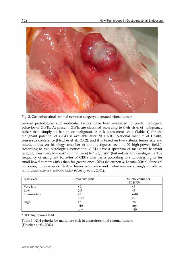
Histopathologic appearances of GISTs can be divided into three main categories: 1) spindle cell type, containing oval-shaped spindle cells with pale eosinophilic cytoplasm and uniform ovoid nuclei; 2) epitheloid cell type, consisting of round cells with variable eosinophilic to clear cytoplasm; 3) mixed spindle cells - epitheloid cells type (Corless et al., 2004). Spindle cell type is the most common (70%), followed by epithelioid cell (20%), and mixed spindle cell-epithelioid pattern (10%). Small bowel GISTs often (40%) contain distinctive extracellular collagen fibers (skeinoid fibers) (Min, 1992). Macroscopically, GISTs are gray or white tumors, well-circumscribed with a pseudocapsule, typically oval and smooth, with normal (rarely dimpled or ulcerated) overlying mucosa (Figure 2).
A B
www.intechopen.com
New Techniques in Gastrointestinal Endoscopy
152
Fig. 2. Gastrointestinal stromal tumor at surgery: ulcerated jejunal tumor
Several pathological and molecular factors have been evaluated to predict biological behavior of GISTs. At present, GISTs are classified according to their risks of malignancy rather than simply as benign or malignant. A risk assessment scale (Table 1) for the malignant potential of GISTs is available after 2001 NIH (National Institute of Health) consensus conference (Fletcher et al., 2002), and it is based on two criteria: tumor size and mitotic index on histology (number of mitotic figures seen in 50 high-power fields). According to this histologic classification, GISTs have a spectrum of malignant behavior ranging from “very low risk” (but not zero) to “high risk” (but not certainly malignant). The frequency of malignant behavior of GISTs also varies according to site, being higher for
small bowel tumors (40%) than for gastric ones (20%) (Miettinen Lasota, 2006b). Survival outcomes, tumor-specific deaths, tumor recurrence and metastases are strongly correlated with tumor size and mitotic index (Crosby et al., 2001).
Risk level Tumor size (cm) Mitotic count per 50 HPF*
Very low <2 <5 Low 2-5 <5 Intermediate <5 6-10 5-10 <5 High >5 >5 >10 any any >10
* HPF: high-power field
Table 1. NIH criteria for malignant risk in gastrointestinal stromal tumors (Fletcher et al., 2002)
www.intechopen.com
Small Bowel Stromal Tumors: Approach by Capsule Endoscopy
153
7. Clinical presentation
Usually, small bowel GISTs grow slowly and remain asymptomatic for many years. The symptoms and signs are not disease-specific. Typically, they are discovered incidentally during radiologic or endoscopic investigations and at surgery for other conditions. When symptomatic, the most common presenting symptom is gastrointestinal bleeding (melena), followed by bowel obstruction. Usually the bleeding is occult, resulting in long-standing anemia and associated symptoms before a small bowel GIST is diagnosed. Other presenting symptoms are abdominal pain or discomfort, altered bowel function, and abdominal fullness. On physical examination, findings based on the tumor size may include a palpable abdominal mass. Approximately half of small bowel GISTs have a metastatic component (liver, peritoneum) at the time of diagnosis.
8. Diagnostic procedures
8.1 Immunohistochemistry
The final diagnosis of small bowel GISTs depends on histological and immunochemistry examinations. The hallmark of small bowel GISTs is their positivity for KIT (CD117), confirmed by immunohistochemical staining usually using purified polyclonal antibodies. The antibodies against CD34 (hematopoietic progenitor cell antigen) is expressed in about 50% of small bowel GISTs, and can be used as an adjunct marker for diagnosis. Protein kinase C theta are used as secondary immunohistochemical markers in KIT negative GISTs (less than 3%) and for differential diagnosis.
8.2 Imaging studies
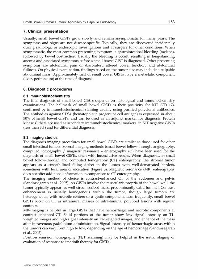
The diagnosis imaging procedures for small bowel GISTs are similar to those used for other small intestinal tumors. Several imaging methods (small bowel follow-through, angiography, computed tomography / magnetic resonance – enterography etc) have been used for the diagnosis of small bowel GISTs, often with inconclusive results. When diagnostic, at small bowel follow-through and computed tomography (CT) enterography, the stromal tumor appears as a smooth-lined filling defect in the lumen with well-demarcated borders, sometimes with focal area of ulceration (Figure 3). Magnetic resonance (MR) enterography does not offer additional information in comparison to CT-enterography. The imaging method of choice is contrast-enhanced CT of the abdomen and pelvis (Sandrasegaran et al., 2005). As GISTs involve the muscularis propria of the bowel wall, the tumor typically appear as well-circumscribed mass, predominantly extra-luminal. Contrast enhancement is usually homogenous within the tumor, though large tumors are heterogeneous, with necrotic centers or a cystic component. Less frequently, small bowel GISTs occur on CT as intramural masses or intra-luminal polypoid lesions with regular contours. MR-imaging is helpful in large GISTs that have hemorrhagic and necrotic components at contrast enhanced-CT. Solid portions of the tumor show low signal intensity on T1-weighted images and high signal intensity on T2-weighted images, and enhance of the mass after intravenous gadolinium administration. Signal intensity of hemorrhagic areas within the tumors can vary from high to low, depending on the age of hemorrhage (Sandrasegaran et al., 2005). Positron emission tomography (PET scanning) may be helpful in the initial staging or evaluation of response to imatinib therapy for GISTs .
www.intechopen.com

New Techniques in Gastrointestinal Endoscopy
154
Fig. 3. CT findings of small bowel stromal tumor: a 43/28/28 mm well-defined mass with central necrosis in the proximal jejunum (image courtesy of D. Negru, MD)
Endoscopic ultrasonography is helpful in other sites (esophagus, stomach, duodenum, rectum), although the method with special probes has been used during single - or double balloon - enteroscopy in small bowel GISTs (Matsui et al., 2008). Angiography may be the initial radiologic procedure in a patient with significant bleeding; angiographically, small bowel GISTs are characterized by irregular or ball-like vessels with neovascularity (Fang et al., 2004). Double-balloon enteroscopy (DBE) or single balloon enteroscopy have proved effective for the diagnosis of small bowel stromal tumors (Lin et al., 2008); typically, they appear as a submucosal mass (Figure 4) with normal lining mucosa and may be dimpled or ulcerated. Biopsies rarely yield diagnostic material; moreover, biopsy entails the risk of bleeding and seeding (National Comprehensive Cancer Network, 2008).
Fig. 4. Small bowel stromal tumor: enteroscopy shows submucosal mass with normal overlying mucosa, located in distal duodenum (A) and proximal jejunum (B)
A B
www.intechopen.com
Small Bowel Stromal Tumors: Approach by Capsule Endoscopy
155
GISTs encountered in duodenum and the last centimeters of ileum are diagnosed during upper gastrointestinal endoscopy and, respectively, colonoscopy, having the same features described at enteroscopy.
9. Capsule endoscopy
Traditionally, small bowel stromal tumors have been difficult to diagnose due to their nonspecific clinical symptoms, combined with inadequate methodologies for examining the small bowel. The advent of CE has revolutionized the investigation of patients with suspected SBTs, including GISTs. Conventional methods of investigating the small bowel (upper gastrointestinal endoscopy, colonoscopy, small bowel follow-through, CT- or MR- enterography) have a low diagnostic yield for GISTs. Small bowel is the second most frequent site for GISTs (20-30%), which are the most frequent tumor type identified by CE (Rondonotti, 2008). Within the small bowel, 50% of the stromal tumors are located in the jejunum, 25% in duodenum, and 25% in ileum. Usually, all patients with small bowel GISTs undergo several investigations prior to CE without a definitive diagnosis being made. The average work-up prior to capsule endoscopy is reported to range between 3 and 5 previous negative procedures per patient (Spada et al., 2008). Thus, the diagnosis of small bowel GISTs is often delayed with the use of traditional diagnostic modalities and, consequently, such tumors often are discovered late, approximately half having already metastasized at the moment of diagnosis. In this context, the sooner we use capsule endoscopy in the investigation of symptomatic patients the earlier we can establish a diagnosis, with a positive impact on patient management and improved outcome. The CE findings of SBTs, and particularly of small bowel GISTs are seldom described in details in the published papers probably due to the absence of universally accepted terminology, the terms usually being “tumor”, “polypoid mass”, “submucosal mass”, “tumor mass”, “bleeding polypoid mass”, “ulcerated mass lesion” and “irregular ulcer”. We have found that a polypoid lesion with normal appearance of overlying mucosa or, sometimes, with central ulceration is strongly suggestive of small bowel GIST (Figure 5). Nevertheless, any elevated lesion with normal overlying mucosa or a bleeding mucosa without a clear identified lesion should to be suspected to hide a GIST. A second CE examination may be necessary, or a balloon assisted enteroscopy should be performed when the first CE did not reveal a diagnosis and a clinical suspicion persists. During CE reading, an inexperienced physician might miss a lesion with no clear-cut features or could misinterpret a bleeding lesion as a NSAID ulcer or angiodysplasia. A second reading at a slower speed (frame to frame) is mandatory in any lesions, even in those without features of tumor. Even an endoscopist with a long experience in traditional endoscopy could also be mislead by images provided by CE. A special kind of training requiring patience and visual skills rather than manual skills is needed for this time consuming procedure. It should be emphasized that CE findings are of uncertain significance in the most of small bowel GISTs, and the final diagnosis is established by further diagnostic/therapeutic procedures (balloon assisted enteroscopy, surgery) after CE. Moreover, CE cannot reliably distinguished between benign and malignant tumors as CE is unable to provide histological confirmation to the diagnosis. However, it should be stressed that in case of suspected GISTs biopsy entails the risk of bleeding and seeding. Most authors consider that biopsy should be avoided in patients with resectable small bowel GISTs (Casali et al., 2006).
www.intechopen.com

New Techniques in Gastrointestinal Endoscopy
156
Fig. 5. Capsule endoscopy findings of small bowel stromal tumors: A) submucosal mass in
distal duodenum; B) ulcerated jejunal polipoid lesion; C, D) jejunal submucosal mass with
normal overlying mucosa; E, F) ileal submucosal mass with normal overlying mucosa
A B
C D
E F
www.intechopen.com

Small Bowel Stromal Tumors: Approach by Capsule Endoscopy
157
In all published series about CE in the diagnosis of SBTs, obscure gastrointestinal bleeding was the leading indication for capsule endoscopy (Rondonotti et al., 2008; Soufleris et al., 2008). CE is also important to establish the location of small bowel tumors, including GISTs. Most frequently, they are located in the jejunum, and it has been agreed that in 90% of cases the location as assessed by CE coincides with that found by further diagnostic and/or therapeutic work-up (Rondonotti et al., 2008). As reported in the literature, the most frequent small bowel tumors types identified by CE was small bowel gastrointestinal stromal tumors (Rondonotti et al., 2008). In the largest database published so far on SBTs detected by CE, Rondonotti et al (Rondonotti et al., 2008)
found that GISTs accounted for 32% of all cases. Schwartz and Barkin (Schwartz Barkin, 2007) found that small bowel GISTs were the most common benign tumors and CE was the diagnostic procedure of choice in patients with suspected small bowel tumors. In our series SBTs were detected in 4.9% of patients undergoing CE and the main tumor type was GIST
(Trifan et al., 2010). Recently, Sidhu and McAlindon (Sidhu McAlindon, 2011) reported that CE is an important modality in the diagnostic work-up of patients with small bowel tumors and it has a positive impact on patient management. Similar, Riccioni et al. (Riccioni et al., 2010) found CE an effective and sensitive diagnostic modality in GISTs in comparison to traditional radiology, having an important role in the algorithm for diagnostic work-up in suspected small bowel tumors. Capsule endoscopy has some limitations and risks. The first is inability to provide histological confirmation of the diagnosis. However, in case of suspected GISTs, biopsy is unnecessary as it may cause bleeding and seeding. Another is that CE cannot be maneuvered, and it has no means to allow prolonged examination or reexamination of questionable or poorly seen areas. Moreover, CE is not a therapeutic tool. Capsule retention remains the most significant complication and a major concern to both physicians and patients as it has the potential to cause small bowel obstruction which can lead to surgical intervention (Lin et al., 2007; Repici et al., 2008). The incidence of capsule retention varies widely depending on the indications for the examination. Absence or low rate (1-2%) of capsule retention was documented in studies with very strict exclusions criteria (Li et al., 2008), while high rates (5-21%) occurred in patients with suspected partial small bowel
obstruction (Cheifetz Lewis, 2006). Among the main causes of capsule retention are small bowel tumors (Toy et al., 2008). So far, there is no safe method of avoiding capsule retention. Radiological examination and even the newer imaging techniques entero-CT/MR have a low diagnostic yield and tend to underestimate small bowel strictures. Therefore, a patency capsule was developed to assess whether patients with suspected small bowel strictures could undergo CE. The patency capsule is a self-dissolving capsule, with the same size as the conventional capsule. The limitations of the first-generation patency capsule have been overcome by the second-generation (the Agile patency capsule), although its role in predicting retention needs to be further documented (Herrerias et al., 2008). Once the capsule has been retained only endoscopic (including double-balloon enteroscopy) and surgical intervention have been shown to be effective in removing the capsule (Baichi et al., 2006; Van Weyenberg et al., 2010). Currently, there are different view points regarding capsule retentions, some authors considering it as a feared complication of CE (Karagiannis et al., 2009), while others have suggested clear benefits from retentions by identifying and treating the underlying disease (Mason et al., 2008; Yang et al., 2009). Particularly, capsule retention in a patient with small bowel tumor is not a major clinical problem since the tumor will require surgical treatment and the capsule can be retrieved at the time of surgery.
www.intechopen.com

New Techniques in Gastrointestinal Endoscopy
158
10. Prognosis
Prognosis of GISTs is variable, determined by the malignant potential of the tumor. The best documented prognostic markers are tumor size and mitotic activity (Table 1). Tumors that show low mitotic frequency (< 5 mitosis per 50 HPF) usually have a benign behavior; however, a low mitotic index does not rule out a malignant behavior (Franquemont, 1995). A combination of low mitotic rate and small size (< 5 cm) is a more accurate predictor of a benign behavior. The small bowel GISTs have a markedly worse prognosis than gastric GISTs; thus, small bowel GISTs >10 cm but with a low mitotic rate have 52% metastatic rate,
whereas gastric GISTs with similar parameters metastasize in 12% of cases (Miettinen Lasota, 2006a). The 5-year survival of all patients with curative resection range from 20% to 80%. Tumor recurrence after surgical resection is frequent within 2-5 years. The median survival after palliative resection is about 10 months (DeMatteo et al., 2000).
11. Treatment
11.1 Localized resectable disease Surgery is the main type of treatment in patients with localized and potentially resectable
disease (Ho Blanke, 2011). The surgeon must avoid intraoperative tumor rupture, which is associated with high risk of peritoneal seeding. Small bowel GISTs often require segmental resection. Neoadjuvant therapy for patients with resectable disease is not recommended. However, preoperative imatinib may be considered for patients with potentially resectable
disease but with a risk of significant morbidity (Eisenberg Judson, 2004). The recurrence rate remains high after resection of a small bowel GIST, and some questions remain regarding post-operative or adjuvant therapy. Imatinib was approved by US Food and Drug Administration for patients with resected GISTs > 3cm in size (Cohen et al., 2010).
11.2 Unresectable or metastatic disease
Management of inoperable GISTs has changed radically with discovery and introduction in practice of molecularly targeted agents. Imatinib mesylate (Gleevec, Novartis Pharmaceuticals, Basel, Switzerland) is the first effective drug in patients with unresectable or metastatic GISTs (Cohen et al., 2009). Imatinib is a selective competitive inhibitor of protein tyrosine kinases including ABL (Abelson proto-oncogene), KIT and PDGFR. By competing with ATP for the kinase-binding site, imatinib inhibits the receptor activation and disables downstream cascades (Heinrich et al., 2000). Currently, for patients with marginally resectable, metastatic, progressive or recurrent disease, the recommended first-line therapy is imatinib. The use of imatinib can be guided by genotyping of KIT mutations: KIT exon 11 mutants respond well to imatinib, while KIT exon 9 mutants, which occur predominantly in small bowel GISTs are less sensitive to imatinib. Therefore, in these patients the starting dose of imatinib is 400 mg once daily and then is increased to 800 mg daily, if tolerated, over one month. The present recommendations are for life-long treatment with imatinib for patients with metastatic GISTs. The most frequent side effects of imatinib include leg edema, nausea, diarrhea, myalgias, fatigue and skin rash. Several clinical trials have demonstrated tumor regression and improved survival in patients treated with imatinib (Le Cesne et al., 2009). Sunitinib malate, a multitargeted tyrosine kinase inhibitor, is considered the standard second-line therapy for advanced GISTs (Demetri et al., 2006). It has been used in patients who did not respond to imatinib or who could not tolerate imatinib.
www.intechopen.com
Small Bowel Stromal Tumors: Approach by Capsule Endoscopy
159
Several drugs with potential activity against GISTs have been developed and tested in recent years. Second-generation tyrosine-kinase inhibitors (nilotinib, dasatinib), sorafenib (Nexavar) and other similar drugs (AZD 2171, XL-820) are in clinical testing (Dewaele et al., 2009; Montemurro et al., 2009).
12. Conclusion
Gastrointestinal stromal tumors have been an active area of gastrointestinal oncology over the last decade with remarkable progress in diagnostic modalities and new treatments. Small bowel is the second most frequent site for GISTs, and their diagnosis is often delayed with the use of traditional diagnostic methods. Usually, all patients with small bowel GISTs undergo several investigations prior to CE, without a final diagnosis being made. The advent of CE has revolutionized the investigation of patients with suspected small bowel tumors, including GISTs which are the most frequent tumor type identified by CE. Despite its limitations, CE may be the most reasonable initial diagnostic strategy to evaluate patients with suspected small bowel stromal tumors. In addition, CE has the potential of shortening the diagnostic work-up of small bowel stromal tumors. Furthermore, it may be expected that CE could identify such tumors at an early stage and thus the prognosis of the patients would be improved. Even more, CE could be used to assess patients after surgery or the efficacy of medical therapy.
13. References
Arakawa, D., Ohmiya, N., Nakamura, M., et al. (2009). Outcome after enteroscopy for patients with obscure gastrointestinal bleeding : Diagnostic comparison between double-balloon endoscopy and videocapsule endoscopy. Gastrointest Endosc, Vol. 69, No. 4, (April 2009), pp. 866-874, ISSN 0016-5107
Baichi, M.M., Arifuddin, R.M. Mantry, P.S. (2006). What we have learned from 5 cases of permanent capsule retention. Gastrointest Endosc, Vol.64, No. 2, (August 2006), pp. 283-287, ISSN 0016-5107
Blay, J.Y., Bonvalot, S., Casali, P., et al. (2005). Consensus meeting for the management of gastrointestinal stromal tumors. Report of the GIST Consensus Conference of 20-21 March 2004, under the auspices of ESMO. Ann Oncol, Vol. 16, No. 4, (April 2004), pp. 566-578, ISSN 0923-7534
Carney, J.A. (1999). Gastric stromal sarcoma, pulmonary chondroma, and extra-adrenal paraganglioma (Carney Triad): natural history, adrenocortical component, and possible familial occurrence. Mayo Clin Proc, Vol. 74, No. 6, (June 2006), pp. 543-552, ISSN 0025-6196
Casali, P.G., Jost, L., Sleijfer, S., et al. (2006).Soft tisuue sarcomas. ESMO clinical recommendations for diagnosis, treatment and follow up. Ann Oncol, Vol.19, Suppl. 2, pp. 90-93, ISSN 0923-7534
Cheifetz, A.S. Lewis, B.S. (2006). Capsule endoscopy: is it a complication? J Clin Gastroenterol, Vol. 40, No. 8, (September 2006), pp. 688–691, ISSN 0192-0790
Cobrin, G.M., Pittman, R.H., Lewis, B.S. (2006). Increased diagnostic yield of small bowel tumors with capsule endoscopy. Cancer, Vol. 107, No. 1, (July 2006), pp.22-27, ISSN 1097-0142
www.intechopen.com

New Techniques in Gastrointestinal Endoscopy
160
Cohen, M.H., Farrell, A.T., Justice, R., et al. (2009).Approval summary: Imatinib mesylate in the treatment of metastatic and/or unresectable malignant gastrointestinal stromal tumors. The Oncologist, Vol. 14, No. 2, (February 2009), pp. 174-180, ISSN 1083-7159
Cohen, M.H., Cortazar, P., Justice, R., et al. (2010). Approval Summary: Imatinib Mesylate in the Adjuvant Treatment of Malignant Gastrointestinal Stromal Tumors. The Oncologist, Vol. 15, No. 3, (March 2010), pp. 300-307, ISSN 1083-7159
Corless, C.L., Fletcher, J.A. & Heinrich, M.C. (2004). Biology of gastrointestinal stromal tumors. J Clin Oncol, Vol. 22, No. 18, (September 2004), pp. 3813-3825, ISSN 2218-4333
Costamagna, G., Shah, S.K., Riccioni, M.E., et al. (2002). A prospective trial comparing small bowel radiographs and video capsule endoscopy for suspected small bowel disease. Gastroenterology, Vol. 123, No. 4, (October 2002), pp. 999-1005, ISSN 0016-5085
Crosby, J.A., Catton, C.N., Davis, A., et al. (2001). Malignant gastrointestinal stromal tumors of the small intestine: a review of 50 cases from a prospective database. Ann Surg Oncol, Vol.8, No. 1, (January 2001), pp. 50-59, ISSN 1068-9265
DeMatteo, R.P., Lewis, J.J., Leung, D., et al. (2000). Two hundred gastrointestinal stromal tumors: recurrence patterns and prognostic factors for survival. Ann Surg, Vol. 232, No. 1, (January 2000), pp. 51-58, ISSN 0003-4932
Demetri, G.D., Oosterom, A., Garrett, C.R. et al. (2006). Efficacy and safety of sunitinib in patients with advanced gastrointestinal stromal tumour after failure of imatinib: a randomised controlled trial. The Lancet, Vol.368, No. 9544, (October 20006), pp. 1329-1338, ISSN 0140-6736
Dewaele, B., Wasag, B., Cools, J., et al. (2008). Activity of dasatinib, a dual SRC/ABL kinase inhibitor, and IPI-504, a heat shock protein 90 inhibitor, against gastrointestinal stromal tumor-associated PDGFRAD842V mutation. Clin Cancer Res, Vol. 14, No. 18, (September 2008), pp. 5749-5758, ISSN 1078-0432.
Eisenberg, B.L. Judson, I. (2004). Surgery and Imatinib in the Management of GIST: Emerging Approaches to Adjuvant and Neoadjuvant Therapy. Annals of Surgical Oncology, Vol. 11, No. 5, (May 2004), pp. 465-475, ISSN 1068-9265
Eliakim, R., Suissa, A., Yasin K, et al. (2004). Wireless capsule video endoscopy compared to barium follow-through and computerized tomography scan in patients with suspected Crohn's disease. Dig Liver Dis, Vol.36, No. 8, (August 2004), pp. 519-522, ISSN 1590-8658
Estevez, E., Gonzalez-Conde, B., Vazquez-Iglesis, J.L., et al. (2007). Incidence of tumoral pathology according to study using capsule endoscopy for patients with obscure gastrointestinal bleeding. Surg Endosc, Vol. 21, No. 10, (October 2010), pp. 1776-1780, ISSN 0930-2794
Fang, S.H., Dong, D.J. & Zhang, S.Z. (2004).Angiographic findings of gastrointestinal stromal tumor. Jin M. World J Gastroenterol, Vol.10, No. 19, (October 2004), pp. 2905-2907, ISSN 1007-9327
Fletcher, C.D., Berman, J.J., Corless, C., et al. (2002). Diagnosis of gastrointestinal stromal tumors: A consensus approach. Hum Pathol, Vol. 33, No. 5, (May 2002), pp. 459-65, ISSN 2218-4333
Franquemont, D.W. (1995). Differentiation and risk assessment of gastrointestinal stromal tumors. Am J Clin Pathol, Vol. 103, No. 1, (January 1995), pp. 41-47, ISSN 0002-9173
www.intechopen.com

Small Bowel Stromal Tumors: Approach by Capsule Endoscopy
161
Fukumoto, A., Tanaka, S., Shishido, T., et al. (2009). Comparison of detectability of small-bowel lesions between capsule endoscopy and double-balloon endoscopy for patients with suspected small-bowel disease. Gastrointest Endosc, Vol. 69, No. 4, (April 2009), pp.857-865, ISSN 0016-5107
Gay, C. Delvaux, M. (2008). Small-bowel endoscopy. Endoscopy, Vol. 40, No. 2, (February 2008), pp. 140-146, ISSN 0013-726X
Golder, S.K., Schreyer, A.G., Endlicher, E., et al. (2006). Comparison of capsule endoscopy and magnetic resonance (MR) enteroclysis in suspected small bowel disease. Int J Colorectal Dis, Vol. 21, No. 2, (March 2006), pp. 97-104, ISSN 0179-1958
Heinrich, M.C., Griffith, D.J., Druker, B.J., et al (2000). Inhibition of c-kit receptor tyrosine kinase activity by STI 571, a selective tyrosine kinase inhibitor. Blood, Vol. 96, No. 3, (August 2000), pp. 925-932, ISSN 0006-4971
Heinrich, M.C., Corless, C.L., Duensing, A., et al. (2003). PDGFRA activating mutations in gastrointestinal stromal tumors. Science, Vol. 299, No. 5607, (January 2003), pp. 708-710, ISSN 0036-8075
Herrerias, J.M., Leighton, J.A., Costamagna, G., et al. (2008). Agile patency system eliminates risk of capsule retention in patients with known gastrointestinal strictures who undergo capsule endoscopy. Gastrointest Endosc, Vol. 67, No. 6, (May 2008), pp. 902-909, ISSN 0016-5107
Ho, M.Y. Blanke, C.D. (2011). Gastrointestinal stromal tumors: disease and treatment update. Gastroenterology, Vol. 140, No. 5, (May 2011), pp. 1372-1376, ISSN 0016-5085
Jensen, M.D., Nathan, T., Rafaelsen, S.R., Kjeldsen, J. (2011). Diagnostic accuracy of capsule endoscopy for small bowel Crohn’s disease is superior to that of MR enterography or CT enterography. Clin Gastroenterol Hepatol, Vol. 9, No. 2 (February 2001), pp. 124-129, ISSN 1542-3565
Karagiannis, S., Faiss, S. Mavrogiannis, C. (2009). Capsule retention: a feared complication of wireless capsule endoscopy. Scand J Gastroenterol, Vol. 44, No. 10, (2009), pp. 1158-1165, ISSN 0036-5521
Le Cesne, A., Van Glabbeke, M., Verweij, J., et al. (2009). Absence of progression as assessed by response evaluation criteria in solid tumors predicts survival in advanced GI stromal tumors treated with imatinib mesylate: the intergroup EORTC-ISG-AGITG phase III trial. J Clin Oncol, Vol. 27, No. 24, (august 2009), pp. 3969-3974, ISSN 2218-4333
Li, F., Gurudu, S.R., De Petris, G., et al. (2008). Retention of the capsule endoscope: a single-center experience of 1000 capsule endoscopy procedures. Gastrointest Endosc, Vol. 68, No. 1, (July 2008), pp. 174-180, ISSN 0016-5107
Lin, M.B., Yin, L., Li, J.W., et al. (2008). Double-balloon enteroscopy reliably directs surgical intervention for patients with small intestinal bleeding. World J Gastroenterol, Vol. 14, No. 12, (March 2008), pp. 1936-40, ISSN 1007-9327.
Lin, O.S., Brandabur, J.J., Schembre, D.B., et al. (2007). Acute symptomatic small bowel obstruction due to capsule impaction. Gastrointest Endosc, Vol. 65, No. 4, (April 2007), pp. 725-728, ISSN 0016-5107
Mason, M., Swain, J., Matthews, B.D., et al. (2008). Use of video capsule endoscopy in the setting of recurrent subacute small-bowel obstruction. J Laparoendosc Adv Surg Tech A, Vol. 18, No. 5, (October 2008), pp. 713-716, ISSN 1092-6429
www.intechopen.com

New Techniques in Gastrointestinal Endoscopy
162
Matsui, N., Akahoshi, K., Motomura, Y., et al. (2008). Endosonographic detection of dumbbell-shaped jejunal GIST using double balloon enteroscopy. Endoscopy, Vol.40, Suppl 2, (September 2008), E38-39, ISSN 0013-726X
Mazur, M.T. Clark, H.B. (1983). Gastric stromal tumors. Reappraisal of histogenesis. Am J Surg Pathol, Vol. 7, No. 6, (September 1983), pp. 507–519, ISSN 0147-5185
Miettinen, M., Monihan, J.M., Sarlomo-Rikala, M., et al. (1999). Gastrointestinal stromal tumors/smooth muscle tumors (GISTs) primary in the omentum and mesentery: clinicopathologic and immunohistochemical study of 26 cases. Am J Surg Pathol, Vol. 23, No. 9, (September 1999), pp. 1109-1118, ISSN 0147-5185
Miettinen, M. Lasota, J. (2001). Gastrointestinal stromal tumors – definition, clinical, histological, immunohistochemical, and molecular genetic features and differential diagnosis. Virchows Arch, Vol. 438, No. 1, (January 2001), pp. 1-12, ISSN 0340-6075
Miettinen, M., Majidi, M. & Lasota, J. (2002). Pathology and diagnostic criteria of gastrointestinal stromal tumors (GISTs): a review. Eur J Cancer, Vol. 38, Suppl 5, (September 2002), S39-51, ISSN 1359-6349
Miettinen, M. Lasota, J. (2006a). Pathology and prognosis of gastrointestinal stromal tumors. Semin Diagn Pathol, Vol. 23, No. 2, (May 2006), pp. 70-83, ISSN 0740-2570
Miettinen, M. Lasota, J. (2006b). Gastrointestinal stromal tumors: review on morphology, molecular pathology, prognosis, and differential diagnosis. Arch Pathol Lab Med, Vol.130, No. 10, (October 2006), pp.1466–1478, ISSN 0003-9985
Miettinen, M., Makhlouf, H., Sobin, L.H. Lasota, J. (2006). Gastrointestinal stromal tumors of the jejunum and ileum: a clinicopathologic, immunohistochemical, and molecular genetic study of 906 cases before imatinib with long-term follow-up. Am J Surg Pathol, Vol. 30, No. 4, (April 2006), pp. 477-89, ISSN 0147-5185
Min, K.W. (1992). Small intestinal stromal tumors with skeinoid fibers. Clinicopathological, immunohistochemical, and ultrastructural investigations. Am J Surg Pathol, Vol. 16, No. 2, (February 1992), pp. 145-155, ISSN 0147-5185
Montemurro, M., Schöffski, P., Reichardt, P., et al. (2009). Nilotinib in the treatment of advanced gastrointestinal stromal tumours resistant to both imatinib and sunitinib. Eur J Cancer, Vol. 45, No. 13, (September 2009), pp. 2293-2297, ISSN 1359-6349
Mylonaki, M., Fritscher-Ravens, A., Swain, P. (2003). Wireless capsule endoscopy: a comparison with push enteroscopy in patients with gastroscopy and colonoscopy negative gastrointestinal bleeding. Gut, Vol. 52, No. 8, (August 2003), pp.1122-1126, ISSN 0017-5749
NCCN Clinical Practice Guidelines in Oncology. V2.2008. Soft Tissue Sarcomas. Nilsson, B., Bümming, P., Meis-Kindblom, J.M., et al. (2005). Gastrointestinal stromal
tumors: the incidence, prevalence, clinical course, and prognostication in the preimatinib mesylate era--a population-based study in western Sweden. Cancer, Vol. 103, No. 4, (February 2005), pp. 821-829, SSN 1097-0142
Nishida, T., Hirota, S., Taniguchi, M., et al. (1998). Familial gastrointestinal stromal tumours with germline mutation of the KIT gene. Nat Genet, Vol.19, No. 4, (August 1998), pp.323–324, ISSN 1061-4036
Nowain, A., Bhakta, H., Pais, S., Kanel, G., Verma, S. (2005). Gastrointestinal stromal tumors: clinical profile, pathogenesis, treatment strategies and prognosis. J Gastroenterol Hepatol, Vol. 20, No. 6, (June 2005), pp. 818-824, ISSN 0815-9319
www.intechopen.com
Small Bowel Stromal Tumors: Approach by Capsule Endoscopy
163
Pasha, S.F., Leighton, J.A., Das, A., et al. (2008). Double-balloon enteroscopy and capsule endoscopy have comparable diagnostic yield in small-bowel disease : a meta-analysis. Clin Gastroenterol Hepatol, Vol. 6, No. 6, (June 2008), pp.:671-676, ISSN 1542-3565
Pennazio, M. (2005). Diagnosis of small-bowel diseases in the era of capsule endoscopy. Expert Rev Med Devices, Vol. 2, No. 5, (September 2005), pp. 587-598, ISSN 1743-4440
Repici, A., Barbon, V., De Angelis, C., et al. (2008). Acute small-bowel perforation secondary to capsule endoscopy. Gastrointest Endosc, Vol. 67, No. 1, (January 2008), pp. 180-183, ISSN 0016-5107
Riccioni, M.E., Urgesi, R., Spada, C., et al. (2010). W1191 Increased Diagnostic Yield of Small Bowel Tumors With PillCam: the Role of Capsule Endoscopy in Diagnosis and Treatment of Gastrointestinal Stromal Tumours (GIST). Italian Single-Centre Experience. Gastroenterology, Vol. 138, No. 5, (May 2010), Suppl 1, S-670-S-671, ISSN 0016-5085
Rondonotti, E., Pennazio, M., Toth, E., et al. (2008). Small-bowel neoplasms in patients undergoing video capsule endoscopy : a multicenter European study. Endoscopy, Vol. 40, No. 6, (June 2008), pp. 488-495, ISSN 0013-726X
Sandrasegaran, K., Rajesh, A., Rushing, D.A., et al. (2005). Gastrointestinal stromal tumors: CT and MRI findings. Eur Radiol, Vol. 15, No. 7, (July 2005), pp. 1407-1414, ISSN 0938-7994
Saperas, E., Dot, J., Videla, S., et al. (2007). Capsule endoscopy versus computed tomographic or standard angiography for the diagnosis of obscure gastrointestinal bleeding. Am J Gastroenterol, Vol. 102, No. 4, (April 2007), pp. 731-737, ISSN 0002-9270
Schwartz, G.D. & Barkin, J.S. (2007). Small-bowel tumors detected by Wireless Capsule Endoscopy. Dig Dis Sci, Vol. 52, No. 4, (April 2007), pp. 1026-1030, ISSN 0163-2116
Sidhu, R., McAlindon, M.E. (2011). The use of capsule endoscopy for the diagnosis of small bowel tumours: the first single centre UK experience. Gut 2011;60:A91-A92 doi:10.1136/gut.2011.239301.189, ISSN 1468-3288
Soufleris, K., Chatzimavroudis, G., Pilpilidis, J., et al. (2008). Five years missed small jejunal stromal tumor (GIST) causing recurrent episodes of bleeding: Successful diagnosis by capsule endoscopy. Annals of Gastroenterology, Vol. 21, No. 3, pp. 201-204, ISSN 1108-7471
Spada, C., Riccioni, M.E., Familiari, P., et al. (2008). Video capsule endoscopy in small-bowel tumours: a single centre experience. Scand J Gastroenterol, Vol. 43, No.4, pp.497-505, ISSN 0036-5521
Toy, E., Rojany, M., Sheikh, R., et al. (2008). Capsule endoscopy’s impact on clinical management and outcomes: a single-center experience with 145 patients. Am J Gastroenterol, Vol. 103, No. 12, (December 2008), pp. 3022-3028, ISSN 0002-9270
Tran, T., Davila, J.A. El-Serag, H.B. (2005). The epidemiology of malignant gastrointestinal stromal tumors: an analysis of 1,458 cases from 1992 to 2000. Am J Gastroenterol, Vol. 100, No. 1, (January 2005), pp. 162-168, ISSN 0002-9270
Trifan, A., Singeap, A.M., Cojocariu, C., et al. (2010). Small bowel tumors in patients undergoing capsule endoscopy: a single center experience. J Gastrointestin Liver Dis, Vol. 19, No. 1, (March 2010), pp. 21-25, ISSN 1841-8724
www.intechopen.com
New Techniques in Gastrointestinal Endoscopy
164
Van Weyenberg, S.J., Van Turenhout, S.T., Bouma, G., et al. (2010). Double-balloon endoscopy as the primary method for small-bowel video capsule endoscope retrieval. Gastrointest Endosc, Vol.71, No. 3, (March 2010), pp. 535-541, ISSN 0016-5107
Yang, X.Y., Chen, C.X., Zhang, B.L., et al. (2009). Diagnostic effect of capsule endoscopy in 31 cases of subacute small bowel obstruction. World J Gastroenterol, Vol. 15, No. 19, (May 2009), pp. 2401-2405, ISSN 1007-9327
www.intechopen.com
New Techniques in Gastrointestinal EndoscopyEdited by Prof. Oliviu Pascu
ISBN 978-953-307-777-2Hard cover, 310 pagesPublisher InTechPublished online 30, September, 2011Published in print edition September, 2011
InTech EuropeUniversity Campus STeP Ri Slavka Krautzeka 83/A 51000 Rijeka, Croatia Phone: +385 (51) 770 447 Fax: +385 (51) 686 166www.intechopen.com
InTech ChinaUnit 405, Office Block, Hotel Equatorial Shanghai No.65, Yan An Road (West), Shanghai, 200040, China
Phone: +86-21-62489820 Fax: +86-21-62489821
As result of progress, endoscopy has became more complex, using more sophisticated devices and hasclaimed a special form. In this moment, the gastroenterologist performing endoscopy has to be an expert inmacroscopic view of the lesions in the gut, with good skills for using standard endoscopes, with goodexperience in ultrasound (for performing endoscopic ultrasound), with pathology experience for confocalexamination. It is compulsory to get experience and to have patience and attention for the follow-up ofthousands of images transmitted during capsule endoscopy or to have knowledge in physics necessary forautofluorescence imaging endoscopy. Therefore, the idea of an endoscopist has changed. Examinationsmentioned need a special formation, a superior level of instruction, accessible to those who have alreadygained enough experience in basic diagnostic endoscopy. This is the reason for what these new issues ofendoscopy are presented in this book of New techniques in Gastrointestinal Endoscopy.
How to referenceIn order to correctly reference this scholarly work, feel free to copy and paste the following:
Anca Trifan, Ana Maria Singeap and Carol Stanciu (2011). Small Bowel Stromal Tumors: Approach by CapsuleEndoscopy, New Techniques in Gastrointestinal Endoscopy, Prof. Oliviu Pascu (Ed.), ISBN: 978-953-307-777-2, InTech, Available from: http://www.intechopen.com/books/new-techniques-in-gastrointestinal-endoscopy/small-bowel-stromal-tumors-approach-by-capsule-endoscopy

© 2011 The Author(s). Licensee IntechOpen. This chapter is distributedunder the terms of the Creative Commons Attribution-NonCommercial-ShareAlike-3.0 License, which permits use, distribution and reproduction fornon-commercial purposes, provided the original is properly cited andderivative works building on this content are distributed under the samelicense.