small molecule inhibitors of ercc1-xpf...
TRANSCRIPT
1521-0111/84/1/12–24$25.00 http://dx.doi.org/10.1124/mol.112.082347MOLECULAR PHARMACOLOGY Mol Pharmacol 84:12–24, July 2013Copyright ª 2013 by The American Society for Pharmacology and Experimental Therapeutics
Small Molecule Inhibitors of ERCC1-XPF Protein-ProteinInteraction Synergize Alkylating Agents in Cancer Cells s
Lars Petter Jordheim, Khaled H. Barakat, Laurence Heinrich-Balard, Eva-Laure Matera,Emeline Cros-Perrial, Karima Bouledrak, Rana El Sabeh, Rolando Perez-Pineiro,David S. Wishart, Richard Cohen, Jack Tuszynski, and Charles DumontetUniversité de Lyon (L.P.J., L.H.-B., E.-L.M., E.C.-P., K.B., R.E.S., R.C., C.D.), Université Lyon 1 (L.P.J., L.H.-B., E.-L.M., E.C.-P.,K.B., R.E.S., R.C., C.D.), INSERM U1052, Centre de Recherche en Cancérologie de Lyon (L.P.J., E.-L.M., E.C.-P., K.B., R.E.S.,C.D.), and CNRS UMR 5286, Centre de Recherche en Cancérologie de Lyon, Lyon, France (L.P.J., E.-L.M., E.C.-P., K.B., R.E.S.,C.D.); Department of Physics, University of Alberta, Edmonton, Alberta, Canada (K.H.B., J.T.); Department of EngineeringMathematics and Physics, Fayoum University, Fayoum, Egypt (K.H.B.); MATEIS UMR-CNRS 5510, ISPB Faculté de Pharmacie,Lyon, France (L.H.-B., R.C.); Department of Biological Sciences (R.P.-P., D.S.W.) and Department of Computing Sciences,University of Alberta, Edmonton, Alberta, Canada (R.P.-P., D.S.W.); Hospices Civils de Lyon, Hôpital Edouard Herriot,Laboratoire de Biochimie et de Biologie Moléculaire, Lyon Cedex 03, France (R.C.); and Department of Oncology, University ofAlberta, Edmonton, Alberta, Canada (J.T.)
Received September 11, 2012; accepted April 11, 2013
ABSTRACTThe benefit of cancer chemotherapy based on alkylating agentsis limited because of the action of DNA repair enzymes, whichmitigate the damage induced by these agents. The interactionbetween the proteins ERCC1 and XPF involves two majorcomponents of the nucleotide excision repair pathway. Here, novelinhibitors of this interaction were identified by virtual screeningbased on available structures with use of the National CancerInstitute diversity set and a panel of DrugBank small molecules.Subsequently, experimental validation of the in silico screeningwasundertaken. Top hits were evaluated on A549 and HCT116 cancercells. In particular, the compound labeledNSC130813 [4-[(6-chloro-
2-methoxy-9-acridinyl)amino]-2-[(4-methyl-1-piperazinyl)methyl]]was shown to act synergistically with cisplatin and mitomycin C;to increase UVC-mediated cytotoxicity; to modify DNA repairas indicated by the staining of phosphorylated H2AX; andto disrupt interaction between ERCC1 and XPF in cells. Inaddition, using the Biacore technique, we showed that thiscompound interacts with the domain of XPF responsible forinteraction with ERCC1. This study shows that small moleculestargeting the protein-protein interaction of ERCC1 and XPF canbe developed to enhance the effects of alkylating agents oncancer cells.
IntroductionPlatinum derivatives are major components of the chemo-
therapeutic arsenal available to treat many patients withcancer. Clinical outcomes for patients treated with thesedrugs depend not only on the tumor type and stage but alsoon the biology of the targeted cancer cells. Indeed, severalcellular parameters involved in pre–DNA damage mecha-nisms (e.g., transport deficiency and detoxification by cyto-plasmic proteins), in the production and maintenance of DNA
damage (e.g., DNA repair), and in the induction of cell death(e.g., DNA damage recognition and apoptotic protein expres-sion) influence the outcome of platinum-based chemotherapy(Galluzzi et al., 2012). The clinical relevance of these mod-ifications is not always clearly established, but a growing bodyof evidence indicates that proteins involved in nucleotide ex-cision repair (NER) of DNA modulate the clinical activity ofcisplatin (Hubner et al., 2011).NER is a multistep DNA repair mechanism involving more
than 30 different proteins to excise approximately 30 bases ona damagedDNAstrand and to synthesize newDNA (Nouspikel,2009). NER is activated to repair different types of DNAdamage, including UV-induced bond breaks and adductsformed by platinum derivatives. By doing so, NER decreasesthe damage induced by platinum-based drugs, such ascisplatin, and, thus, is associated with resistance to treatment.Several NER proteins are essential for a correct DNA repair,
This work was supported by Ligue Contre le Cancer comité du Rhône; Liseog Arnfinn Hejes fond (to L.P.J.); the Allard Foundation; the Canadian BreastCancer Foundation; and the Alberta Advanced Education and Technology (toJ.A.T.).
L.P.J. and K.H.B. contributed equally to this work.dx.doi.org/10.1124/mol.112.082347.s This article has supplemental material available at molpharm.
aspetjournals.org.
ABBREVIATIONS: CI95, 95% combination index; DMSO, dimethylsulfoxide; ICL, interstrand crosslink; MD, molecular dynamics; MMC, mitomycin C;MTT, methylthiazoletetrazolium; NCIDS, National Cancer Institute Diversity Set; NER, nucleotide excision repair; NSC 130813, 4-[(6-chloro-2-methoxy-9-acridinyl)amino]-2-[(4-methyl-1-piperazinyl)methyl]; NSC 303812, [5,13-dihydroxy-6-(hydroxymethyl)-2,6-dimethyltetracyclo[10.3.1.01,10.02,7]hexadec-13-yl]methyl glycinate; NSC 254681, 3-acetyl-11-amino-3,6-dihydroxy-10-methoxy-5,12-dioxo-1,2,3,4,5,12-hexahydro-1-tetracenyl 3-amino-2,3,6-trideoxyhexopyranoside; PBS, phosphate-buffered saline.
12
http://molpharm.aspetjournals.org/content/suppl/2013/04/11/mol.112.082347.DC1Supplemental material to this article can be found at:
at ASPE
T Journals on M
ay 27, 2018m
olpharm.aspetjournals.org
Dow
nloaded from

and cell lines deficient in any of them are highly sensitive toUVirradiation and platinum agents. On the basis of this, it hasbeen hypothesized that the inhibition of NER should sensitizecancer cells to these DNA-damaging agents, thus increasingtheir activity and eventually improving clinical outcomes.The inhibition of NER could be achieved by several
approaches, such as the development of small moleculeligands specific to the enzymes involved in the NER pathway.Another approach aims at blocking the interaction betweenthe different elements of the NER pathway, thus preventingthe repair from being completed. Indeed, the interactionsamong NER proteins seem to be essential for accomplishingrepair, as indicated by the inhibition of NER by a peptidecorresponding to the domain of XPA interacting with ERCC1(Tsodikov et al., 2007). Such protein-protein interactionsproved to be viable as druggable targets for cancer treat-ments, as indicated by the p53-MDM2 interaction (Shangaryand Wang, 2008) and the interaction between Bcl-2 and pro-apoptotic proteins through the BH3 domain (Azmi andMohammad, 2009). Here, we continue our in silico–basedsearch for inhibitors targeting protein-protein interactions byfocusing on those interactions that involve ERCC1 (Barakatet al., 2009, 2012). In the present study, we identified inhibitorsfor the ERCC1-XPF interaction. In addition to playing a keyrole in NER, the ERCC1-XPF complex is involved in in-terstrand crosslink (ICL) repair, particularly through theinteraction with SLX4. Cisplatin induces mostly intrastrandadducts that are repaired by NER, but also 2–3% of ICL thatare mainly responsible for the cytotoxic effect of cisplatin(Jamieson and Lippard, 1999). Other compounds inducing ICLare alkylating agents, such as mitomycin C (MMC), in whichactivity is also expected to be increased by the inhibition of theERCC1-XPF interaction.
Materials and MethodsMolecular Dynamics Simulations. The model of the ERCC1-
XPF C-terminal complex that we generated was based on the PDB ID1Z00 (Tripsianes et al., 2005). The available structure represents onlythe interaction between the C-termini of the two proteins. Moleculardynamics (MD) simulations used the NAMD program (Phillips et al.,2005) at a mean (physiologic) temperature of 310 K, pH of 7, and theall-hydrogen AMBER99SB force-field. Protonation states of all ioniz-able residues were calculated using PDB ID 2PQR (Dolinsky et al.,2007). After parameterization, the XPF protein alone or in complexwith ERCC1 was immersed in the center of a TIP3P water cube afteradding hydrogen atoms to the initial protein structure. Only oneERCC1-XPF complex model was used for binding energy analysis.However, 20 distinct unbound XPF structures were prepared throughMD simulations for statistically relevant docking analysis. The watercube’s dimensions were chosen to provide at least a 20 Å buffer ofwater molecules around the molecular system. To neutralize andprepare the XPF-bound and XPF-free systems, respectively, undera physiologic ionic concentration, chloride and sodium ions wereadded by replacing water molecules with the highest electrostaticenergies on their oxygen atoms. The number of counter ions for eachcase was calculated by first estimating the number of ions that areneeded to simulate the system at pH of 7, followed by adding thenumber of chloride ions required to bring its charge to zero. The fullysolvated proteins were then minimized and heated to the simulationtemperature, with heavy restraints placed on all backbone atoms.After heating, the systems were equilibrated using periodic boundaryconditions for 100 picoseconds and energy restraints reduced to zeroin successive steps of the MD simulation. The simulations continued
for 10 nanoseconds, during which atomic coordinates were saved tothe trajectory every 2 picoseconds. The root-mean-square deviationsand fluctuation coefficients (B-factors) for the protein backbone werecomputed over the last 5 nanoseconds of the MD simulation with useof the PTRAJ utility in AMBER10 (Case et al., 2005). A hydrogen bondwas defined by a heavy donor–heavy acceptor distance # 3.4 Å, a lightdonor–heavy acceptor distance # 2.5 Å, and a deviation of less than 660° from linearity. For the ERCC1-XPF complex, snapshots wereextracted every 2 picoseconds and used for the subsequent molecularmechanics Poisson-Boltzmann surface area binding energy analysis.
Binding Energy Analysis. Binding energy calculations used themolecular mechanics Poisson-Boltzmann surface area technique(Kollman et al., 2000; Kuhn et al., 2005). The total free energy is thesum of the mean molecular mechanical gas-phase energies (EMM),solvation-free energies (Gsolv), and entropy contributions (2TSsolute)to the binding reaction:
G5EMM 1Gsolv 2TSsolute (1)
The total molecular mechanical energies can be further decom-posed into contributions from electrostatic (Eele), van der Waals(Evdw), and internal energies (Eint):
EMM 5Eele 1Evdw 1Eint (2)
Furthermore, the solvation-free energy can be expressed as a sumof nonelectrostatic and electrostatic contributions:
DGsolv � DGnonelesolv 1DGele
solv (3)
The nonelectrostatic part was approximated by a linear function ofthe solvent-accessible surface area:
DGnonelesolv 5g � SASA; where g 5 7:2 cal=mol=A2 (4)
The molecular mechanical (EMM) energy of each snapshot structurewas calculated using the SANDER module of AMBER with allpairwise interactions included using a dielectric constant («) of 1. Thesolvation-free energy (DGsolv) is estimated as the sum of electrostaticsolvation-free energy, calculated using the finite-difference solution ofthe Poisson-Boltzmann equation in the Adaptive Poisson-BoltzmannSolver program. The nonpolar solvation-free energy is directlyproportional to the solvent-accessible surface area of the target. Thesolute entropy is approximated using normal mode analysis. Thebinding-free energy can be approximated by:
DGo 5DGERCC12XPFgas 1DGERCC12XPF
solv 2nDGXPF
solv 1DGERCC1solv
o(5)
where (DGERCC12XPFgas ) represents the free energy per mole of the
noncovalent association of the ERCC1-XPF complex in vacuum (gasphase) at 310 K, and (2DGsolv) stands for the work required to transfera molecule from its solution conformation to the same conformation invacuum at 310 K (assuming that the binding conformation of theprotein-protein complex is the same in solution and in vacuum).
Selection of a Ligand Database. The National Cancer InstituteDiversity Set (NCIDS) and a panel of DrugBank small molecules(Kale et al., 1999) were used. From the approximately 2000compounds in the NCIDS collection, a number of ligands containingrare earth elements could not be properly parameterized and wereexcluded, leaving a total of 1883 compounds for docking analysis. Weused a version of the NCIDS formatted for use in AUTODOCK, whichwas prepared by the AUTODOCK Scripps team (http://autodock.scripps.edu/resources/databases). The DrugBank small moleculeslibrary is a set of 1488 Food andDrugAdministration–approved drugsdownloaded from the ZINC database that was expanded to 1566 byincluding several protonation states of some compounds.
Docking. Docking simulations on the identified site (see Results)of XPF were performed using the software package AUTODOCK,version 4.0 (The Scripps Research Institute, La Jolla, CA) (Morris
Inhibitors of ERCC1-XPF Interaction 13
at ASPE
T Journals on M
ay 27, 2018m
olpharm.aspetjournals.org
Dow
nloaded from

et al., 1998). Hydrogen atoms were added to the protein and ligands,and partial atomic charges were then assigned using the Gasteiger-Marsili method (Gasteiger and Marsili, 1980). Atomic solvationparameters were assigned to the atoms of the protein using theAUTODOCK 4.0 utility ADDSOL. Docking grid maps with 70 � 80 �66 points and grid-point spacing of 0.275 Å were centered on thebinding site with use of the AUTOGRID 4.0 program (Morris et al.,1998). Rotatable bonds of each ligand were then automaticallyassigned using the AUTOTORS utility of AUTODOCK 4.0. Dockingwas performed using the Lamarckian Genetic Algorithmmethod withan initial population of 400 random individuals, a maximum numberof 10 � 106 energy evaluations, 100 trials, 35,000 maximumgenerations, a mutation rate of 0.02, a crossover rate of 0.80, and therequirement that only one individual can survive into the nextgeneration (Morris et al., 1998). A total of 20 independent virtualscreening runs were performed against the full set of docked ligandswith all residues of the binding site set rigid during docking exper-iments. This set of XPF models comprises the 20 equilibrated NMRstructures (see above).
Clustering and Ranking. We used an iterative clusteringtechnique that was developed and successfully applied in our previousstudies (Barakat et al., 2009, 2010; Barakat and Tuszynski, 2011). Inbrief, we used an automated approach to the elbow criterion (Shaoet al., 2007) with the clusteringmodule of PTRAJ utility of AMBER10.This method exploits the fact that the percentage of varianceexhibited by the data (l) will plateau for cluster counts exceedingthe optimal number.
The percentage of variance is defined by:
l5SSR=SST (6)
where SSR denotes the sum-of-squares regression from each clustersummed over all clusters and SST is the total sum of squares.We usedthe SOM algorithm as implemented in the PTRAJ utility of theAMBER10 program to cluster the docking results. This modifiedclustering program increases the number of clusters required untilthe percentage of variance exhibited by the data (l) plateaus. This canbe determined by calculating the first and second derivatives ofthe percentage of variance with respect to the clusters number(dl=dN and d2l=dN2) after each attempt to increase the clustercounts. The clustering process stops at an acceptable value for thesederivatives that is close to 0. Consequently, the clustering proceduredepends only on the system itself and adjusts itself to arrive at theoptimal clustering pattern for that specific system.
Ranking of Hits. For each virtual screening experiment, weranked significant poses for each of the molecules contained in thedatabase by using the results from the elbow criterion and the lowestenergy that corresponds to the most populated cluster. After allposes from each ligand entry were clustered, we filtered all clustersso that only those containing at least 25% of the total populationwere considered as top hits. These were collected from the 20 com-putational experiments by first extracting the largest clusterfrom each individual screening, followed by ranking the clusters ac-cording to their binding energies. This produced a set of nonre-dundant hits ranked by their binding energies of the most populatedcluster.
Cytotoxicity Assays. Human lung cancer (A549) and coloncancer (HCT116) cells were purchased from American Type CultureCollection (Manassas, VA). Cytotoxicity assays were conducted aspreviously described with use of the reducing activity of methylthia-zoletetrazolium (MTT) to provide an estimation of cell survival(Jordheim et al., 2005). The NSC compounds (obtained from the DrugSynthesis and Chemistry Branch, Developmental Therapeutics Pro-gram, Division of Cancer Treatment and Diagnosis, National CancerInstitute, Bethesda, MD) were assessed at a final concentration of100 mM, and the cytotoxicity was expressed as the percentage of liv-ing cells, compared with unexposed control cells. The chemical stru-cture of F06 was confirmed by proton NMR, mass spectrometry, and
high-resolution mass spectrometry (Supplemental Fig. 1). Its puritywas 89.4%, as determined by high-performance liquid chromatography.
Evaluation of Synergy Effects. All compounds inducing at leastan 80% decrease in cell survival at 100 mMwere evaluated for synergywith cisplatin and MMC. Cells were seeded and adhered in the samemanner as for cytotoxicity assays; then, different concentrations ofa chemotherapeutic compound alone (0.01–4.5 mM MMC and 0.19–135 mM cisplatin for A549, 0.006–4.5 mM MMC and 0.25–180 mMcisplatin for HCT116), inhibitor alone, or a mixture with a fixedconcentration ratio of the two compounds were added, and cells wereincubated for another 72 hours before living cells were quantified withthe MTT assay. Ratios of the two compounds used were approxi-mately equal to IC50,drug /IC50,inhibitor. Values for IC50 and 95%combination index (CI95) were calculated using CompuSyn software1.0 (ComboSyn, Inc., Paramus, NJ). Synergy was defined as CI95,0.9, additivity for 0.9, CI95 ,1.1, and antagonism as CI95 .1.1(Chou and Talalay, 1984).
For human breast cancer MDA-MB-231 and MDA-MB-436 cells,similar experiments were performed with 3000 cells seeded per well,olaparib (0.25–180 mM), and F06 (0.2–810 mM) alone or in fixedconcentration ratio.
UV Cell Survival Assay. Cells were seeded and adhered in 96-well plates as described above. Then, media were removed and cellswere washed with phosphate-buffered saline (PBS) and exposed toUVC irradiation (40 or 80 J/m2) in a Spectrolinker XL 1000(Spectronics Corporation, Westbury, NY). Media without or with 0.1,1, or 10 mM inhibitors, respectively, were added, and cells wereincubated for another 72 hours before living cells were quantifiedusing the MTT assay. Sensitization was identified as a statisticallysignificant decrease in living cells in the presence of an inhibitor,compared with UVC-irradiated cells only. Statistical analysis wasperformed using Student’s t test, and results are indicated with * forP , 0.05; ** for P , 0.01; and *** for P , 0.001.
Biacore Experiments. All experiments were performed usingsynthetic peptides corresponding to the interacting domains betweenERCC1 and XPF (i.e., ERCC1220-297 and XPF814-905; Proteogenix,Oberhausbergen, France). XPF814-905 was immobilized at flow cell 2of a CM5 sensor chip (GE Healthcare, Little Chalfont, UK) with anamine coupling kit (GE Healthcare), as indicated by the manufac-turer, using XPF814-905 (0.05 g/l) in sodium acetate 10 mM (pH 5.5),leaving flow cell 1 as reference. ERCC1220-297 was immobilized at flowcell 4 of the same chip andwith an amine coupling using ERCC1220-297
(0.15 g/l) in sodium acetate 10 mM (pH 4.0), leaving flow cell 3 asreference. Immobilization level was 1264 RU in flow cell 2 and 1188RU in flow cell 4. Experiments were performed on a Biacore 2000 (GEHealthcare) with use of a PBS 5% dimethylsulfoxide (DMSO) runningbuffer for small molecule experiments and PBS for ERCC1-XPFinteraction. Analyses were performed with a point at the end of theassociation phase with the maximum signal and compared with thesame point in the reference channel (Fc2-Fc1 or Fc4-Fc3). A solventcorrection was performed because of the presence of DMSO in therunning buffer for experiments with inhibitors (Frostell-Karlssonet al., 2000).
Steady-State Fluorescence and Synchronous FluorescenceExperiments. All fluorescence spectra were collected on a PTIMODEL-MP1 spectrofluorimeter using 10-mm path length cell. Forthe steady-state fluorescence experiments, an excitation wavelengthof 271 nm and a scan range of 282–350 nm were used. The intrinsicfluorescence of the tyrosine of XPF (2 mM; PBS buffer, pH 7.3) wasmonitored in the presence of increasing concentrations of F06 (10–30 mM). The data obtained from the quenching of the tyrosine intrinsicfluorescence of XPF by F06 was used to estimate the apparent bindingconstant for the protein-ligand complex by using the Stern-Volmerequation (van de Weert and Stell, 2011):
ðFo 2FÞ=F5KA½L�a (7)
where Fo and F are the fluorescence intensities in the absence and inthe presence of quencher, respectively; KA is the formation constant of
14 Jordheim et al.
at ASPE
T Journals on M
ay 27, 2018m
olpharm.aspetjournals.org
Dow
nloaded from

the donor-acceptor (quencher-fluorogen) complex; and [L]a is theconcentration of the ligand added. Excitation and emission slits wereset at 7 nm. All spectra were collected with samples having finaloptical densities (1 cm) less than 0.3 at maximum absorbance of addedligand and were corrected for the inner filter effect according to Eq. 8(Lakowicz, 1999):
Fcorr 5Fobs � 10 ½̂ðAexc 1 AemÞ=2� (8)
where Fcorr is the corrected fluorescence, Fobs is the measuredfluorescence, Aexc is the absorption value at the excitation wavelength(295 nm), and Aem is the absorption value at the emission wavelength(336 nm). From the slope of the linear plot of [(Fo – F) / F] versus [L]a,the binding constant value was estimated. The result is expressed asmean value 6 S.D. (n 5 6).
The value for the bimolecular rate quenching constant (Kq) wascalculated from Eq. 9 (Lakowicz, 1999):
KA 5 Kqt (9)
where KA is the association constant and t is the fluorescence lifetimeof the unquenched fluorophore.
Synchronous fluorescence spectra were measured at Dl 5 20 nmin a synchronous fluorescence wavelength range of 250–350 nm.Spectral data were collected using fluorescence software, and dataanalysis was performed using OriginPro 7.5 software (OriginLab,Northampton, MA).
Proximity Ligation Assay. A549 cells were incubated in theabsence or presence of F06 (2 mM) and cisplatin (20 mM) for 24 hoursand subsequently treated for Duolink assay (Olink Bioscience,Uppsala, Sweden), as indicated elsewhere (Coste et al., 2010), withuse of ERCC1 antibody (FL-297, 1 mg; Santa Cruz Biotechnology,Santa Cruz, CA) and XPF antibody (LS-C33719, 1/1000; LifeSpanBioSciences, Seattle, WA). Dots corresponding to protein-proteininteraction were counted using the Blobfinder software (The Centerfor Image Analysis, Uppsala University, Uppsala, Sweden) in at least150 cells from at least five microscopic fields, and results are meanvalues from two independent experiments.
Immunoprecipitation. Proteins were extracted from A549 andHCT116 cells by lysis in a buffer containing 20 mM Tris-HCl (pH 6.8),1 mM MgCl2, 2 mM EGTA, 0.5% Nonidet P-40, soybean trypsininhibitor (10 mg/ml), leupeptin (1 mg/ml), aprotinin (1 mg/ml), benzami-dine (300 mg/ml), phenylmethylsulfonyl potassium (75 mg/ml), N-tosyl-L-pheylalanyl chloromethyl ketone (10 mg/ml), and subsequentcentrifugation. Proteins (400 mg) were incubated with DMSO or F06at 100, 200, or 500 mM at 37°C for 1 hour and then incubated with
protein A/G PLUS-agarose (Santa Cruz Biotechnology) for 30 minutesat 4°C. After centrifugation and recovery of supernatant and a secondincubation with A/G PLUS-Agarose, the supernatant was incubatedwith ERCC1 antibody (FL-297, 1 mg; Santa Cruz Biotechnology) for2 hours at 4°C. A/G PLUS-Agarose (20 ml) was added, and the mixturewas incubated overnight at 4°C on a turning wheel. After centrifu-gation and washing in lysis buffer, proteins were eluted twice fromthe beads in 15 ml of Laemmli solution at 95°C during 3 minutes.Proteins were then separated by SDS-PAGE using 10% acrylamideand transferred onto nitrocellulose membrane using iBlot system(Life Technologies, Carlsbad, CA). Membranes were incubated withXPF antibody (LS-C33719, 1/1000; LifeSpan BioSciences) and anti-murin antibody (IRdye 800CW, 1/5000; LI-COR Biosciences, Lincoln,NE), and protein expression was visualized using the Odyssey infraredsystem (LI-COR Biosciences).
gH2AX Immunocytochemistry. A549 cells were seeded (60,000cells per well) in 6-well plates containing cover slips and incubated for24 hours at 37°C; exposed to MMC (1 mM), cisplatin (50 mM), or freshmedia for 1 hour; rinsed with PBS; and incubated with F06 (1 mM) ormedia alone for 48 hours. Cells were then washed with PBS, fixedwith paraformaldehyde 4% for 15 minutes at room temperature,washed again with PBS, and blocked for 3 minutes with a buffercontaining 300 mM sucrose, 3 mM MgCl2, 20 mM Hepes (pH 7),50 mM NaCl, and 0.5% Triton X-100. After washing with PBS andincubation with 1:800 dilution of anti–phospho H2AXser139 antibody(05636; Millipore, Billerica, MA) for 40 minutes at 37°C, cells wererinsed and incubated with a 1:100 dilution of a fluorescein isothio-cyanate-conjugated secondary anti-mouse antibody (F0232; DakoDenmark, Glostrup, Denmark) for 20 minutes at 37°C. Finally, cellswere rinsed with PBS and mounted with one drop of Vectashield(Vector Laboratories, Burlingame, CA). Foci were counted usinga Leica DMI3000 microscope (Leica Microsystems, Wetzlar, Ger-many) in at least 100 cells for each condition by a person who wasblinded for experimental conditions. Statistical analysis was per-formed using Student’s t test, and results are indicated with * for P,0.05.
ResultsMD Simulations and Binding Energy Analysis. A
comparison of atomic fluctuations (as revealed by thecorresponding B factor values) between the unbound-XPFand the bound-XPF structures is shown in Fig. 1. Almost allXPF residues are rigid in the bound case, compared with the
Fig. 1. Flexibility of the XPF residues. Atomic fluctua-tions for the free and bound XPF proteins. Bindingof ERCC1 to XPF considerably stabilized the protein,indicating a wide range of protein-protein interaction.
Inhibitors of ERCC1-XPF Interaction 15
at ASPE
T Journals on M
ay 27, 2018m
olpharm.aspetjournals.org
Dow
nloaded from

free structure. This indicates a massive interaction betweenERCC1 and XPF in which almost every residue from XPF iseither interacting or being affected by an interaction withERCC1 residues. The most flexible regions in XPF includeresidues 828–835, 859–862, 878–882, and 892–905. Thesehave almost no flexibility in the bound structure, demon-strating a contribution to binding with ERCC1.The enthalpic contribution to the binding-free energy
between the two proteins was calculated and found to beexceptionally large (2123 kcal/mol). Although the solvationenergy contributed passively to the interaction (298 kcal/mol),compensation from the electrostatic and van der Waalsinteractions dominated the overall interaction (2238 kcal/moland2184 kcal/mol, respectively). This enthalpic term is brokendown into residue-binding energy contributions, which areshown in Table 1. The ERCC1 residues shared ∼50% of thetotal energy, with Phe293 being the residue that contributesthe most to the ERCC1-XPF interaction (211 kcal/mol). On theXPF side, Phe894 has been found to be the most stronglyinteracting residue by contributing27.7 kcal/mol to the binding-free energy. With the exception of Asp839 from XPF, whichdisfavored the interaction by ∼1 kcal/mol, the indicated residuesfavored the binding between ERCC1 and XPF.By correlating the individual contributions with the
positions of the different residues, one can characterize threebinding interactions for XPF (Fig. 2), with only one bindingpocket suitable for fitting drug candidates. When mappingthese sites onto the ERCC1 side, we did not find a singlepocket that would be able to fit small molecule inhibitors, evenafter carefully inspecting the trajectory of the XPF-ERCC1MD simulation.The first interaction site on the XPF protein (site I) includes
residues Tyr833, Asn834, Pro837, Gln838, Met856, Lys860,Asn861, and Ile862. On the ERCC1 protein, residues 292–294interact directly with this XPF pocket. In addition, the ERCC1residues Val288, His290, Lys295, and Leu300 interact with
residues that are close to site I. Combining the free energycontributions from the two sides shows that this portioncontributes more than 250 kcal/mol. The second interactionsite on XPF (site II) includes Asp839, Phe840, Lys843, Pro845,and Ala863. These residues interact with Arg234, Cys238, andThr241 from ERCC1, and this portion contributes about228 kcal/mol to the binding-free energy. The final XPF site(site III) comprises residues Asp888, Phe889, Thr892, Ser893,Phe894, Ala895, Val898, and Gly901, interacting with ERCC1residues Phe231, Val232, Val235, Leu254, Gly258, Ser259,Leu260, and Glu261. These ERCC1-XPF residues contributeabout246 kcal/mol to the binding-free energy. After a carefulinspection of the shapes and contributions to the binding-freeenergy for the three different sites, we selected site I for ourvirtual screening.Relaxed Complex Scheme Virtual Screening. Here,
we used a comprehensive approach that incorporates proteinflexibility during the screening of ERCC1-XPF inhibitors. The
TABLE 1Binding energy decomposition (BED) for the ERCC1-XPF interactionBinding energy is decomposed into residue contributions. Only residues thatcontributed at least 1 kcal/mol are shown.
ERCC1 XPF
Residue BED Residue BED
kcal/mol kcal/mol
Met219 21.3 Tyr833 22.3Phe231 22.8 Asn834 24.1Val232 22.5 Pro837 23.9Arg234 22.4 Gln838 21.2Val235 22.3 Asp839 21.2Cys238 24.5 Phe840 26.9Thr241 22.7 Lys843 24.6Leu254 21.2 Pro845 23.3Gly258 21.9 Met856 21.5Ser259 22.9 Lys860 22.3Leu260 26.3 Asn861 23.3Glu261 21.0 Ile862 23.6Ile264 23.0 Ala863 22.1Val288 22.1 Asp888 21.1His290 21.4 Phe889 24.5Pro292 24.7 Thr892 23.6Phe293 211.1 Ser893 21.4Leu294 24.2 Phe894 27.7Lys295 22.6 Ala895 21.8Leu300 21.8 Val898 21.7
Gly901 23.6
Fig. 2. The XPF-ERCC1 complex. (A) The two proteins have a massiveinteraction. (B) Binding energy decomposition identified three interactionsites on XPF (see text for details). The most effective site and the one thatwe targeted in this study is interaction-site I (red).
16 Jordheim et al.
at ASPE
T Journals on M
ay 27, 2018m
olpharm.aspetjournals.org
Dow
nloaded from

method has been referred to as the relaxed complex scheme (Linet al., 2002; Amaro et al., 2008). We performed 20 independentvirtual screening runs against the 20 XPF structures present inthe NMR ensemble after their free-energy equilibration usingMD simulations. Top hits from each screening simulation wereextracted by first clustering of their binding modes with use ofthe elbow criterion (see Materials and Methods), then rankingthem through their binding energies. We defined a hit as
a compound that has at least 25% of its binding modes in thesame cluster and has a binding energy that is lower than26 kcal/mol. A nonredundant list of 73 hits (Table 2) is thenused for binding mode inspection and experimental validation.Because there are currently no known inhibitors of the ERCC1-XPF interaction, wewere not able to use any positive controls inour relative ranking of the compounds. The binding modes ofthe three hits F02, F03, and F06 (Fig. 3) indicate that the
TABLE 2Results from virtual screening (VS) of hit compounds
Code Database ID VS Score Code Database ID VS Score Code Database ID VS Score
kcal/mol kcal/mol kcal/mol
F01 NSC 113909 29.82 F26 NSC 37245 29.77 F50 NSC 53396 29.29F02 NSC 117271 28.31 F27 NSC 45208 210.03 F51 NSC 74702 210.04F03 NSC 120672 28.43 F28 NSC 45582 29.78 F52 NSC 79563 210.01F04 NSC 120686 28.50 F29 NSC 601364 29.12 F53 NSC 95090 29.68F05 NSC 128437 210.13 F30 NSC 633406 29.49 F54 NSC 96021 29.12F06 NSC 130813 28.23 F31 NSC 658142 28.9 F55 NSC 108697 210.64F07 NSC 143099 28.28 F32 NSC 72254 29.46 F56 NSC 115448 29.17F08 NSC 143491 29.07 F33 NSC 7524 210.14 F57 NSC 117268 29.09F09 NSC 148354 28.70 F34 NSC 82147 28.89 F58 NSC 123994 210.51F10 NSC 15226 28.38 F35 NSC 93241 29.29 F59 NSC 127133 29.23F11 NSC 153625 28.50 F36 NSC 94810 28.72 F60 NSC 135371 29.23F12 NSC 16087 29.41 F37 NSC 95278 28.34 F61 NSC 145031 29.43F13 NSC 169534 29.41 F38 NSC 97345 29.39 F62 NSC 162404 210.73F14 NSC 169874 29.01 F39 NSC 99804 210.31 F63 NSC 168197 29.42F15 NSC 194598 28.82 F40 NSC 45576 29.07 F64 NSC 254681 210.23F16 NSC 211332 28.74 F41 NSC 45583 29.70 F65 NSC 282027 29.41F17 NSC 228148 28.94 F42 ZINC03881958 28.69 F66 NSC 303812 29.79F18 NSC 270156 28.43 F43 ZINC00538312 29.31 F67 NSC 305787 210.09F19 NSC 299589 29.82 F44 NSC 3354 29.53 F68 NSC 305821 29.28F20 NSC 303769 28.78 F45 NSC 12181 29.39 F69 NSC 307241 29.83F21 NSC 328087 28.83 F46 NSC 13975 29.24 F70 NSC 371878 210.01F22 NSC 339614 28.87 F47 NSC 26645 29.35 F71 NSC 372280 29.34F23 NSC 357777 29.64 F48 NSC 35949 210.19 F72 NSC 402959 29.17F24 NSC 371880 28.8 F49 NSC 45382 29.33 F73 NSC 407807 29.45F25 NSC 372294 29.81
Fig. 3. Binding modes of the three hits F02 (A), F03 (B), and F06 (C) and their chemical structures (D). Atomic colors are as follows: carbon in yellow,nitrogen in blue, oxygen in red, and chlorine in green. Molecular surface grid is shown in pink around the compounds to demonstrate their fitting withinthe interaction site I of XPF.
Inhibitors of ERCC1-XPF Interaction 17
at ASPE
T Journals on M
ay 27, 2018m
olpharm.aspetjournals.org
Dow
nloaded from

compounds fit well in the XPF-binding site, suggesting a highdegree of shape and feature complementarity.Synergy between Inhibitors and Cisplatin or MMC.
We chose to perform synergy studies with potential inhibitorsand cisplatin on A549 andHCT116 cells in a classicMTT assay.These cell lines were chosen as models for lung and coloncancer, which represent two cancers treated with platinumderivatives. As synergy studies require compounds withintrinsic activity (here representing cytotoxicity), we onlyconducted these on compounds inducing at least an 80%decrease in cell survival at 100 mM. Of the 73 compoundsobtained from the NCI and assessed for cell survival data, only
26 were retained for synergy analysis (unpublished data).Determination of IC50 indicated various activity of potentialinhibitors on A549 cells (IC50 5 0.23–107 mM) and HCT116cells (IC50 5 0.053–65.2 mM) (Supplemental Table 1). InA549 cells, we observed synergy between cisplatin andthe compounds F02, F03, F06, F62, F64, F66, F67, and F68,whereas in HCT116, we observed synergy with F06, F08,F19, F48, and F64 (Fig. 4). All synergies were moderate(0.7 , CI95 , 0.85) or firm (0.3 , CI95 , 0.7). In contrast,antagonism with cisplatin was observed in A549 cells withF11, F14, F17, F19, F20, F32, F48, F53, F61, F63, F69, andF73 and in HCT116 cells with F11, F14, F17, F20, F23, F32,
Fig. 4. CI95 of inhibitors and cisplatin or MMC in A549 and HCT116 cells. Results are mean values from at least seven experiments with various ratiosof compounds, and error bars are S.E.M. Dotted horizontal lines indicate limits for synergy (,0.9), additivity (0.9, CI95 , 1.1), and antagonism (.1.1).
18 Jordheim et al.
at ASPE
T Journals on M
ay 27, 2018m
olpharm.aspetjournals.org
Dow
nloaded from

F44, F53, F61, F62, F63, F66, F67, and F68, whereas allother combinations were additive.Some compounds were also analyzed for synergy with
MMC, another ICL-inducing agent. Here, we observedsynergy with F03 and F06 and a slight synergy with F02 inA549 cells, as well as synergy with F03 and moderatesynergies with F02, F06, F49, and F66 in HCT116 cells(Fig. 4).Inhibitors Sensitize Cancer Cells to UVC Irradiation.
UVC irradiation induces DNA adducts that are repaired byNER. Therefore, we used cell survival after exposure to UVCas an indirect measure of NER activity in cells incubated withor without potential inhibitors of the ERCC1-XPF interaction.Cell survival after exposure to 40 or 80 J/m2 indicated thatsome compounds sensitize these cells to UVC light. Inparticular, for compounds showing synergy, A549 cells weresensitized by F02 (3.8-fold to 40 J/m2; P5 0.007), F06 (1.8-foldto 80 J/m2; P 5 0.02), and F66 (1.3-fold to 40 and 80 J/m2;
P 5 0.04; P 5 0.02), whereas HCT116 cells only weresensitized by F02 (3.9-fold to 40 J/m2; P 5 0.03) (Fig. 5).Other compounds, such as F51 and F56, induced statisticallysignificant sensitization in both cell lines at both doses of UVC(Supplemental Fig. 2).Inhibitors Interact with Target Protein Fragments.
We first confirmed the correctness of our XPF814-905 model byshowing interaction with the fragment ERCC1220-297 (Fig.6A). We then evaluated the interaction between selectedcompounds and XPF814-905 and observed an importantsignature of interaction for F67 (Fig. 6B). Interaction was alsoobserved for F06 and a weaker signal for F02 and F03.However, no interaction was observed for F15 and F16, twocompounds added as negative control based on the cellsurvival studies, or for compounds F49 and F66. Because ourSPR model did not allow us to calculate binding constants,we used fluorescence-quenching experiments. The incubationof XPF814-905 with different concentrations of F06 decreased
Fig. 5. Cell survival of A549 and HCT116 cells exposed to 40 and 80 J/m2 UVC irradiation and subsequently incubated in presence or absence ofpotential inhibitors of ERCC1-XPF interaction. Inhibitors were used at 10 mM, except for F06 (1 mM). Results aremean values of ratios of cell survival forcells exposed and unexposed to inhibitors from four independent experiments, and error bars are S.E.M. *P , 0.05; **P , 0.01; and ***P , 0.001,comparing survival without and with inhibitor using Student’s t test. For clarity, only molecules inducing significant difference in at least one cell modelare presented. Data on all molecules are available in Supplemental Fig. 1.
Inhibitors of ERCC1-XPF Interaction 19
at ASPE
T Journals on M
ay 27, 2018m
olpharm.aspetjournals.org
Dow
nloaded from

the intrinsic fluorescence caused by the tyrosine residue inXPF814-905, as shown both by steady-state fluorescence and bysynchronous fluorescence spectroscopy (Fig. 7, A and B).Binding constant (KA) and dissociation constant (KD) betweenXPF814-905 and F06 were calculated from the Stern-Volmerplot (Fig. 7C) and found to be 3.31 � 104 6 0.0598 l/mol and30 � 1026 M, respectively. The specificity of the signal wasconfirmed by the calculation of the bimolecular quenchingrate constant (Kq), which was found to be 9.19 � 1012 Ms21
and, thus, higher than Kq values obtained with nonspecificquenchers and the biopolymer (2 � 1010 Ms21).NER Inhibitor Modifies ERCC1-XPF Interaction in
Cells. To confirm modification of protein-protein interactionin a cellular context, we performed proximity ligation assayin A549 cells incubated with F06 and/or cisplatin. Asexpected, cisplatin increased the mean interactions per cells,as determined by the count of dots per cell (15.2 versus 35.3)(Supplemental Fig. 3; Table 3). The addition of F06 decreasedthe interaction between ERCC1 and XPF, both in absence (6.9dots/cell) and in presence of cisplatin (15.2 dots/cell), showingthat F06 is a potent inhibitor of the ERCC1-XPF interactionin whole cells. We confirmed this with immunoprecipitation
assay in cell extracts from A549 cells incubated with differentconcentrations of F06 or DMSO as control. Using anti-ERCC1as the precipitating antibody, we observed that F06 decreasedthe quantity of co-immunoprecipitated XPF from these cellextracts (Fig. 8), indicating that F06 is able to inhibit theinteraction between XPF and ERCC1 in an environment withall cellular proteins, although at higher concentrations thanexpected from in vitro experiments. Similar experiments inHCT116 cells gave less concluding results (unpublisheddata).NER Inhibitor Modifies Double-Stranded Break Re-
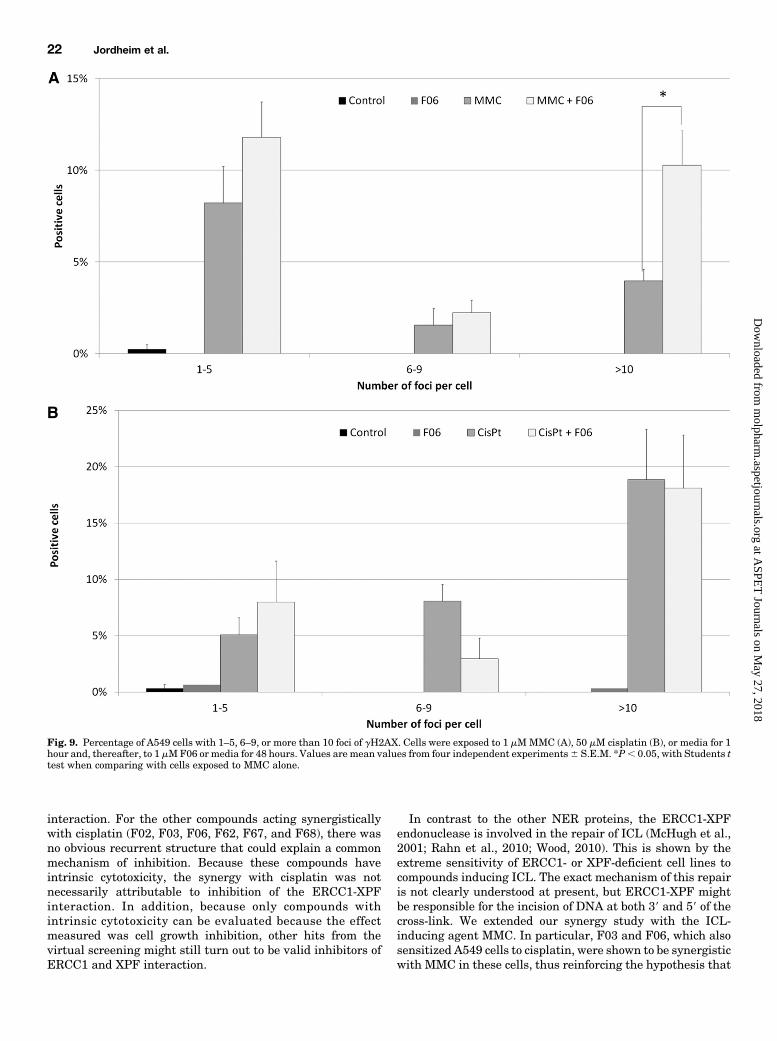
pair. The repair of ICL involves the production of double-strand breaks that can be visualized by immunofluorescencewith antibody against the phosphorylated histone H2AX. Aspreviously shown, the inhibition of ERCC1 or XPF withsiRNA modifies the kinetics of gH2AX positivity in cancercells (Arora et al., 2010). Thus, we studied this marker inA549 cells exposed to MMC or cisplatin and incubated in thepresence or absence of F06, respectively. Preliminary experi-ments showed that MMC and cisplatin induced positivity forgH2AX staining that was easily quantified after 48 hours(Supplemental Fig. 4). For cells exposed to MMC, the addition
Fig. 6. (A) Sensorgram for interaction between XPF814-905 and ERCC1220-297. ERCC1220-297 was injected at 10 mM in PBS containing 1mMdithiothreitolon a chip with immobilized XPF814-905. Dotted line indicates time of injection of ERCC1220-297. (B) Interaction between inhibitors and XPF814-905. Resultsare mean values of (corrected signal � 100/molecular weight)inhibitor 2 (corrected signal � 100/molecular weight)negative control from at least threeindependent experiments, and error bars are S.E.M. Inhibitors were diluted at 30 mM in PBS with a final concentration of 5% DMSO. PBS 5% DMSO isconsidered as negative control.
Fig. 7. Fluorescence experiments for the interaction between XPF814-905 and F06. (A) Emission spectra of XPF814-905 (2 mM) alone (X) and in thepresence of different concentrations of F06 (A: 10, B: 15, C: 20, D: 25, E: 27, and F: 30 mM). lexcit = 271 nm and excitation and emission slit width were7 nm. (B) Synchronous fluorescence spectra of XPF814-905 (2 mM) alone (X) and in the presence of different concentrations of F06 (A: 10, B: 15, C: 20, D: 25,E: 27, and F: 30 mM) at Dl = 20 nm. (C) The Stern-Volmer plots of fluorescence quenching of XPF814-905 by compound F06. The value of KA (3.31 � 104 60.0598 l mol21) was calculated from the slope of the linear regression analysis, r = 0.9783. a.u., arbitrary unit.
20 Jordheim et al.
at ASPE
T Journals on M
ay 27, 2018m
olpharm.aspetjournals.org
Dow
nloaded from

of 1 mM F06 in the culture media statistically increased thepercentage of cells with more than 10 foci after 48 hours,compared with cells incubated only with cell media (10%versus 4%; P 5 0.019) (Fig. 9A). However, for cells exposedto cisplatin, no such difference was observed (Fig. 9B). Theincubation with F06 alone did not induce any gH2AX stainingin these conditions.Synergy with PARP Inhibitor in BRCA1-Mutated
Cells. Olaparib (AZD2281) is a PARP inhibitor that hasshown interesting activity both alone and in combination withplatinum derivatives in BRCA1-mutated breast cancer cells,based on the principle of synthetic lethality (Rottenberg et al.,2008). We assumed that F06 would act synergistically witholaparib in BRCA1-negative cells, because there would be aninhibition of homologous recombination, NER, ICL repair,and nonhomologous end-joining. We performed synergyanalysis in both wild-type (MDA-MB-231) and BRCA1-mutated (MDA-MB-436) human breast cancer cells. Asexpected, sensitivity to olaparib was different in the two celllines (IC50 5 1356 25 mM versus 226 6 mM in MDA-MB-231and MDA-MB-436, respectively), whereas the sensitivity toF06 was similar in the two cell lines (2.9 6 0.9 mM versus3.1 6 0.7 mM). However, we observed antagonism betweenolaparib and F06 in wild-type MDA-MB-231 cells (CI95 51.7) and synergy in BRCA1-mutated MDA-MB-436 cells(CI95 5 0.7).
DiscussionCisplatin is a widely used cancer chemotherapy drug that is
active in several tumor types, and in particular, it inducesresponse rates above 90% in testicular cancer. However, manypatients discontinue cisplatin-based treatment because of theassociated toxicity or drug resistance. Therefore, the de-velopment of strategies to increase the sensitivity of resistantcancer cells to cisplatin promises to offer an enhanced clinicalbenefit.The involvement of NER proteins in the clinical activity of
cisplatin is now clearly established, thus constituting a targetfor the sensitization of cancer cells to platinum derivatives.This has been confirmed in vitro, as for example, by theinduction of ERCC1 by H-Ras in human breast cancer cells(Youn et al., 2004). There, resistance to cisplatin andoxaliplatin was reversed by ERCC1-specific siRNA. Similarresults were observed with the over-expression of SNAIL inhuman head and neck squamous cell carcinoma cell lines(FaDu, PECM-1, and CAL-27) (Hsu et al., 2010). Reduction inapoptosis in cancerous testicular germ cells (833 K) trans-fected with plasmid coding both ERCC1 and XPF exposed tocisplatin was also reported (Usanova et al., 2010). The sameauthors showed that siRNA against ERCC1 sensitized humanbladder cancer cells (MGH-U1) to cisplatin with modified
kinetics of gH2AX staining and increased proportion of cellsin subG1. This was confirmed by individual or concomitantsiRNA inhibition of ERCC1 or XPF in several human lungcancer cells (Arora et al., 2010). These studies clearly showthat the modulation of ERCC1-XPF in cancer cells is associatedwith modified sensitivity to platinum derivatives and in gH2AXstaining. Therefore, as recently suggested by others (McNeiland Melton, 2012), we used human cancer cells to evaluate theactivity of potential ERCC1-XPF inhibitors and their effects onthe cytotoxicity of cisplatin.A major obstacle was encountered while identifying
ERCC1-XPF inhibitors, namely an immense enthalpic con-tribution to the binding energy between the two proteins.Disrupting this interaction requires a quantitative under-standing of its different contributions. Consequently, we firstanalyzed the binding mode to isolate a druggable binding sitesuitable for virtual screening simulations. On the basis of theenergetic contributions of the different residues and theirability to form a deep pocket, we chose site I for the virtualscreening. The fact that the studied inhibitors target only thissite might explain why we did not observe pharmacologicalmodifications at a level comparable to ERCC1- or XPF-deficient cells in the cells exposed to the selected inhibitors.To identify potential inhibitors, we performed 20 virtual
screening simulations with use of the 20 available NMRsnapshots (Lin et al., 2002; Amaro et al., 2008). From ourexperience, the NCI diversity set and DrugBank providea good starting point, especially for screening against targetswith no known inhibitors. These libraries include ∼3500distinct structures that can serve as scaffolds for optimiza-tion. These structures were docked individually to the 20 XPFtargets, and the top 73 hits were selected for experimentalvalidation. An important feature present in most of our hitsis the side chain that protrudes within the cavity producedby XPF-residues Tyr833, Asn834, and Pro837 that forma deep pocket, which buries the key residue Phe293. Theactual interaction between compounds and these residues ofXPF could be confirmed by in vitro experiments with mutantprotein, and we plan to perform these experiments in thefuture.For biological evaluation, we performed synergy assays
to evaluate the sensitization of cisplatin by the potentialinhibitors. We found several compounds that had synergisticactivity with cisplatin in A549 cells and two in HCT116 cells.Two compounds are expected to be synergistic independentlyof ERCC1 and XPF [i.e., F66 (NSC 303812)] (Damia et al.,1992; O’Dwyer et al., 1994) and F64 (NSC 254681), a de-rivative of daunorubicin with IC50 of 0.23 and 0.05 mM onA549 and HCT116 cells, respectively. This effect is mostprobably not attributable to the inhibition of the ERCC1-XPF
TABLE 3Results from proximity ligation assayThe numbers indicate the spots due to ERCC1-XPF interaction in A549 cells alone orincubated with 2 mM F06 and/or 20 mM cisplatin for 24 hours. Representativemicroscopic images are available in Supplemental Fig. 3.
Condition NT F06 Cisplatin Cisplatin + F06
Spots per cell 15.2 6.9 35.3 15.2
Fig. 8. Immunoprecipitation in cell extracts from A459 cells incubatedwith 100, 200, or 500 mM F06. The image shows a representative result ofthree independent experiments. D, DMSO.
Inhibitors of ERCC1-XPF Interaction 21
at ASPE
T Journals on M
ay 27, 2018m
olpharm.aspetjournals.org
Dow
nloaded from
interaction. For the other compounds acting synergisticallywith cisplatin (F02, F03, F06, F62, F67, and F68), there wasno obvious recurrent structure that could explain a commonmechanism of inhibition. Because these compounds haveintrinsic cytotoxicity, the synergy with cisplatin was notnecessarily attributable to inhibition of the ERCC1-XPFinteraction. In addition, because only compounds withintrinsic cytotoxicity can be evaluated because the effectmeasured was cell growth inhibition, other hits from thevirtual screening might still turn out to be valid inhibitors ofERCC1 and XPF interaction.
In contrast to the other NER proteins, the ERCC1-XPFendonuclease is involved in the repair of ICL (McHugh et al.,2001; Rahn et al., 2010; Wood, 2010). This is shown by theextreme sensitivity of ERCC1- or XPF-deficient cell lines tocompounds inducing ICL. The exact mechanism of this repairis not clearly understood at present, but ERCC1-XPF mightbe responsible for the incision of DNA at both 39 and 59 of thecross-link. We extended our synergy study with the ICL-inducing agent MMC. In particular, F03 and F06, which alsosensitized A549 cells to cisplatin, were shown to be synergisticwith MMC in these cells, thus reinforcing the hypothesis that
Fig. 9. Percentage of A549 cells with 1–5, 6–9, or more than 10 foci of gH2AX. Cells were exposed to 1 mMMMC (A), 50 mM cisplatin (B), or media for 1hour and, thereafter, to 1 mMF06 or media for 48 hours. Values are mean values from four independent experiments6 S.E.M. *P, 0.05, with Students ttest when comparing with cells exposed to MMC alone.
22 Jordheim et al.
at ASPE
T Journals on M
ay 27, 2018m
olpharm.aspetjournals.org
Dow
nloaded from

these molecules act by inhibition of the interaction betweenERCC1 and XPF.To reveal the mechanism of action of the potential inhib-
itors of the ERCC1-XPF interaction, we evaluated the in-teraction between these compounds and XPF with use of afragment of XPF corresponding to the targeted domain ofthe interaction with ERCC1. Biacore experiments are basedon the detection and the quantification of the interactionbetween one compound immobilized on a sensor chip (here,the XPF fragment) and another compound solubilized ina running buffer (here, the inhibitors). Our results show that,in particular, F67 and F06 interact with XPF814-905, suggest-ing that the effect observed on the cell models with F06 isattributable to an interaction with NER proteins. F06 wasalso shown to interact with ERCC1, although at higherconcentrations than for XPF. F67 induced a strong signal ofinteraction with XPF814-905, even though it showed onlysynergy with cisplatin in A549 cells and not with othercompounds or in HCT116 cells. This indicates that itsinteraction with the XPF fragment is not necessarily in-hibiting the protein-protein interaction. Concerning F06,we were able to show a specific and saturable interactionwith XPF and to determine the binding parameters of theinteraction by fluorescence quenching experiments. Thefact that we were not able to quantify this interaction bySPR might be attributable to the differences in structuralconformation of the XPF fragment immobilized on the sensorchip, compared with the soluble XPF fragment used influorescence quenching experiments. The role of F06 on theinhibition of the interaction between ERCC1 and XPF wasfinally confirmed both by proximity ligation assays and byimmunoprecipitation. The proximity ligation assay clearlyconfirmed the ability of this compound to act on ERCC1 andXPF in cells.There is a negative correlation between cell survival after
cisplatin exposure and the gH2AX expression in cancer cells(Olive and Banath, 2009). Niedernhofer et al. (2004) showedin ERCC1-deficient mouse embryonic stem cells that ERCC1-XPF is not required for the formation of double-strand breaksafter exposure to ICL-inducing agents. However, ICL repairis largely slowed down in ERCC1-deficient cells, and thekinetics of the gH2AX signal was largely modified, lasting formore than 72 hours in ERCC1-deficient cells, compared with12–24 hours in wild-type cells. Such variations were alsoobserved in cancer cells transfected with siRNA againstERCC1 or XPF and exposed to cisplatin (Arora et al., 2010;Usanova et al., 2010). Our results on gH2AX staining areconsistent with previous observations concerning the down-regulation of ERCC1 and XPF, even though the effect issomehow weaker. This can be attributable to the weakerinhibition caused by F06, compared with siRNA againstERCC1 or XPF, which further motivates us to search for moreactive analogs of F06.Synthetic lethality is a concept based on targeting a path-
way that has become crucial in a cell line because of theinactivation of another pathway. A well-described exampleinvolves PARP in BRCA1- or BRCA2-deficient cells. Be-cause ERCC1-XPF is involved in DNA repair other thanPARP and BRCA, NER inhibition should additionally in-crease the activity of PARP inhibitors in BRCA-mutatedcells. As shown, the association of olaparib and F06 is onlysynergistic in BRCA1-deficient breast cancer cells. This is
the first observation of such an additional effect by tar-geting NER. Confirmation of the role of ERCC1-XPF in thissynergy is still needed.We did not observe a clear correlation between the results
obtained with different techniques. This discrepancy amongintrinsic cytotoxicity, synergistic potential, UVC sensitiza-tion, and interaction with the XPF fragment can be explainedby the nonspecificity of the compounds. Indeed, somecompounds might have several distinct molecular effects oncancer cells and elicit various responses. Hypothetically, theinteraction between some compounds and XPF could increasethe affinity for ERCC1, thus inducing antagonsimwith cisplatinor MMC. Although specific molecules are most often preferredin studies of this type, the most important finding reported hereis that our hit compound is able to increase the cytotoxicity ofcisplatin in cancer cells.We have focused on inhibitors of NER acting through the
inhibition of a protein-protein interaction and demonstratedthat F06 (NSC 130813, NERI02) interacts with XPF, synergizescancer cells to cisplatin and MMC, modifies DNA repair,interacts with ERCC1 and XPF in vitro, and decreases theinteraction between ERCC1 and XPF in cells and cell ex-tracts. This confirms the hypothesis that the inhibition ofNER by targeting protein-protein interactions sensitizes thecytotoxic activity of alkylating agents. NERI02 should besubjected to derivatization to optimize its action.
Acknowledgments
The authors thank Nicolas Foray for assistance on gH2AXassessment, Amandine Roux for Biacore experiments, and SuzannePeyrottes for structure and purity determinations. NAMD wasdeveloped by the Theoretical and Computational Biophysics Groupin the Beckman Institute for Advanced Science and Technology at theUniversity of Illinois at Urbana-Champaign.
Authorship Contributions
Participated in the research design: Jordheim, Barakat, Heinrich-Balard, Matera, Cros-Perrial, Bouledrak, El Sabeh, Perez-Pineiro,Wishart, Cohen, Tuszynski, Dumontet.
Conducted experiments: Jordheim, Barakat, Heinrich-Balard,Matera, Cros-Perrial, Bouledrak, El Sabeh, Perez-Pineiro.
Performed data analysis: Jordheim, Barakat, Heinrich-Balard,Matera, Cros-Perrial, Bouledrak, El Sabeh, Perez-Pineiro, Tuszynski,Dumontet.
Wrote or contributed to the writing of the manuscript: Jordheim,Barakat, Heinrich-Balard, Wishart, Cohen, Tuszynski, Dumontet.
References
Amaro RE, Baron R, and McCammon JA (2008) An improved relaxed complexscheme for receptor flexibility in computer-aided drug design. J Comput Aided MolDes 22:693–705.
Arora S, Kothandapani A, Tillison K, Kalman-Maltese V, and Patrick SM (2010)Downregulation of XPF-ERCC1 enhances cisplatin efficacy in cancer cells. DNARepair (Amst) 9:745–753.
Azmi AS and Mohammad RM (2009) Non-peptidic small molecule inhibitors againstBcl-2 for cancer therapy. J Cell Physiol 218:13–21.
Barakat K, Mane J, Friesen D, and Tuszynski J (2010) Ensemble-based virtualscreening reveals dual-inhibitors for the p53-MDM2/MDMX interactions. J MolGraph Model 28:555–568.
Barakat K and Tuszynski J (2011) Relaxed complex scheme suggests novel inhibitorsfor the lyase activity of DNA polymerase beta. J Mol Graph Model 29:702–716.
Barakat KH, Jordheim LP, Perez-Pineiro R, Wishart D, Dumontet C, and TuszynskiJA (2012) Virtual screening and biological evaluation of inhibitors targeting theXPA-ERCC1 interaction. PLoS ONE 7:e51329.
Barakat KH, Torin Huzil J, Luchko T, Jordheim L, Dumontet C, and Tuszynski J(2009) Characterization of an inhibitory dynamic pharmacophore for the ERCC1-XPA interaction using a combined molecular dynamics and virtual screening ap-proach. J Mol Graph Model 28:113–130.
Case DA, Cheatham T, Darden T, Gohlke H, Luo R, Merz KM Jr, Onufriev A,Simmerling C, Wang B, and Woods R (2005) The Amber biomolecular simulationprograms. J Comput Chem 26:1668–1688.
Inhibitors of ERCC1-XPF Interaction 23
at ASPE
T Journals on M
ay 27, 2018m
olpharm.aspetjournals.org
Dow
nloaded from

Chou TC and Talalay P (1984) Quantitative analysis of dose-effect relationships: thecombined effects of multiple drugs or enzyme inhibitors. Adv Enzyme Regul 22:27–55.
Coste I, Le Corf K, Kfoury A, Hmitou I, Druillennec S, Hainaut P, Eychene A,Lebecque S, and Renno T (2010) Dual function of MyD88 in RAS signaling andinflammation, leading to mouse and human cell transformation. J Clin Invest120:3663–3667.
Damia G, Tagliabue G, Zucchetti M, Davoli E, Sessa C, Cavalli F, and D’Incalci M(1992) Activity of aphidicolin glycinate alone or in combination with cisplatin ina murine ovarian tumor resistant to cisplatin. Cancer Chemother Pharmacol 30:459–464.
Dolinsky TJ, Czodrowski P, Li H, Nielsen JE, Jensen JH, Klebe G, and Baker NA(2007) PDB2PQR: expanding and upgrading automated preparation of bio-molecular structures for molecular simulations. Nucleic Acids Res 35 (Web Serverissue):W522-5.
Frostell-Karlsson A, Remaeus A, Roos H, Andersson K, Borg P, Hämäläinen M,and Karlsson R (2000) Biosensor analysis of the interaction between immobilizedhuman serum albumin and drug compounds for prediction of human serum albu-min binding levels. J Med Chem 43:1986–1992.
Galluzzi L, Senovilla L, Vitale I, Michels J, Martins I, Kepp O, Castedo M,and Kroemer G (2012) Molecular mechanisms of cisplatin resistance. Oncogene 31:1869–1883.
Gasteiger J and Marsili M (1980) Iterative partial equalization of orbital electro-negativity: a rapid access to atomic charges. Tetrahedron 36:3219–3228.
Hsu DS, Lan HY, Huang CH, Tai SK, Chang SY, Tsai TL, Chang CC, Tzeng CH, WuKJ, and Kao JY et al. (2010) Regulation of excision repair cross-complementationgroup 1 by Snail contributes to cisplatin resistance in head and neck cancer. ClinCancer Res 16:4561–4571.
Hubner RA, Riley RD, Billingham LJ, and Popat S (2011) Excision repair cross-complementation group 1 (ERCC1) status and lung cancer outcomes: a meta-analysis of published studies and recommendations. PLoS ONE 6:e25164.
Jamieson ER and Lippard SJ (1999) Structure, Recognition, and Processing ofCisplatin-DNA Adducts. Chem Rev 99:2467–2498.
Jordheim LP, Guittet O, Lepoivre M, Galmarini CM, and Dumontet C (2005) In-creased expression of the large subunit of ribonucleotide reductase is involved inresistance to gemcitabine in human mammary adenocarcinoma cells. Mol CancerTher 4:1268–1276.
Kale L, Skeel R, Bhandarkar M, Brunner R, Gursoy A, Krawetz N, Phillips J,Shinozaki A, Varadarajan K, and Schulten K (1999) NAMD2: Greater Scalabilityfor Parallel Molecular Dynamics. J Comput Phys 151:283–312.
Kollman PA, Massova I, Reyes C, Kuhn B, Huo S, Chong L, Lee M, Lee T, Duan Y,and Wang W et al. (2000) Calculating structures and free energies of complexmolecules: combining molecular mechanics and continuum models. Acc Chem Res33:889–897.
Kuhn B, Gerber P, Schulz-Gasch T, and Stahl M (2005) Validation and use of theMM-PBSA approach for drug discovery. J Med Chem 48:4040–4048.
Lakowicz JR(1999) Principles of fluorescence spectroscopy, Second ed. KluwerAcademic/Plenum, New York.
Lin JH, Perryman AL, Schames JR, and McCammon JA (2002) Computational drugdesign accommodating receptor flexibility: the relaxed complex scheme. J AmChem Soc 124:5632–5633.
McHugh PJ, Spanswick VJ, and Hartley JA (2001) Repair of DNA interstrandcrosslinks: molecular mechanisms and clinical relevance. Lancet Oncol 2:483–490.
McNeil EM and Melton DW (2012) DNA repair endonuclease ERCC1-XPF as a noveltherapeutic target to overcome chemoresistance in cancer therapy. Nucleic AcidsRes 40:9990–10004.
Morris GM, Goodsell DS, Halliday RS, Huey R, Hart WE, Belew RK, and Olson AJ(1998) Automated docking using a Lamarckian genetic algorithm and an empiricalbinding free energy function. J Comput Chem 19:1639–1662.
Niedernhofer LJ, Odijk H, Budzowska M, van Drunen E, Maas A, Theil AF, de Wit J,Jaspers NG, Beverloo HB, and Hoeijmakers JH et al. (2004) The structure-specificendonuclease Ercc1-Xpf is required to resolve DNA interstrand cross-link-induceddouble-strand breaks. Mol Cell Biol 24:5776–5787.
Nouspikel T (2009) DNA repair in mammalian cells : Nucleotide excision repair:variations on versatility. Cell Mol Life Sci 66:994–1009.
O’Dwyer PJ, Moyer JD, Suffness M, Harrison SD, Jr, Cysyk R, Hamilton TC,and Plowman J (1994) Antitumor activity and biochemical effects of aphidicolinglycinate (NSC 303812) alone and in combination with cisplatin in vivo. Cancer Res54:724–729.
Olive PL and Banáth JP (2009) Kinetics of H2AX phosphorylation after exposure tocisplatin. Cytometry B Clin Cytom 76:79–90.
Phillips JC, Braun R, Wang W, Gumbart J, Tajkhorshid E, Villa E, Chipot C, SkeelRD, Kale L, and Schulten K (2005) Scalable molecular dynamics with NAMD. JComput Chem 26:1781–1802.
Rahn JJ, Adair GM, and Nairn RS (2010) Multiple roles of ERCC1-XPF in mam-malian interstrand crosslink repair. Environ Mol Mutagen 51:567–581.
Rottenberg S, Jaspers JE, Kersbergen A, van der Burg E, Nygren AO, Zander SA,Derksen PW, de Bruin M, Zevenhoven J, and Lau A et al. (2008) High sensi-tivity of BRCA1-deficient mammary tumors to the PARP inhibitor AZD2281alone and in combination with platinum drugs. Proc Natl Acad Sci USA 105:17079–17084.
Shangary S and Wang S (2008) Targeting the MDM2-p53 interaction for cancertherapy. Clin Cancer Res 14:5318–5324.
Shao J, Tanner SW, Thompson N, and Cheatham TE, 3rd (2007) Clustering Molec-ular Dynamics Trajectories: 1. Characterizing the Performance of Different Clus-tering Algorithms. J Chem Theory Comput 3:2312–2334.
Tripsianes K, Folkers G, Ab E, Das D, Odijk H, Jaspers NG, Hoeijmakers JH,Kaptein R, and Boelens R (2005) The structure of the human ERCC1/XPF in-teraction domains reveals a complementary role for the two proteins in nucleotideexcision repair. Structure 13:1849–1858.
Tsodikov OV, Ivanov D, Orelli B, Staresincic L, Shoshani I, Oberman R, Schärer OD,Wagner G, and Ellenberger T (2007) Structural basis for the recruitment ofERCC1-XPF to nucleotide excision repair complexes by XPA. EMBO J 26:4768–4776.
Usanova S, Piée-Staffa A, Sied U, Thomale J, Schneider A, Kaina B, and KöberleB (2010) Cisplatin sensitivity of testis tumour cells is due to deficiency ininterstrand-crosslink repair and low ERCC1-XPF expression. Mol Cancer 9:248.
van de Weert M and Stell L (2011) Fluorescence quenching and ligand binding: Acritical discussion of a popular methodology. J Mol Struct 998:144–150.
Wood RD (2010) Mammalian nucleotide excision repair proteins and interstrandcrosslink repair. Environ Mol Mutagen 51:520–526.
Youn CK, Kim MH, Cho HJ, Kim HB, Chang IY, Chung MH, and You HJ (2004)Oncogenic H-Ras up-regulates expression of ERCC1 to protect cells from platinum-based anticancer agents. Cancer Res 64:4849–4857.
Address correspondence to: Lars Petter Jordheim, Equipe Anticorps-Anticancer, Centre de Recherche en Cancérologie de Lyon, INSERM U1052–CNRS UMR 5286, Faculté Rockefeller, 8 Avenue Rockefeller, 69008 Lyon,France. E-mail: [email protected]
24 Jordheim et al.
at ASPE
T Journals on M
ay 27, 2018m
olpharm.aspetjournals.org
Dow
nloaded from