small ubiquitin-like modifier alters ifn response · small ubiquitin-like modifier alters ifn ......
TRANSCRIPT

of June 4, 2018.This information is current as
ResponseSmall Ubiquitin-like Modifier Alters IFN
Chelbi-AlixLaurent Dianoux, Sébastien Nisole and Mounira K. Ghizlane Maarifi, Mohamed Ali Maroui, Jacques Dutrieux,
http://www.jimmunol.org/content/195/5/2312doi: 10.4049/jimmunol.1500035July 2015;
2015; 195:2312-2324; Prepublished online 29J Immunol
MaterialSupplementary
5.DCSupplementalhttp://www.jimmunol.org/content/suppl/2015/07/29/jimmunol.150003
Referenceshttp://www.jimmunol.org/content/195/5/2312.full#ref-list-1
, 18 of which you can access for free at: cites 36 articlesThis article
average*
4 weeks from acceptance to publicationFast Publication! •
Every submission reviewed by practicing scientistsNo Triage! •
from submission to initial decisionRapid Reviews! 30 days* •
Submit online. ?The JIWhy
Subscriptionhttp://jimmunol.org/subscription
is online at: The Journal of ImmunologyInformation about subscribing to
Permissionshttp://www.aai.org/About/Publications/JI/copyright.htmlSubmit copyright permission requests at:
Email Alertshttp://jimmunol.org/alertsReceive free email-alerts when new articles cite this article. Sign up at:
Print ISSN: 0022-1767 Online ISSN: 1550-6606. Immunologists, Inc. All rights reserved.Copyright © 2015 by The American Association of1451 Rockville Pike, Suite 650, Rockville, MD 20852The American Association of Immunologists, Inc.,
is published twice each month byThe Journal of Immunology
by guest on June 4, 2018http://w
ww
.jimm
unol.org/D
ownloaded from
by guest on June 4, 2018
http://ww
w.jim
munol.org/
Dow
nloaded from

The Journal of Immunology
Small Ubiquitin-like Modifier Alters IFN Response
Ghizlane Maarifi, Mohamed Ali Maroui, Jacques Dutrieux, Laurent Dianoux,
Sebastien Nisole, and Mounira K. Chelbi-Alix
IFNs orchestrate immune defense through induction of hundreds of genes. Small ubiquitin-like modifier (SUMO) is involved in
various cellular functions, but little is known about its role in IFN responses. Prior work identified STAT1 SUMOylation as an
important mode of regulation of IFN-g signaling. In this study, we investigated the roles of SUMO in IFN signaling, gene
expression, protein stability, and IFN-induced biological responses. We first show that SUMO overexpression leads to STAT1
SUMOylation and to a decrease in IFN-induced STAT1 phosphorylation. Interestingly, IFNs exert a negative retrocontrol on their
own signaling by enhancing STAT1 SUMOylation. Furthermore, we show that expression of each SUMO paralog inhibits IFN-
g–induced transcription without affecting that of IFN-a. Further, we focused on IFN-induced gene products associated to
promyelocytic leukemia (PML) nuclear bodies, and we show that neither IFN-a nor IFN-g could increase PML and Sp100
protein expression because they enhanced their SUMO3 conjugation and subsequent proteasomal degradation. Because it is
known that SUMO3 is important for the recruitment of RING finger protein 4, a poly–SUMO-dependent E3 ubiquitin ligase,
and that PML acts as a positive regulator of IFN-induced STAT1 phosphorylation, we went on to show that RING finger protein 4
depletion stabilizes PML and is correlated with a positive regulation of IFN signaling. Importantly, inhibition of IFN signaling by
SUMO is associated with a reduction of IFN-induced apoptosis, cell growth inhibition, antiviral defense, and chemotaxis. Con-
versely, inhibition of SUMOylation results in higher IFN-g–induced STAT1 phosphorylation and biological responses. Altogether, our
results uncover a new role for SUMO in the modulation of IFN response. The Journal of Immunology, 2015, 195: 2312–2324.
Interferons are a family of cytokines that exhibit diverse bi-ological activities. Identified and named for their antiviralproperties, IFNs have also immunomodulatory, antiproliferative,
and apoptotic activities (1). IFNs are successfully used in therapy totreat viral infections, cancer, or multiple sclerosis. However, the useof IFN is limited and some patients are resistant to treatment. Thus,progress remains to be done to better understand the mechanism ofaction of IFNs.Based on their structure and interaction with distinct receptor
complexes, IFNs are subdivided into three distinct types. IFNsconsist in multiple type I species (including IFN-a and IFN-b), onetype II (IFN-g), and three members of type III species (IFN-ls,also known as IL-28 and -29) (2). IFNs act on cells by binding totheir respective receptors (IFN-a receptor [IFNAR] for IFN-a/b,IFNGR for IFN-g, or IFNLR for IFN-l) and activating the
JAK/STAT pathways to trigger the transcription of .300 IFN-stimulated genes (ISGs), the products of which are the media-
tors of their biological effects (2). The interaction of IFN-a/b with
IFNAR leads to the activation of the JAK tyrosine kinases (Tyk2
and JAK1) that phosphorylate STAT1 and STAT2. Phosphorylated
STATs heterodimerize and form with the DNA binding protein
IFN regulatory factor 9 (IRF9), a complex called IFN-stimulated
growth factor 3 (ISGF3). ISGF3 translocates into the nucleus to
induce ISGs harboring an IFN-stimulated response element
(ISRE). The binding of IFN-g to its receptor, IFNGR, results in
the phosphorylation of STAT1 by JAK1 and JAK2. p-STAT1 on
Tyr701 forms homodimers that migrate to the nucleus and bind to
a DNA element termed g-activated sequence (GAS) in the pro-
moter of specific ISGs. Accordingly, transcriptional responses to
IFN-g are dominated by the activity of pSTAT1 homodimers.
Finally, type III IFNs that are structurally and genetically distinct
from type I IFNs bind to different receptors, but activate the same
signal transduction pathway (2).The signal transduction induced by IFNs is transient because it
is inhibited by negative regulators of IFN signaling that include
phosphotyrosine phosphatases (Src homology region 2 domain-
containing phosphatases, CD45, and PTP1B/TC-PTP), suppressors
of cytokine signaling, and protein inhibitors of activated STATs
(PIAS), which ensure the proper termination of IFN response (3).Ubiquitin or ubiquitin-like proteins, such as SUMO or ISG15,
modify many ISGs or key regulators of IFN signaling (4). However,
the role of SUMO on IFN-induced signaling, cell growth inhibi-
tion, apoptosis, and antiviral activity remains to be elucidated.
SUMOylation is the posttranslational covalent but reversible con-
jugation of SUMO to proteins. In humans, the SUMO protein
family consists of SUMO1 and two highly homologous proteins
SUMO2 and SUMO3 (collectively called SUMO2/3), which cannot
be distinguished by currently available Abs. SUMO2 and SUMO3
share 97% sequence identity and are expressed at much higher
levels than SUMO1, with which they only share ∼50% identity (5).
INSERM Unite Mixte de Recherche S 1124, Universite Paris Descartes, 75006Paris, France
Received for publication January 12, 2015. Accepted for publication June 17, 2015.
This work was supported by the Agence Nationale de la Recherche (ANR11BSV3002803). M.A.M. is supported by the Agence Nationale de la Recherche.J.D. is supported by the Agence Nationale de la Recherche sur le SIDA et lesHepatites Virales.
Address correspondence and reprint requests to Dr. Sebastien Nisole and Dr. MouniraK. Chelbi-Alix, INSERM Unite Mixte de Recherche S 1124, Universite Paris Des-cartes, 45 rue des Saints-Peres, 75006 Paris, France. E-mail addresses: [email protected] (S.N.) and [email protected] (M.K.C.-A.)
The online version of this article contains supplemental material.
Abbreviations used in this article: EMCV, encephalomyocarditis virus; F, forward;GAS, g-activated sequence; IFNAR, IFN-a receptor; IP-10, IFN-g–induced protein10; IRF, IFN regulatory factor; ISG, IFN-stimulated gene; ISGF3, IFN-stimulatedgrowth factor 3; ISRE, IFN-stimulated response element; MOI, multiplicity of infec-tion; NB, nuclear body; PI, propidium iodide; PIAS, protein inhibitor of activatedSTATs; PKR, RNA-dependent protein kinase; PML, promyelocytic leukemia; qRT-PCR, quantitative RT-PCR; R, reverse; RNF4, RING finger protein 4; siRNA, smallinterfering RNA; SUMO, small ubiquitin-like modifier; TAP1, transporter associatedwith Ag processing 1; TCID50, 50% tissue culture infective dose; wt, wild-type.
Copyright� 2015 by TheAmericanAssociation of Immunologists, Inc. 0022-1767/15/$25.00
www.jimmunol.org/cgi/doi/10.4049/jimmunol.1500035
by guest on June 4, 2018http://w
ww
.jimm
unol.org/D
ownloaded from

SUMO modification occurs through the formation of an iso-peptide bond between the a-amino group of a lysine residuefrom the substrate and the C terminus COOH group of SUMO.SUMOylation involves a complex network of SUMO-activatingenzymes (1 and 2), conjugating enzyme (Ubc9), and SUMO-E3ligases (PIAS1, PIAS3, PIASxa, PIASxb, PIASy, RanBP2, andPc2) (6). The dynamic protein SUMOylation is counterbalancedby SUMO-specific proteases, which cleave SUMO moieties on
specific substrates. SUMOylation leads to significant structuraland conformational changes of the substrate by masking or con-ferring additional scaffolding surfaces for protein interactions.This posttranslational modification is involved in the regulation ofintracellular trafficking, cell cycle, DNA repair, cell signaling, andprotein degradation (5).At present, little is known about the role of SUMO on IFN re-
sponses. STAT1 was found to be conjugated to SUMO on Lys703 byPIAS (7–9). Several studies reported that the activity of STAT1could be inhibited by SUMO conjugation, because a SUMOylation-deficient STAT1 mutant is hyperphosphorylated and has higherDNA binding on STAT1-responsive gene promoters (8, 10, 11).However, whether increased cellular protein conjugation to SUMOcould alter IFN signaling, stability of ISG products or IFN re-sponses remains unknown. To better understand the role ofSUMOylation in type I and II IFN pathways, we stably overex-pressed each SUMO paralog in human cells and investigated theconsequences on IFN signaling and IFN transcriptional responses.We also tested the capacity of IFN to regulate its own signaling andanalyzed the fate of some ISG products, including promyelocyticleukemia (PML) nuclear body (NB)–associated proteins. Finally,
we assessed the effects of SUMO on IFN-induced biological ac-tivities such as cell growth and viral inhibition, apoptosis, and IFN-g–induced protein 10 (IP-10)–induced chemotaxis.
Materials and MethodsMaterials
Recombinant human IFN-a2 was from Schering, human IFN-g fromRoussel Uclaf (Romainville, France), and ginkgolic acid from Merck.Mouse monoclonal anti-PML (sc-966) and anti–IP-10 (sc-101500) Absand rabbit polyclonal Abs raised against PML (Sc-5621), STAT1(C-STAT1 Ab) (sc-345), STAT1 phosphotyrosine 701 (sc-7988), RNA-dependent protein kinase (PKR; sc-707), IRF1 (sc-497), SUMO1(sc-9060), and transporter associated with Ag processing 1 (TAP1; sc-20930) were from Santa Cruz Biotechnology. Rabbit anti-STAT2 andanti-STAT2 phosphotyrosine 689 Abs were obtained from Upstate Bio-technology. Monoclonal anti-STAT1 Ab (MA1-19371) (N-STAT1Ab)recognizing an epitope included within aa 8–23 of STAT1 was from LifeTechnologies. Mouse anti-6His Abs were from Clontech and rabbit anti-SUMO2/3 Abs used for Western blot from Invitrogen. Peroxidase-coupledsecondary Abs were purchased from Santa Cruz Biotechnology. Mouseanti-6His Abs used for immunofluorescence were from Thermo Scientific.Rabbit anti-SUMO2/3 Abs used for immunofluorescence were a gift fromMary Dasso and Maia Ouspenskaia (National Institutes of Health,Bethesda, MD). Rabbit anti-Sp100 Abs were a gift from Hans Will(Leibniz Institute for Experimental Virology, Hamburg, Germany), andrabbit anti-EMCV Abs were from Ann Palmenberg (University of Wis-consin–Madison, Madison, WI). Secondary Abs conjugated to AlexaFluor were purchased from Molecular Probes. Encephalomyocarditisvirus (EMCV) was produced as previously described (12). Plasmidtransfections were performed using Fugene 6 (Promega). Small inter-fering RNA (siRNA) targeting PML, Ubc9, or RING finger protein 4(RNF4) were purchased from GE Healthcare (ON-TARGETplus siRNASMARTpool) and transfected into cells using HiperFect transfectionreagent (Qiagen). IP-10 was quantified in cell culture medium usingCXCL10/IP-10 Immunoassay (Quantikine ELISA; R&D Systems). Lu-ciferase assays were performed using the Luciferase Assay System(Promega) following the manufacturer’s instructions. Cell proliferationwas evaluated using the Cell Proliferation Kit I (MTT) purchased fromRoche. Confocal laser microscopy was performed on a Zeiss LSM 710microscope (Carl Zeiss).
Cells
Human glioblastoma astrocytoma U373MG, cervical cancer HeLa, andhepatocellular carcinoma HepG2 cells were grown at 37˚C in DMEMsupplemented with 10% FCS. His-SUMO constructs were generated byinserting each cDNA encoding SUMO paralog in pcDNA3.1. In accor-dance with the National Center for Biotechnology Information database,we refer to the entry P63165 as SUMO1, P61956 as SUMO2, and P55854as SUMO3. HeLa and U373MG cells stably expressing each SUMOparalog were obtained by transfection with pcDNA SUMO constructs andsubsequent neomycine selection (0.5 mg/ml).
Purification of His6-tagged SUMO conjugates
Cells (107) untreated or treated with 1000 U/ml IFN for 30 min were lysedin denaturating buffer A (6 mol guanidinium-HCl, 0.1 mol Na2HPO4/NaH2PO4, 0.01 mol Tris-HCl [pH 8], 5 mmol imidazole, and 10 mmol2-ME). After sonication, the lysates were mixed with 50 ml Ni-NTA-agarosebeads (Qiagen) for 3 h at room temperature. The beads were successivelywashed with buffer B (0.1% Triton X-100, 8 mol urea, 0.1 mol Na2HPO4/NaH2PO4, 0.01 mol Tris-HCl [pH 6.3], and 10 mmol 2-ME), and subse-quently eluted with 200 mmol imidazole in 0.15 mol Tris-HCl (pH 6.7),30% glycerol, and 0.72 mol 2-ME.
EMSA
Control cells and cells stably expressing SUMO1 or SUMO3 were leftuntreated or treated with 1000 U/ml IFN-g or IFN-a for 30 min. Cells (33107) were harvested, and nuclear cell extracts were prepared using theNucBuster Protein Extraction Kit from Novagen. Proteins were examinedby EMSA with a 32P-labeled GAS probe or 32P-labeled ISRE probe. TheGAS probe was generated with the following duplex oligonucleotide: 59-TACAACAGCCTGATTTCCCCGAAATGACGC-39 (the GAS-like site isitalicized). The ISRE probe was generated with the duplex oligonucleotide59-AAAGGGAAAGTGAAACTAGAAAGTGAAAGA-39. The presenceof specific GAF or ISGF3 complexes was confirmed with specific anti-STAT1 or anti-STAT2 Abs. The reaction products were analyzed byelectrophoresis in a 4% nondenaturing polyacrylamide gel. The gel wasdried and analyzed by PhosphorImager.
Virus stocks and cell infection
EMCV (4 3 108 PFU/ml) titer was determined by standard plaque assays.HeLa-wt, HeLa-SUMO1, or HeLa-SUMO3 cells were grown on glasscoverslips in six-well plates (from 50–80% confluence) and infected withEMCV at the multiplicities of infection (MOIs) and at times postinfectionindicated in the figure legends. Viral titers were determined on HeLa cellsby measuring the 50% tissue culture infective dose (TCID50).
Real-time PCR
Total RNAs were extracted using RNeasy Mini Kit (Qiagen) following themanufacturer’s instructions. RNA samples were converted to cDNA usingthe RevertAid H Minus First Strand cDNA Synthesis Kit (Thermo Sci-entific). Real-time PCR reactions were performed in duplicates using 5 mlcDNA diluted 10 times in water using Takyon ROX SYBR MasterMixblue dTTP (Eurogentec). The following program was used on a 7900HTFast Real-Time PCR System (Applied Biosystems): 3 min at 95˚C fol-lowed by 35 cycles of 15 s at 95˚C, 25 s at 60˚C, and 25 s at 72˚C. Valuesfor each transcript were normalized to expression levels of RPL13A (60Sribosomal protein L13a) using the 2-DDCt method. Primers used forquantification of transcripts by real-time quantitative PCR are as follows:STAT1 forward (F), 59-CAGAGCCAATGGAACTTGATGG-39 and STAT1reverse (R), 59-TCCGAGACACCTCGTCAAACTC-39; IRF1-F, 59-ACTTTC-GCTGTGCCATGAACTC-39 and IRF1-R, 59-CGGCTGGACTTCGACTTT-CTTT-39; PML-F, 59- ACACCAGTGGTTCCTCAAGCA-39 and PML-R, 59-CTCGGCAGTAGATGCTGGTCA-39; PKR-F, 59-GCGATACATGAGCC-CAGAACAG-39 and PKR-R, 59-CTGAGATGATGCCATCCCGTAG-39; TAP1-F,59-TCCTGGTGGTCCTCTCCTCTCT-39 and TAP1-R, 59-CACTGCACTGGC-TATGGTGAGA-39; and IP-10–F, 59-CGCTGTACCTGCATCAGCAT-39 and IP-10–R, 59-GCAATGATCTCAACACGTGGAC-39.
Immunofluorescence analysis
Cells grown on glass coverslip were fixed 20 min with 4% paraformal-dehyde in PBS and permeabilized for 5 min in 0.1% Triton X-100 in PBS.Cells were then prepared for immunofluorescence staining using the ap-propriate primary Ab and the corresponding secondary Ab conjugated toAlexa Fluor (Molecular Probes). Cells were mounted onto glass slides byusing Immu-Mount (Shandon) containing DAPI. Confocal laser microscopywas performed on a Zeiss LSM 710 microscope (Carl Zeiss).
The Journal of Immunology 2313
by guest on June 4, 2018http://w
ww
.jimm
unol.org/D
ownloaded from

Apoptosis
Cell apoptosis was assessed using the Annexin V-FITC/PI Kit (BD Bio-sciences). Briefly, HepG2 cells were transfected with 0.5 ng SUMO3-YFPor pcDNA3 constructs. After 24 h, cells were left untreated or treated with1000 U/ml IFN-a or IFN-g for 72 h, and HeLa-wt, HeLa-SUMO1, andHeLa-SUMO3 were untreated or treated with 1000 U/ml IFN-g for 72 h.Cells were collected, resuspended in 100 ml PBS, and stained withAnnexin V for 15 min at 4˚C, followed by propidium iodide (PI) staining.Ten thousand cells were analyzed by flow cytometry on an FACSCalibur(BD Biosciences).
In vitro chemotaxis assay
HeLa cells or cells stably expressing SUMO1 or SUMO3were seeded at 105
cells/well CELLSTAR 12-well cell culture plates. Cells were left untreatedor treated with 1000 U/ml IFN-g. Seventy-two hours later, 12-well Thin-Cert cell culture inserts (Greiner Bio-One) were inserted into the wells.CD3/CD28-activated human CD4 T lymphocytes (5 3 105) were added toeach well culture insert and incubated for 6 h. Cells that migrated into thelower chambers were collected and counted on a flow cytometer. Cellmigration rates were determined by calculating the percentage of inputcells that migrated into the lower chamber.
ResultsIFN-induced STAT1 phosphorylation is reduced by SUMO
In order to investigate the impact of SUMO on the cellular responseto IFN, we generated HeLa cells stably expressing His-SUMO1
(HeLa-SUMO1) or His-SUMO3 (HeLa-SUMO3) and U373MG
cells stably expressing His-SUMO2 (U373MG-SUMO2) or His-
SUMO3 (U373MG-SUMO3). Immunofluorescence and Western
blot analyses confirmed an enrichment of the expression of each
SUMO paralog compared with wild-type (wt) cells (Supplemental
Fig. 1 and data not shown). Western blot analysis showed a marked
increase of free SUMO expression as well as the proportion of
SUMO-conjugated proteins in HeLa (Supplemental Fig. 1A) and
U373MG (Supplemental Fig. 1B) cells. Immunofluorescence anal-
ysis revealed that SUMO1, SUMO2, or SUMO3 were found in
nuclear speckles named PML NBs, as expected (Supplemental
Fig. 1C, 1D).Next, we sought to determine the effect of SUMO expression on
IFN signaling by analyzing STAT1 tyrosine phosphorylation. HeLaand U373MG wt cells and cells overexpressing SUMO1, SUMO2,or SUMO3 were treated with IFN-a or IFN-g for 30 min and thecorresponding protein extracts analyzed by Western blot. As an-ticipated, IFN-g (Fig. 1A, 1B) and IFN-a (Fig. 1C, 1D) induceda robust increase in p-STAT1 in wt cells. The induction of pSTAT1in response to IFN-g was markedly reduced in cells expressingSUMO1 (Fig. 1A), SUMO2 (Supplemental Fig. 1E), or SUMO3(Fig. 1B). A similar reduction in STAT1 phosphorylation wasobserved in cells overexpressing SUMO1 (Fig. 1C), SUMO2(Supplemental Fig. 1E), or SUMO3 (Fig. 1D) in response toIFN-a. In contrast, the IFN-a–induced phosphorylation of STAT2was insensitive to SUMO (Fig. 1C, 1D, Supplemental Fig. 1E).In a converse experiment, we examined the impact of the in-
hibition of SUMOylation on IFN-induced STAT1 phosphorylation.For this purpose, HeLa-wt cells were either treated with ginkgolicacid (Fig. 1E, 1F) that directly binds E1 and inhibits the formation
FIGURE 1. Effect of SUMO1 or SUMO3 expression on STAT1 phosphorylation in response to IFN-g or IFN-a. HeLa-wt, HeLa-SUMO1 (A and C), or
HeLa-SUMO3 (B and D) cells were treated for 30 min with IFN-g (A and B) or IFN-a (C and D) at 1000 U/ml. (E and F) IFN-g was added for different
times to HeLa-wt cells pretreated with 100 mmol ginkgolic acid. In all cases, cells were treated with ginkgolic acid for 6 h. (G and H) HeLa-wt cells
transfected with siRNA scramble (Scr) or siRNA Ubc9 were untreated or treated 2 d later with 1000 U/ml of IFN-g for 30 min. Equal amounts of cell
extracts were analyzed by Western blot for the expression of p-STAT1, p-STAT2, STAT1, STAT2, SUMO2/3, Ubc9, or Actin. P-STAT1/Actin ratios were
quantified using Image J software (National Institutes of Health) (H, right panel). (I) HeLa-wt cells were treated with IFN-g for the indicated times. Whole-
cell lysates were immunoprecipitated with anti-STAT1 Abs and analyzed by SDS-PAGE using anti-SUMO2/3 Abs. HeLa-His-SUMO1 and HeLa-SUMO3
cells were untreated or treated for 30 min with 1000 U/ml of IFN-a (J) or IFN-g (K). Cell extracts were purified on Ni-NTA–agarose beads. The inputs and
the purified extracts were analyzed by Western blotting using anti-STAT1 Abs.
2314 SUMO ALTERS IFN RESPONSE
by guest on June 4, 2018http://w
ww
.jimm
unol.org/D
ownloaded from

of the E1-SUMO intermediate (13) or were depleted for Ubc9(Fig. 1G, 1H), the unique E2-conjugating enzyme for SUMOylation.Both treatments resulted in a decrease in the level of SUMO2/3-modifed proteins (Fig. 1E, 1G) and most notably in a higher level ofSTAT1 phosphorylation in response to IFN-g or IFN-a (Fig. 1F, 1Hand data not shown).Because STAT1 is a SUMO substrate, these results prompted us
to evaluate the modification of STAT1 by SUMO in cells treated ornot with IFN-a or IFN-g for 30 min. First, we were able to observean interaction between endogenous STAT1 and endogeneousSUMO2/3 by coimmunoprecipitation. Interestingly, this interac-tion increased in cells treated 30 min with IFN-g and reacheda maximum at 2 h (Fig. 1I and data not shown). In addition, wefound that STAT1 was highly conjugated to SUMO3 in Ni-NTA–purified extracts from untreated HeLa-SUMO3 cells and thattreatment with IFN-a (Fig. 1J) or IFN-g (Fig. 1K) caused an in-crease of STAT1 conjugation to SUMO3. In contrast, we were notable to detect STAT1 modification by SUMO1 in Ni-NTA–purifiedextracts from HeLa-SUMO1 cells, although a slower migratingform of STAT1 with an expected size for its conjugation toSUMO1 was detected in the input (Fig. 1J). This latter observationis in line with the notion that SUMO1 conjugation is much moredifficult to detect than SUMO2/3 conjugation (14).Taken together, these results show that stable expression of
SUMO results in STAT1 SUMOylation and in a decrease of IFN-induced STAT1 phosphorylation. They also suggest that IFN exertsa negative retrocontrol on its own signaling by increasing STAT1SUMOylation.
SUMO1 or SUMO3 expression decreases p-STAT1 nuclearlocalization and DNA binding in response to IFN
Latent STAT1 resides mainly in the cytoplasm of unstimulated cellsand undergoes a rapid and transient nuclear accumulation after IFNstimulation (2). In order to analyze the effect of SUMO on IFN-induced STAT1 nuclear accumulation, we first performed indirectimmunofluorescence staining to monitor the subcellular distribu-tion of p-STAT1 after stimulation of HeLa-wt, HeLa-SUMO1, andHeLa-SUMO3 cells with IFN.As anticipated, both IFN-a and IFN-g promoted an increase
in p-STAT1 staining in wt cells (Fig. 2A and 2B). The level ofnuclear p-STAT1 staining was much lower when SUMO1 orSUMO3 were overexpressed (Fig. 2A). In line with the absenceof effect of SUMO on IFN-a–induced STAT2 phosphorylation,IFN-a promoted a marked increase in nuclear p-STAT2 staining,irrespective of SUMO expression (Fig. 2B).Next, we investigated the effect of SUMO on the downstream
intranuclear step of IFN signaling. In the IFN-g pathway, p-STAT1homodimers (called GAF) are translocated to the nucleus, wherethey bind to a DNA element termed GAS to induce the tran-scription of ISGs (2). We therefore performed EMSA to analyzethe binding capacity of p-STAT1 homodimers to GAS (Fig. 2C).HeLa-wt, HeLa-SUMO1, and HeLa-SUMO3 cells were treated ornot with IFN-g for 30 min, and nuclear extracts were subjected toEMSA with a (a-[32P]) ATP-labeled GAS probe. As expected,upon exposure to IFN-g, a band corresponding to the slower-migrating product predicted to be a GAF complex was apparentin extracts of HeLa-wt cells (Fig. 2C). The incubation of extractsfrom IFN-treated HeLa-wt cells with the anti-STAT1 Ab prior toincubation with the probe revealed the presence of a supershiftedband, confirming that the GAF complex was composed of the p-STAT1 homodimers (Fig. 2C). In the extracts from HeLa-SUMO1and HeLa-SUMO3 cells, the amount of GAF complexes waslower, indicating that SUMO overexpression interferes with theIFN-g–induced DNA binding of p-STAT1 (Fig. 2C).
For the IFN-a–dependent cascade, the transcriptional responserelies on the formation of the ISGF3 complex formed by STAT1–STAT2 heterodimers in association with IRF9. Thus, EMSAanalysis was performed to analyze the effect of SUMO on thecomplex formation of ISGF3 with DNA, using an ISRE probe. Innuclear extracts from IFN-a–treated HeLa-wt cells, we observedan increase in ISRE binding activity, which was reversed in thepresence of Abs specific for STAT1 and STAT2 (Fig. 2D). Thus, inline with data published previously (15), IFN-a promotes theformation of an ISRE binding complex associating STAT1,STAT2, and IRF9 in wt cells. Quite unexpectedly, nuclear extractsfrom HeLa-SUMO1 or HeLa-SUMO3 cells treated with IFN-adisplayed an ISGF3-like complex ISRE binding activity. Theformation of this complex was highly reduced by Abs againstSTAT2, whereas Abs against STAT1 recognizing the C-terminalregion of STAT1 did not (Fig. 2D). To further confirm theseresults, the EMSA analysis was performed using Abs againstSTAT1 recognizing either the C- or the N-terminal region ofSTAT1 (Supplemental Fig. 2). In nuclear extracts from IFN-a–treated HeLa-wt cells, the formation of the ISGF3 complex wasreversed in the presence of Abs against STAT1 recognizing eitherthe C- or the N-terminal region of STAT1 (Supplemental Fig. 2).In contrast, none of these anti-STAT1 Abs was able to alter thecomplex formation in nuclear extracts from IFN-a–treated HeLa-SUMO3 cells (Supplemental Fig. 2). These observations suggestthat in SUMO-overexpressing cells, IFN-a mobilizes a STAT1-independent STAT2-containing complex for ISRE binding.As a whole, our results indicate that stable expression of SUMO1
or SUMO3 decreases IFN-a– and IFN-g–induced activation andnuclear redistribution of STAT1. This process correlates withlower levels of STAT1 binding to GAS in response to IFN-g,without altering the binding of an ISGF3-like complex to ISRE inresponse to IFN-a.
SUMO1 or SUMO3 expression reduces IFN-g but not IFN-atranscriptional responses
Having shown that SUMO expression alters IFN signaling byreducing IFN-induced STAT1 activation, we next investigatedwhether SUMO would also affect IFN transcriptional responses.To this purpose, we quantified the activity of ISRE- and GAS-luciferase reporters in HeLa-wt, HeLa-SUMO1, or HeLa-SUMO3cells in response to IFN-a and IFN-g, respectively.As expected, in HeLa-wt cells, IFN-a treatment resulted in
a robust induction of ISRE-luciferase reporter gene activity rela-tive to untreated cells (Fig. 3A, left panel). Expression of SUMO1or SUMO3 did not affect IFN-a response transcription, in linewith the lack of impact of SUMO expression on the IFN-a–me-diated binding of the ISGF3 complex to DNA. Similarly, IFN-gtreatment enhanced GAS-luciferase activity in HeLa-wt cells(Fig. 3A, right panel). In this case, the IFN-g transcriptional re-sponse was sensitive to SUMO, with an inhibition of 35 and 85%in cells overexpressing SUMO1 and SUMO3, respectively (Fig. 3A,right panel).
Selective effects of SUMO on ISG expression in response toIFN-g
It was previously shown that a SUMOylation-deficient STAT1mutant is hyperphosphorylated and has higher DNA binding onSTAT1-responsive gene promoters (8, 10, 11), resulting in a selec-tive upregulation of certain IFN-g–induced ISGs such as TAP1 (8).We went to characterize the physiological effect of SUMOylation
on some well-characterized IFN-g–induced genes, such as IP-10(also known as CXCL10), TAP1, STAT1, PKR, IRF1, and PML byquantifying their mRNA by quantitative RT-PCR (qRT-PCR) in the
The Journal of Immunology 2315
by guest on June 4, 2018http://w
ww
.jimm
unol.org/D
ownloaded from

extracts from HeLa-wt, HeLa-SUMO1, and HeLa-SUMO3 cellstreated with IFN-a or IFN-g (Fig. 3B). In accord with the lit-erature, the expression of PML, STAT1, PKR, and TAP1 mRNAswas induced by both IFN-a and IFN-g, whereas that of IRF1 andIP-10 was only sensitive to IFN-g treatment (Fig. 3B). The up-regulation of PML, STAT1, PKR, and IRF1 mRNA expressioninduced by IFN-a or IFN-g was insensitive to SUMO (Fig. 3B).In contrast, SUMO overexpression dramatically reduced the IFN-g–mediated induction of IP-10 and TAP1 mRNA levels (Fig. 3B).These observations are reminiscent of the selective upregulationof particular IFN-g–induced ISGs, such as the TAP1, which wasreported in cells expressing the SUMOylation-deficient STAT1mutant (8).
Assuming that the differential sensitivity of ISGs to SUMO mayrelate to differential STAT1 binding affinity to their promoter,we submitted cells to increasing doses of IFN to compare theirsensitivity to IFN-dependent induction. For this, HeLa-wt, HeLa-SUMO1, and HeLa-SUMO3 cells were treated or not with IFN-gor IFN-a for 24 h, and the protein expression of STAT1, PKR,IRF1, IRF9, IP-10, and TAP1 was analyzed by Western blot.These experiments showed that, at low doses of IFN-g (10 U/ml),STAT1, PKR, and IRF1 proteins were increased, whereas IP-10and TAP1 proteins can only be detected at higher concentrationsof IFN-g, at 100 and 1000 U/ml, respectively (Fig. 3C). Con-firming our qRT-PCR results, the IFN-g–induced expression ofSTAT1, PKR, and IRF1 was unaffected by SUMO. In contrast,
FIGURE 2. Effect of SUMO1 or SUMO3 on STAT1 localization and DNA binding in response to IFN. HeLa-wt, HeLa-SUMO1, and HeLa-SUMO3
cells were treated or not with 1000 U/ml of IFN-g or IFN-a for 30 min. (A) Untreated cells (Control) and cells treated with IFN-g or IFN-a were stained
with anti–p-STAT1 and DAPI. Images obtained in IFN-treated cells were quantified using Image J software (National Institutes of Health). Resulting
relative values corresponding to p-STAT1 nuclear fluorescence intensity are shown in histograms below (n = 10). Student t test was performed to determine
the p value (***p , 0.001). (B) Untreated cells (Control) or cells treated with IFN-a were stained with p-STAT2 and DAPI. Images obtained in IFN-
a–treated cells were quantified using Image J software. Resulting relative values corresponding to p-STAT2 nuclear fluorescence intensity are shown in
histograms below (n = 10). (C) Nuclear extracts of HeLa-wt, HeLa-SUMO1, and HeLa-SUMO3 cells untreated or treated 30 min with IFN-g were analyzed
by EMSA using a GAS a-32P–labeled probe. The specificity of complex formation was confirmed by adding anti-STAT1 Abs or cold probe to the sample.
(D) Nuclear extracts of HeLa-wt, HeLa-SUMO1, and HeLa-SUMO3 cells untreated or treated with IFN-a for 30 min were examined by EMSAwith a32P–labeled ISRE probe. The specificity of complex formation in the extracts from HeLa-wt and HeLa-SUMO3 cells was confirmed by adding anti-STAT1
or anti-STAT2 Abs to the sample. Quantifications of GAF and ISGF3 signal intensities in the extracts from IFN-treated cells performed using Image J
software (National Institutes of Health) are provided below each gel.
2316 SUMO ALTERS IFN RESPONSE
by guest on June 4, 2018http://w
ww
.jimm
unol.org/D
ownloaded from

IP-10 and TAP1 could not be enhanced, even at high concen-trations of IFN-g, when SUMO1 or SUMO3 were overexpressed(Fig. 3C). The inhibition of IFN-induced TAP1 expression bySUMO was specific to the IFN-g pathways, because IFN-a hadcomparable effects on TAP1 expression irrespective of SUMOexpression (Fig. 3C).Altogether, these results show that SUMO1 and SUMO3 dras-
tically inhibit the induction of particular ISGs by IFN-g, whereasthe induction of other ISGs is unaffected. The fact that highconcentrations of IFN-g are necessary for inducing IP-10 andTAP1 favors the view that SUMO selectively downregulates theinduction of genes requiring high amounts of p-STAT1.
The increase of PML and Sp100 protein expression in responseto IFN is impaired by their SUMO3 but not SUMO1conjugation and subsequent proteasomal degradation
PML is an ISG product required for the formation of PML NBs,which also contain another permanent constituent, the ISG productSp100 (16–18). We thus assessed the expression of PML and
Sp100 and their association with PML NBs in HeLa-wt, HeLa-SUMO1, and HeLa-SUMO3 cells treated or not with IFN-a orIFN-g for 18 h by Western blot (Fig. 4A, Supplemental Fig. 3A)and immunofluorescence (Supplemental Fig. 3B). As expected,treatment of wt cells with IFN-a or IFN-g increased the expres-sion of PML and Sp100 proteins, resulting in an increase of thenumber of PML NBs. This increase was not affected by SUMO1expression but was impaired by SUMO3 expression (Fig. 4A,Supplemental Fig. 3A, 3B). Because SUMO3 expression abro-gated the increase in PML and Sp100 protein expression in re-sponse to IFN without altering their mRNA level (Fig. 3B and datanot shown), we hypothesized that SUMO3 may promote theirdegradation. In accord with this hypothesis, increased conjugationof PML to SUMO2/3 was shown to result in its proteasome-dependent degradation (19). To assess the implication of theproteasome pathway, HeLa-wt and HeLa-SUMO3 cells incubatedwith IFN-g were left untreated or treated with the proteasomeinhibitor MG132 (Fig. 4B, 4C). Treatment with MG132 signifi-cantly enhanced PML and Sp100 protein levels in IFN-g–treated
FIGURE 3. Effects of SUMO on IFN
transcriptional responses and increase
of ISG products. (A) HeLa-wt, HeLa-
SUMO1, and HeLa-SUMO3 cells were
transfected with ISRE-luciferase or GAS-
luciferase reporter plasmids. One day
posttransfection, cells were left untreated
or treated with IFN-a or IFN-g for 24 h
prior to lysis and luciferase assays. (B)
HeLa-wt, HeLa-SUMO1, and HeLa-
SUMO3 cells were untreated or treated
with IFN-a or IFN-g for 8 h. Total RNA
was extracted and mRNAs encoding PML,
STAT1, PKR, IRF1, IP-10, TAP1, and
RPL13A were quantified by qRT-PCR.
Means and SDs of three independent ex-
periments are shown. Student t test was per-
formed to determine the p value. (C) HeLa-
wt, HeLa-SUMO1, and HeLa-SUMO3 cells
were left untreated or treated for 24 h
with IFN-a or IFN-g at the indicated con-
centrations. Cell extracts were analyzed
by Western blot for the expression of
STAT1, PKR, IRF1, IP-10, TAP1, or Actin.
**p , 0.01, ***p , 0.001.
The Journal of Immunology 2317
by guest on June 4, 2018http://w
ww
.jimm
unol.org/D
ownloaded from

wt cells, as revealed by Western blot (Fig. 4B and data not shown)and double immunofluorescence (Fig. 4C). Interestingly, in SUMO3-overexpressing cells, MG132 treatment restored the capacity ofIFN-g to enhance PML and Sp100 protein expression, due to theabrogation of their SUMO3-induced degradation. Thus, we canconclude that SUMO3 targets these two ISG products for pro-teasomal degradation in IFN-treated cells.We next asked whether this proteosomal degradation depends on
the increased conjugation of PML or Sp100 to SUMO3. Therefore,we evaluated the capacity of IFN in SUMO3-expressing cells toenhance SUMOylation of endogenous PML and Sp100 proteins.To do this, HeLa-SUMO3 cells were left untreated or treated withIFN-a or IFN-g for 30 min. Cell extracts purified on Ni-NTA–agarose beads and analyzed by immunoblot revealed that both IFNsincreased PML and Sp100 conjugation to SUMO3 (Fig. 4D,Supplemental Fig. 3C).Our results show that in SUMO3-expressing cells, treatment with
IFN-a or IFN-g results in a rapid and high increase of PML andSp100 SUMOylation that is followed by their proteasomal deg-
radation. Therefore, SUMO3 expression impairs the capacity ofIFN to enhance PML and Sp100 proteins, resulting in a loss ofPML NBs.
RNF4 depletion stabilizes PML, leading to positive regulationof IFN-g signaling and transcriptional response
SUMO2/3 can act as a signal for the recruitment of RNF4, whichacts as a SUMO-dependent E3 ubiquitin-ligase (19). RNF4 bind-ing to SUMO2/3-modified PML leads to its ubiquitination andsubsequent degradation upon cell treatment with arsenic triox-ide (As2O3) (19, 20). RNF4 preferentially degrades SUMO2/3-modified PML as compared with SUMO1-PML (19). We there-fore tested whether downregulation of RNF4 by RNA interferencecould stabilize PML protein expression in IFN-treated HeLa-wtand HeLa-SUMO3 cells. Depletion of RNF4 in HeLa-wt cells(Fig. 5A) resulted in a marked enhancement of PML proteinexpression in untreated and IFN-treated cells compared withuntransfected cells or cells transfected with a scramble siRNA(Fig. 5B, top panel). Similarly, RNF4 depletion in IFN-treated
FIGURE 4. IFNs increase PML SUMOyla-
tion in SUMO3-expressing cells resulting in
its proteasome-dependent degradation. HeLa-wt,
HeLa-SUMO1, and HeLa-SUMO3 cells were un-
treated or treated for 18 h with IFN-a or IFN-g
at 1000 U/ml. (A) Cell extracts were analyzed
by Western blot for the expression of PML and
Actin. PML/Actin ratios were quantified using
Image J software (National Institutes of Health).
(B and C) HeLa-wt and HeLa-SUMO3 cells
were untreated or treated with 1000 U/ml of
IFN-g in the presence or not of 10 mmol of
MG132. Cell extracts were analyzed by immu-
noblot for PML and Actin (B), and immunoflu-
orescence analysis was performed for PML
and Sp100 staining (C). Relative values corre-
sponding to PML and Sp100 nuclear fluores-
cence intensity in cells treated with IFN-g in the
absence or the presence MG132 are shown in
histograms (n = 10) (C, bottom panel). Student t
test was performed to determine the p value
(***p , 0.001). (D) HeLa-SUMO3 cells were
untreated or treated for 30 min with 1000 U/ml
of IFN-a or IFN-g. Cell extracts were purified
on Ni-NTA–agarose beads. The inputs and pu-
rified extracts were analyzed by Western blot-
ting using anti-PML Abs.
2318 SUMO ALTERS IFN RESPONSE
by guest on June 4, 2018http://w
ww
.jimm
unol.org/D
ownloaded from
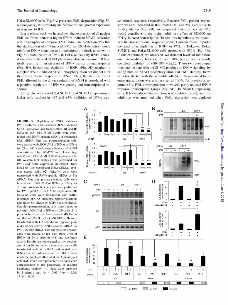
HeLa-SUMO3 cells (Fig. 5A) prevented PML degradation (Fig. 5B,bottom panel), thus restoring an increase of PML protein expressionin response to IFN.In a previous work, we have shown that expression of all nuclear
PML isoforms induces a higher IFN-g–induced STAT1 activationand transcriptional response (21). Thus, our prediction was thatthe stabilization of IFN-induced PML by RNF4 depletion wouldreinforce IFN-g signaling and transcription. Indeed, as shown inFig. 5C, stabilization of PML in HeLa-wt cells by RNF4 knock-down led to enhanced STAT1 phosphorylation in response to IFN-g,itself resulting in an increase of IFN-g transcriptional response(Fig. 5D). In contrast, depletion of RNF4 (Fig. 5D) resulted ina higher IFN-a–induced STAT1 phosphorylation but did not alterthe transcriptional response to IFN-a. Thus, the stabilization ofPML allowed by the downregulation of RNF4 is correlated witha positive regulation of IFN-g signaling and transcriptional re-sponse.In Fig. 3A, we showed that SUMO1 and SUMO3 expression in
HeLa cells resulted in ∼35 and 85% inhibition of IFN-g tran-
scriptional response, respectively. Because PML protein expres-sion was not increased in IFN-treated HeLa-SUMO3 cells due toits degradation (Fig. 4B), we suspected that this lack of PMLwould contribute to the higher inhibitory effect of SUMO3 onIFN-g–induced transcription. To test this hypothesis, we quanti-fied the transcriptional response of the GAS-luciferase reporterconstruct after depletion of RNF4 or PML in HeLa-wt, HeLa-SUMO1, and HeLa-SUMO3 cells treated with IFN-g (Fig. 5E).In this experiment, we observed two different levels of inhibition:one intermediate, between 30 and 50% (gray), and a nearlycomplete inhibition of ∼80–90% (black). These two phenotypesillustrate the dual effect of SUMO paralogs on IFN-g signaling, byacting both on STAT1 phosphorylation and PML stability. In wtcells transfected with the scramble siRNA, IFN-g–induced lucif-erase transcription was arbitrary set to 100%. As previously re-ported (21), PML downregulation in wt cells partly reduced IFN-gresponse transcription (gray) (Fig. 5E). In SUMO1-expressingcells, IFN-g–induced transcription was inhibited (gray), and thisinhibition was amplified when PML expression was depleted
FIGURE 5. Depletion of RNF4 stabilizes
PML isoforms and enhances IFN-g–induced
STAT1 activation and transcription. (A and B)
HeLa-wt and HeLa-SUMO3 cells were trans-
fected with RNF4-specific siRNA or scrambled
(Scr) siRNA. One day posttransfection, cells
were treated with 1000 U/ml of IFN-a or IFN-g
for 24 h. (A) Knockdown efficiency of RNF4
was estimated by qRT-PCR in HeLa-wt (top
panel) and HeLa-SUMO3 (bottom panel) cells.
(B) Western blot analysis was performed for
PML and Actin expression in extracts from
HeLa-wt (top panel) and HeLa-SUMO3 (bot-
tom panel) cells. (C) HeLa-wt cells were
transfected with RNF4-specific siRNA or Scr
siRNA. One day posttransfection, cells were
treated with 1000 U/ml of IFN-a or IFN-g for
30 min. Western blot analysis was performed
for PML, p-STAT1, and Actin expression. (D)
HeLa-wt cells were transfected with ISRE-
luciferase or GAS-luciferase reporter plasmids
and either Scr siRNA or RNF4-specific siRNA.
One day posttransfection, cells were treated or
not with 1000 U/ml of IFN-a or IFN-g for 24 h
prior to lysis and luciferase assays. (E) HeLa-
wt, HeLa-SUMO1, or HeLa-SUMO3 cells were
transfected with GAS-luciferase reporter plas-
mid and Scr siRNA, RNF4-specific siRNA, or
PML-specific siRNA. One day posttransfection,
cells were treated or not with 1000 U/ml of
IFN-g for 24 h prior to lysis and luciferase
assays. Results are represented as the percent-
age of Luciferase activity compared with cells
transfected with Scr siRNA and treated with
IFN-g that was arbitrarily set to 100%. Under-
neath the graph are illustrated the 3 phenotypes
obtained, which are represented as a color code
corresponding to the percentage of residual
Luciferase activity. All data were analyzed
by Student t test. *p , 0.05, **p , 0.01,
***p , 0.001.
The Journal of Immunology 2319
by guest on June 4, 2018http://w
ww
.jimm
unol.org/D
ownloaded from

(black). Finally, in cells overexpressing SUMO3, the inhibition ofGAS-driven transcription in response to IFN-g was nearly com-plete (black), because it resulted from a 2-fold effect of SUMO3:the inhibition of STAT1 phosphorylation combined to PML deg-radation. In agreement, PML depletion by siRNA in SUMO3-expressing cells did not lead to further inhibition. In contrast,RNF4 downregulation, which prevents PML degradation, ledto a partial recovery of IFN-g transcriptional response (gray)(Fig. 5E).Altogether, these results show that both SUMO1 and SUMO3
inhibit IFN-g transcriptional response and that this inhibitioncould be further amplified in SUMO3-expressing cells due toPML degradation.
SUMO1 and SUMO3 impair IFN-induced biological responses
Finally, we sought to evaluate the role of SUMO on IFN-inducedbiological activities. We analyzed the capacity of IFN-a and IFN-gto inhibit EMCV production in wt cells and in cells stablyexpressing SUMO1 or SUMO3. To test whether SUMO alters the
antiviral effect of IFN, it is important to infect cells with a virusfor which replication is not impaired by SUMO alone. Note thatSUMO1 or SUMO3 expression did not alter EMCV protein ex-pression and EMCV production in the absence of IFN treatment(Fig. 6A), which is not the case for other viruses such as vesicularstomatitis virus or HSV-1 (data not shown). Cells were treatedwith 100 U/ml of IFN-a or IFN-g for 24 h prior to infection withEMCV at an MOI of 5 for 8 h (Fig. 6A). Cell extracts were an-alyzed by Western blot, and viral titers were determined bymeasuring the TCID50. After IFN-a or IFN-g treatment, EMCVproteins were no longer detectable in HeLa-wt or HeLa-SUMO1cells. In contrast, SUMO3 overexpression reduced the capacity ofIFN-a and IFN-g to inhibit viral proteins and EMCV production(Fig. 6A). These results are consistent with the degradation ofPML in IFN-treated HeLa-SUMO3 cells (Fig. 4A) and with ourprevious observation that PML depletion decreases the anti-EMCV effect of IFN (12).Next, to obtain a more complete picture of the biological con-
sequences of SUMOylation, we investigated another well-defined
FIGURE 6. Effects of SUMO on IFN-
induced biological responses. (A) Effects
of SUMO on IFN-induced anti-EMCV
activity. HeLa-wt cells and cells stably
expressing SUMO1 (S1) or SUMO3 (S3)
were treated with 100 U/ml of IFN-a or
IFN-g for 24 h then infected with EMCVat
an MOI of 5 for 8 h. Cell extracts were
analyzed for viral proteins and Actin (left
panel), and culture supernatants were used
for the determination of viral titers by
measuring the TCID50 (right panel). (B)
HeLa-wt, HeLa-SUMO1, or HeLa-SUMO3
cells were treated or not with 1000 U/ml of
IFN-a or IFN-g. The number of viable cells
after 4 d was estimated using an MTT assay.
Results are presented as the percentage
of cells in IFN-treated cells compared
with untreated cells that was arbitrary set
to 100%. (C) Effects of SUMO on IFN-
induced apoptosis. HepG2 and HeLa wt
cells or expressing SUMO3 were treated
with 1000 U/ml of IFN-a or IFN-g for 3 d.
The proportion of apoptotic cells stained
with FITC labeled Annexin V and PI was
estimated by FACS. Results are expressed
as the ratio of apoptotic cells in IFN-treated
cells compared with untreated cells that
was arbitrary set to 1. (D) HeLa-wt, HeLa-
SUMO1, or HeLa-SUMO3 cells were
treated or not with 1000 U/ml of IFN-g for
3 d, and the amount of IP-10 secreted in the
medium was quantified by ELISA. (E)
HeLa-wt, HeLa-SUMO1, or HeLa-SUMO3
cells cultivated in the bottom chamber of
a transwell plate were left untreated or
treated with 1000 U/ml of IFN-g. Seventy-
two hours later, 5 3 105 activated human
T lymphocytes was added in the upper
chamber. After 8 h, the number of T cells
that migrated to the lower chamber was
determined by flow cytometry. All data were
analyzed by Student t test. **p , 0.01,
***p , 0.001.
2320 SUMO ALTERS IFN RESPONSE
by guest on June 4, 2018http://w
ww
.jimm
unol.org/D
ownloaded from

property of IFNs, which is their antiproliferative/proapoptoticactivity. HeLa-wt, HeLa-SUMO1, and HeLa-SUMO3 cells weregrown for 4 d in the presence of 1000 U/ml of IFN-a or IFN-g, andthe number of living cells was estimated by an MTT assay. Asshown in Fig. 6B, in wt cells, IFN-a induced a reduction of cellproliferation of ∼30%. A similar 30% reduction in cell prolifer-ation was monitored in HeLa-SUMO1 cells, whereas SUMO3overexpression rescued cell proliferation to ∼100% (Fig. 6B).Because neither SUMO1 nor SUMO3 affect IFN-a transcriptionalresponse, whereas SUMO3 (but not SUMO1) promotes PMLdegradation, this selective effect of SUMO3 is probably the con-sequence of the proteasome-dependent degradation of PML,which is a well-known tumor suppressor. In contrast, bothSUMO1 and SUMO3 expression attenuated the antiproliferativeeffect of IFN-g (Fig. 6B), further confirming that inhibition ofIFN-g signaling by SUMO paralogs had biological consequences.Next, we went on to investigate the consequences of SUMO3expression on IFN-a–induced apoptosis in the hepatoma cell lineHepG2. Indeed, IFN-a–induced apoptosis in HepG2 cells isknown to involve PML (22), and our above data show that PMLprotein expression is not enhanced by IFN in cells expressingSUMO3. HepG2 cells expressing SUMO3-YFP were treated for
3 d with 1000 U/ml IFN-a or IFN-g, and the proportion of apo-ptotic cells was estimated by flow cytometry using Annexin V/PIstaining on both SUMO3-YFP–negative and SUMO3-YFP–positive subpopulations (Fig. 6C, left panel). The results show thatSUMO3 expression reduced the rate of apoptosis in HepG2 cellstreated either with IFN-a or IFN-g. This further confirms thatSUMO3 blocked IFN response via PML degradation. Further-more, IFN-g–induced apoptosis was also inhibited by SUMO3expression in HeLa cells (Fig. 6C, right panel), suggesting thatPML may also be involved, although the involvement of other ISGproducts cannot be ruled out.Finally, we assessed the biological consequences of the specific
inhibition of the IFN-g–mediated induction of defined ISGs bySUMO3. We focused on IP-10, for which IFN-g–induced expres-sion was particularly affected (Fig. 3B, 3C). We first compared theamount of IP-10 secreted in response to IFN-g by HeLa-wt, HeLa-SUMO1, or HeLa-SUMO3 cells through ELISA. In line with ourqRT-PCR (Fig. 3B) and Western blot (Fig. 3D) data, both SUMO1and SUMO3 expression led to a dramatic decrease of IP-10 se-cretion in response to IFN-g (Fig. 6D). IP-10 is also known asCXCL10 and is able to attract monocytes/macrophages, T cells, NKcells, and dendritic cells (23). Thus, we tested the effect of SUMO
FIGURE 7. Ubc9 depletion in wt
cells increases IFN-g–induced ISG
products and biological response. (A)
Western blot for IP10 and Ubc9 ex-
pression in wt-cells depleted for Ubc9
and treated with 100 U/ml of IFN-g for
24 h. (B) The same experiment was
performed as in Fig. 6D in wt-cells de-
pleted for Ubc9 and treated with
100 U/ml of IFN-g for 3 d. (C) The same
experiment was performed as in Fig. 6E
in wt-cells depleted for Ubc9 and treated
with 100 U/ml of IFN-g for 3 d. (D)
Extracts from wt-cells depleted for
Ubc9 and treated with 100 U/ml of IFN-g
were analyzed by Western blot for
PML, STAT1, TAP1, IRF1, Ubc9, and
Actin expression. (E) HeLa-wt trans-
fected with siRNA scramble (siScr) or
siRNA Ubc9 (siUbc9) were treated or
not with 100 U/ml of IFN-g. The num-
ber of viable cells after 4 d was esti-
mated using an MTT assay. Results are
presented as the percentage of cells in
IFN-g–treated cells compared with un-
treated cells that was arbitrary set to
100%. (F) Cells were treated as in (E)
for 3 d. The proportion of apoptotic cells
stained with FITC-labeled Annexin V
and PI was estimated by FACS. Results
are expressed as the ratio of apoptotic
cells in IFN-treated cells compared with
untreated cells that was arbitrary set to
1. (G) HeLa-wt transfected with siRNA
Scr or siRNA Ubc9 were treated or not
with 10 U/ml of IFN-g for 20 h before
infection with EMCVat an MOI of 5 for
8 h. Culture supernatants were used for
the determination of viral titers by
measuring the TCID50 (right panel). All
data were analyzed by Student t test.
*p , 0.05, **p , 0.01, ***p , 0.001.
The Journal of Immunology 2321
by guest on June 4, 2018http://w
ww
.jimm
unol.org/D
ownloaded from

on chemotaxis using activated human T lymphocytes. As shown inFig. 6E, expression of SUMO1 or SUMO3 abrogated the capacityof IFN-g to promote T cell chemoattraction. Although we cannotrule out the involvement of other chemokines, this result illustratesthe fact that the inhibition of IFN-g–induced expression of IP-10 bySUMO paralogs correlates with the inhibition of chemotaxis. Tofurther confirm these results, we depleted Ubc9 in wt cells. As
shown in Fig. 1H, Ubc9 depletion enhanced IFN-g–induced STAT1phosphorylation and also IFN-g–induced IP10 expression (Fig. 7A)and secretion (Fig. 7B), which are correlated with a significant in-crease of chemotaxis (Fig. 7C). In addition, Ubc9 depletion in IFN-g–treated cells enhanced the induction of ISG products such asPML, IRF1, TAP1, and STAT1 (Fig. 7D), the anti–cell growth effect(Fig. 7E), the induction of apoptosis (Fig. 7F), and the inhibition of
FIGURE 8. Model for SUMO1- and SUMO2/3-mediated inhibition of IFN signaling. (A) The interaction of IFN-a with IFNAR leads to the activation of
the JAK tyrosine kinases (Tyk2 and JAK1) that phosphorylate STAT1 and STAT2. p-STAT1 and p-STAT2 heterodimerize and translocate into the nucleus to
induce ISGs harboring an ISRE. The binding of IFN-g to its receptor, IFNGR, results in the phosphorylation of STAT1 by JAK1 and JAK2. p-STAT1
homodimers migrate to the nucleus and bind to a DNA element termed GAS in the promoter of specific ISGs such as IRF-1, TAP1, IP-10, Sp100, and PML.
In return, PML positively regulates IFN-g signaling (21). (B) SUMO1 overexpression leads to an inhibition of STAT1 phosphorylation in response to IFN.
Whereas the IFN-a transduction pathway is unaffected by this inhibition, because STAT2 homodimers can compensate the lack of STAT1 phosphorylation,
the IFN-g transduction pathway is impaired, as illustrated by the reduced capacity of STAT1 to bind GAS. This leads to a dramatic decrease of the ex-
pression of certain IFN-g–induced genes, such as TAP1 and IP-10. The inhibition of IFN-g transduction pathway is partly compensated by PML, for which
modification by SUMO1 does not reduce its activity. (C) In response to IFN, SUMO3 overexpression also leads to an inhibition of STAT1 phosphorylation
and binding to GAS resulting in an inhibition of TAP1 and IP-10 mRNA expression. In addition, IFN-induced PML modification by poly-SUMO3 chains
leads to its RNF4-dependent ubiquitination and its subsequent degradation by the proteasome. Thus, the effect of hyper-SUMOylation by SUMO3 leads to
a more pronounced phenotype, because PML cannot exert its positive activity on IFN-g signaling due to its proteasomal degradation.
2322 SUMO ALTERS IFN RESPONSE
by guest on June 4, 2018http://w
ww
.jimm
unol.org/D
ownloaded from

EMCV production (Fig. 7G). Therefore, our results show that in-hibition of SUMOylation results in a higher IFN-g–induced STAT1phosphorylation that is correlated with enhanced biological re-sponses. Our results are summarized in an illustration depictingSUMO1 and SUMO2/3-mediated inhibition of IFN signaling(Fig. 8).
DiscussionIn this study, we report that SUMO1 and SUMO3 selectively impairIFN-g signaling by inhibiting STAT1 phosphorylation and there-fore downstream events. Also, we show that IFN exerts a negativeretrocontrol on its own signaling by enhancing STAT1 SUMOy-lation. Furthermore, we found that SUMO3, but not SUMO1,further inhibits IFN-g transcriptional response through SUMO3-dependent PML degradation.First, we showed that the stable expression of SUMO1 or
SUMO3 in different human cell lines alters IFN-g action at thelevel of signaling and transcription of ISGs. Indeed, SUMO1 orSUMO3 overexpression led to a lower STAT1 phosphorylation. Atthe opposite, inhibition of SUMOylation in wt cells treated withginkgolic acid or depleted for Ubc9 resulted in an increased IFN-g–induced STAT1 activation. It was found that STAT1 is conju-gated to SUMO1 on K703 and that the corresponding mutantexhibits a higher transcriptional activity than the wt protein (8, 9).However, the physiologic function of STAT1 SUMOylation onIFN responses was not evaluated. In this study, we demonstratedthat stable expression of SUMO1 or SUMO3 in human cells re-duced GAS-luciferase activity in response to IFN-g, whereasquantitative qRT-PCR revealed a selective reduction of STAT1-mediated ISG mRNA expression. Indeed, expression of SUMO1or SUMO3 reduced IFN-g–induced mRNA expression of IP-10and TAP1 but did not affect mRNA levels of IRF1, PKR, STAT1,and PML. We were able to confirm these observations at theprotein level because IFN-g did not increase TAP1 and IP-10protein expression in SUMO1- or SUMO3-expressing cells. Thisinhibition has biological consequences, because we found thatSUMO overexpression causes a dramatic reduction of IP-10–induced chemotaxis and that opposite results were obtained in wtcells depleted for Ubc9.In wt cells, whereas IRF1, STAT1, or PKR were increased at low
doses of IFN-g (10 U/ml), the increase of IP-10 and TAP1 ex-pression required higher concentrations of IFN-g. This suggeststhat the ISGs that are induced by low doses of IFN are not affectedby the expression of SUMO, whereas those induced by high dosesare affected, probably due to the fact that there is less availableactivated STAT1. Such a selective inhibition of the induction ofcertain ISGs by SUMO has previously been observed, because theexpression of a SUMOylation-deficient STAT1 mutant resulted inthe selective increase of CBP1 and TAP1 expression, whereasother ISGs such as IRF1 were not affected (8).In addition, we showed that although overexpression of SUMO1
or SUMO3 decreased STAT1 activation in response to IFN-a, it didnot alter STAT2 phosphorylation, binding of ISGF3 to the ISRE,or transcriptional response. Type I IFN signaling is a complexprocess, as they can activate several kinases in addition to JAKs,other STATs in addition to STAT1 and STAT2, and even othertranscription factors (24–26). In addition, accumulating evidencesupports the existence of alternative STAT2 signaling pathwaysthat are independent from STAT1 (27). Indeed, in response toIFN-a, STAT2 was shown to form, independently from STAT1,a complex with IRF9 (28) that binds ISRE and mediates ISG ex-pression (29). Also, it was shown that whereas STAT1 cooperativityis essential for IFN-g response, it is dispensable for IFN-a signaling(30). Very recently, it has been reported that IFN-a activates the
STAT2/IRF9 complex that forms an ISGF3-like response withoutSTAT1 and generates an antiviral response in the absence of STAT1(31). Accordingly, in our report, we show that although SUMOdecreased STAT1 phosphorylation in response to IFN-a, it did notalter the formation of ISGF3-like complex and the transcriptionalresponse. Therefore, it is likely that SUMO overexpression has noeffect on IFN-a signaling, because alternative complexes, such asp-STAT2 homodimers (Fig. 8), can compensate the reduction ofSTAT1 phosphorylation.PML and Sp100 proteins are the major permanent constituents of
PML NBs and are ISG products (16–18). It is established that PMLis modified by SUMO under normal growth conditions (32, 33)and that As2O3 enhances PML SUMOylation and promotes itsinteraction with RNF4, a poly–SUMO-dependent ubiquitin E3ligase responsible for proteasome-mediated PML degradation (19,20). We demonstrated in this report that in SUMO3-expressingcells, IFN-a and IFN-g increased PML and Sp100 SUMOylation(Fig. 4), induced their mRNA (Fig. 3B and data not shown)without enhancing their protein level due to their proteosomaldegradation (Fig. 4), and thus cancelled the IFN-mediated increasein PML and Sp100 protein expression (Fig. 8). Accordingly,treatment of SUMO3-expressing cells with the proteasome in-hibitor (Fig. 4B) or depletion of RNF4 (Fig. 5B) restored the in-crease of PML proteins in response to IFN-a and IFN-g. It shouldbe noted that the endogenous level of PML is similar in wt cells orcells expressing SUMO3 (Fig.3A), demonstrating that SUMO3expression alone did not alter PML protein level. We cannot ruleout that targets for IFN-induced SUMOylation and degradation inSUMO3-overexpressing cells may include proteins distinct fromPML and Sp100. However, IFNs can enhance protein SUMOylationwithout resulting in their downregulation because we showed thatIFNs enhanced STAT1 SUMOylation in SUMO3-expressing cellswithout altering its protein level (Fig. 3C).In line with the selective effect of SUMO3, whereas SUMO1
expression induced a modest reduction of IFN-g transcriptionalresponse of ∼35%, this reduction reached 85% with SUMO3.We were able to demonstrate that the higher inhibition of ISGtranscription in response to IFN-g in SUMO3- versus SUMO1-expressing cells results from the enhanced degradation of PML.Accordingly, stabilization of PML by RNF4 depletion in HeLa-wtcells results in an increase of IFN-g–induced STAT1 phosphory-lation and transcriptional responses. Also, depletion of RNF4 re-duced the inhibitory effect of SUMO3 on IFN-g transcriptionalresponse (Fig. 8). These results recall our previous report, showingthat PML positively regulates IFN-g signaling resulting in an in-creased induction of ISGs (21).It is now well established that PML plays an important role in
antiviral defense and IFN response (34–36). In this study, we reportthat PML degradation correlates with a decrease in IFN-inducedantiviral state. Indeed, in SUMO3-expressing cells, the IFN-induced increase in PML expression is impaired, resulting ina decrease of IFN-induced anti-EMCV activity. These observa-tions perfectly fit with our previous report that PML depletion inhuman cells boosts EMCV production and reduced the capacity ofIFN to protect them from EMCV infection (12).In addition to IP-10–induced chemotaxis and PML-mediated
EMCV protection, our data shed light on the consequences ofSUMO expression on the antiproliferative and proapoptotic effectof IFN. Whereas only SUMO3 counteracts the antiproliferativeactivity of IFN-a, probably due to PML degradation, bothSUMO1 and SUMO3 could rescue cell proliferation induced byIFN-g. Finally, we showed that in HepG2 cells, in which IFN-induced apoptosis is known to depend on PML, SUMO3 expres-sion was able to reduce the apoptotic process. More surprisingly,
The Journal of Immunology 2323
by guest on June 4, 2018http://w
ww
.jimm
unol.org/D
ownloaded from

we found that SUMO3 could also rescue HeLa cells from IFN-g–induced apoptosis, suggesting either that PML is also implicated orthat other ISG products can be affected by SUMO3 overexpression.Taken together, our results uncover an unprecedented role of
SUMOylation in attenuating cell sensitivity to IFN-g by decreasingSTAT1 activation, its binding to DNA, and the transcription ofspecific ISGs. Conversely, inhibition of SUMOylation resulted ina higher IFN-g–induced STAT1 phosphorylation and enhancedbiological responses. Also, we show that by IFN exerts a negativeretrocontrol on its own signaling by enhancing STAT1 SUMOy-lation. Furthermore, we provide evidence for a specific action ofSUMO3, which abrogates the increase of NB-associated PML andSp100 protein expression in response to type I and II IFNs byinducing their proteasome-mediated degradation. We cannot ex-clude that SUMOylation may alter other factors involved in sig-naling, transcription, or protein stability. It will be interesting toidentify which proteins are conjugated to SUMO in response toIFN in future studies. As a conclusion, our work allows includingSUMO to the list of negative regulators of IFN signaling known todate and posits SUMO as a new regulator of IFN response.
AcknowledgmentsWe thank Jeanne Wietzerbin for comments and Sophie Mouillet-Richard
for critical reading of the manuscript. We also thank the FACS and cell
sorting platform (S2C) of the Saints-Peres Center, Paris Descartes
University, for FACS analyses.
DisclosuresThe authors have no financial conflicts of interest.
References1. Borden, E. C., G. C. Sen, G. Uze, R. H. Silverman, R. M. Ransohoff,
G. R. Foster, and G. R. Stark. 2007. Interferons at age 50: past, current and futureimpact on biomedicine. Nat. Rev. Drug Discov. 6: 975–990.
2. Schneider, W. M., M. D. Chevillotte, and C. M. Rice. 2014. Interferon-stimulated genes: a complex web of host defenses. Annu. Rev. Immunol. 32:513–545.
3. Schindler, C., D. E. Levy, and T. Decker. 2007. JAK-STAT signaling: frominterferons to cytokines. J. Biol. Chem. 282: 20059–20063.
4. Oudshoorn, D., G. A. Versteeg, and M. Kikkert. 2012. Regulation of the innateimmune system by ubiquitin and ubiquitin-like modifiers. Cytokine GrowthFactor Rev. 23: 273–282.
5. Gareau, J. R., and C. D. Lima. 2010. The SUMO pathway: emerging mecha-nisms that shape specificity, conjugation and recognition. Nat. Rev. Mol. CellBiol. 11: 861–871.
6. Geiss-Friedlander, R., and F. Melchior. 2007. Concepts in sumoylation: a decadeon. Nat. Rev. Mol. Cell Biol. 8: 947–956.
7. Rogers, R. S., C. M. Horvath, and M. J. Matunis. 2003. SUMO modification ofSTAT1 and its role in PIAS-mediated inhibition of gene activation. J. Biol.Chem. 278: 30091–30097.
8. Ungureanu, D., S. Vanhatupa, J. Gronholm, J. J. Palvimo, and O. Silvennoinen.2005. SUMO-1 conjugation selectively modulates STAT1-mediated gene re-sponses. Blood 106: 224–226.
9. Ungureanu, D., S. Vanhatupa, N. Kotaja, J. Yang, S. Aittomaki, O. A. Janne,J. J. Palvimo, and O. Silvennoinen. 2003. PIAS proteins promote SUMO-1conjugation to STAT1. Blood 102: 3311–3313.
10. Gronholm, J., S. Vanhatupa, D. Ungureanu, J. Valiaho, T. Laitinen, J. Valjakka,and O. Silvennoinen. 2012. Structure-function analysis indicates that sumoyla-tion modulates DNA-binding activity of STAT1. BMC Biochem. 13: 20.
11. Begitt, A., M. Droescher, K. P. Knobeloch, and U. Vinkemeier. 2011. SUMOconjugation of STAT1 protects cells from hyperresponsiveness to IFNg. Blood118: 1002–1007.
12. Maroui, M. A., M. Pampin, and M. K. Chelbi-Alix. 2011. Promyelocytic leu-kemia isoform IV confers resistance to encephalomyocarditis virus via the se-questration of 3D polymerase in nuclear bodies. J. Virol. 85: 13164–13173.
13. Fukuda, I., A. Ito, G. Hirai, S. Nishimura, H. Kawasaki, H. Saitoh, K. Kimura,M. Sodeoka, and M. Yoshida. 2009. Ginkgolic acid inhibits protein SUMOy-
lation by blocking formation of the E1-SUMO intermediate. Chem. Biol. 16:133–140.
14. Saitoh, H., and J. Hinchey. 2000. Functional heterogeneity of small ubiquitin-related protein modifiers SUMO-1 versus SUMO-2/3. J. Biol. Chem. 275: 6252–6258.
15. Kessler, D. S., S. A. Veals, X. Y. Fu, and D. E. Levy. 1990. Interferon-alpharegulates nuclear translocation and DNA-binding affinity of ISGF3, a multimerictranscriptional activator. Genes Dev. 4: 1753–1765.
16. Chelbi-Alix, M. K., L. Pelicano, F. Quignon, M. H. Koken, L. Venturini,M. Stadler, J. Pavlovic, L. Degos, and H. de The. 1995. Induction of the PMLprotein by interferons in normal and APL cells. Leukemia 9: 2027–2033.
17. Stadler, M., M. K. Chelbi-Alix, M. H. Koken, L. Venturini, C. Lee, A. Saıb,F. Quignon, L. Pelicano, M. C. Guillemin, C. Schindler, et al. 1995. Tran-scriptional induction of the PML growth suppressor gene by interferons is me-diated through an ISRE and a GAS element. Oncogene 11: 2565–2573.
18. Grotzinger, T., K. Jensen, and H. Will. 1996. The interferon (IFN)-stimulatedgene Sp100 promoter contains an IFN-gamma activation site and an imperfectIFN-stimulated response element which mediate type I IFN inducibility. J. Biol.Chem. 271: 25253–25260.
19. Tatham, M. H., M. C. Geoffroy, L. Shen, A. Plechanovova, N. Hattersley,E. G. Jaffray, J. J. Palvimo, and R. T. Hay. 2008. RNF4 is a poly-SUMO-specificE3 ubiquitin ligase required for arsenic-induced PML degradation. Nat. CellBiol. 10: 538–546.
20. Lallemand-Breitenbach, V., M. Jeanne, S. Benhenda, R. Nasr, M. Lei, L. Peres,J. Zhou, J. Zhu, B. Raught, and H. de The. 2008. Arsenic degrades PML or PML-RARalpha through a SUMO-triggered RNF4/ubiquitin-mediated pathway. Nat.Cell Biol. 10: 547–555.
21. El Bougrini, J., L. Dianoux, and M. K. Chelbi-Alix. 2011. PML positivelyregulates interferon gamma signaling. Biochimie 93: 389–398.
22. Herzer, K., T. G. Hofmann, A. Teufel, C. C. Schimanski, M. Moehler,S. Kanzler, H. Schulze-Bergkamen, and P. R. Galle. 2009. IFN-alpha-inducedapoptosis in hepatocellular carcinoma involves promyelocytic leukemia proteinand TRAIL independently of p53. Cancer Res. 69: 855–862.
23. Taub, D. D., A. R. Lloyd, K. Conlon, J. M. Wang, J. R. Ortaldo, A. Harada,K. Matsushima, D. J. Kelvin, and J. J. Oppenheim. 1993. Recombinant humaninterferon-inducible protein 10 is a chemoattractant for human monocytes andT lymphocytes and promotes T cell adhesion to endothelial cells. J. Exp. Med.177: 1809–1814.
24. Sizemore, N., A. Agarwal, K. Das, N. Lerner, M. Sulak, S. Rani, R. Ransohoff,D. Shultz, and G. R. Stark. 2004. Inhibitor of kappaB kinase is required to ac-tivate a subset of interferon gamma-stimulated genes. Proc. Natl. Acad. Sci. USA101: 7994–7998.
25. Gil, M. P., E. Bohn, A. K. O’Guin, C. V. Ramana, B. Levine, G. R. Stark,H. W. Virgin, and R. D. Schreiber. 2001. Biologic consequences of Stat1-independent IFN signaling. Proc. Natl. Acad. Sci. USA 98: 6680–6685.
26. van Boxel-Dezaire, A. H., M. R. Rani, and G. R. Stark. 2006. Complex mod-ulation of cell type-specific signaling in response to type I interferons. Immunity25: 361–372.
27. Steen, H. C., and A. M. Gamero. 2012. The role of signal transducer and acti-vator of transcription-2 in the interferon response. J Interferon Cytokine Res 32:103–110.
28. Martinez-Moczygemba, M., M. J. Gutch, D. L. French, and N. C. Reich. 1997.Distinct STAT structure promotes interaction of STAT2 with the p48 subunit ofthe interferon-alpha-stimulated transcription factor ISGF3. J. Biol. Chem. 272:20070–20076.
29. Bluyssen, H. A., and D. E. Levy. 1997. Stat2 is a transcriptional activator thatrequires sequence-specific contacts provided by stat1 and p48 for stable inter-action with DNA. J. Biol. Chem. 272: 4600–4605.
30. Begitt, A., M. Droescher, T. Meyer, C. D. Schmid, M. Baker, F. Antunes,K. P. Knobeloch, M. R. Owen, R. Naumann, T. Decker, and U. Vinkemeier.2014. STAT1-cooperative DNA binding distinguishes type 1 from type 2 inter-feron signaling. Nat. Immunol. 15: 168–176.
31. Blaszczyk, K., A. Olejnik, H. Nowicka, L. Ozgyin, Y. L. Chen, S. Chmielewski,K. Kostyrko, J. Wesoly, B. L. Balint, C. K. Lee, and H. A. Bluyssen. 2015.STAT2/IRF9 directs a prolonged ISGF3-like transcriptional response and anti-viral activity in the absence of STAT1. Biochem. J. 466: 511–524.
32. Duprez, E., A. J. Saurin, J. M. Desterro, V. Lallemand-Breitenbach, K. Howe,M. N. Boddy, E. Solomon, H. de The, R. T. Hay, and P. S. Freemont. 1999.SUMO-1 modification of the acute promyelocytic leukaemia protein PML:implications for nuclear localisation. J. Cell Sci. 112: 381–393.
33. Kamitani, T., K. Kito, H. P. Nguyen, H. Wada, T. Fukuda-Kamitani, andE. T. Yeh. 1998. Identification of three major sentrinization sites in PML. J. Biol.Chem. 273: 26675–26682.
34. Geoffroy, M. C., and M. K. Chelbi-Alix. 2011. Role of promyelocytic leukemiaprotein in host antiviral defense. J Interferon Cytokine Res 31: 145–158.
35. Nisole, S., M. A. Maroui, X. H. Mascle, M. Aubry, and M. K. Chelbi-Alix. 2013.Differential Roles of PML Isoforms. Front. Oncol. 3: 125.
36. Everett, R. D., and M. K. Chelbi-Alix. 2007. PML and PML nuclear bodies:implications in antiviral defence. Biochimie 89: 819–830.
2324 SUMO ALTERS IFN RESPONSE
by guest on June 4, 2018http://w
ww
.jimm
unol.org/D
ownloaded from