smgr up - smgebooks.comsmgebooks.com/different-aspects-rhinosinusitis/... · the standard...
TRANSCRIPT

1Different Aspects of Rhinosinusitis | www.smgebooks.comCopyright Otilia F.This book chapter is open access distributed under the Creative Commons Attribution 4.0 International License, which allows users to download, copy and build upon published articles even for commercial purposes, as long as the author and publisher are properly credited.
Gr upSMUltrasound of Maxillary Sinus
ABSTRACTAcute rhinosinusitis may sometimes present non-specific signs and symptoms. The imaging
techniques usually used for its diagnosis are invasive and/or expensive (computed tomography, magnetic resonance imaging, standard radiography) and should be performed only in selected cases. Ultrasonography of the maxillary sinuses is seldom mentioned in literature as a diagnosis method of sinusitis.
The objective of this material is to introduce maxillary sinuses ultrasonography as a possible and useful method in the diagnosis of acute maxillary sinusitis as well as to present the normal aspects of the maxillary sinuses and the ultrasonographic findings of acute maxillary sinusitis (fluid collections, mucosal thickening).
Ultraosnography of the maxillary sinuses is not widely accepted and it is not used on a large scale, but, as the material will demonstrate, almost any alteration of the normal ultrasonographic image is highly suggestive of a pathological process within the sinusal cavity.
The role of computed tomography and magnetic resonance imaging in the acute and chronic pathology of the maxillary sinuses remains undoubted, but we believe ultrasonography can play an important role in the diagnosis of uncomplicated acute maxillary sinusitis and its use may reduce costs and avoid unnecessary radiation exposure and antibiotic treatment.
Fufezan Otilia* and Asavoaie CarmenEmergency Children Hospital, Romania
*Corresponding author: Fufezan Otilia, Emergency Children Hospital, Str. Campeni 2, 400217 Cluj-Napoca, Romania, Tel: +40264534848; Email: [email protected]
Published Date: December 29, 2015

2Different Aspects of Rhinosinusitis | www.smgebooks.comCopyright Otilia F.This book chapter is open access distributed under the Creative Commons Attribution 4.0 International License, which allows users to download, copy and build upon published articles even for commercial purposes, as long as the author and publisher are properly credited.
LEARNING OBJECTIVESTo introduce maxillary sinuses ultrasonography and its role in diagnosing acute maxillary
sinusitis.
To present the normal aspects of the maxillary sinuses and the ultrasonographic findings of acute maxillary sinusitis.
BACKGROUNDAcute sinusitis represents a frequent pathology among the pediatric population, accounting
for a large amount of consultations and representing one of the diagnoses for which antibiotics are frequently administered [1].
The American Academy of Pediatrics published in 2001 a guideline for the management of sinusitis where it is stated that the diagnosis of acute sinusitis is basically a clinical one. Yet the signs and symptoms of sinusitis are nonspecific and usually overlapping those of other upper respiratory tract infections, often requiring an imaging confirmation for a certain diagnosis [2].
The plain radiograph of the paranasal sinuses is no longer routinely in the diagnosis of acute maxillary sinusitis.
Computed tomography is now considered the criterion standard for the evaluation of all the sinuses and it is required in cases where surgery is indicated. Magnetic resonance imaging on the other hand is recommended in specific clinical situations where complications are suspected, especially extension of the infectious process into surrounding structures or thrombosis of the superior sagittal sinus [3,4]. Ultrasonography was first mentioned in the seventies in studies regarding the diagnosis of sinusitis. Mann W et al. published a comparative study about the liability of ultrasound in maxillary sinus disease and the confidence of diagnosis in maxillary sinus pathology ranges up to 90% [5,6].
Today it is rarely mentioned as a diagnostic technique for the assessment of the maxillary sinuses and therefore it is not widely accepted. Maxillary sinus ultrasound is mostly used as first examination in the emergency department [7,8].
In pediatrics there is an ongoing concern in avoiding and decreasing exposure to x rays as well as sedation or contrast media administration. That is one of the reasons maxillary ultrasound may represent a useful method in pediatric patients with suspicion of sinusitis.
IMAGING FINDINGS OR PROCEDURE DETAILSMaxillary Sinuses Ultrasonography
The ultrasonographic evaluation of the maxillary sinuses is performed with the patient sitting and facing the sonographer and with the head slightly tilted forward (Figure 1).

3Different Aspects of Rhinosinusitis | www.smgebooks.comCopyright Otilia F.This book chapter is open access distributed under the Creative Commons Attribution 4.0 International License, which allows users to download, copy and build upon published articles even for commercial purposes, as long as the author and publisher are properly credited.
Figure 1: Maxillary sinus ultrasonography examination – patient and probe position.
The transducer is placed on each side of the nose, under the inferior wall of the orbit, on the anterior wall of the maxillary sinus. Then the sinus is scanned in a cranio-caudal and medio-lateral manner by gently moving and angulating the transducer [9].
The exam is performed in a transverse scan and the normal aspect of the maxillary sinus is very similar with that of the normal lung. The first layer is represented by the skin and subcutaneous tissue, than there is a fine, continuous, hyperechoic line produced by the anterior wall of the maxillary sinus. Since the normal sinus contains air, its ultrasonographic appearance is produced by the reflection of the sound waves back to the probe and consists of fine, parallel, echoic lines. This appearance is similar with that of the normal lung (Figure 2).
Figure 2a: Normal ultrasonographic appearance of the maxillary sinus compared with the normal ultrasonographic aspect of the lung. Examination was performed with a convex transducer.

4Different Aspects of Rhinosinusitis | www.smgebooks.comCopyright Otilia F.This book chapter is open access distributed under the Creative Commons Attribution 4.0 International License, which allows users to download, copy and build upon published articles even for commercial purposes, as long as the author and publisher are properly credited.
Figure 2b: Normal ultrasonographic appearance of the maxillary sinuses. Examination was performed with a linear transducer.
Ultrasonography Findings in Maxillary Sinusitis
Acute maxillary sinusitis is defined as an inflammation of the sinusal mucosa in response to bacterial, viral or fungal infection, but also in the context of allergy or chronic nasal conditions. This inflammation is translated into swelling of the mucosa and accumulation of fluid into the sinuses. Sometimes polyps or cysts may also be visualized inside the sinus [8,9].
The ultrasonographic aspect of these alterations has been described in detail in literature by various authors [4, 8,10,11]. When inside the sinus cavity there is more than air the sound waves are no longer reflected back to the transducer because fluid and mucus are able to transport the sound waves to the back wall of the sinus and thus produce an image. The most frequent findings associated with acute maxillary sinusitis are presented in (Table 1).
Normal sinus Sound waves return from 1 cm or lessMucosal thickening Distance between first echoes and air echoes is increased
Sound waves from the posterior bony wall usually presentFluid collection Strong, Back Wall Echo (BWE) or complete sinusogram
Distance from the first echoes to the BWE ≥ 3.5 cm (2 cm in children)Polypoid mucosal thickening Intense echoes produced by air and multiple echoic images generated by the enlarged mucos
“Sinusogram”
A “sinusogram” is defined as a real-time ultrasonographic sign significant for sinusal walls and cavity visualization. It represents a well-defined, triangular, homogeneous, hypoechoic or transonic image of the maxillary sinus and it is diagnostic for a fluid collection (Figure 3). This term was descriebed by Lichtenstein D in 1998 [8].

5Different Aspects of Rhinosinusitis | www.smgebooks.comCopyright Otilia F.This book chapter is open access distributed under the Creative Commons Attribution 4.0 International License, which allows users to download, copy and build upon published articles even for commercial purposes, as long as the author and publisher are properly credited.
Figure 3: Complete “sinusogram” – the posterior, medial and lateral walls of the maxillary sinus are visualized.
The “sinusogram” is considered complete if the internal, external and posterior walls are clearly visible, and incomplete if the walls are only partially visualized [8].
Hypoechoic or echoic images that do not have a triangular shape (incomplete sinusogram) and a fine contour are interpreted as mucosal thickening (Figure 4).
Figure 4: Bilateral incomplete “sinusogram”. The aspect found on the right was interpreted as thickening of the mucosa and the one on the left as fluid collection (distance from the back wall echo to the first echo
was 3.1 cm).

6Different Aspects of Rhinosinusitis | www.smgebooks.comCopyright Otilia F.This book chapter is open access distributed under the Creative Commons Attribution 4.0 International License, which allows users to download, copy and build upon published articles even for commercial purposes, as long as the author and publisher are properly credited.
“Back Wall Echo” (BWE)
The back wall echo (BWE) is an intense, hyperechoic image generated by the posterior bony wall of the maxillary sinus. When visualized, the BWE is a sign of pathological content within the sinus (Figure 5).
Figure 5: Bilateral Back Wall Echo (BWE) at different depths from the first echo (interpreted as fluid collection on the right side and mucosal thickening on the left side).
If the distance from the first intensity echo to the BWE is ≥ 3.5 cm in adults and 2 cm in children the aspect is consistent with a fluid collection [8].
TYPES OF TRANSDUCERSThe evaluation can be performed with any type of transducer, but the most useful diameters
proved to be between 8 and 12 mm and the most useful frequencies from 3 to 6 MHz [9].
The images presented in (Figure 6) were obtained using a convex, a microconvex (pediatric), a linear transducer and also a probe used in echocardiography (Figure 6). The images were acquired during the same examination. In all situations a fluid collection, with a complete “sinusogram” in both maxillary sinuses, was visualized.

7Different Aspects of Rhinosinusitis | www.smgebooks.comCopyright Otilia F.This book chapter is open access distributed under the Creative Commons Attribution 4.0 International License, which allows users to download, copy and build upon published articles even for commercial purposes, as long as the author and publisher are properly credited.
Figure 6: The ultrasonographic appearance of the maxillary sinus using various types of transducers (same patient, same examination). Observe that the quality of the images improves
as the diameter of the transducer decreases.
CORRESPONDENCE BETWEEN US AND PLAIN RADIOGRAPHYIn our center, plain radiography and ultrasonography were the most frequently used imaging
techniques for the evaluation of the maxillary sinuses in the past few years. In selected cases (chronic or associated pathology) an MRI exam was performed [9].
Therefore in most cases the ultrasonographic results were compared with the radiographic ones [9-11] (Figure 7).
Figure 7: Ultrasonographic - X-ray correlations. Ultrasonography reveals changes suggestive for mucosal thickening on the right and for fluid collection on the left. X-ray shows concentric opacification of
the right maxillary sinus and complete opacification of the left maxillary sinus.

8Different Aspects of Rhinosinusitis | www.smgebooks.comCopyright Otilia F.This book chapter is open access distributed under the Creative Commons Attribution 4.0 International License, which allows users to download, copy and build upon published articles even for commercial purposes, as long as the author and publisher are properly credited.
The standard projection for the evaluation of the paranasal sinuses is the occipitomental projection (Water’s view).
Ultrasonographic and radiographic findings correlated as follows:
•A complete sinusogram (back wall echo at more than 2 cm from initial echo) on US corresponded to a complete opacification on the radiograph
•An incomplete sinusogram on US was consistent with either an air/fluid level or a concentric mucosal thickening on the radiograph.
•Sometimes a normal aspect on the ultrasound correlated with mild concentric mucosal thickening on the radiograph, while normal X ray exams were found in patients with incomplete sinusograms on the US [9].
Several studies report an agreement of about 80% between ultrasonographic and radiographic findings in the evaluation of the maxillary sinuses. The results are even more consistent for the normal aspects and for the detection of fluid collections (complete opacity on the X ray and complete sinusogram on US) [4,9].
CORRESPONDENCE BETWEEN US AND CT SCAN OR MRIThere are not too many studies comparing maxillary sinus ultrasonography with computed
tomography. Hilbert G et al. reported a sensitivity of 100%, a specificity of 96.7% of ultrasonography findings compared with computed tomography and 97% agreement between the two methods [12]. Similar results were obtained by Karantanas AH et al.. - 66.7% sensitivity and 94.9% specificity of ultrasonography [13]. In a study published in 2014 was reported a sensitivity of 92.6%, a specificity of 100% of ultrasonography compared with computed tomography results [14].
Puhakka et al.. reported a sensitivity of 64% and a specificity of 95% of US when compared with magnetic resonance imaging [11].
CASE PRESENTATION(Figures 8-10) present the case of a 9 years old boy who presented with symptoms suggestive
for acute maxillary sinusitis (purulent nasal discharge, headache, coughing). The radiography acquired in Water’s view projection revealed complete opacification of the right maxillary sinus consistent with fluid collection and eccentric opacification of the left maxillary sinus suggestive for mucosal thickening (Figure 7).
The ultrasound performed in the same day showed bilateral complete sinusogram, with a back wall echo present at more than 2.5 cm from the initial echo. The ultrasonographic findings were interpreted as bilateral fluid collection (Figure 8).

9Different Aspects of Rhinosinusitis | www.smgebooks.comCopyright Otilia F.This book chapter is open access distributed under the Creative Commons Attribution 4.0 International License, which allows users to download, copy and build upon published articles even for commercial purposes, as long as the author and publisher are properly credited.
Figure 8: Water’s view projection of the skull reveals opacification of the right maxillary sinus and eccentric opacification of the left maxillary sinus walls.
Figure 9: First ultrasonographic exam (upper right) reveals bilateral complete “sinusogram” consistent with bilateral maxillary fluid collection. Note that the aspect improves on the follow-up examinations, but
does not resolve completely. On the last examination (lower left) there is still a Back Wall Echo (BWE) present on the right, but there is a normal aspect on the left.
10Different Aspects of Rhinosinusitis | www.smgebooks.comCopyright Otilia F.This book chapter is open access distributed under the Creative Commons Attribution 4.0 International License, which allows users to download, copy and build upon published articles even for commercial purposes, as long as the author and publisher are properly credited.
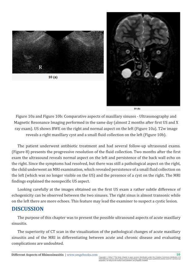
Figure 10a and Figure 10b: Comparative aspects of maxillary sinuses - Ultrasonography and Magnetic Resonance Imaging performed in the same day (almost 2 months after first US and X ray exam). US shows BWE on the right and normal aspect on the left (Figure 10a). T2w image
reveals a right maxillary cyst and a small fluid collection on the left (Figure 10b).
The patient underwent antibiotic treatment and had several follow-up ultrasound exams. (Figure 8) presents the progressive resolution of the fluid collection. Two months after the first exam the ultrasound reveals normal aspect on the left and persistence of the back wall echo on the right. Since the symptoms had resolved, but there was still a pathological aspect on the right, the child underwent an MRI examination, which revealed persistence of a small fluid collection on the left (which was no longer visible on the US) and the presence of a cyst on the right. The MRI findings explained the nonspecific US aspect.
Looking carefully at the images obtained on the first US exam a rather subtle difference of echogenicity can be observed between the two sinuses. The right sinus is almost transonic while on the left there are more echoes. This feature may lead the examiner to suspect a cystic lesion.
DISCUSSIONThe purpose of this chapter was to present the possible ultrasound aspects of acute maxillary
sinusitis.
The superiority of CT scan in the visualization of the pathological changes of acute maxillary sinusitis and of the MRI in differentiating between acute and chronic disease and evaluating complications are undoubted.

11Different Aspects of Rhinosinusitis | www.smgebooks.comCopyright Otilia F.This book chapter is open access distributed under the Creative Commons Attribution 4.0 International License, which allows users to download, copy and build upon published articles even for commercial purposes, as long as the author and publisher are properly credited.
The place of ultrasonography alone is restricted to uncomplicated cases and considered useful as it is easily accessible, cheaper, avoids radiation exposure (important especially in children and pregnant women) and may reduce unnecessary antibiotic drugs prescription.
The basic idea is that almost any alteration of the normal US aspect of the maxillary sinus should raise suspicion that there may be a pathological content within the sinusal cavity (exceptions are found in patients with decreased bone density or other bony structure changes).
Like all ultrasonographic exams maxillary sinuses US is highly examiner dependent and the findings must always be correlated with the clinical status of the patient, with the lab studies and other investigations or associated pathologies [9].
Techniques usually used in the diagnosis of maxillary sinusitis, such as the conventional radiograph, computed tomography or magnetic resonance imaging, are invasive and/or expensive imaging modalities. Ultrasonography may be a reliable alternative.
All imaging investigations may lead to a false positive diagnosis of infection when mucosal thickening, polyps, sinus cysts or anatomical anomalies are present [4].
In the past CT was considered to be the gold standard in the diagnosis of sinusitis. But this radiation exposing imaging technique should be considered only in certain situations (such as recurrent sinusitis, chronic sinusitis and no response to therapy) or if the MRI studies are not available [3,15,16].
Radiologic exams used to confirm the diagnosis of uncomplicated sinusitis are less and less recommended in the literature and just for children older than 6 years [2,17]. There are some literature data, which suggests imaging investigations are not necessary in uncomplicated acute rhinosinusitis [18,19].
There are relatively few studies in literature evaluating the role of ultrasonography in the diagnosis of rhinosinusitis in adults or in children. One dimension A-mode or two dimensions B-mode were used to evaluate maxillary sinuses. Studies of ultrasound in pediatric maxillary sinusitis revealed conflicting results [10,20-22].
The use of ultrasonography compared with the standard radiograph in acute sinusitis was evaluated in a comparative study on 197 young adults showing common cold symptoms for 48 hours. From the initial study group a number of 40 patients were randomly selected and examined through MRI. In this study ultrasonography compared with MRI had 64% sensitivity and 95 % specificity. If the radiograph was also evaluated (two steps diagnosis) the sensitivity increased up to 86% without a drop of the specificity (95%) [11].
Happaviemi J detected 77 % sensitivity and a 49% specificity of the ultrasound in a study published in 2001. The study evaluated 209 maxillary sinuses and compared ultrasonography and maxillary antral lavage [20].

12Different Aspects of Rhinosinusitis | www.smgebooks.comCopyright Otilia F.This book chapter is open access distributed under the Creative Commons Attribution 4.0 International License, which allows users to download, copy and build upon published articles even for commercial purposes, as long as the author and publisher are properly credited.
In a prospective and referenced study mentioned before (Lichtenstein D et al., Intensive Care Med 1998; 24:1057-61), 100 maxillary sinuses were evaluated by ultrasound and radiography. The “sinusogram” was correlated with the X-ray’s findings. Therefore, the sinusogram was present in 21 of the 21 opaque maxillary sinuses found on the radiograph, in 2 of 21 sinuses with an air-fluid level, in 8/21 cases of mucosal thickening and in 1/1 giant nasal polyp. The author proposes ultrasonography as a first-line investigation for the diagnosis of radiologically detected maxillary sinusitis [8].
Another study, performed on an adult population, assessed the accuracy of ultrasonography compared with conventional radiograph and maxillary sinuses sinoscopy in detecting acute or chronic inflammation. A number of 90 sinuses were evaluated in the study, a radiograph, an ultrasound and a sinoscopy being performed in each case. The value of ultrasonography was analyzed using McNemar’s test for paired data. Compared with radiography the sensitivity of the ultrasound was 93% and the specificity was 60%. Compared with sinoscopy the ultrasound presented a 93 % sensitivity and 74% specificity [23].
In a study performed on 56 adult patients, with a clinical and/or radiological diagnosis of acute maxillary sinusitis, a sensitivity of 66.7% and a specificity of 94.9% of the ultrasound compared with computed tomography was detected [13].
Ionannidis et al. carried out in 2001 a meta-analysis of the existing studies on rhinosinusitis imaging in literature until up that point. Two out of 8 comparative studies evaluated ultrasonography as a rhinosinusitis diagnosis method. This meta-analysis showed there is a moderate agreement between the results of the paranasal sinuses radiograph and the clinical diagnosis. The agreement between the two depended on the definition of the clinical diagnosis. Another conclusion reached in this study was that the results of the other imaging methods (including ultrasound) did not correlate with the clinical data [24].
Sheid DC and Hamm RM published a review of the comparative imaging studies in the diagnosis of rhinosinusitis in adults. In this review the ultrasonography as compared with sinus puncture showed an 84% (with a range from 54% to 98%) sensitivity and 69% (values between 30% and 94%) specificity [18].
In another review performed in 2000 by Veronen, 7 studies, assessing ultrasonography versus sinsus puncture, were analyzed, revealing 85% sensitivity and 82% specificity of the ultrasound exam [4].
There are studies in literature, which revealed similar results about the low accuracy of ultrasound in diagnosis of maxillary sinus mucosal thickening [9,25-27]. In our study published in 2010 the ultrasound exam of maxillary sinus was compared with the conventional X ray exam, performed in occipitomental view. The concordance between these two imaging methods was present in 83.5%. Regarding all situations (fluid collection in the maxillary sinus and mucosal thickening), the sensitivity of ultrasound was 94.8% and the specificity 98%.

13Different Aspects of Rhinosinusitis | www.smgebooks.comCopyright Otilia F.This book chapter is open access distributed under the Creative Commons Attribution 4.0 International License, which allows users to download, copy and build upon published articles even for commercial purposes, as long as the author and publisher are properly credited.
If there were evaluated in a divided interpretation for the normal aspect, the fluid collection and the mucosal thickening, the error was very low for the normal aspect and for the fluid collection (1.58 % irrespectively 5.12%), but the error for the thickening of the mucosa was high (59.37%). This result entitles for the conclusion that ultrasonography is not suitable in a confident evaluation of the mucosal thickening [9].
CONCLUSIONMaxillary sinuses ultrasonography is possible due to the presence of air in the sinusal cavity,
the normal image being produced by the reflected echoes returning from the posterior wall of the sinus.
The presence of a “sinusogram” or of a back wall echo at various depths of the sinus are possible US aspects produced by fluid collection and/or mucosal thickening, which are pathological features of acute maxillary sinusitis.
Ultrasonography can be considered a trustworthy imaging technique in the diagnosis of uncomplicated acute sinusitis in children, thus avoiding unnecessary radiation exposure and antibiotic drugs prescription.
References1. Rosenfeld RM, Andes D, Bhattacharyya N, Cheung D, Eisenberg S, Ganiats TG, et al. Clinical practice guideline: adult sinusitis.
Otolaryngol Head Neck Surg. 2007; 137: S1-31.
2. American Academy of Pediatrics Subcommittee on Management of Sinusitis and Committee on Quality Improvement.. Clinical practice guideline: management of sinusitis. Pediatrics. 2001; 108: 798-808.
3. Osguthorpe JD. Adult rhinosinusitis: diagnosis and management. Am Fam Physician. 2001; 63: 69-76.
4. Varonen H, Mäkelä M, Savolainen S, Läärä E, Hilden J. Comparison of ultrasound, radiography, and clinical examination in the diagnosis of acute maxillary sinusitis: a systematic review. J Clin Epidemiol. 2000; 53: 940-948.
5. Mann W. [Comparison of x-ray, echography and sinumanometry findings in paranasal sinusitis (author’s transl). Laryngol Rhinol Otol (Stuttg). 1977; 56: 759-765.
6. Mann W, Beck C, Apostolidis T. Liability of ultrasound in maxillary sinus disease. Arch Otorhinolaryngol. 1977; 215: 67-74.
7. Blanco P, Do Pico JL, Ciotta R. Maxillary sinusitis diagnosed by ultrasound. Med Intensiva. 2015; 39: 391.
8. Lichtenstein D, Biderman P, Mezière G, Gepner A. The “sinusogram”, a real-time ultrasound sign of maxillary sinusitis. Intensive Care Med. 1998; 24: 1057-1061.
9. Fufezan O, Asavoaie C, Cherecheş Panta P, Mihuţ G, Bursaşiu E, Anca I, et al. The role of ultrasonography in the evaluation of maxillary sinusitis in pediatrics. Med Ultrason. 2010; 12: 4-11.
10. Revonta M, Kuuliala I. The diagnosis and follow-up of pediatric sinusitis: Water’s view radiography versus ultrasonography. Laryngoscope. 1989; 99: 321-324.
11. Puhakka T, Heikkinen T, Mäkelä MJ, Alanen A, Kallio T, Korsoff L, et al. Validity of ultrasonography in diagnosis of acute maxillary sinusitis. Arch Otolaryngol Head Neck Surg. 2000; 126: 1482-1486.
12. Hilbert G, Vargas F, Valentino R, Gruson D, Chene G, et al. Comparison of B-mode ultrasound and computed tomography in the diagnosis of maxillary sinusitis in mechanically ventilated patients. Crit Care Med. 2001; 29:1337-1342.
13. Karantanas AH, Sandris V. Maxillary sinus inflammatory disease: ultrasound compared to computed tomography. Comput Med Imaging Graph. 1997; 21: 233-241.
14. Mori A, Nakayama T, Tsukidate T, Hirabayashi H, Haruna S. Comparison of B-mode ultrasonography and computed tomography in the evaluation of maxillary sinusitis in pediatric patients. Nihon Jibiinkoka Gakkai Kaiho. 2014; 117: 26-33.
15. McAlister WH, Kronemer K. Imaging of sinusitis in children. Pediatr Infect Dis J. 1999; 18: 1019-1020.

14Different Aspects of Rhinosinusitis | www.smgebooks.comCopyright Otilia F.This book chapter is open access distributed under the Creative Commons Attribution 4.0 International License, which allows users to download, copy and build upon published articles even for commercial purposes, as long as the author and publisher are properly credited.
16. Okuyemi KS, Tsue TT. Radiologic imaging in the management of sinusitis. Am Fam Physician. 2002; 66: 1882-1886.
17. Wald ER. Sinusitis. Long SS, Pickering LK, Prober CG, editors. In: Principles and practice of pediatric infectious diseases. New York, Churchill Livingstone. 2003: 205-210
18. Scheid DC, Hamm RM. Acute bacterial rhinosinusitis in adults: part I Evaluation. Am Fam Physician. 2004; 70: 1685-1692.
19. Esposito S, Bosis S, Bellasio M, Principi N. From clinical practice to guidelines: how to recognize rhinosinusitis in children. Pediatr Allergy Immunol. 2007; 18: 53-55.
20. Haapaniemi J. Comparison of ultrasound and X-ray maxillary sinus findings in school-aged children. Ear Nose Throat J. 1997; 76: 102-106.
21. Reilly JS, Hotaling AJ, Chiponis D, Wald ER. Use of ultrasound in detection of sinus disease in children. Int J Pediatr Otorhinolaryngol. 1989; 17: 225-230.
22. Shapiro GG, Furukawa CT, Pierson WE, Gilbertson E, Bierman CW. Blinded comparison of maxillary sinus radiography and ultrasound for diagnosis of sinusitis. J Allergy Clin Immunol. 1986; 77: 59-64.
23. Risavi R, Klapan I, Barcan T, Simović S. Effectiveness of ultrasonography in diagnosis of maxillary sinus disease: a prospective comparison with radiographic and sinusoscopic examinations. Croat Med J. 1998; 39: 45-48.
24. Ioannidis JP, Lau J. Technical report: evidence for the diagnosis and treatment of acute uncomplicated sinusitis in children: a systematic overview. Pediatrics. 2001; 108: E57.
25. Berg O, Carenfelt C. Etiological diagnosis in sinusitis: ultrasonography as clinical complement. Laryngoscope. 1985; 95: 851-853.
26. Jensen C, von Sydow C. Radiography and ultrasonography in paranasal sinusitis. Acta Radiol. 1987; 28: 31-34.
27. Vento SI, Ertama LO, Hytönen ML, Malmberg CH. A-mode ultrasound in the diagnosis of chronic polypous sinusitis. Acta Otolaryngol. 1999; 119: 916-920.