smooth muscle-specific expression of calcium-independent
TRANSCRIPT

Eastern Kentucky UniversityEncompass
Biological Sciences Faculty and Staff Research Biological Sciences
5-2012
Smooth Muscle-specific Expression of Calcium-independent Phospholipase A2 (iPLA2 )Participates in the Initiation and Early Progressionof Vascular Inflammation and NeointimaFormationShu LiuCapital Medical University
Zhongwen XieUniversity of Kentucky
Qingwei David ZhaoUniversity of Texas Health Science Center, San Antonio
Huan PangUniversity of Kentucky
John TurkWashington University, St. Louis
This Article is brought to you for free and open access by the Biological Sciences at Encompass. It has been accepted for inclusion in Biological SciencesFaculty and Staff Research by an authorized administrator of Encompass. For more information, please contact [email protected].
Recommended CitationLiu, S., Xie, Z., Zhao, Q., Pang, H., Turk, J., Calderon, L., . . . Guo, Z. (2012). Smooth Muscle-specific Expression of Calcium-independent Phospholipase A2 (iPLA2 ) Participates in the Initiation and Early Progression of Vascular Inflammation and NeointimaFormation. Journal of Biological Chemistry, 287(29), 24739-24753. doi:10.1074/jbc.m112.340216

See next page for additional authors
Follow this and additional works at: http://encompass.eku.edu/bio_fsresearch
Part of the Biology Commons, and the Medicine and Health Sciences Commons

AuthorsShu Liu, Zhongwen Xie, Qingwei David Zhao, Huan Pang, John Turk, Lindsay Calderon, Wen Su, GuogangZhao, Haifei Xu, Ming Gong, and Zhenheng Guo
This article is available at Encompass: http://encompass.eku.edu/bio_fsresearch/26
Smooth Muscle-specific Expression of Calcium-independentPhospholipase A2� (iPLA2�) Participates in the Initiation andEarly Progression of Vascular Inflammation and NeointimaFormation*□S
Received for publication, January 5, 2012, and in revised form, May 20, 2012 Published, JBC Papers in Press, May 25, 2012, DOI 10.1074/jbc.M112.340216
Shu Liu‡1, Zhongwen Xie§1, Qingwei Zhao§1, Huan Pang§, John Turk¶, Lindsay Calderon§, Wen Su‡, Guogang Zhao‡,Haifei Xu‡, Ming C. Gong§, and Zhenheng Guo‡§2
From the Departments of ‡Internal Medicine and §Physiology, University of Kentucky School of Medicine, Lexington, Kentucky40536 and the ¶Department of Medicine, Washington University School of Medicine, St. Louis, Missouri 63110
Background: The role of iPLA2� as a regulator of inflammatory signaling and neointima formation is unknown.Results: Smooth muscle-specific expression of iPLA2� exacerbates proinflammatory cytokine production, macrophage infil-tration, and neointima formation.Conclusion: Smooth muscle-specific iPLA2� participates in the initiation and early progression of vascular inflammation andneointima formation.Significance: iPLA2� may represent a novel therapeutic target for attenuating vascular inflammation and restenosis.
Whether groupVIA phospholipaseA2 (iPLA2�) is involved invascular inflammation and neointima formation is largelyunknown. Here, we report that iPLA2� expression increases inthe vascular tunica media upon carotid artery ligation and thatneointima formation is suppressed by genetic deletion ofiPLA2� or by inhibiting its activity or expression via perivascu-lar delivery of bromoenol lactone or of antisense oligonucleo-tides, respectively. To investigate whether smooth muscle-spe-cific iPLA2� is involved in neointima formation, we generatedtransgenic mice in which iPLA2� is expressed specifically insmooth muscle cells and demonstrate that smooth muscle-spe-cific expression of iPLA2� exacerbates ligation-inducedneointima formation and enhanced both production of proin-flammatory cytokines and vascular infiltration bymacrophages.With cultured vascular smooth muscle cell, angiotensin II,arachidonic acid, and TNF-� markedly induce increasedexpression of IL-6 and TNF-� mRNAs, all of which were sup-pressed by inhibiting iPLA2� activity or expression with bro-moenol lactone, antisense oligonucleotides, and genetic dele-tion, respectively. Similar suppression also results from geneticdeletion of 12/15-lipoxygenase or inhibiting its activity withnordihydroguaiaretic acid or luteolin. Expression of iPLA2�protein in cultured vascular smooth muscle cells was found todependon the phenotypic state and to rise upon incubationwith
TNF-�. Our studies thus illustrate that smoothmuscle cell-spe-cific iPLA2� participates in the initiation and early progressionof vascular inflammation and neointima formation and suggestthat iPLA2� may represent a novel therapeutic target for pre-venting cardiovascular diseases.
Neointima formation is a common feature of restenosis afterballoon angioplasty, transplantation of vessels and organs, cor-onary artery bypass grafting, percutaneous transluminal coro-nary angioplasty, and atherosclerosis (1–5). Neointima forma-tion has been extensively studied because of itsmultiple clinicalimplications. The persistently high rates of restenosis after vas-cular interventions indicate that the current understanding ofthe molecular mechanisms responsible for neointima forma-tion is incomplete, however, and the clinical significance ofneointima formation calls for identification of new therapeutictargets.The vascular smoothmuscle cell (VSMC)3 is a major cellular
component of the blood vessel wall, and its primary physiolog-ical functions are to maintain homeostasis of blood flow andblood pressure within normal ranges. In healthy mature bloodvessels, the VSMC exhibits a quiescent contractile phenotypeand expresses a unique repertoire of smoothmuscle contractileproteins. Upon various injurious stimuli, theVSMCdedifferen-tiates, rapidly switches from a contractile phenotype to a syn-thetic phenotype, and migrates from the medial to the intimal* This work was supported, in whole or in part, by National Institutes of Health
HL088389 and HL088389-02S1 (to Z. G.), HL082791 (to M. G.), P20RR021954 from NCRR and P20 GM103527 from NIGMS, and USPHS GrantsR37-DK34388, P41-RR00954, P60-DK20579, and P30-DK56341 (to J. T.).This work was also supported by the Commonwealth of Kentucky DiabetesResearch Trust Fund (to Z. G.) and a postdoctoral fellowship from theAmerican Heart Association (to S. L.).
□S This article contains supplemental Experimental Procedures, Figs. 1–5,Table 1, and additional references.
1 These authors contributed equally to this work.2 To whom correspondence should be addressed: University of Kentucky, 515
Wethington Bldg., 900 South Limestone, Lexington, KY 40536. Tel.: 859-323-4933 (Ext. 81416); Fax: 859-257-3565; E-mail: [email protected].
3 The abbreviations used are: VSMC, vascular smooth muscle cell; iPLA2, cal-cium-independent phospholipase A2; AA, arachidonic acid; LPC, 1-radyl,2-lyso-glycerophosphocholine; BEL, bromoenol lactone; Ang II, angioten-sin II; SM-iPLA2�-Tg, iPLA2� smooth muscle-specific transgenic mice;SM�A, smooth muscle cell �-actin; SMMHC, smooth muscle heavy chain;MCP-1, monocyte chemotactic protein-1; NF�B, nuclear factor �-light-chain-enhancer of activated B cell; PCNA, proliferating cell nuclear antigen;LO, lipoxygenase; COX, cyclooxygenase; CYP, cytochrome P450-depen-dent epoxygenase; PNPLA, phospholipase domain-containing protein.
THE JOURNAL OF BIOLOGICAL CHEMISTRY VOL. 287, NO. 29, pp. 24739 –24753, July 13, 2012© 2012 by The American Society for Biochemistry and Molecular Biology, Inc. Published in the U.S.A.
JULY 13, 2012 • VOLUME 287 • NUMBER 29 JOURNAL OF BIOLOGICAL CHEMISTRY 24739
at EA
STE
RN
KE
NT
UC
KY
UN
IV on M
ay 9, 2016http://w
ww
.jbc.org/D
ownloaded from
at E
AST
ER
N K
EN
TU
CK
Y U
NIV
on May 9, 2016
http://ww
w.jbc.org/
Dow
nloaded from
at EA
STE
RN
KE
NT
UC
KY
UN
IV on M
ay 9, 2016http://w
ww
.jbc.org/D
ownloaded from
at E
AST
ER
N K
EN
TU
CK
Y U
NIV
on May 9, 2016
http://ww
w.jbc.org/
Dow
nloaded from
at EA
STE
RN
KE
NT
UC
KY
UN
IV on M
ay 9, 2016http://w
ww
.jbc.org/D
ownloaded from
at E
AST
ER
N K
EN
TU
CK
Y U
NIV
on May 9, 2016
http://ww
w.jbc.org/
Dow
nloaded from
at EA
STE
RN
KE
NT
UC
KY
UN
IV on M
ay 9, 2016http://w
ww
.jbc.org/D
ownloaded from
at E
AST
ER
N K
EN
TU
CK
Y U
NIV
on May 9, 2016
http://ww
w.jbc.org/
Dow
nloaded from
at EA
STE
RN
KE
NT
UC
KY
UN
IV on M
ay 9, 2016http://w
ww
.jbc.org/D
ownloaded from
at E
AST
ER
N K
EN
TU
CK
Y U
NIV
on May 9, 2016
http://ww
w.jbc.org/
Dow
nloaded from
at EA
STE
RN
KE
NT
UC
KY
UN
IV on M
ay 9, 2016http://w
ww
.jbc.org/D
ownloaded from
at E
AST
ER
N K
EN
TU
CK
Y U
NIV
on May 9, 2016
http://ww
w.jbc.org/
Dow
nloaded from
at EA
STE
RN
KE
NT
UC
KY
UN
IV on M
ay 9, 2016http://w
ww
.jbc.org/D
ownloaded from
at E
AST
ER
N K
EN
TU
CK
Y U
NIV
on May 9, 2016
http://ww
w.jbc.org/
Dow
nloaded from

layer of the vessel wall where it proliferates to form neointima(1–6). In addition tomigration and proliferation, VSMCwith asynthetic phenotype can produce various proinflammatorycytokines in vitro and in vivo (7–9). Paradoxically, the initiationand early progression of vascular inflammation in restenosishas been attributed largely to interactions amongmacrophages,lymphocytes, and endothelial cells (1–5), despite the fact thatthe large number of VSMC in the vessel wall are capable ofproducing significant amounts of cytokines that could contrib-ute to the evolution of the inflammatory process.Phospholipases A2 (PLA2) comprise a family of enzymes that
hydrolyze esterified fatty acid residues from the sn-2 position ofglycerophospholipids to produce a free fatty acid (e.g. arachi-donic acid (AA)) and a lysophospholipid (e.g. 1-radyl, 2-lyso-glycerophosphocholine (LPC)) (10). Based on their cellularlocation and the Ca2� requirement for enzymatic activity,PLA2s are classified into three subfamilies as follows: secretoryPLA2, cytosolic PLA2, and calcium-independent PLA2 (iPLA2).The iPLA2 enzymes recognized so far are located within cells,do not require Ca2� for enzymatic activity, and are subject toirreversible inhibition by the suicide substrate bromoenol lac-tone (BEL) at concentrations that do not inhibit secretory PLA2or cytosolic PLA2 enzymes (11).
The iPLA2 enzymes are also members of a larger family oflipases designated the patatin-like phospholipase domain-con-taining proteins (PNPLA), of which the human genomeexpresses nine members (PNPLA1–9) (12). PNPLA familymembers contain a protein domain discovered initially in pata-tin, which is a lipid hydrolase that is the most abundant proteinof the potato tuber. Mammalian PNPLAs include lipid hydro-lases with specificities for diverse substrates such as triacylglyc-erols, phospholipids, and retinol esters. PNPLA9 correspondsto group VIA PLA2 (iPLA2�), and its recognition predates thatof the PNPLA family as a whole. Of the iPLA2 enzymes, iPLA2�was the first recognized, the most extensively studied, and thebest characterized member. iPLA2� is ubiquitously expressedand is distributed mainly in cytoplasm under resting condi-tions, but upon cellular stimulation, it can translocate to mem-branous organelles where it hydrolyzes phospholipids to gen-erateAA and LPC (13, 14), among other products. BothAA andLPC have intrinsic second messenger functions in some set-tings, can also bemetabolized into diverse bioactive lipidmedi-ators, and have been implicated in a variety of physiopatholog-ical processes (15).We and others have shown that iPLA2� is expressed in cul-
tured VSMC in vitro and in blood vessels in vivo (16–20), thatiPLA2� enzymatic activity increases upon incubating VSMCwith angiotensin II (Ang II), vasopressin, thrombin, and highconcentrations of glucose in culture (16, 17, 20–22), and thatagonist-induced release of free AA fromVSMC is largely medi-ated by iPLA2� (21–23). Moreover, smoothmuscle iPLA2� hasbeen functionally implicated in Ca2� influx (18), proliferation(17, 23), transcriptional regulation (16, 24), Ca2� sensitizationof smooth muscle contraction (19), and diabetes-associatedvascular hypercontractility (20).Whether iPLA2� plays a role in vascular inflammation and
neointima formation has so far not been examined in any ani-malmodels of whichwe are aware. Here, we report that smooth
muscle cell-specific iPLA2� responds to vascular injury andparticipates in the initiation and early progression of vascularinflammation and neointima formation in a murine carotidartery ligation model.
EXPERIMENTAL PROCEDURES
Materials and Animals—The antibody against iPLA2� wasgenerated in our laboratory as described previously (16, 20).The antibodies against smoothmuscle cell�-actin (SM�A) andFLAGwere purchased from Sigma. The antibody against CD31was purchased from BD Biosciences. The antibody againstF4/80 was purchased from AbD Serotec (Raleigh, NC). Theantibody against �-actin and PCNA was purchased from CellSignaling (Danvers,MA). The antibody against cPLA2 was pur-chased from Santa Cruz Biotechnology (Santa Cruz, CA). Theantibodies against IL-6 and NF�B p65 were purchased fromAbcam (Cambridge, MA). The antibody against TNF-� waspurchased from IHC World (Woodstock, MD). Recombinantmouse TNF-� was purchased from R&D Systems (Minneapo-lis, MN). BEL, 17-octadecynoic acid, MK886, baicalein, andluteolin were purchased from Cayman (Ann Arbor, MI). Nor-dihydroguaiaretic acid and indomethacin were purchased fromBiomol (Plymouth Meeting, PA). Other chemicals and materi-als were purchased from Sigma or Fisher unless indicatedotherwise.C57BL/6 and 12/15-lipoxygenase-null mice were purchased
from The Jackson Laboratory (Bar Harbor, ME). The iPLA2�-null mice were generated in the laboratory of Dr. John Turk, asdescribed elsewhere (25). All animals used in this study were8–10-week-old male mice. All animal studies were performedin accordance with the “Guidelines for the Care and Use ofExperimental Animals,” American Association for Accredita-tion of Laboratory Animal Care, and were approved by theInstitutional Animal Care andUseCommittee at theUniversityof Kentucky.Cloning of Rabbit Smooth Muscle Myosin Heavy Chain Pro-
moter andMouse iPLA2� Promoter—Nested PCR was used forcloning of the rabbit smooth muscle myosin heavy chain(SMMHC) promoter. Briefly, the first pair of external primers(rabbit SMMHC-MluI-F1 and rabbit SMMHC-SpeI-R1, seesupplemental Table 1) was used to amplify a 2,305-bp rabbitSMMHC promoter from the rabbit brain genomic DNA. Thesecond pair of internal primers (rabbit SMMHC (�2251)-F2,rabbit SMMHC (�18)-R2, see supplemental Table 1) was usedto amplify a 2,234-bp fragment (�2,251 to �18 bp relative tothe transcription start site) using the first PCR product as atemplate. The 2,234-bp PCR product was sequenced and foundto be almost identical to the published rabbit SMMHC pro-moter sequence (26). A 14-mer oligonucleotide correspondingto the�17 to�4 bp of the rabbit SMMHCpromoterwas addedto the 3�-end of the 2,234-bp fragment by PCR to generate a2,248-bp rabbit SMMHC promoter (�2251 to �4 bp).A mouse bacterial artificial chromosome clone (RP23-
300M4) containing iPLA2� gene was purchased from Invitro-gen and used as PCR template. A 0.952-kb PCR fragment(�1,411 bp to �459 bp relative to the translational start site)containing a predicated iPLA2� promoter (�1,278 to �460 bp,analyzed by Genomatix MatInspector software) was amplified
iPLA2�, Vascular Inflammation, and Neointima Formation
24740 JOURNAL OF BIOLOGICAL CHEMISTRY VOLUME 287 • NUMBER 29 • JULY 13, 2012
at EA
STE
RN
KE
NT
UC
KY
UN
IV on M
ay 9, 2016http://w
ww
.jbc.org/D
ownloaded from

by PCR using a pair of primers (supplemental Table 1). Afterverification by DNA sequencing, this putative 0.952-kb iPLA2�promoter was subcloned into a pGL3 basic vector (Promega,Madison, WI) at KpnI and XhoI sites to generate iPLA2� pro-moter-Luc reporter.Generation of Smooth Muscle-specific iPLA2� Transgenic
Mice—Four sequential steps were taken to construct a smoothmuscle- specific transgenic vector as described below. First, anadditional 28-mer (�3 to �25 bp) oligonucleotide containingthe rabbit SMMHC transcriptional start site was added to the3�-end of 2,248-bp SMMHC promoter by PCR to generate a2,276-bp SMMHC promoter (�2251 to �25 bp). Second, aNotI enzyme site in pCI vector (Promega, Madison, WI) wasremoved by NotI and SmaI enzyme digestion followed by large(Klenow) fragment of DNA polymerase and blunt ligation. Themodified pCI vector was then cut by PstI and BamH1 to gener-ate an �500-bp fragment containing a chimeric intron, a mul-tiple cloning site, and a SV40 late poly(A). The �500-bp frag-mentwas ligated into PCR-Blunt vector (Invitrogen) at PstI andBamH1 enzyme sites to generate an “intermediate vector 1.”Third, an �2,400-bp rat iPLA2� cDNA (19), containing aKozak sequences at its 5�-end and a FLAG tag at its 3�-end, wasamplified by PCR and then ligated into the intermediate vector1 at NheI and SalI enzyme sites to generate an “intermediatevector 2.” Finally, the 2,276-bp rabbit SMMHC promoter wasligated into the “intermediate construct 2” at NotI and EcoRVenzyme sites to generate an iPLA2� smooth muscle-specifictransgenic vector containing a rabbit SMMHCpromoter, a chi-meric intron derived frompCI vector, a rat iPLA2�-Flag cDNA,and SV40 late poly(A) derived from pCI vector (Fig. 2A).The iPLA2� smooth muscle-specific transgenic vector was
linearized by NsiI enzyme to remove the PCR-Blunt vectorbackbone. The linear DNA fragment was microinjected intozygotes from B6C3F1 mice (Harlan Laboratories, Indianapolis,IN) by the University of Kentucky Transgenic Mouse Facility.Pups derived from the microinjected embryos were screenedfor the presence of the iPLA2� transgene by mouse tail geno-typing PCR using two sets of primers (supplemental Table 1) asfollows: the first set of primers, Trans-iPLA2-up and Trans-iPLA2-down, was used to amplify a fragment from the 3�-end ofiPLA2� to the 5�-endof the FLAG tag; the second set of primers,MHCP-Intron-F1 and iPLA2-R1, was used to amplify a frag-ment from 3�-end of a chimeric intron to 5�-end of iPLA2�.Seven independent founders were identified to be positive toboth sets of PCR screenings. Pups derived from the sevenfounders were further subjected toWestern blot using an anti-FLAG mAb. Three of seven founders were found expressingiPLA2�-FLAG tag protein in vascular smooth muscle tissues.Based upon levels of iPLA2� protein expression and iPLA2enzymatic activity (data not shown), two independent founderswith different levels of exogenous iPLA2� were retained in thelaboratory and were backcrossed with C57BL/6J mice at leasteight generations for the current studies.Murine Carotid Artery Complete Ligation Model—Mice
were anesthetizedwith an intraperitoneal injection of ketamine(100 mg/kg) and xylazine (10 mg/kg) in sterile saline. Thecarotid arteries were exposed through a small midline incisionin the neck. The left common artery was ligated with a 5-0
suture just near its bifurcation to completely disrupt the bloodflow (27). The right common carotid artery was used as a sham-operated control by passing the same suture below withoutligation.Local Administration of BEL or Antisense Oligonucleotide to
Carotid Artery by Pluronic Gel—We used pluronic gel, anestablished local drug delivery method (28), to deliver BEL orantisense oligonucleotide to the carotid artery to inhibitiPLA2� and avoid potential systemic side effects. BEL or vehicle(Me2SO2) was mixed with 30% F-127 pluronic gel at 4 °C. Thefinal concentration of BEL in pluronic gel was 91 �M. iPLA2�antisense or sense oligonucleotide was mixed with Lipo-fectamine 2000 reagent (Invitrogen) and then suspended in30% F-127 pluronic gel at 4 °C. The final concentration of Lipo-fectamine 2000 reagent and oligonucleotides was 1% and 50�g/ml, respectively. Immediately after left carotid artery liga-tion, 200 �l of F-127 pluronic gel containing BEL or vehicle or100 �l of F-127 pluronic gel containing antisense or sense oli-gonucleotides were distally applied to the external surface ofthe carotid artery relative to the ligation site.Morphometric Analysis—At 3 or 28 days after carotid artery
ligation,mice were euthanized and perfused with PBS for 5minfollowed by Formalde-Fresh solution (Fisher) for 30 minthrough the left ventricle under physiological pressure. Theperfusion-fixed left carotid arterieswere excised and embeddedin paraffin orTissue-TekOCTcompound. Serial 5-�mparaffincross-sections or 10�mcross-cryosectionswere obtained fromeach mouse, which covers 500–2,500 �m of carotid artery rel-ative to the ligation site. Cross-sections were stained with theElastic Stain kit (Fisher) or hematoxylin and eosin. All stainedsections were photographed by an Olympus IX70 microscopeequipped with Olympus DP70 digital camera. The circumfer-ence of the lumen, the internal elastic lamina, and the externalelastic lamina were determined by Olympus MicroSuitTM-B3software. The areas surrounded by the luminal surface, internalelastic lamina, and external elastic lamina were then calculated.The neointimal area was calculated by subtracting the lumenarea from the area inside the internal elastic lamina. Themedialarea was calculated by subtracting the area inside the internalelastic lamina from the area inside the external elastic lamina.Immunocytochemistry—Paraffin cross-sections were depar-
affinized with xylene and rehydrated in a graded ethanol seriesand unmasked by antigen unmasking solution (Vector Labora-tories, Burlingame, CA) or proteinase K (Invitrogen). Endoge-nous peroxidaseswere quenched by 3%hydrogen peroxide, andnonspecific binding sites were blocked by using an avidin/bio-tin blocking kit (Vector Laboratories), followed by 10% normalgoat serum. Slides were incubated with the following concen-trations of primary antibodies overnight at 4 °C: anti-iPLA2�Ab (1:5,000 dilution), anti-FLAG Ab (1:100 dilution), anti-IL-6Ab (1:800 dilution), anti-TNF-� Ab (no dilution), anti-NF�BP65 Ab (1:4,000 dilution), anti-F4/80 Ab (1:50 dilution), andanti-PCNA Ab (1:16,000 dilution). Slides were then subjectedto the procedure of Vectastain Elite ABC system (Vector Lab-oratories). Immunoreactivity was visualized by 3,3�-diamino-benzidine (DAKO North America Inc., Carpinteria, CA) or 3-amino-9-ethyl carbazole (Biomeda Corp., Foster City, CA),followed by counterstaining with hematoxylin.
iPLA2�, Vascular Inflammation, and Neointima Formation
JULY 13, 2012 • VOLUME 287 • NUMBER 29 JOURNAL OF BIOLOGICAL CHEMISTRY 24741
at EA
STE
RN
KE
NT
UC
KY
UN
IV on M
ay 9, 2016http://w
ww
.jbc.org/D
ownloaded from

Western Blot Analysis—To obtain sufficient amount of pro-teins for immunoblotting, the ligated or nonligated carotidarteries from twomicewere pooled for one sample preparation.Carotid arteries were frozen with liquid nitrogen and subjectedtoWestern blot analysis as described previously (16, 19, 20, 24,29–32).Real Time PCR—Primer sequences used for quantification of
mRNA levels frommouse carotid arteries by real time PCR arelisted in supplemental Table 1 except for 18 S rRNA that hasbeen described previously (16, 20, 24, 29, 33). The procedures ofreal time PCR were described previously (16, 20, 24, 29, 33).iPLA2� Promoter Activity Analysis—2- or 9-passage rat aor-
tic VSMCwere grown in 12-well cell culture plates. When theyachieved 70–80% confluence, cells were co-transfectedwith aniPLA2�-Luc reporter and pRL-TK vector (Promega) usingLipofectamine-Plus reagent. iPLA2� promoter activity wasanalyzed as described previously (24).iPLA2 Assay—The iPLA2 activity was assayed using 14C-la-
beled 1-palmitoyl-2-[1-14C]palmitoyl-sn-glycero-3-phosphor-ylcholine (GE Healthcare), as described previously (19), orusing arachidonoyl thio-PC as described previously (16, 20).Primary VSMC Culture—The procedure for isolation and
culture of primary aortic VSMC frommaleNewZealandWhiterabbits, Sprague-Dawley rats, iPLA2�-null mice, 12/15 lipoxy-genase-null mice, and wild-type littermates was described pre-viously (16, 19, 20, 24, 29–32). The usage of cultured rabbit, rat,and mouse VSMC was specifically indicated under the“Results” and in the figure legends.Statistical Analysis—Each experiment was repeated inde-
pendently at least three times. Data were expressed as mean �
S.E. Statistical analysis was performed by using unpaired ttests for two groups and one- or two-way analysis of variancewith repeated measurement for multiple groups (GraphPadPrism 4).
RESULTS
iPLA2� Up-regulation in Response to Carotid Artery Liga-tion Precedes Neointima Formation—To determine whetheriPLA2� participates in neointima formation, we first exam-ined iPLA2� protein expression in a widely used model ofvascular injury that involves ligation of the carotid artery(27). Immunoblotting analyses with our recently developediPLA2� antibody (16) revealed a substantial increase inexpression of iPLA2� protein in carotid arteries at 28 daysafter ligation compared with that in nonligated vessels fromcontrol mice (Fig. 1, A and B).To examine the temporal relationship of increased expres-
sion of iPLA2� protein and formation of neointima in responseto carotid artery ligation, the arteries were isolated at 3 daysafter ligation and subjected to immunostaining analyses withour iPLA2� antibody. No neointima was observed 3 days afterligation, but increased expression of iPLA2� protein was clearlyapparent in the tunica media of ligated vessels compared withthat in nonligated vessels (supplemental Fig. 1A). To identifythe cells responsible for increased expression of iPLA2� pro-tein, immunostaining of carotid arteries 3 days after ligationwas performed with our iPLA2� antibody and antibodiesdirected against markers for smooth muscle cells, for endothe-lial cells, and for macrophages. The supplemental Fig. 1B illus-trates that iPLA2� largely co-localized with the smooth muscle
FIGURE 1. iPLA2� is up-regulated in carotid artery by ligation and inhibition of iPLA2� with BEL, antisense oligonucleotide, and genetic deletionattenuates neointima formation. Ligated (injury) and nonligated (sham) carotid arteries were isolated from C57BL/6J mice at 3 days (C), 14 days (F and G), or28 days (A and B, D and E, and H and I) after ligation and then subjected to Western blot analysis (A and B), real time PCR (C), or morphometric analysis (D–I).A, representative Western blots (each sample represents carotid arteries from two mice). B, summary of Western blot results shown in A (n � 5). C, summary ofreal time PCR results (n � 7). D and E, summary of neointimal area (D) and neointimal/medium ratio (E) from four pairs of mice that were perivascularly treatedwith BEL or vehicle (Me2SO2). F and G, summary of neointimal area (F) and neointimal/medium ratio (G) from nine pairs of mice that were perivascularly treatedwith iPLA2� antisense or sense oligonucleotides. H and I, summary of neointimal area (H) and neointimal/medium ratio (I) from 10 pairs of iPLA2�-null miceand WT littermates. Results are expressed as mean � S.E. of four cross-sections that are 200 �m apart and cover 1,000 –1,600 �m (D–G) and 1,000 –1,400 �m(H and I) along the carotid artery from the ligation site. *, p � 0.05; **, p � 0.01; ***, p � 0.001.
iPLA2�, Vascular Inflammation, and Neointima Formation
24742 JOURNAL OF BIOLOGICAL CHEMISTRY VOLUME 287 • NUMBER 29 • JULY 13, 2012
at EA
STE
RN
KE
NT
UC
KY
UN
IV on M
ay 9, 2016http://w
ww
.jbc.org/D
ownloaded from

marker �-actin (SM�A). Lesser amounts of iPLA2�were foundto co-localize with the endothelial cell marker CD31 and themacrophage marker F4/80 (supplemental Fig. 1, C and D).These results indicate that increased expression of iPLA2� pro-tein precedes neointima formation and may arise mainly fromresident VSMC rather than from endothelial cells ormacrophages.To investigate the mechanism by which carotid artery liga-
tion leads to increased iPLA2� protein expression, we exam-ined iPLA2�mRNA levels by real time PCR, andwe found themto be increased in carotid arteries 3 days after ligation (Fig. 1C),which may account in part for the increased expression ofiPLA2� protein.Inhibiting iPLA2� Activity or Expression by Perivascular
Delivery of BEL or Antisense Oligonucleotides, Respectively,Suppresses Neointima Formation Induced by Carotid ArteryLigation, as Does Genetic Deletion of iPLA2�—To determinewhether increased iPLA2� expression after carotid artery liga-tion plays a causal role in neointima formation, the iPLA2�inhibitor BEL (11) was delivered into the perivascular space inthermoreversible F127 pluronic gel (supplemental Fig. 1E) (28).The BEL concentration in the gel was 91 �M, but release ofinhibitors from pluronic gel is a continuous and relatively slowprocess (28). The effective concentration of BEL that enters thecarotid artery in vivo under these conditions is thus probablycomparable with that used in vitrowith culturedVSMC (16, 17,20–22). Carotid arteries were isolated 28 days after ligation andsliced in serial sections to determine the effect of BEL onneointima formation (supplemental Fig. 1F). Representativeimages of Verhoeff-VanGieson staining (supplemental Fig. 1G)and quantitative data indicate that perivascular delivery of BELresulted in significant reduction of the neointimal area (Fig. 1D)and of the ratio of the neointimal area to the medial area (Fig.1E).BEL inhibits all iPLA2 isoforms (34), and any of them (e.g.
iPLA2� versus iPLA2�) might account for the effect of BEL tosuppress neointima formation. BEL may also inhibit otherunrecognized targets (35).We therefore examined the effects ofan iPLA2� antisense oligonucleotide that we and others havepreviously demonstrated to selectively suppress iPLA2� pro-tein expression and function effectively in cultured VSMC in aselective manner (16–18, 24). Antisense oligonucleotide iscompletely released from pluronic gel after 3 days (28), and wetherefore harvested carotid arteries 14 days rather than 28 daysafter ligation to examine the effect of antisense oligonucleotideon neointima formation. Less neointima formation wasobserved 14 days after ligation compared with that at 28 daysafter ligation (e.g. Fig. 1, D versus F). Nonetheless, the iPLA2�antisense oligonucleotide inhibited neointima formation in amanner similar to BEL (Fig. 1, F and G; supplemental Fig. 1H).These results suggest that iPLA2� may play a causal role inneointima formation.We also examined the effect of genetic deletion of iPLA2� on
neointima formation 28 days after carotid artery ligation. Thearea of newly formed neointima in the wild-type (WT) litter-mates of iPLA2�-null mice was �3-fold lower than that inC57BL/6J mice (e.g. Fig. 1, D versus H), which probably reflectsmouse strain differences because the iPLA2�-null mice were
derived from 129/SvJmouse embryonic stem cells (25). 129/SvJmice are known to be more resistant to vascular injuryresponses to carotid artery ligation than are C57B/6J mice (36).Nonetheless, a significant decrease in neointimal area and inthe ratio of the neointimal area to themedial area was observedfor iPLA2�-null mice compared with theirWT littermates (Fig.1, H and I).Development of a Novel Smooth Muscle-specific iPLA2�
Transgenic MouseModel (SM-iPLA2�-Tg)—Because iPLA2� isubiquitously expressed (13), it is unclear what cell type (e.g.VSMC versus endothelial cell) expresses the pool of iPLA2�involved in neointima formation. To address this issue, we cre-ated transgenic mice that overexpress iPLA2� specifically insmooth muscle cells, which is similar to the increased iPLA2�expression that occurs in the media of the vascular wall inresponse to carotid artery ligation (Fig. 1,A–C, and supplemen-tal Fig. 1, A–D).
To create these mice, we cloned a 2,276-bp SMMHC pro-moter from rabbit genomic DNA by nested PCR. Dual-Lucifer-ase assay demonstrated that the cloned SMMHC promoteractivity in cultured VSMC was 8–10-fold higher than that incultured HeLa cells or GH3 cells (data not shown). As illus-trated in Fig. 2A, the construct used to generate SM-iPLA2�-Tgmice is composed of a rabbit SMMHC promoter, a chimericintron, a full-length rat iPLA2� cDNA coding sequence, aFLAG tag, and a poly(A) tail. Insertion of an intron between asmoothmuscle-specific promoter and cDNA in transgenic vec-tors has been shown to increase transgene expression (37).Inclusion of a FLAG tag in the C terminus of iPLA2� allowedexogenous and endogenous iPLA2� to be distinguishedwithoutinterfering with iPLA2� function (16, 19, 20).
Three independent founder lines of SM-iPLA2�-Tg micewere obtained, and the one that exhibited the highest level ofiPLA2� expression was further characterized. First, to deter-mine whether exogenous iPLA2� is expressed specifically insmoothmuscle cells in SM-iPLA2�-Tgmice, transgene expres-sion in various tissues was examined by immunoblotting withan anti-FLAG antibody. Fig. 2B illustrates that the FLAG-iPLA2� fusion protein product of the transgene was detectableonly in smoothmuscle cell-enriched organelles, such as arteriesand colon. Interestingly, overexpression of exogenous iPLA2�in smooth muscle did not alter endogenous cPLA2� proteinexpression in these tissues.Second, to verify that the FLAG-iPLA2� fusion protein arises
only from smoothmuscle cells, we performed immunostainingwith an anti-FLAG antibody and observed that FLAG-express-ing cells were readily apparent in the vascular smooth musclecell layers of coronary arteries, renal arteries, carotid arteries,and aortas, in addition to the visceral smoothmuscle cell layersof bladder, rectum, and ileum of the SM-iPLA2�-Tg mice butnot control mice (supplemental Fig. 2).Third, to examine the expression levels of iPLA2� from the
endogenous gene and from the transgene, immunoblotting wasperformed with our iPLA2� antibody (16). Increased iPLA2�protein expression was observed in tissues of the transgenicmice of at least 2.5-fold in aorta, 10-fold in mesenteric arteries,and 2.3-fold in carotid arteries compared with WT littermates(Fig. 2, C and D).
iPLA2�, Vascular Inflammation, and Neointima Formation
JULY 13, 2012 • VOLUME 287 • NUMBER 29 JOURNAL OF BIOLOGICAL CHEMISTRY 24743
at EA
STE
RN
KE
NT
UC
KY
UN
IV on M
ay 9, 2016http://w
ww
.jbc.org/D
ownloaded from

Fourth, to determine whether iPLA2� protein arising fromthe transgene is enzymatically active, wemeasured iPLA2 activ-ity using a radiolabeled phospholipid substrate and followingrelease of the radiolabeled fatty acid product (19). The iPLA2-specific activities in aorta and mesenteric arteries ofSM-iPLA2�-Tg mice were found to be significantly greaterthan those of WT littermates (Fig. 2E). These results are con-cordant with those from the immunoblotting studies (Fig. 2, Cand D) and verify that iPLA2� that arises from the transgene isenzymatically active.Smooth Muscle-specific Expression of iPLA2� Exacerbates
Neointima Formation in Response to Carotid Artery Ligation—To determine whether smooth muscle-specific expression ofiPLA2� affects neointima formation, we examined carotidarteries from SM-iPLA2�-Tg mice and WT littermates 28days after ligation. No neointima was observed in eitherSM-iPLA2�-Tg mice or WT littermates in the absence ofcarotid ligation (Fig. 3A). This result suggests that smoothmus-cle-specific expression of iPLA2� is insufficient to induceneointima formation. Therefore, only ligated carotid arterieswere subjected to quantitative analysis of neointima formation,which revealed that smooth muscle-specific expression ofiPLA2� exacerbates ligation-induced increases in the neointi-mal area (Fig. 3B) and in the ratio of the neointimal and totalarea (Fig. 3C).
To determine whether smoothmuscle-specific expression ofiPLA2� affects vascular remodeling, we compared areas of thelumen,media, and total vessel in SM-iPLA2�-Tgmice and theirWT littermates at 28 days after carotid artery ligation. Smoothmuscle-specific expression of iPLA2� did not affect the luminalarea (supplemental Fig. 3A) but was associated with signifi-cantly increased total vessel area andmedial area (supplementalFig. 3, B and C). This suggests that smooth muscle-specificexpression of iPLA2� causes positive (expansive) vascularremodeling inwhich an increase in the neointimal area does notnecessarily result in a decrease in the luminal area due to simul-taneous vessel enlargement (27, 38).To exclude the possibility that the exacerbated neointima
formation in SM-iPLA2�-Tg mice is due to a nonspecific ran-dom insertion of the transgenic construct into chromosomes,we analyzed neointima formation in a second line ofSM-iPLA2�-Tg mice derived from a different founder with alower level of iPLA2� expression comparedwith that of the firstSM-iPLA2�-Tg line studied. Similar results were obtained withthe second transgenic line (Fig. 3, D and E).SmoothMuscle-specific Expression of iPLA2� Promotes Inflam-
matory Cytokine Production, Macrophage Infiltration, andVSMC Proliferation in Response to Carotid Artery Ligation—To gain insight into the mechanism by which smooth muscleiPLA2� mediates neointima formation in response to carotid
FIGURE 2. iPLA2� is specifically expressed in smooth muscle cells in SM-iPLA2�-Tg mice. A, schematic diagram of the DNA construct used for generationof SM-iPLA2�-Tg mice. B, representative Western blots of various tissues isolated from SM-iPLA2�-Tg mice (�) and WT littermates (�). C, representativeWestern blots of aorta, mesentery artery (MA), and carotid artery (CA) isolated from SM-iPLA2�-Tg mice (Tg) and WT littermates (WT). D, summary of Westernblots shown in C from 7 to 10 pairs of mice. E, summary of iPLA2 assay results (n � 3). *, p � 0.05; ***, p � 0.0001 versus WT.
iPLA2�, Vascular Inflammation, and Neointima Formation
24744 JOURNAL OF BIOLOGICAL CHEMISTRY VOLUME 287 • NUMBER 29 • JULY 13, 2012
at EA
STE
RN
KE
NT
UC
KY
UN
IV on M
ay 9, 2016http://w
ww
.jbc.org/D
ownloaded from

artery ligation, we measured mRNA levels of proinflammatorycytokines in carotid arteries fromSM-iPLA2�-Tgmice andWTlittermates at 28 days after ligation. Fig. 4, A–D, illustrates thatsmooth muscle-specific expression of iPLA2� exacerbated theincrease in mRNA levels for tumor necrosis factor-� (TNF-�),interleukin-6 (IL-6), interleukin-1� (IL-1�), and monocytechemotactic protein-1 (MCP-1) that occurred in response tocarotid artery ligation, although basal expression was unaf-fected. Expression of RhoA (Fig. 4E) and ROCK2 (data notshown) mRNA was also unaffected.To examine effects of iPLA2� on initiation and early progres-
sion of vascular inflammation, we determined TNF-� proteinexpression in carotid arteries from SM-iPLA2�-Tg mice andWT littermates at 3 days after ligation. This time point wasselected because expression of iPLA2� increases at 3 days, butneointima formation has not yet begun (supplemental Fig. 1A).In nonligated vessels, little TNF-� immunostaining was
detected for either SM-iPLA2�-Tg mice or their WT litter-mates (Fig. 4F). In contrast, a dramatic increase in TNF-�immunostaining was observed 3 days after carotid ligation inboth genotypes, and smooth muscle-specific expression ofiPLA2� amplified this increase. Similar effects were observedwith immunostaining for IL-6 (supplemental Fig. 4A).The fact that smooth muscle-specific expression of iPLA2�
affects expression of multiple proinflammatory cytokines (Fig.4,A–F) suggests the possibility that iPLA2� might affect a mas-ter regulator of inflammatory cytokine expression, such asNF�B. To test this possibility, we examined NF�B p65 immu-nostaining in carotid arteries from SM-iPLA2�-Tg mice andWT littermates at 3 days after ligation and found that theNF�Bp65 immunostaining pattern (supplemental Fig. 4B) was simi-lar to that of TNF-� (Fig. 4F) and IL-6 (supplemental Fig. 4A).To investigate whether increased proinflammatory cytokine
production by smooth muscle-specific expression of iPLA2�
FIGURE 3. Smooth muscle-specific expression of iPLA2� exacerbates carotid ligation-induced neointima formation. Ligated (injury) and nonligated(sham) carotid arteries were isolated from one line (A–C) or an independent line (D and E) of SM-iPLA2�-Tg mice (Tg) and WT littermates (WT) at 28 days afterligation and then subjected to morphometric analysis. A, representative photographs of Verhoef-Van Gieson staining. B–E, summary of neointimal area (B andD) and neointimal/total ratio (C and E). Results were analyzed by two-way analysis of variance.
iPLA2�, Vascular Inflammation, and Neointima Formation
JULY 13, 2012 • VOLUME 287 • NUMBER 29 JOURNAL OF BIOLOGICAL CHEMISTRY 24745
at EA
STE
RN
KE
NT
UC
KY
UN
IV on M
ay 9, 2016http://w
ww
.jbc.org/D
ownloaded from

might result in elaboration of chemotactic signals that attractmacrophagemigration into the lesion site, carotid arteries wereisolated from SM-iPLA2�-Tg mice and WT littermates at 3days after ligation. Macrophages were identified by F4/80immunostaining and hematoxylin staining of their distinctivelarge nuclei. In nonligated vessels, no macrophages wereobserved in either SM-iPLA2�-Tgmice or theirWT littermates(Fig. 4G). In contrast, macrophages that had infiltrated the vas-cular wall were readily detectable after carotid ligation, and it
was noteworthy that most of these macrophages were attachedto the vessel wall, although some were observed in the mediaand adventitia of the vessels. This observation suggests that the3-day time point represents an early stage in the process ofmacrophage infiltration in which attachment to the vessel hasbegunbut penetration into the vesselwall has just begun.None-theless, substantially more macrophages were associated withligated vessels of SM-iPLA2�-Tgmice comparedwith theirWTlittermates (Fig. 4H).
FIGURE 4. Effects of smooth muscle-specific expression of iPLA2� or genetic deletion of iPLA2� on carotid ligation-induced proinflammatory cytokineproduction and macrophage infiltration. A–E, summary of real time PCR results in ligated (Injury) and nonligated (Sham) carotid arteries from SM-iPLA2�-Tgmice (Tg) and WT littermates (WT) at 28 days after ligation (n � 3– 6). F, representative TNF-� immunostaining of carotid artery cross-sections from Tg and WTmice at 3 days after ligation. G, representative F4/80 immunostaining of carotid artery cross-sections from Tg and WT mice at 3 days after ligation. Attachedmonocytes/macrophages were identified by F4/80-positive immunostaining, hematoxylin staining of distinct large nuclei, and tethered on the luminal surfaceof the vessel wall (vector arrow). Infiltrated monocytes/macrophages were F4/80-positive cells that localize in the media of the vessel wall. H, summary ofF4/80-positive cells shown in G, n � 5. I, representative F4/80 immunostaining of carotid artery cross-sections from iPLA2� knock-out mice (KO) and WTlittermates (WT) at 3 days after ligation. J, summary of F4/80-positive cells shown in I, n � 5. *, p � 0.05; **, p � 0.01; ***, p � 0.001 versus sham in A–E or WT inH and J. †, p � 0.05; ††, p � 0.01; †††, p � 0.001 versus WT/injury in A–E. NS, no significance.
iPLA2�, Vascular Inflammation, and Neointima Formation
24746 JOURNAL OF BIOLOGICAL CHEMISTRY VOLUME 287 • NUMBER 29 • JULY 13, 2012
at EA
STE
RN
KE
NT
UC
KY
UN
IV on M
ay 9, 2016http://w
ww
.jbc.org/D
ownloaded from

Macrophage vascular infiltrationwas also examined at 3 daysafter carotid artery ligation in iPLA2�-null mice and their WTlittermates (Fig. 4, I and J), and the number of macrophagesattached to the vessel wall was reduced in the former, althoughthe number of macrophages that had infiltrated the vessel walldid not differ between those two genotypes.Effects of smooth muscle-specific expression of iPLA2� on
VSMC proliferation in vivowere examined by immunostainingfor the proliferation marker PCNA in carotid arteries fromSM-iPLA2�-Tg mice and their WT littermates at 3 days afterligation. The supplemental Fig. 5A illustrates that smoothmus-cle-specific expression of iPLA2� had no effect on PCNAimmunostaining in nonligated vessels, but it was associatedwith a dramatic increase in PCNA immunostaining in ligatedcarotid arteries. VSMC migration and proliferation were alsoexamined in aortic explants in fibrin gels. Representativemicrographs andquantitative data illustrate that at least 10-foldmore SM�A-positive cells migrated and/or proliferated from
vessel explants from SM-iPLA2�-Tg mice compared with theirWT littermates (supplemental Fig. 5, B and C).12/15-Lipoxygenase Is Selectively Coupled to iPLA2� in Ang II-
and AA-induced IL-6 mRNA Expression in Cultured VSMC—Results described so far demonstrate that smooth muscle-spe-cific expression of iPLA2� is involved in the initiation and earlyprogression of vascular inflammation and neointima formationin a murine carotid artery ligation model, but they do not iden-tify the molecular mechanisms underlying these events.To address these issues, we examined whether iPLA2� is
involved in Ang II-induced IL-6 mRNA expression in culturedrat aortic VSMC. A dramatic increase in IL-6 mRNA expres-sion was observed in VSMC treated with Ang II compared withunstimulated cells (Fig. 5A). Pretreatment of VSMC with BELpotently inhibited Ang II-induced IL-6mRNAup-regulation ina concentration-dependent manner.To ensure that the effect of BEL on Ang II-induced IL-6
mRNAexpression is attributable to inhibition of iPLA2�, rather
FIGURE 5. Effects of inhibiting the iPLA2�/COX/LO/CYP pathway on Ang II- or AA-induced IL-6 mRNA expression in cultured VSMC. Rat aortic VSMC (A,B, D, and E) and mouse aortic VSMC (C and F) were pretreated with various inhibitors for 30 min and/or stimulated with Ang II (100 nM, 1 h) or AA (10 �M, 1 h).A, effects of inhibiting iPLA2 with various concentrations of BEL on Ang II-induced IL-6 mRNA expression. B, effects of iPLA2� antisense or sense oligonucleo-tides on Ang II-induced IL-6 mRNA expression. C, effects of genetic deletion of iPLA2� on Ang II-induced IL-6 mRNA expression. D, effects of COX2 inhibitorindomethacin (indo, 50 �M), LO inhibitor nordihydroguaiaretic acid (NDGA) (30 �M), and CYP inhibitor 17-octadecynoic acid (10 �M) on AA-induced IL-6 mRNAexpression. E, effects of 5-LO inhibitor MK886 (1 �M), P-12-LO inhibitor baicalein (10 �M), and 12/15-LO inhibitor luteolin (50 �M) on AA-induced IL-6 mRNAexpression. F, effects of genetic deletion of 12/15-LO on AA-induced IL-6 mRNA expression. Results are expressed as mean � S.E. from at least three indepen-dent experiments. **, p � 0.01; ***, p � 0.001 versus basal in A–C, or vehicle (Veh, Me2SO2) in D–F. †††, p � 0.001 versus Ang II only in A. NS, no significance versusantisense/basal in B or iPLA2�-KO/basal in C.
iPLA2�, Vascular Inflammation, and Neointima Formation
JULY 13, 2012 • VOLUME 287 • NUMBER 29 JOURNAL OF BIOLOGICAL CHEMISTRY 24747
at EA
STE
RN
KE
NT
UC
KY
UN
IV on M
ay 9, 2016http://w
ww
.jbc.org/D
ownloaded from

than to another BEL-sensitive enzyme (34), rat aortic VSMCwere preincubated with a well characterized iPLA2� antisenseoligonucleotide that effectively suppresses iPLA2� expression(16, 17, 20–22). Fig. 5B illustrates that the iPLA2� antisenseoligonucleotide, but not the corresponding sense oligonucleo-tide, abolished Ang II-induced IL-6 mRNA up-regulation.The role of iPLA2� in Ang II-induced IL-6 mRNA up-regu-
lation was also examined in aortic VSMC isolated from iPLA2�knock-out mice andWT littermates. Fig. 5C demonstrates thatAng II-induced IL-6 mRNA expression was also markedly sup-pressed in iPLA2�-deficientmouse VSMC comparedwithWT.The products of iPLA2� action on phospholipids include a
free fatty acid (e.g.AA) and a 2-lysophospholipid (e.g. LPC). AAcan be further metabolized to a variety of biologically activeeicosanoids via lipoxygenases (LO), cyclooxygenases (COX),and cytochrome P450-dependent epoxygenases (CYP) (15). Todetermine whether AA itself or an AAmetabolite is involved inAng II-induced and iPLA2�-mediated IL-6 mRNA expression,VSMC were pretreated with the LO inhibitor nordihydroguai-aretic acid, the COX inhibitor indomethacin, or the CYP inhib-itor 17-octadecynoic acid, respectively, before addition of AA.We used AA rather than Ang II as a stimulus because ago-
nist-induced AA release is largely mediated by iPLA2� inVSMC (21–23), and Ang II simultaneously activates multipleintracellular signaling pathways in VSMC, which makes it dif-ficult to evaluate whether the iPLA2� activation pathway issolely responsible for subsequent events. Fig. 5D shows thatincubation of rat aortic VSMC with AA did induce a markedrise in IL-6 mRNA levels. Pretreating the cells with the LOinhibitor nordihydroguaiaretic acid completely prevented thisresponse to AA, but the COX inhibitor indomethacin and theCYP inhibitor 17-octadecynoic acid hadno effect. This suggeststhat AAmetabolism by the LO pathway but not by the COX orCYP pathways is involved in Ang II-induced accumulation ofIL-6 mRNA in cultured VSMC.Of the LO isozymes, rat aortic VSMC express 5-LO, platelet-
type 12-LO (P-12-LO), and leukocyte-type 12-LO (L-12-LO,which is highly homologous to human and rabbit 15-LO) (20).To determine which of these LO enzymes is involved in AA-in-duced IL-6 mRNA accumulation, rat aortic VSMC were pre-treated with MK886 (a selective 5-LO inhibitor), baicalein (aselective P-12-LO inhibitor), or luteolin (a selective 12/15-LOinhibitor) (16, 20, 24), before addition ofAA. Luteolin abolishedAA-stimulated IL-6 mRNA accumulation, but baicalein andMK886 had no effect (Fig. 5E).The potential role of 12/15-LO in AA-induced IL-6 gene
transcription was also examined in aortic VSMC from 12/15-LO-null mice, which exhibited no rise in IL-6 mRNA levelsupon incubation with AA, although basal levels were similar toWT.Thus, results from studies involving pharmacological inhi-bition and genetic ablation of 12/15-LO suggest that metabo-lites from that pathway produced from AA released by iPLA2�are involved in the signaling pathway through which Ang IIstimulates IL-6 mRNA accumulation in VSMC.Role of iPLA2� and 12/15-LO in TNF-�-induced TNF-� and
IL-6 mRNA Expression in Cultured VSMC—To determinewhether iPLA2� and 12/15-LO are involved in proinflamma-tory cytokine production by VSMC in response to other ago-
nists, rat aortic VSMCwere incubated with TNF-�. TNF-�wasselected because TNF-� is markedly up-regulated in carotidarteries 3 days after ligation (Fig. 4F) and is implicated inneointima formation in response to carotid artery ligation (39).An approximate 5-fold increase was found in iPLA2 specificactivity in cells stimulated with TNF-� as compared withunstimulated cells (Fig. 6A).TNF-�-induced iPLA2-specific activity (Fig. 6A) is associ-
ated with its dramatic effect on TNF-� mRNA accumulation(Fig. 6B). To determine whether iPLA2� and 12/15-LO arerequired for TNF-�-induced TNF-� mRNA accumulation, rataortic VSMCwere pretreated with the iPLA2 inhibitor BEL, the12/15-LO inhibitor luteolin (Fig. 6B), and the iPLA2� antisenseoligonucleotide (Fig. 6C), respectively. It was found that inhib-iting iPLA2� with BEL or antisense oligonucleotide and inhib-iting 12/15-LO with luteolin markedly suppressed TNF-�-in-duced TNF-� mRNA accumulation.The effect of iPLA2� on TNF-�-induced TNF-� mRNA
accumulation was also examined in mouse aortic VSMC iso-lated from iPLA2�-null mice andWT littermates. TNF-� couldpotently stimulate TNF-� mRNA expression in mouse aorticWTVSMC, but this response was also significantly suppressedin iPLA2�-deficient cells (Fig. 6D). These results indicate thatthe iPLA2�/12/15-LO pathway is involved in TNF-�-inducedaccumulation of TNF-� mRNA in VSMC.
TNF-� was also found to strongly stimulate IL-6 mRNAaccumulation in rat aortic VSMC (Fig. 6E), and this responsewas also suppressed by the iPLA2� inhibitor BEL (Fig. 6E), byiPLA2� antisense oligonucleotide (Fig. 6F), by iPLA2� geneticdeletion (data not shown), or by the 12/15-LO inhibitor luteolin(Fig. 6E). These results suggest that the iPLA2�/12/15-LOpath-way may serve as a common component of a signaling networkgoverning production of inflammatory cytokines by VSMC.VSMC iPLA2� Expression Varies with Phenotypic State and
Increases in Response to TNF-�—Immunohistological analysesof control nonligated carotid arteries in supplemental Fig. 1,A–D suggest that healthy VSMC with a contractile phenotypeexpress only low levels of iPLA2�. Examination of iPLA2� pro-tein expression levels in early and late passage VSMC isolatedfrom mice, rats, and rabbits revealed that in each speciesiPLA2� protein expression levels were higher in late passagethan in early passage VSMC (Fig. 7A). In contrast, expressionlevels of the contractile proteins SM22� and SM�Awere lowerin late passage than in early passage VSMC. These results sug-gest that the iPLA2� protein expression level of the VSMC var-ies with the phenotypic state of the cells.To investigate the mechanism that may underlie iPLA2�
up-regulation in late passage VSMC, we cloned a 0.952-kbmouse iPLA2� promoter from a bacterial artificial chromo-some clone and examined iPLA2� promoter activity in earlyand late passage rat aortic VSMC. We found that iPLA2�promoter activity in late passage VSMC was about 3-foldhigher than that in early passage VSMC (Fig. 7B). This resultsuggests that transcriptional up-regulation of iPLA2� is, atleast in part, responsible for the rise in iPLA2� protein levelsin late passage VSMC.Proinflammatory cytokines are known to be able to alter the
VSMC phenotype (9). To explore the possibility that such
iPLA2�, Vascular Inflammation, and Neointima Formation
24748 JOURNAL OF BIOLOGICAL CHEMISTRY VOLUME 287 • NUMBER 29 • JULY 13, 2012
at EA
STE
RN
KE
NT
UC
KY
UN
IV on M
ay 9, 2016http://w
ww
.jbc.org/D
ownloaded from

effects include regulation of iPLA2� expression, rat aorticVSMC were incubated with and without TNF-�. As illustratedin Fig. 7, C and D, TNF-� did cause an increase in VSMCiPLA2� protein levels.
DISCUSSION
Although iPLA2� was once thought to serve as a housekeep-ing enzyme involved in phospholipid remodeling (40), subse-quent evidence from our laboratory (16, 19, 20, 24), and manyothers (13, 14, 41), over the last 15 years has demonstrated thatiPLA2� expression is regulated and that it can be activated inresponse to a variety of physiological stimuli in a number of celltypes. Moreover, it is now clear that iPLA2� participates insignaling pathways that underlie processes that include insulinsecretion, cell proliferation, apoptosis, gene expression, Ca2�
influx, and Ca2� sensitization of vascular smooth muscle con-
traction (13, 14, 41). Importantly, alterations of iPLA2� expres-sion or activity have been linked to many human diseases,including cancer, diabetes, neurodegenerative disorders, andBarth syndrome (an X-linked cardioskeletal myopathy) (13, 14,41).To examine the role of iPLA2� in vascular physiological and
pathophysiological processes, we have created a transgenicmouse line in which iPLA2� is overexpressed in smoothmusclecells. Using these mice in conjunction with other approachesthat include pharmacological inhibition of iPLA2� activity,suppression of iPLA2� expression with antisense oligonucleo-tides, and genetic deletion with iPLA2�-null mice, we haverevealed a previously unrecognized role for iPLA2� in vascularinflammation and neointima formation in a carotid artery liga-tion model. We have found that vascular expression of iPLA2�
FIGURE 6. Effects of inhibiting iPLA2� or 12/15-LO on TNF-�-induced TNF-� or IL-6 mRNA expression in cultured VSMC. Rat aortic VSMC (A–C, E, and F)and mouse aortic VSMC (D) were pretreated with various inhibitors for 30 min and/or stimulated with TNF-� (10 ng/ml) for 5 min (A) or 1 h (B–F). A, effects ofTNF-� stimulation on iPLA2-specific activity. B, effects of BEL (10 �M) or luteolin (50 �M) on TNF-�-induced TNF-� mRNA expression. C, effects of iPLA2�antisense or sense oligonucleotides on TNF-�-induced TNF-� mRNA expression. D, effects of genetic deletion of iPLA2� on TNF-�-induced TNF-� mRNAexpression. E, effects of BEL (10 �M) or luteolin (50 �M) on TNF-�-induced IL-6 mRNA expression. F, effects of iPLA2� antisense or sense oligonucleotides onTNF-�-induced IL-6 mRNA expression. Results are expressed as mean � S.E. from at least three independent experiments. *, p � 0.05; ***, p � 0.001 versus basal.†††, p � 0.001 versus vehicle (Veh, Me2SO2) in B and E or sense/TNF-� in C and F. NS, no significance.
iPLA2�, Vascular Inflammation, and Neointima Formation
JULY 13, 2012 • VOLUME 287 • NUMBER 29 JOURNAL OF BIOLOGICAL CHEMISTRY 24749
at EA
STE
RN
KE
NT
UC
KY
UN
IV on M
ay 9, 2016http://w
ww
.jbc.org/D
ownloaded from

protein increases markedly in response to carotid artery liga-tion and that this precedes the neointima formation (Fig. 1,A–C; supplemental Fig. 1, A–D).
Upon vascular injury, the concentrations of many moleculesthat promote neointima formation increase in the lesion site(1). Among them, TNF-� is of particular interest because inmice that lack functional TNF-�, the area of neointima formedin response to carotid artery ligation is 14-fold lower than thatof WT controls (39). In addition, TNF-� stimulates iPLA2�activity in adult rat ventricularmyocytes (42). Here, we demon-strate that expression of TNF-� and iPLA2� increases in a tem-porally and spatially related manner in carotid arteries 3 daysafter ligation (Fig. 4F and supplemental Fig. 1,A–D). Incubationof VSMC with TNF-� in culture induces increased expressionof iPLA2� activity and protein (Figs. 6A and 7, C andD), whichsuggests that TNF-� might also increase iPLA2� expression invivo. This would represent a novel mechanism by which proin-flammatory cytokines influence neointima formation through asignaling pathway that involves iPLA2�.
Our studies also demonstrate expression of TNF-� itself isincreased in response to TNF-� via a signaling pathway thatinvolves iPLA2� (Figs. 4, A and F, and 6, B–D). This is a poten-tially important finding because it suggests the existence of apositive feedback loop that is initiated by proinflammatorycytokines such as TNF-�, perhaps derived from endothelialcells or infiltrating leukocytes, followed by activation of iPLA2�inVSMC, and then resulting in robust TNF-�production. Sucha positive feedback loop could explain why TNF-� and iPLA2�are up-regulated coordinately, and these could be pivotal eventsin the initiation and early progression of vascular inflammation.The mechanism by which iPLA2� protein expression is up-
regulated in response to carotid artery ligation has not yet beenelucidated. In particular, information regarding regulation ofiPLA2� gene transcription levels in VSMC and vascular endo-thelial cells is limited. In cultured Chinese hamster ovary(CHO) cells, Seashols et al. (43) cloned a 1-kb human iPLA2�promoter and demonstrated that sterol regulator element-binding protein-2 (SREBP-2) binds to the iPLA2� promoterand is responsible for sterol depletion-induced stimulation ofiPLA2� promoter activity. In cultured pancreatic islet �-cells,Lei et al. (44) reported that both basal and thapsigargin-inducediPLA2� expression is suppressed by a dominant negativeSREBP-1 mutant. In cultured VSMC, we reported that iPLA2�mRNA is up-regulated in response to high concentrations ofglucose in a time-dependent manner (20). A report by Zhou etal. (45) that SREBP-1 protein expression is enhanced in theinjured vascular wall, especially within the neointima, and co-localizes with SM�A-positive cells raises interest in the possi-bility that SREBP is involved in increased iPLA2� expression inresponse to carotid artery ligation. It is tempting to speculatethat carotid artery ligation may lead to increased SREBP-1expression that in turn causes an increase in iPLA2� mRNAexpression, and this possibility deserves further examination inthe future.It is of interest that smooth muscle-specific expression of
iPLA2� alone is insufficient to induce neointima formation(Fig. 3A), proinflammatory cytokine production (Fig. 4, A–F),or macrophage infiltration (Fig. 4,G andH). These results sug-gest that iPLA2� remains inactive in the absence of vascularinjury. Some cytosolic proteins, e.g. calmodulin, can interactwith iPLA2� tomaintain its inactive state, and some stimuli, e.g.
FIGURE 7. iPLA2� protein expression levels are higher in late passagethan in early passage VSMC and can be further up-regulated by TNF-� incultured VSMC. A, representative Western blots show expressions of iPLA2�,SM22�, SM�A, and GAPDH in cultured early and late passage aortic VSMCisolated from mice (5 passage versus 10 passages), rats (1 passage versus 9passages), and rabbit (2 passage versus 9 passages). E, early passage; L, latepassage. B, summary of a 0.952-kb mouse iPLA2� promoter activity in cul-tured early and later rat aortic VSMC (2 passage versus 9 passages). ***, p �0.001 versus early passage VSMC. C, representative iPLA2� Western blots fromrat aortic VSMC stimulated with or without TNF-� (10 ng/ml, 24 h). D, quanti-tative data shown in C. n � 8.
iPLA2�, Vascular Inflammation, and Neointima Formation
24750 JOURNAL OF BIOLOGICAL CHEMISTRY VOLUME 287 • NUMBER 29 • JULY 13, 2012
at EA
STE
RN
KE
NT
UC
KY
UN
IV on M
ay 9, 2016http://w
ww
.jbc.org/D
ownloaded from

thapsigargin, induce release of iPLA2� from the calmodulin-iPLA2� complex to activate iPLA2� (18, 46). It is therefore pos-sible that increased TNF-� levels may result in iPLA2� activa-tion through a mechanism that involves disassociation of aninhibitory complex. This may explain why both carotid arteryligation and increased expression of iPLA2� in smooth musclecells are required for development of vascular inflammationand neointima formation in SM-iPLA2�-Tg mice.
Perhaps the most novel finding from this study is that wedemonstrate that activation of iPLA2� in VSMC in response tocarotid artery ligation is involved in the initiation and earlyprogression of vascular inflammation. Several independentlines of evidence support this conclusion. First, in the absenceof neointima formation, iPLA2�, TNF-�, and IL-6 protein lev-els increase in a temporally and spatially coordinatedmanner inresponse to carotid artery ligation (supplemental Figs. 1, A–D,and 4A and 4F ). Second, smooth muscle-specific expression ofiPLA2� results in increased levels of mRNA for several proin-flammatory cytokines (Fig. 4,A–E). Third, smoothmuscle-spe-cific expression of iPLA2� elevates TNF-� and IL-6 proteins inthe absence of neointima formation (Fig. 4F; supplemental Fig.4A). Fourth, macrophage infiltration, which is a hallmark ofearly vascular inflammation, is enhanced by smooth muscle-specific expression of iPLA2� (Fig. 4,G andH) and is attenuatedin vessels from iPLA2�-null mice (Fig. 4, I and J). Finally, inhi-bition of iPLA2� activity with BEL, suppression of iPLA2�expression with antisense oligonucleotide, and genetic deletionof iPLA2� each resulted in suppression of accumulation ofmRNA for IL-6 and TNF-� in VSMC in response to incubationwith Ang II or TNF-� (Figs. 5, A–C, and 6, B–F).
We have previously demonstrated that, in cultured VSMC,12/15-LO is downstream of iPLA2� in the signaling pathwaysunderlying Ang II-induced RGS2 transcriptional activation(16) and cAMP-response element-binding protein phosphory-lation (24) and RhoA/Rho-kinase/CPI-17 phosphorylationinduced by incubationwith high concentrations of glucose (20).Consistent with those reports, our current studies illustratethat 12/15-LO is also a component downstream of iPLA2� in asignaling pathway that underlies increases in levels of mRNAfor IL-6 and TNF-� in VSMC incubated with AA or TNF-�(Figs. 5, D–F, and 6, B and E).Consistent with a role for 12/15-LO in production of proin-
flammatory cytokines, Natarajan et al. (47) reported that the12/15-LO product hydroperoxyoctadecadienoic acid is apotent stimulator of NF�B activity in primary cultured porcineVSMC. Dwarakanath et al. (48) reported that hydroper-oxyoctadecadienoic acid causes increased transcription of theMCP-1 andTNF-� genes in cultured humanVSMC in anNF�Bp65-dependent manner. Similarly, Chava et al. (49) reportedthat the 12/15-LO product hydroxyeicosatetraenoic acid stim-ulates IL-6mRNAaccumulation in primary cultured rat VSMCin a cAMP-response element-binding protein-dependentman-ner. Concordant with those reports, our current studies dem-onstrate that smooth muscle-specific expression of iPLA2�results in a marked increase in NF�B p65 protein immuno-staining in response to carotid artery ligation (supplementalFig. 4B), which suggests that NF�B p65may serve as a commontranscriptional factor that links the iPLA2�/12/15-LO signaling
pathway to production of multiple proinflammatory cytokines,although the detailed mechanism by which this pathway leadsto NF�B p65 is the subject of ongoing inquiry.A role for iPLA2� in vascular inflammation has not been well
recognized previously. An early study byWalev et al. (50) indi-cated that the iPLA2� inhibitor BEL diminishes LPS-inducedIL-1� secretion by inhibiting the inflammasome in mononu-clear cells, but a more recent study by Franchi et al. (51) dem-onstrated that this is an off-target effect of BEL that does notresult from iPLA2� inhibition. Whether iPLA2� is involved intranscriptional regulation of IL-1� or other proinflammatorycytokines is a question that has been largely unexplored in anycell type.Our findings that iPLA2� is involved in proinflammatory
cytokine production in VSMC provides new insight into inter-actions among cellular participants in vascular inflammation,which has long been thought to involve mainly monocytes/macrophages, other leukocytes, and endothelial cells, despitethe fact that VSMC can also produce proinflammatory cyto-kines both in vitro and in vivo (7–9) and that this could be asignificant source of cytokines in view of the large number ofVSMC in the vascular wall. Evidence from our current studiesindicates that VSMC are not merely passive responders to sig-nals from macrophages or endothelial cells but rather activelyinteract with other cells in the vascular wall and participate inproinflammatory cytokine production in a coordinatedmannerthat leads to the initiation and early progression of vascularinflammation.Involvement of iPLA2� in cell proliferation has been demon-
strated in several types of cultured cells (41), including VSMC(17, 23), but there has been little attention to this issue in animalmodels that aremore relevant to physiological and pathologicalprocesses in vivo. Our current studies are the first of which weare aware to address these issues with four independent lines ofinvestigation that include the following: 1) pharmacologicalinhibition of iPLA2� with BEL (Fig. 1, D and E); 2) suppressionof iPLA2� expression with antisense oligonucleotides (Fig. 1, FandG); 3) genetic deletion of iPLA2� by homologous recombi-nation in iPLA2�-null mice (Fig. 1, H and I); and 4) smoothmuscle-specific expression of iPLA2� (Fig. 3, A–E; supplemen-tal Fig. 5, A and B). Findings from each approach are comple-mentary and consistently indicate that iPLA2� plays a criticalrole in neointima formation.The pharmacological iPLA2� inhibitor BEL used in our in
vitro and in vivo experiments has off-target effects that includeinhibition of other serine lipases (34), serine proteases (51), anda number of other enzymes (35). Conclusions based on exper-iments involving BEL thus require confirmation by experi-ments from independent lines of investigation. AlthoughiPLA2� global knock-out mice and iPLA2� antisense oligonu-cleotides are generally thought to be more specific than BEL,such reagents do not discriminate among cell types. Smoothmuscle cell-specific iPLA2� transgenic mice that are describedhere are useful in that regard, but there is always the concern ofwhether overexpressed iPLA2� is same as endogenous iPLA2�.Conditional iPLA2� knock-out mice that selectively fail toexpress iPLA2� only in smooth muscle cells are required toclarify these issues.
iPLA2�, Vascular Inflammation, and Neointima Formation
JULY 13, 2012 • VOLUME 287 • NUMBER 29 JOURNAL OF BIOLOGICAL CHEMISTRY 24751
at EA
STE
RN
KE
NT
UC
KY
UN
IV on M
ay 9, 2016http://w
ww
.jbc.org/D
ownloaded from

In summary, our results demonstrate that activation ofiPLA2� in VSMC is involved in the initiation and early progres-sion of vascular inflammation and neointima formation in amouse carotid artery ligation model. Fig. 8 summarizes amodel that integrates our major findings and experimentalapproaches and proposes a sequence of biochemical events inVSMC signaling pathways. Activation of iPLA2� by carotidartery ligation is proposed to liberate AA that is metabolized bythe 12/15-LO enzyme to produce eicosanoid mediators thatelicit a train of events that lead to production of inflammatorycytokines, infiltration of macrophages into the vascular wall,proliferation of VSMC, formation of neointima, and stenosis orrestenosis of the vessel to produce luminal compromise orocclusion. Our results indicate that smooth muscle iPLA2�may represent a novel therapeutic target for development ofnew therapeutic agents to attenuate or prevent such vaso-oc-clusive events in human cardiovascular diseases.
Acknowledgment—We thank Ming Zhang for excellent technicalassistance in breeding of iPLA2�-Tg mice and WT littermates.
REFERENCES1. Schwartz, S. M., deBlois, D., and O’Brien, E. R. (1995) The intima. Soil for
atherosclerosis and restenosis. Circ. Res. 77, 445–4652. Ross, R. (1999) Atherosclerosis is an inflammatory disease. Am. Heart J.
138, S419–S4203. Libby, P. (2002) Inflammation in atherosclerosis. Nature 420, 868–8744. Costa, M. A., and Simon, D. I. (2005) Molecular basis of restenosis and
drug-eluting stents. Circulation 111, 2257–22735. Glass, C. K., and Witztum, J. L. (2001) Atherosclerosis. The road ahead.
Cell 104, 503–5166. Owens, G. K., Kumar, M. S., and Wamhoff, B. R. (2004) Molecular regu-
lation of vascular smooth muscle cell differentiation in development anddisease. Physiol. Rev. 84, 767–801
7. Schober, A., andWeber, C. (2005) Mechanisms of monocyte recruitmentin vascular repair after injury. Antioxid. Redox. Signal. 7, 1249–1257
8. Doran, A. C., Meller, N., and McNamara, C. A. (2008) Role of smoothmuscle cells in the initiation and early progression of atherosclerosis. Ar-terioscler. Thromb. Vasc. Biol. 28, 812–819
9. Raines, E. W., and Ferri, N. (2005) Thematic review series. The immunesystem and atherogenesis. Cytokines affecting endothelial and smoothmuscle cells in vascular disease. J. Lipid Res. 46, 1081–1092
10. Dennis, E. A. (1994) Diversity of group types, regulation, and function ofphospholipase A2. J. Biol. Chem. 269, 13057–13060
11. Hazen, S. L., Zupan, L. A., Weiss, R. H., Getman, D. P., and Gross, R. W.(1991) Suicide inhibition of caninemyocardial cytosolic calcium-indepen-dent phospholipase A2. Mechanism-based discrimination between calci-um-dependent and -independent phospholipases A2. J. Biol. Chem. 266,7227–7232
12. Kienesberger, P. C., Oberer, M., Lass, A., and Zechner, R. (2009)Mamma-lian patatin domain containing proteins. A family with diverse lipolyticactivities involved in multiple biological functions. J. Lipid Res. 50,S63–S68
13. Ma, Z., and Turk, J. (2001) Themolecular biology of the group VIA Ca2�-independent phospholipase A2. Prog. Nucleic Acids Res. Mol. Biol. 67,1–33
14. Murakami, M., Taketomi, Y., Miki, Y., Sato, H., Hirabayashi, T., andYamamoto, K. (2011) Recent progress in phospholipaseA2 research. Fromcells to animals to humans. Prog. Lipid Res. 50, 152–192
15. Natarajan, R., and Nadler, J. L. (2004) Lipid inflammatory mediators indiabetic vascular disease. Arterioscler. Thromb. Vasc. Biol. 24, 1542–1548
16. Xie, Z., Gong, M. C., Su, W., Turk, J., and Guo, Z. (2007) Group VIAphospholipase A2 (iPLA2�) participates in angiotensin II-induced tran-scriptional up-regulation of regulator of G-protein signaling-2 in vascularsmooth muscle cells. J. Biol. Chem. 282, 25278–25289
17. Yellaturu, C. R., and Rao, G. N. (2003) A requirement for calcium-inde-pendent phospholipase A2 in thrombin-induced arachidonic acid releaseand growth in vascular smooth muscle cells. J. Biol. Chem. 278,43831–43837
18. Smani, T., Zakharov, S. I., Csutora, P., Leno, E., Trepakova, E. S., andBolotina, V.M. (2004) A novel mechanism for the store-operated calciuminflux pathway. Nat. Cell Biol. 6, 113–120
19. Guo, Z., Su, W., Ma, Z., Smith, G. M., and Gong, M. C. (2003) Ca2�-independent phospholipase A2 is required for agonist-induced Ca2� sen-sitization of contraction in vascular smooth muscle. J. Biol. Chem. 278,1856–1863
20. Xie, Z., Gong, M. C., Su, W., Xie, D., Turk, J., and Guo, Z. (2010) Role ofcalcium-independent phospholipase A2� in high glucose-induced activa-tion of RhoA, Rho kinase, and CPI-17 in cultured vascular smoothmusclecells and vascular smooth muscle hypercontractility in diabetic animals.J. Biol. Chem. 285, 8628–8638
21. Jenkins, C. M., Han, X., Mancuso, D. J., and Gross, R. W. (2002) Identifi-cation of calcium-independent phospholipase A2 (iPLA2) �, and notiPLA2�, as themediator of arginine vasopressin-induced arachidonic acidrelease in A-10 smooth muscle cells. Enantioselective mechanism-baseddiscrimination of mammalian iPLA2s. J. Biol. Chem. 277, 32807–32814
22. Lehman, J. J., Brown, K. A., Ramanadham, S., Turk, J., and Gross, R. W.(1993) Arachidonic acid release from aortic smooth muscle cells inducedby [Arg8]vasopressin is largely mediated by calcium-independent phos-pholipase A2. J. Biol. Chem. 268, 20713–20716
23. Moon, S. H., Jenkins, C. M., Mancuso, D. J., Turk, J., and Gross, R. W.(2008) Smooth muscle cell arachidonic acid release, migration, and pro-liferation are markedly attenuated in mice null for calcium-independentphospholipase A2�. J. Biol. Chem. 283, 33975–33987
24. Xie, Z., Liu, D., Liu, S., Calderon, L., Zhao, G., Turk, J., and Guo, Z. (2011)Identification of a cAMP-response element in the regulator of G-proteinsignaling-2 (RGS2) promoter as a key cis-regulatory element for RGS2transcriptional regulation by angiotensin II in cultured vascular smoothmuscles. J. Biol. Chem. 286, 44646–44658
25. Bao, S., Miller, D. J., Ma, Z., Wohltmann, M., Eng, G., Ramanadham, S.,
FIGURE 8. Model for vascular injury-induced and iPLA2�-mediated vas-cular inflammation and neointima formation. See text for details.
iPLA2�, Vascular Inflammation, and Neointima Formation
24752 JOURNAL OF BIOLOGICAL CHEMISTRY VOLUME 287 • NUMBER 29 • JULY 13, 2012
at EA
STE
RN
KE
NT
UC
KY
UN
IV on M
ay 9, 2016http://w
ww
.jbc.org/D
ownloaded from

Moley, K., and Turk, J. (2004) Male mice that do not express group VIAphospholipase A2 produce spermatozoa with impaired motility and havegreatly reduced fertility. J. Biol. Chem. 279, 38194–38200
26. Kallmeier, R. C., Somasundaram, C., and Babij, P. (1995) A novel smoothmuscle-specific enhancer regulates transcription of the smooth musclemyosin heavy chain gene in vascular smooth muscle cells. J. Biol. Chem.270, 30949–30957
27. Kumar, A., and Lindner, V. (1997) Remodeling with neointima formationin the mouse carotid artery after cessation of blood flow. Arterioscler.Thromb. Vasc. Biol. 17, 2238–2244
28. Villa, A. E., Guzman, L. A., Poptic, E. J., Labhasetwar, V., D’Souza, S.,Farrell, C. L., Plow, E. F., Levy, R. J., DiCorleto, P. E., and Topol, E. J. (1995)Effects of antisense c-Myb oligonucleotides on vascular smooth musclecell proliferation and response to vessel wall injury.Circ. Res. 76, 505–513
29. Guo, Z., Su, W., Allen, S., Pang, H., Daugherty, A., Smart, E., and Gong,M. C. (2005) COX-2 up-regulation and vascular smoothmuscle contract-ile hyper-reactivity in spontaneous diabetic db/db mice. Cardiovasc. Res.67, 723–735
30. Pang, H., Guo, Z., Su, W., Xie, Z., Eto, M., and Gong, M. C. (2005) RhoA-Rho kinase pathway mediates thrombin- and U-46619-induced phos-phorylation of a myosin phosphatase inhibitor, CPI-17, in vascularsmooth muscle cells. Am. J. Physiol. Cell Physiol. 289, C352–C360
31. Pang, H., Guo, Z., Xie, Z., Su,W., andGong,M. C. (2006) Divergent kinasesignaling mediates agonist-induced phosphorylation of phosphatase in-hibitory proteins PHI-1 andCPI-17 in vascular smoothmuscle cells.Am. J.Physiol. Cell Physiol 290, C892–C899
32. Xie, Z., Su, W., Guo, Z., Pang, H., Post, S. R., and Gong, M. C. (2006)Up-regulation of CPI-17 phosphorylation in diabetic vasculature and highglucose cultured vascular smooth muscle cells. Cardiovasc. Res. 69,491–501
33. Su, W., Guo, Z., Randall, D. C., Cassis, L., Brown, D. R., and Gong, M. C.(2008) Hypertension and disrupted blood pressure circadian rhythm intype 2 diabetic db/db mice. Am. J. Physiol. Heart Circ. Physiol. 295,H1634–H1641
34. Jenkins, C. M., Mancuso, D. J., Yan,W., Sims, H. F., Gibson, B., and Gross,R. W. (2004) Identification, cloning, expression, and purification of threenovel human calcium-independent phospholipase A2 family memberspossessing triacylglycerol lipase and acylglycerol transacylase activities.J. Biol. Chem. 279, 48968–48975
35. Song, H., Ramanadham, S., Bao, S., Hsu, F. F., and Turk, J. (2006) A bro-moenol lactone suicide substrate inactivates group VIA phospholipase A2
by generating a diffusible bromomethyl keto acid that alkylates cysteinethiols. Biochemistry 45, 1061–1073
36. Harmon, K. J., Couper, L. L., and Lindner, V. (2000) Strain-dependentvascular remodeling phenotypes in inbred mice. Am. J. Pathol. 156,1741–1748
37. Stec, D. E., Morimoto, S., and Sigmund, C. D. (2001) Vectors for high levelexpression of cDNAs controlled by tissue-specific promoters in trans-genic mice. BioTechniques 31, 256–260
38. Kumar, A., Hoover, J. L., Simmons, C. A., Lindner, V., and Shebuski, R. J.(1997) Remodeling and neointimal formation in the carotid artery of nor-
mal and P-selectin-deficient mice. Circulation 96, 4333–434239. Rectenwald, J. E., Moldawer, L. L., Huber, T. S., Seeger, J. M., and Ozaki,
C. K. (2000) Direct evidence for cytokine involvement in neointimal hy-perplasia. Circulation 102, 1697–1702
40. Balsinde, J., and Dennis, E. A. (1997) Function and inhibition of intracel-lular calcium-independent phospholipase A2. J. Biol. Chem. 272,16069–16072
41. Hooks, S. B., and Cummings, B. S. (2008) Role of Ca2�-independent phos-pholipase A2 in cell growth and signaling. Biochem. Pharmacol. 76,1059–1067
42. Liu, S. J., and McHowat, J. (1998) Stimulation of different phospholipaseA2 isoforms by TNF-� and IL-1� in adult rat ventricular myocytes. Am. J.Physiol. 275, H1462–H1472
43. Seashols, S. J., del Castillo Olivares, A., Gil, G., and Barbour, S. E. (2004)Regulation of group VIA phospholipase A2 expression by sterol availabil-ity. Biochim. Biophys. Acta 1684, 29–37
44. Lei, X., Zhang, S., Barbour, S. E., Bohrer, A., Ford, E. L., Koizumi, A., Papa,F. R., and Ramanadham, S. (2010) Spontaneous development of endoplas-mic reticulum stress that can lead to diabetes mellitus is associated withhigher calcium-independent phospholipase A2 expression. A role for reg-ulation by SREBP-1. J. Biol. Chem. 285, 6693–6705
45. Zhou, R. H., Pesant, S., Cohn, H. I., and Eckhart, A. D. (2008) Enhancedsterol response element-binding protein in postintervention restenoticblood vessels plays an important role in vascular smooth muscle prolifer-ation. Life Sci. 82, 174–181
46. Wolf, M. J., Wang, J., Turk, J., and Gross, R. W. (1997) Depletion of intra-cellular calcium stores activates smoothmuscle cell calcium-independentphospholipase A2. A novel mechanism underlying arachidonic acid mo-bilization. J. Biol. Chem. 272, 1522–1526
47. Natarajan, R., Reddy,M. A.,Malik, K. U., Fatima, S., andKhan, B. V. (2001)Signaling mechanisms of nuclear factor-�B-mediated activation of in-flammatory genes by 13-hydroperoxyoctadecadienoic acid in culturedvascular smooth muscle cells. Arterioscler. Thromb. Vasc. Biol. 21,1408–1413
48. Dwarakanath, R. S., Sahar, S., Reddy, M. A., Castanotto, D., Rossi, J. J., andNatarajan, R. (2004) Regulation of monocyte chemoattractant protein-1by the oxidized lipid, 13-hydroperoxyoctadecadienoic acid, in vascularsmooth muscle cells via nuclear factor-�B (NF-�B). J. Mol. Cell. Cardiol.36, 585–595
49. Chava, K. R., Karpurapu, M., Wang, D., Bhanoori, M., Kundumani-Srid-haran, V., Zhang, Q., Ichiki, T., Glasgow, W. C., and Rao, G. N. (2009)CREB-mediated IL-6 expression is required for 15(S)-hydroxyeicosatetra-enoic acid-induced vascular smooth muscle cell migration. Arterioscler.Thromb. Vasc. Biol. 29, 809–815
50. Walev, I., Klein, J., Husmann, M., Valeva, A., Strauch, S., Wirtz, H.,Weichel, O., and Bhakdi, S. (2000) Potassium regulates IL-1� processingvia calcium-independent phospholipase A2. J. Immunol. 164, 5120–5124
51. Franchi, L., Chen, G., Marina-Garcia, N., Abe, A., Qu, Y., Bao, S., Shay-man, J. A., Turk, J., Dubyak, G. R., and Núñez, G. (2009) Calcium-inde-pendent phospholipase A2� is dispensable in inflammasome activationand its inhibition by bromoenol lactone. J. Innate Immun. 1, 607–617
iPLA2�, Vascular Inflammation, and Neointima Formation
JULY 13, 2012 • VOLUME 287 • NUMBER 29 JOURNAL OF BIOLOGICAL CHEMISTRY 24753
at EA
STE
RN
KE
NT
UC
KY
UN
IV on M
ay 9, 2016http://w
ww
.jbc.org/D
ownloaded from
Supplementary Data for Liu et al.
- 1 -
Smooth Muscle-Specific Expression of iPLA2β Participates in the Initiation and Early Progression of Vascular Inflammation and Neointima Formation
Shu Liu1, Zhongwen Xie2, Qingwei Zhao2, Huan Pang2, John Turk3, Lindsay Calderon2, Wen Su1, Guogang Zhao1, Haifei Xu1, Ming C. Gong2, and Zhenheng Guo1,2
From 1Department of Internal Medicine and 2Department of Physiology, University of Kentucky School of Medicine, Lexington, KY 40536 and 3Department of Medicine, Washington University School of Medicine, St. Louis, Missouri 63110
Address correspondence to: Zhenheng Guo, University of Kentucky, 515 Wethington Building, 900 South Limestone, Lexington, KY 40536; Tel. 859-323-4933; ext. 81416; Fax: 859-257-3565; E-mail:
[email protected]. SUPPLEMENTAL EXPERIMENTAL PROCEDURES Coimmunofluorescence − Ligated and non-ligated carotid arteries were isolated at 3 days after ligation from 10 week-old C57BL/6 mice, snap-frozen by quenching in liquid nitrogen, and embedded in Tissue-Tek O.C.T. (Fisher Scientific). 10 µm thickness cross cryosections of carotid arteries were dried at room temperature (RT) for 20 min before they were fixed by chilled 4% formaldehyde in 150 mM sodium phosphate buffer (PH 7.4) for 30 min. Sections were then blocked with 5% normal goat serum in PBS at RT for 1 hour. Sections were incubated with a rabbit anti-iPLA2β polyclonal Ab (1:50 dilution) that was generated by our laboratory as described (1) at 4°C over night. Sections were washed and then incubated with Alexa Fluor 488 goat anti-rabbit IgG (Invitrogen, 1:100) at RT for 30 min. Sections were washed and then incubated with the following Abs in blocking buffer at RT for 1 hour: 1) a mouse monoclonal anti smooth muscle α-Actin (SMαA, Sigma-Arldrich, 1:100); 2) a rat anti-mouse CD31 (BD pharmagen, 1:50); 3) a rat anti-mouse F4/80 (AbD Serotec, 1:100). After washes, sections were incubated with M.O.M.™ Mouse Ig Blocking Reagent (Vector Lab, Burlingame, CA) and goat anti-mouse Alexa Fluor 594 for SMαA or goat anti-rat Alexa Fluor 594 (Invitrogen) for CD31 and F4/80 at RT for 30 min. Sections were then stained with DAPI nuclear counterstain at RT for 3 min. Sections were mounted in glycerol gelatin (Sigma) and photographed by Olympus IX70 microscope as described (2). Aortic Explant Culture – Aortas were isolated from iPLA2β-Tg mice and WT littermates. After removal of blood, fibroadipose tissue and endothelial cells, the aortas were cut into 2 mm2 explants using a ruler under dissection microscope. The aortic explants were placed in a 12-well tissue culture dish containing 15 μl thrombin (0.15 U, Sigma) and 300 μl clotting media (0.3% fibrinogen, 0.5% -amino-n-caproic acid, 50 mg/ml gentamicin (Sigma) in Medium 199 (Invitrogen). The gel was allowed to clot at 24°C for 60 minutes. An equal volume of normal VSMC growth medium (10% FBS in DMEM supplement with ampicillin and streptomycin) was added. The aortic explants were then placed in cell culture incubator. The aortic explants were photographed at day 3, 5, 7, and 14 by Olympus IX70 microscope. The migrated cells were immunostained with an anti-SMαA Ab and over 95% of cells are positive (data not shown). The area of migrated VSMC was traced and analyzed with Olympus MicroSuit™-B3 software. REFERENCES 1. Xie, Z., Gong, M. C., Su, W., Turk, J., and Guo, Z. (2007) J Biol Chem 282, 25278-25289 2. Xie, Z., Liu, D., Liu, S., Calderon, L., Zhao, G., Turk, J., and Guo, Z. (2011) The Journal of
biological chemistry 286, 44646-44658

Supplementary Data for Liu et al.
- 2 -
SUPPLEMENTARY FIGURE LEGEND Supplemental Figure 1. Increased iPLA2β protein expression largely colocalizes with the smooth muscle α-actin and inhibiting iPLA2β with BEL or antisense oligonucleotide inhibits neointima formation. (A) non-ligated (sham) and ligated (injury) carotid arteries were isolated from C57BL/6J mice at 3 days after ligation, and then subjected to iPLA2β antibody immunostaining. Representative photographs show that iPLA2β immunostaining (brown color) is evident in the tunica media of injured carotid arteries. (B through D) Non-ligated (sham) and ligated (injury) carotid arteries were isolated from C57BL/6 mice at 3 days after ligation, embedded in O.C.T., and then were subjected to coimmunostaining with the antibody against iPLA2β with the antibodies against smooth muscle cell marker SMαA, the endothelial cell marker CD31, and monocyte/macrophage marker F4/80. iPLA2β immunostainings (B-D, images iii and iv, red color) were merged with SMαA (B, image i and ii, green color), CD31 (C, images i and ii, green color), and F4/80 (D, images i and ii, green color), respectively, showing that iPLA2β largely colocalized with SMαA (B, images vi and viii) although some iPLA2β was also found to colocalize with CD31 (C, images vi and viii) and monocyte/macrophage marker F4/80 (D, images vi and viii). Of note, DAPI was used as a nuclear counter-stain (blue color) and the white square in images v and vi in (B-D) at lower magnification (LM) indicates the region photographed at higher magnification (HM) in images vii and viii in (B-D). (E) A schematic diagram shows local delivery of BEL or antisense oligonucleotide to carotid arteries by pluronic gel. (F) A schematic diagram outlines a serial of sequential cross-sections. Carotid arteries were cut in serial sections of 5 μm thickness. 4 sections that are 200 μm apart and cover 1,000 μm to 1,800 μm along the carotid artery from the ligation site were collected for morphometric and immunostaining analysis. (G) Representative Verhoeff-Van Gieson elastin staining of paraffin cross-sections shows that 28-day carotid ligation markedly induced neointimal formation, and inhibiting iPLA2 by pluronic gel-mediated perivascular delivery of BEL, but not vehicle (Me2SO2), inhibits neointima formation at 28 days after ligation. (H) Representative Verhoeff-Van Gieson elastin staining of paraffin cross-sections shows that inhibiting iPLA2β by pluronic gel-mediated perivascular delivery of iPLA2β antisense oligonucleotide, but not sense oligonucleotide, inhibits neointima formation at 14 days after ligation. All experiments were repeated at least three times. Supplemental Figure 2. iPLA2β is expressed specifically in smooth muscle cells in various vascular or visceral smooth muscle tissues from SM-iPLA2β mice. Representative photographs show that exogenous iPLA2β protein, as indicated by immunostaining of cross-cryosections with a specific FLAG antibody (red color), is selectively expressed in smooth muscle cells in vascular smooth muscle tissues (coronary artery, renal artery, carotid artery, and aorta) and visceral smooth muscle tissues (bladder, rectum, and ileum). Experiments were repeated at least three times. Supplemental Figure 3. Effects of smooth muscle-specific expression of iPLA2β on lumen area, total vessel area, and media area. Carotid arteries were collected from SM-iPLA2β-Tg mice (Tg) and WT littermates at 28 days after carotid ligation, and then subjected to morphometric analysis. Summary of lumen area (A), total vessel area (B), and media area (C) from 20 sections that cover 500 µm to 2,500 µm along the carotid artery from the ligation site. Results were analyzed by 2-way ANOVA analysis. Supplemental Figure 4. Smooth muscle-specific expression of iPLA2β exacerbates carotid ligation-induced IL-6 and NFκB p65 protein expression. Ligated (injury) and non-ligated (sham) carotid arteries were collected from SM-iPLA2β-Tg mice (Tg) and WT littermates (WT) at 3 days after carotid ligation, and then subjected to immunocytochemistry. Representative photographs show that IL-6 (A) or NFκB p65 (B) immunostainings in SM-iPLA2β-Tg mice (brown color) are more evident than that in WT littermates. All experiments were repeated at least three times.

Supplementary Data for Liu et al.
- 3 -
Supplemental Figure 5. Smooth muscle-specific expression of iPLA2β promotes VSMC proliferation in vivo and ex vivo. (A) Carotid arteries were isolated from SM-iPLA2β-Tg mice (Tg) and WT littermates (WT) at 3 days after carotid ligation, and then subjected to PCNA immunostaining. Representative photographs show that PCNA immunostaining in SM-iPLA2β-Tg mice (brown color) was more evident than that in WT littermates. (B) Aortas were collected from SM-iPLA2β-Tg mice (Tg) and littermates (WT) and then cultured for 7 days. Representative SMαA immunostaining shows that migrated/proliferated cells from explanted aortas are VSMC, and more cells migrated/proliferated from SM-iPLA2β-Tg mice than that from littermates. (C) Summary of results shown in (B). Results are expressed as mean ± SEM of areas that are covered by migrated SMC from 10 segments of aorta. All experiments were repeated at least three times. *, P<0.05.

Supplementary Data for Liu et al.
- 4 -

Supplementary Data for Liu et al.
- 5 -

Supplementary Data for Liu et al.
- 6 -

Supplementary Data for Liu et al.
- 7 -

Supplementary Data for Liu et al.
- 8 -
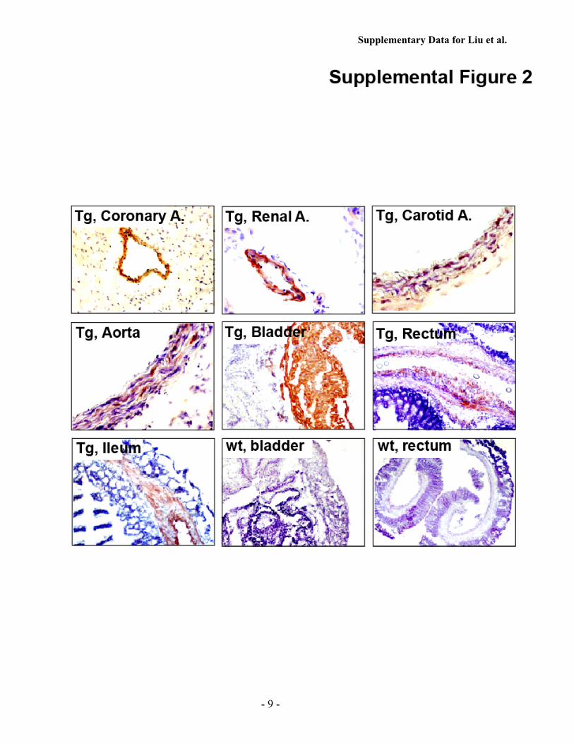
Supplementary Data for Liu et al.
- 9 -
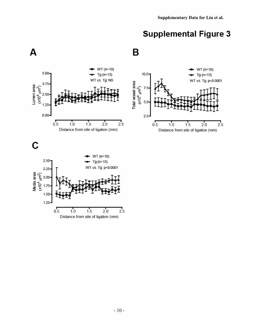
Supplementary Data for Liu et al.
- 10 -

Supplementary Data for Liu et al.
- 11 -

Supplementary Data for Liu et al.
- 12 -

Su, Guogang Zhao, Haifei Xu, Ming C. Gong and Zhenheng GuoShu Liu, Zhongwen Xie, Qingwei Zhao, Huan Pang, John Turk, Lindsay Calderon, Wen
Inflammation and Neointima Formation) Participates in the Initiation and Early Progression of Vascularβ2(iPLA
β2Smooth Muscle-specific Expression of Calcium-independent Phospholipase A
doi: 10.1074/jbc.M112.340216 originally published online May 25, 20122012, 287:24739-24753.J. Biol. Chem.
10.1074/jbc.M112.340216Access the most updated version of this article at doi:
Alerts:
When a correction for this article is posted•
When this article is cited•
to choose from all of JBC's e-mail alertsClick here
Supplemental material:
http://www.jbc.org/content/suppl/2012/05/25/M112.340216.DC1.html
http://www.jbc.org/content/287/29/24739.full.html#ref-list-1
This article cites 51 references, 35 of which can be accessed free at at EA
STE
RN
KE
NT
UC
KY
UN
IV on M
ay 9, 2016http://w
ww
.jbc.org/D
ownloaded from