snakes of papua new guinea - kingsnake.com are widely feared in papua new guinea, ... someone they...
TRANSCRIPT

1
5
Snakebite in Papua New Guinea D a v i d W i l l ia m s
Introduction
Snakes are widely feared in Papua New Guinea, and with very good reason. In many parts of PNG snakebite is an almost daily occurrence and venomous snakebite is a serious public health problem, with localized incidence rates that are among the highest of any region in the world. Medical and epidemiological studies of snakebite in different parts of the country have given us detailed snapshots of some of the outcomes of snakebite, and although there are still many gaps in our knowledge, there is a significant amount of factual data available.
Ask anyone about snakebite and they will undoubtedly have a story to tell about someone they know who was bitten by a snake, or who died of snakebite. It would in fact be very easy to believe that venomous snakes lie in wait for unsuspecting people at every turn, and that right across PNG snakebite is claiming dozens of lives every day. If you ask Papua New Guineans about which snakes are responsible for snakebite one species above all others will feature at the forefront of every conversation; the fearsome ‘Papuan (Pap) black’.
Of course the challenge for clinicians, health workers and scientists when it comes to snakebite is to separate the finely woven threads of fact and fiction:
• Just how many people really are bitten by snakes, and in what parts of the country do these bites occur?
• Are there as many deaths as either the scientific data, or the local people would have us believe?
• Which species are dangerous and bite the most people and which are not? • What types of antivenom will best suit the needs of certain areas of the
country?
We have to be able to answer these and many other questions in order to provide the victims of snakebite with the most appropriate medical care. Having factual data enables resources (including antivenoms) to be distributed to the right places, and most importantly of all, reliable data provides health managers with the information they need to be able to make the right funding decisions to address community needs.
V e n o m o u s b i t e s a n d s t i n g s i n P a p u a N e w G u i n e a
6
At the present time however, the extent of our knowledge lacks national clarity; there are many provinces throughout the country for which we simply have no reliable data. Without even basic information it is extremely difficult to give clinicians and health workers exact information about snakebite in their communities, and it is completely impossible to provide sound advice to health administrators. As you will learn in this chapter, research into the epidemiology and clinical consequences of snakebite is taking place, and we have learned quite a lot about snakebite over the last fifty years. We hope to stimulate your enthusiasm to assist in ongoing research and to develop our knowledge even further.
Local beliefs & perceptions of snakes and snakebite
Snakes occupy an important role in local culture and tradition, and it is these beliefs and perceptions that affect the ways in which Papua New Guineans deal with the issue of snakebite and snakes in general. In many communities across Papua New Guinea snakes are considered to be the mortal shells of bush spirits, demons, or deceased ancestors, enemies and sorcerers (Campbell, 1969). There are numerous legends that tell of fearsome demons which take the natural form of snakes. The Marind-anim people from south-western PNG and eastern Papua believe, for example, that a demonic old woman known as the pathogu steals young children while in the body of a snake. To the Orokolo people living to the west of Kerema in Gulf province, snakes are the homes of mythical bush spirits – dark and dangerous creatures that can cause untold trouble.
Many Mekeo people believe that the dreaded ‘Papuan black’ attacks people at the behest of a sorcerer who may have first stolen something personal from the victim and have placed it into a heated pot containing the body of a snake (O’Shea, 1996). A similar belief is held by the Kiwai people from the Fly River delta; the ove-devenar sorcerer will collect the faeces of his victim and put it into the mouth of a snake model made from wood or clay which he then places in an area that the victim is likely to visit. The model becomes a living snake which hunts and kills the ove-devenar’s target. The Elema people of Gulf province believe in an evil spirit called ove-hahu who is the cause of accidents, serious sudden illnesses and snakebite. Similar traditional beliefs about snakes are held by the Arapesh people from the Sepik region, and by many other clans throughout PNG.
In Central province, the coastal Motu-Koitabu and mountain Koiari peoples often believed that snakebite, as well as being a tool of the vada (sorcerer), was sometimes a punishment meted out by good spirits such as the birava for social and traditional crimes like adultery. The Keakalo people of Marshall Lagoon also believed that

C h a p t e r 1 : S n a k e b i t e e p i d e m i o l o g y
7
snakebite was a common punishment for social transgressions such as adultery, theft, spousal abuse or taboo-breaking. As well as this, their mega mega auri (snake senders) could not only use magic to send out a snake to bite someone, but could also be sought out and paid to administer a cure. Dr Charles Campbell, a physician who conducted the first medical studies of snakebite in PNG, learned that this cure usually involved obtaining a variety of tree bark called paia and a rainforest vine wamela which were chewed by the mega mega auri and then blown onto the face and body of the victim, who would also be massaged with coconut flesh or milk and kiki leaves. Chewing the paia ivoa (ginger leaves) and wamela was also considered to be a way of deterring snakes from biting.
Just as snakes are associated with evil deeds, dark spirits and sudden death, they are also widely held to play an important role in fertility and the guardianship of important traditional sites. The Kamea people (known as the Kukukuku by outsiders) live in the remote highlands of Gulf and Morobe provinces. These small, strongly built people, with an infamous warrior tradition and a fearsome reputation, have many animistic beliefs and some of them believe strongly that an enormous snake keeps the world functioning and protects us all. This creature is guarded by benevolent sorcerers who engage in a constant struggle to protect the snake from evil sorcerers who would kill it, a calamity that would spell the end of the world.
Kiwai people in Western province believe that the mythical maigidubu snake spirit is crucial to the success of yam crops, and that if the maigidubu leaves its tracks through the crop around the time of the yam festival, then success will be assured. Also, their guardian spirits, the etengena, would often take the form of snakes and stand guard over their gardens, biting intruders to send them away. Trobriand Islanders believe that some snakes are sacred and either house the spirits of great chiefs, or are their reincarnations. All the same, finding such as snake in a village was not considered a good sign, and unless the creature could be tempted to leave with prayers or other offerings, sudden illness was likely to befall the entire community. Despite this dreadful possibility, it was taboo to kill the snake, as this would only make the consequences worse.
The Bariai of West New Britain tell the tale of Moro, a snake-man who started the tradition of pig exchanges and other ceremonies in honour of firstborn children, and to give praise to the dead (McPherson, 1994). Legend has it that Moro’s father Kamaia had been killed in a disagreement with his brothers-in-law, and his liver cut out and cooked. Later during his funeral Moro was tricked into eating some of the liver by his cousin-in-law Kaukave whereupon his two legs immediately became

V e n o m o u s b i t e s a n d s t i n g s i n P a p u a N e w G u i n e a
8
fused together and the entire lower half of his body was transformed into that of a snake. Moro’s transformation is ascribed to the vengeful punishment of his father’s ghost for the having consumed the liver, albeit unknowingly. Pursued by the ghost of his father, Moro and his mother escape after Moro uses magic to carve out the Amara River, and then tricks the ghost into trying to cross, only to be eaten by a large crocodile!
Just as perceptions about the supernatural abilities of snakes often influence how people deal with snake, so too do local beliefs about how a snake ‘poisons’ its victims. Throughout much of Western, Gulf and Southern Highlands Provinces it is believed that death adders (Acanthophis spp.) inflict illness by actually stinging the victim with the tip of the tail. This sometimes leads to people attempting to handle these snakes after first cutting off the tail with a bush knife, only to find that the now furious snake is more than happy to content itself with proving them wrong!
In the Wipim district many people will not eat venomous snakes as they belief that this may cause poisoning, while the Kuru people living close to the nearby Oriomo River are very adept snake catchers who are more than happy to make a meal of an errant serpent, venomous or not.
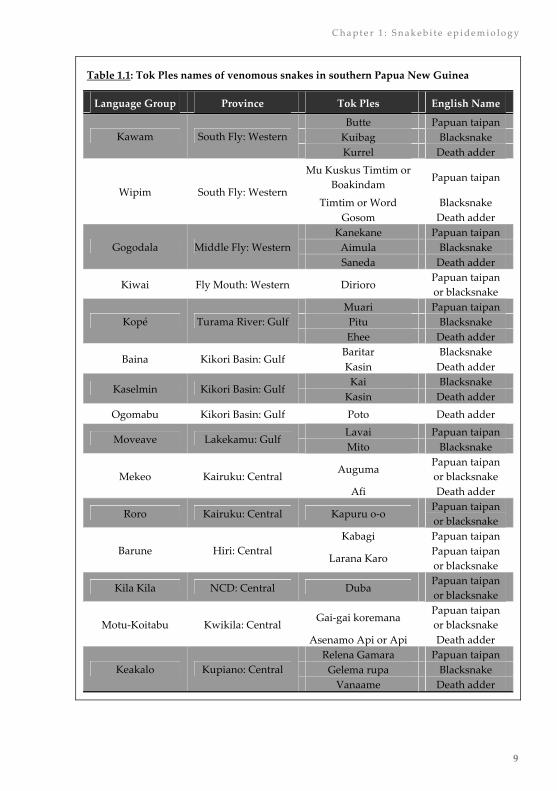
To many of the Papuan people living in southern Papua New Guinea the most important venomous snakes are dark in colour. The early colonial settlers quickly learned from local people that black coloured snakes were very dangerous, and long before any taxonomic labels were applied, the term ‘Papuan black’ came to be synonymous with those snakes that were to be most feared and despised. Such is the strength of this association that today few Papua New Guineans who have been exposed to Caucasian influence use any term other than ‘Pap black’ to describe large venomous snakes. This is despite the fact that nomenclatural distinctions between different types of venomous snakes are made by many of the people living in southern Papua New Guinea. Different names may sometimes even be used by different clans of the same language groups for exactly the same snake, although most groups use just one name to describe each different species of venomous snakes (Table 1.1)
As will be shown later in this book, misidentifying a snake after a snakebite can have serious consequences, especially when it results in the bitten person receiving incorrect or inappropriate treatment. The issue of snake identification is dealt with further in Chapter 2.
C h a p t e r 1 : S n a k e b i t e e p i d e m i o l o g y
9
Table 1.1: Tok Ples names of venomous snakes in southern Papua New Guinea
Language Group Province Tok Ples English Name
Butte Papuan taipan Kuibag Blacksnake Kawam South Fly: Western Kurrel Death adder
Mu Kuskus Timtim or Boakindam
Papuan taipan
Timtim or Word Blacksnake Wipim South Fly: Western
Gosom Death adder Kanekane Papuan taipan
Aimula Blacksnake Gogodala Middle Fly: Western Saneda Death adder
Kiwai Fly Mouth: Western Dirioro Papuan taipan or blacksnake
Muari Papuan taipan Pitu Blacksnake Kopé Turama River: Gulf Ehee Death adder
Baritar Blacksnake Baina Kikori Basin: Gulf
Kasin Death adder Kai Blacksnake
Kaselmin Kikori Basin: Gulf Kasin Death adder
Ogomabu Kikori Basin: Gulf Poto Death adder Lavai Papuan taipan
Moveave Lakekamu: Gulf Mito Blacksnake
Auguma Papuan taipan or blacksnake Mekeo Kairuku: Central
Afi Death adder
Roro Kairuku: Central Kapuru o-o Papuan taipan or blacksnake
Kabagi Papuan taipan Barune Hiri: Central
Larana Karo Papuan taipan or blacksnake
Kila Kila NCD: Central Duba Papuan taipan or blacksnake
Gai-gai koremana Papuan taipan or blacksnake Motu-Koitabu Kwikila: Central
Asenamo Api or Api Death adder Relena Gamara Papuan taipan
Gelema rupa Blacksnake Keakalo Kupiano: Central Vanaame Death adder

V e n o m o u s b i t e s a n d s t i n g s i n P a p u a N e w G u i n e a
10
Neither the Mekeo nor Kila Kila Motu people make a significant distinction between taipans and blacksnakes and use just one name each to describe the two different species. Even the people of the Moveave region in the east of Gulf province, while using different names to describe taipans and blacksnakes, identify both as ‘blacksnakes’; the different names are used to distinguish the snakes on the basis of the type of habitat they live in, rather than on the appearance of the snakes themselves. Lavai is used to describe blacksnakes (actually Papuan taipans) that live in tall grass, while Mito is the name given to blacksnakes (real Papuan blacksnakes!) that inhabit the regions large tracts of Sago Palm forest. In the Wipim district of Western province Timtim is loosely used to describe any dark coloured snake, although the Mu Kuskus Timtim (the blacksnake with a red back) is actually a black Papuan taipan. At the same time, Papuan taipans in Wipim with chocolate brown base colour and a large very prominent reddish dorso-vertebral stripe are given another name, Boakindam; and are especially feared.
The majority of people also distinguish venomous snakes from non-venomous pythons and other snakes. The Motu people know pythons ubiquitously as Navara, while their Koitabu cousins use Lavara, and the Keakalo call large pythons Kapari and may consider some of them sacred spirit animals. The Rumu Hei people living at Ogomabu near the Kikori Government Station in Gulf Province exemplify the ability of Papua New Guineans to distinguish between several species of python and colubrid snakes (Table 1.2). Health workers should familiarise themselves with the local names of different types of snakes; this can be helpful when it comes to making a presumptive identification of the type of snake responsible for a bite.
Table 1.2: Names of non-venomous snakes in the Rumu Hei Tok-Ples (Ogomabu village, Kikori Basin, Gulf province) demonstrating differentiation of species
Rumu Hei Name English Name Scientific Name
Maru New Guinea ground boa Candoia aspera
Miaru Papuan python Apodora papuana
Beramo Green tree python Morelia viridis
Mahe Carpet python or
amethystine python Morelia spilota harrisoni and Morelia
amethistina amethistina
Rudri D’Albert’s python Leiopython albertisi
Horeke Common tree snake Dendrelaphis punctulatus
Kahe Pate Uki Slatey-grey snake Stegonotus cucullatus

C h a p t e r 1 : S n a k e b i t e e p i d e m i o l o g y
1 1
Common misconceptions about snakebite
There are several common misunderstandings and misconceptions about snakes and snakebite in Papua New Guinea:
Belief: The majority of snakebites are caused by the Papuan blacksnake.
Reality: Incorrect
From a medical perspective, this is a particularly dangerous misconception that can seriously harm a snakebite patient. The clinical reality is that there is little evidence to show that the real Papuan blacksnake (Pseudechis papuanus) causes large numbers of snakebites, particularly in Central province where investigations using venom identification assays have shown that only 4.3% of the serious snakebites admitted to Port Moresby General Hospital were caused by this species. In Central province the evidence suggests very strongly that the majority (more than 80%) of serious snakebites admitted to PMGH are caused by Papuan taipans (Oxyuranus scutellatus canni). In the past people have died as a result of the misidentification of taipans as ‘blacksnakes’.
In some parts of Milne Bay and Western provinces, it is still possible that a larger proportion of snakebites are caused by the Papuan blacksnake, but at the present there is very little reliable evidence available. Papuan blacksnakes only occur in the Papuan region; they are not found in the Highlands, in the Mamose region, or in the large island provinces.
Belief: The death adder uses a poison spine on its tail to harm people.
Reality: Incorrect.
The soft spine-like projection on the end of a death adders (Acanthophis spp.) tail contains nothing toxic, and has no role in the injection of venom. The ‘spine’ is nothing more than a lure that is wriggled by the snake in order to attract food – just like dangling a worm on a fishing hook. When a small lizard or frog tries to eat the lure, the snake bites the animal killing it with venom injected by fangs in the mouth.
Belief: The forked tongue of a snake is a poisonous sting.
Reality: Incorrect.
The tongue of a snake is completely harmless. It is a specialised scent organ that is used to collect odours from the air and deposit them in a special organ on the roof of the mouth (Jacobson’s organ) that contains the same types of olfactory cells that

V e n o m o u s b i t e s a n d s t i n g s i n P a p u a N e w G u i n e a
12
occur in the human nose, and which allow us to smell; the tongue does nothing more than help the snake detect smells and odours.
Belief: All snakes are venomous.
Reality: Incorrect.
This could not be further from the truth; of the 112 species of snakes that occur in Papua New Guinea and Papua, only a third are venomous, and of 37 species, there are currently only seven land snake species known to have the ability to cause human fatality. The two species of sea krait (Laticauda spp.) can also cause death, as can several species of true sea snakes; but sea snake related deaths are very rare in PNG. One very good general rule to teach people, is that venomous snakes in PNG very rarely climb off the ground; a snake in a tree, the roof of a house, or a banana plant in the garden is most likely to be non-venomous.
Belief: Snakebite is the work of sorcerers.
Reality: Incorrect.
Traditional beliefs do play an important role in the lives of many Papua New Guineans, and the belief in sorcery persists in many communities. Some people belief that snakebite is caused by sorcery, and that a cure can only be had by paying the vada (sorcerer) or mega mega auri (snake sender) to remove the curse. The belief in sorcery is no different to the belief in Christianity, and people may have very strong feelings of faith in the validity of sorcery. The reality is that snakebite is a result of an accidental encounter between humans and snakes, and sorcery really has nothing to do with it. The proof of this lies in the fact that while sorcery can always cure the bite of a non-venomous snake, the same cannot be said when the snake is a highly venomous Papuan taipan; without antivenom or other expert medical care, all taipan victims die.
Belief: Traditional cures are best for snakebite
Reality: Incorrect.
At this time there is no impartial evidence to show that any of the traditional remedies used for snakebite in PNG actually benefit the patient. Some treatments, such as the use of mango bark juice, which promotes vomiting, may actually be harmful, especially if the patient aspirates vomit into the lungs. Although research overseas suggests that some plant materials may have potential to neutralise snake venom in experiments, their success and safety in human patients is uncertain.

C h a p t e r 1 : S n a k e b i t e e p i d e m i o l o g y
1 3
Epidemiological knowledge of snakebite
The study of a disease, medical condition or illness, its causes, characteristics and the factors that may ultimately help to control it, is called epidemiology. Scientists and doctors use epidemiological information to understand why, when and where diseases, accidents and illness occur. Learning about the epidemiology of snakebite in PNG is an important first step in understanding the problem of snakebite and developing practical ways to ensure that important resources like antivenoms are made available in the right places. Some of the things that we can learn by studying snakebite epidemiology include:
• How many people are the victims of snakebite, and how many become ill or die because of snakebite;
• Who are these people and is snakebite more likely in some particular groups (for example: teenagers and young adults) than it is in others;
• How does snakebite occur: what sorts of activities are more likely to result in snakebite (for example: gardening or hunting);
• How could snakebite be prevented: is there a common human behaviour that if changed, would result in fewer snakebites;
• What types of snakes cause snakebite in different parts of PNG: knowing this makes it possible to supply the right types of antivenoms;
• When is snakebite more likely to happen (for example: are there times of day, or times of year when snakebite is more common);
• What are the consequences of snakebite (for example: what medical problems do snakebite victims develop);
• How can we improve the treatment of snakebite victims.
Studies of snakebite epidemiology first began in PNG more than 40 years ago, and although the first published medical report of snakebite in Papua New Guinea did not appear until 1961, the risk to public health presented by venomous snakes was well known, and snakes generally were (and remain) widely feared.
While the Papua New Guinean perception of snakebite may have revolved around the traditional beliefs of the various cultural and social groups throughout the country, the perceptions of colonial medical officers was tempered by ‘western’ attitudes which had by the start of the 20th Century largely turned away from belief in supernatural forces towards acceptance of ‘scientific’ conclusions that were based on rational hypotheses, demonstrable facts and clinical reality. Significant advances in human understanding of basic physiology and biology provided a vastly different perspective of the causes of the clinical effects seen after snakebite. Rather than being considered as the result of sorcery, colonial doctors had a strong belief

V e n o m o u s b i t e s a n d s t i n g s i n P a p u a N e w G u i n e a
14
that snakebite was the consequence of the physiological changes produced by organic toxins injected by the snakes themselves. Over the last 5 decades there has been considerable interest in the problems associated with snakebite in Papua New Guinea, and a number of epidemiological and clinical studies, aimed at learning more about the consequences of snakebite and the outcomes of various treatment strategies, have been carried out. As a result there exists a considerable body of information to help us in understanding why snakebite occurs, what the conse-quences may be, and what we should be doing to improve the prognosis for snakebite patients.
How many people are bitten by snakes in PNG every year?
Early publications about snakebite in Papua New Guinea give no data on the over-all incidence of snakebite throughout the country, and even today we do not known exactly how many snakebites occur each year. Dr Charles Campbell (Fig. 1.2) who worked at Port Moresby General Hospital (PMGH) during the 1960’s said that from 1961 to 1967 there was an average of 155.5 cases of snakebite admitted to the hospital each year. Snakebite patients made up less than 1% of all patients who came to the hospital for treatment (Campbell, 1969a).
Thirty years later two different studies of snakebite were carried out in Central province. Dr Bart Currie (Fig. 1.4) and his colleagues found that between January 1987 and June 1989 an average of 81.8 envenoming snakebites per 100,000 people occurred in rural Central province each year, compared to 21.8 cases for every 100,000 people living in the urban National Capital District in each year (Currie et al., 1991). Another study by Dr David Lalloo and colleagues (Fig. 1.7) investigated snakebite in different parts of Central province (Lalloo et al., 1995a). The average annual incidence of reported snakebites from 1987 to 1992 at major health centres and the Port Moresby General Hospital was 215.5 cases for every 100,000 people living in the region. Of these, an average of 62.6 per 100,000 people had signs of envenomation (Currie et al., 1991). It was also found that rather than being the same everywhere, the incidence of snakebite even within Central province varied enormously. In the hilly country of Goilala the average yearly rate was 20.6 cases per 100,000 people, while in the lowlands of Kairuku it was 526.2 snakebites per 100,000 people. In real terms the numbers of admissions to health centres ranged from less than five cases a year, to more than 100 (Fig. 1.1) (Lalloo et al., 1995a).
“Snakebite” (reported snakebite): means all cases of either suspected or real snakebite. “Envenomation” (envenoming snakebite): means only cases with positive symptoms and signs of illnesses after a suspected or real snakebite.

C h a p t e r 1 : S n a k e b i t e e p i d e m i o l o g y
1 5
Understanding incidence and mortality rates
Most epidemiological studies try to report the number of cases (incidence), or the number of deaths (mortality), caused by a disease, accident or illness, in a given time period (for example: 1 year) in a standardised population size. This makes it possible to compare the incidence rates of different diseases, or of the same disease in different groups of people, in a meaningful way. Most studies report incidence and mortality as the number of cases occurring over one year for a standard population of 100,000 people.
For example: if there are 140 snakebites in a district over a year, and the average population of the district during the year is 35,000, the incidence rate is calculated as:
Number of snakebites in the year Average population
× 100,000 = incidence rate
140
35,000× 100,000 = 400 per 100,000 per year
The average population is calculated by adding together the number of people living in a place at the start of the year to the number living there at the end of the year, and then dividing by two to find the average. This helps take into account births, deaths and the movement of people into and out of an area over the period of time in question.
New studies conducted by the author over the last four years have found that Mekeo still has one of the highest overall snakebite incidence rates anywhere in the world (561.9 per 100,000 per year) (Williams et al., 2003). What is perhaps more important is the fact that almost 60% of patients in Mekeo develop envenomation (314.6 per 100,000 per year) and that in some of the village communities the envenomation rates are up to 2.5 times higher, while in other villages the rates were many times lower. This means that even in relatively small areas snakebite is a localised problem; while one village may experience lots of cases every year, another close-by village might have very few cases, if any at all. Understanding the reasons for this might one day help to prevent snakebite in those high risk places.
Outside of Central province there is very little information available about the number of cases of snakebite. A study of cases at Modilon Hospital in Madang between 1978 and 1988 found that there were only 3 cases of envenomation among every 100,000 people living in the province each year (Hudson & Pomat, 1988). This study estimated that the average yearly rate for all snakebites was 8.3 cases per 100,000 people. An ongoing study of snakebite in the Kikori Basin of Gulf province estimated the yearly incidence of snakebite from 1999 to 2005 as being between 97.7-132.5 cases per 100,000 people. In the Middle Fly district of Western province there were an average of 32.8 snakebite admissions to Balimo Hospital each year

V e n o m o u s b i t e s a n d s t i n g s i n P a p u a N e w G u i n e a
16
between 1995 and 2004 with an average yearly incidence rate of 123.3 cases per 100,000 people. In 2003 and 2004 other health services in the Gogodala local level government (LLG) area reported an average of an additional 27 cases of snakebites each year. The combined average yearly Balimo urban/Gogodala LLG snakebite incidence in 2003-2004 was 203.8 cases per 100,000 people.
Figure 1.1:
Numbers of admissions to Port Moresby General Hospital and rural health centres in Central province for snakebite between 1987 and 1992.
Not all of these cases resulted in envenomation.
(after Lalloo et al., 1995)
How many people die from snakebite in PNG?
When a group of 62 health workers were asked to answer this question in 2004, the majority (72.3%) believed that no more than 10% of snakebites were fatal. This corresponds well to the available statistical information, but as is the case with the general incidence of snakebite, the mortality rates vary depending on the location and the age of the patients.
Dr Campbell reported a case fatality rate after snakebite of 6.8% at PMGH from 1959 to 1965, and a rate of 3.1% was reported among 192 patients admitted to the hospital between January 1967 and December 1971 (Campbell, 1969b, Price and Campbell, 1979). In the 1980’s the case fatality rate among children at PMGH was 7.7%, but another study soon afterwards found that while the overall case fatality rate between 1987 and 1992 was 4.4%, the rate among children was actually 10.0% while the adult rate was 3.3% (Brian & Vince, 1987; Lalloo et al., 1995a).
Sadly the case fatality rates after snakebite have continued to increase since the early 1990’s. A study of snakebite deaths in the Intensive Care Unit (ICU) at PMGH between 1992 and 2001 found that 87 of 722 patients died (12.0%) but children were much more likely to die after snakebite than adults (McGain et al., 2004). The case fatality rate for children was 14.6% compared to 8.2% among adults. Another study
0
20
40
60
80
100
120
No
of A
dmis
sion
s
PMG
H
Bere
ina
Vei
fa'a
Kw
ikila
Kup
iano
Mor
egui
na
Irun
a
Soge
ri
Tapi
ni
Oth
er H
C

C h a p t e r 1 : S n a k e b i t e e p i d e m i o l o g y
1 7
of ICU cases between 1998 and 2001 found that 13.5% of female snakebite patients died compared to only 7.7% of men (Williams et al., 2002). More recently a 12 month review by the author of snakebite cases admitted to the ICU at PMGH between September 2003 and August 2004 found that the adult case fatality rate was 14.5% and the rate for children was a disturbing 25.9%; 1 in 4 children died!
There is little regional or national information available about snakebite deaths although studies of snakebite have been conducted in several rural health centres. From 1987-1991 the case fatality rates at five centres in Central province were found to range from 2.9% to 30%. Recent studies in Mekeo have shown that the case fatality rate there was 4.3% between 1999 and 2001 (Williams et al., 2003). This translates into an average annual mortality rate of 13.8 deaths per 100,000 people. In the Abau region where antivenom is rarely available, the average annual mortality rate from 1996 to 2003 was 33.0 deaths per 100,000 people. At Balimo Hospital in Western province the annual mortality rate from 1998 to 2004 ranged from 3.3-11.3 deaths per 100,000 people. When deaths at outlying Middle Fly health facilities were taken into account for the years 2003 and 2004, the average annual mortality rate was 16.3 deaths per 100,000 people. In South Fly’s Morehead and Oriomo/ Bituri LLG’s the average annual mortality rate for 2003 and 2004 was 8.9 deaths per 100,000 people. This probably under-estimates the true rate as almost all health centres reported that most snakebite victims did not present for treatment. None of the health centres in South Fly had stocks of antivenom, and because it was commonly known that antivenom was unavailable, people prefer to stay at home to die among family (unpublished data).
In the 1980’s one study of snakebite in Madang suggested a mortality rate of 0.9 deaths per 100,000 people (a case fatality rate of 1.14%) (Hudson & Pomat, 1988). Another study by Dr Currie and his colleagues reported the annual mortality rate for Madang province to have been 3 deaths per 100,000 people from 1978 to 1988 (Currie et al., 1991). In Central province between January 1987 and June 1989 the annual mortality rate in rural areas was 4.3 deaths per 100,000 people, compared to 2.1 deaths per 100,000 people in urban areas (such as Port Moresby). The study conducted by Dr Lalloo and others, including the late Professor Sirus Naraqi (Fig. 1.3), of snakebites that occurred between 1987 and 1992 reported a mortality rate in Central province of 7.9 deaths per 100,000 people; and a combined rate for Central/ NCD of 3.7 deaths per 100,000 people (Lalloo et al., 1995a). As for the national mortality rate, we simply do not currently have enough information to make more than a conservative estimate. Based on rates in Central and Western provinces it seems reasonable to believe that up to 200 people a year may die from snakebite in PNG, but until data is available from all provinces this figure is difficult to confirm.

V e n o m o u s b i t e s a n d s t i n g s i n P a p u a N e w G u i n e a
18
Who are the most likely people to be bitten?
Some people are much more likely to be bitten by a snake than others. One way of grouping people to look at their respective risks of snakebite is to use age and sex, and most of the studies of snakebite in PNG have done exactly this.
A consistent finding by different researchers over the years has been that the average age of a suspected snakebite victim ranges from 20-27 years, and that children 15 years or younger make up from 18-26% of cases (Campbell 1964, 1967a, 1967c; Currie et al., 1991; Lalloo et al., 1995a; Williams et al., 2002). When it comes to cases in which envenomation occurs children are strongly represented. A study of cases from across southern PNG admitted to the Intensive Care Unit at PMGH between 1998 and 2001 found that children up to the age of 15 years accounted for 40% of all cases (Williams et al., 2002). In the study of snakebite deaths at PMGH undertaken by Dr Forbes McGain and colleagues it was found that 47% of those who died were children under 15 years of age (McGain et al., 2004).
In Madang the study of cases from 1977 to 1986 found that men were almost twice as likely to be bitten as women (83 males: 46 females) (Hudson & Pomat, 1988). This has proven to be a common finding right throughout PNG ever since. Another study that included patients treated at both PMGH and at Modilon Hospital also found that men were 1.6 times more likely to be bitten than women (Currie et al., 1991). Of a total of 1,421 patients admitted to health centres in Central province and to PMGH between 1987 and 1991, men outnumbered women by a ratio of 1.4 to 1. An interesting exception was in the Veifa’a region where slightly more women than men became snakebite victims. Throughout Central province males aged 15-29 years were 4 times more likely to be bitten by a snake than females in the same age group (Lalloo et al., 1995a).
While more men than women are bitten by snakes, it is children who are especially at risk of death after snakebite
Where on the body are people most likely to be bitten?
Most rural Papua New Guineans do not wear long trousers/dresses and shoes when they go to the garden, or go out into the bush hunting or collecting firewood. It is no surprise therefore that the majority of people who are bitten by snakes suffer bites to their unprotected lower limbs, ankles or feet. In one of his studies Dr Charles Campbell reported that bites to the lower limbs accounting for 98.1% of cases; made up of bites to the toes (17.3%), feet (32.7%), ankles (15.4%) and legs

C h a p t e r 1 : S n a k e b i t e e p i d e m i o l o g y
1 9
(32.7%) (Campbell, 1964). Bites occurred when the victims were walking on paths near long grass, or through long grass, and 23% actually trod on the snake itself.
In a study of death adder (Acanthophis spp.) bites, Campbell reported that 8 out of 15 patients were bitten after standing on the snake in bare feet (Campbell, 1966). In one case a man laid down on a snake concealed beneath leaf litter and was bitten on the leg, and another was bitten on the finger by a snake he accidentally picked up in a pile of cut grass. The other 13 bites were on either the feet or ankles. In the only other study of proven Acanthophis spp. envenomation in Papua New Guinea it was reported that 30 of 32 cases involved bites on the lower limbs (Lalloo et al., 1996).
A study of proven Papuan taipan (O. s. canni) bites found that 96.4% were on the legs, ankles or feet (Lalloo et al., 1995b). On the northern side of PNG, 81% of a series of 129 patients were bitten on a lower limb; and 87.6% of these were bites on the feet (Hudson & Pomat 1988). Ten of the 129 patients (7.75%) were bitten on the hands. In a large study of patients from both the Papuan and Mamose regions it was reported that out of 334 patients, 316 (94.7%) were bitten on the lower limbs; 204 of these (64.5%) were bites on the foot or the toes (Currie et al., 1991).
Snakebite is more common during the daytime
During the 1960’s the majority of people bitten by venomous snakes suffered their bites during daylight hours (Campbell, 1964). Only 4 out of 15 cases of death adder bite he reported occurred at night; the other 11 cases all occurred between 8.30am and 5.30pm (Campbell, 1966). In a later study of death adder cases 22 of the 32 bites (68.7%) occurred in the daytime (Lalloo et al., 1996). This was not unlike the result reported in a study at Madang where death adders are the most common cause of snakebite envenomation. In that group of patients 62% of bites took place during the day (Hudson & Pomat, 1988).
Dr David Lalloo and colleagues observed that 85% of proven Papuan taipan (O. s. canni) bites occurred during daylight, as was the case in all 9 cases of proven Papuan blacksnake (P. papuanus) envenomation (Lalloo et al., 1994, 1995b). In a case of fatal small-eyed snake envenomation, the patient was bitten at approximately 6.00pm while alone in the bush (Blasco & Hornabrook 1972), and in a later case series of 11 proven bites by this species, 3 of 7 cases for which a time of injury was recorded occurred after 8.00pm at night (Warrell et al., 1996). Looking at the two large epidemiological studies carried out in the early 1990’s, only 15.3% of bites in one study occurred at night (Currie et al., 1991). In the other study 89.4% of the 205 envenomed patients were bitten during daylight compared to just 54% of the 130 non-envenomed patients (Lalloo et al., 1995a).

V e n o m o u s b i t e s a n d s t i n g s i n P a p u a N e w G u i n e a
20
Is there a snakebite season?
In cooler parts of the world, many snakes become inactive and shelter from the weather during the cold part of the year. The incidence of snakebite in these places therefore tends to be seasonal; it is more likely during the warm summer months than during winter. Snakes that live in the tropics are not as likely to follow this pattern. The warmer weather right through the year in PNG means that snakes probably do not have long periods where they are inactive. There are however some differences in snake behaviour associated with weather and climate.
A study led by Dr Bart Currie found that 56.8% of snakebites occurred between the ‘wet season’ months from November to April (Currie et al., 1991). The study by the Lalloo group reported that 57.9% of 221 envenomed patients and 51.6% of 128 non-envenomed patients were bitten between November-April (Lalloo et al., 1995a). Although they compared snakebite incidence to monthly rainfall, there was no evidence that rainfall had a significant impact on the number of cases of snakebite. A study of cases admitted to the PMGH ICU from January 1998 to December 2001 from across PNG found that 54% occurred between November-April (Williams et al., 2002), and 60% of fatal cases at the ICU in the 10 years from January 1992 to December 2001 occurred between November and April (McGain et al., 2004).
Analysis of 739 cases of snakebite in the Mekeo region found that there was no significant difference in the number of bites that resulted in envenomation during the ‘wet’ and ‘dry’ seasons (Williams, 2005). What was discovered is that there was a significant increase in the number of bites without envenomation during the ‘wet season’; this suggests that non-venomous snakes may be more active during this time of year, and come into contact with more people.
What are people doing when they get bitten?
It is important to try and understand why people get bitten by venomous snakes if we hope to prevent snakebite in the future. The type of activity a person undertakes may in some cases be associated with higher risks of snakebite, and since we cannot modify snake behaviour, we can use knowledge of human behaviour to educate people about ways in which they can reduce their own chances of becoming a snakebite victim.
Many people are bitten walking along bush tracks, through long grass, or on paths on their way to the garden (Campbell, 1966, 1967a). In the large study of snakebite undertaken by Dr David Lalloo and others it was found that the three most com-mon activities at the time of a snakebite were; walking (34.5%), working in the garden (19%) or while in the bush (14.8%) (Lalloo et al., 1995a). In a recent study of

C h a p t e r 1 : S n a k e b i t e e p i d e m i o l o g y
2 1
presumed Papuan taipan bite all nine patients had been bitten in the daytime (Williams and Bal 2003). One patient was bitten at home, two were bitten in the garden and the other six cases occurred when patients were walking along bush tracks or dirt roads.
One memorable report was the unusual case of a man who was bitten by the decapitated head of a small-eyed snake (Micropechis ikaheka) that he thought he had killed (Warrell et al., 1996). Two other patients in that study were bitten by snakes they had tried to pick up and handle!
! The majority of snakebites could be prevented if people wore cheap rubber gum boots when walking along bush tracks and paths, or when they go to the garden, or to the bush.
What types of snakes do people get bitten by in PNG?
Throughout Papua New Guinea there are only four species of venomous snake that contribute to the majority of serious envenomations, along with perhaps three other species that can cause human fatality. The British research team led by Drs David Lalloo and Andrew Trevett (Fig. 1.5) used a special diagnostic test (a species-specific venom enzyme immunosorbent assay or EIA) during the early 1990’s to show that the snake responsible for the majority of serious snakebites admitted to PMGH was the Papuan taipan (O. s. canni) and not the Papuan blacksnake (P. papuanus) (Lalloo et al., 1994, 1995a,b). Papuan taipans caused an incredible 83.2% of envenomations compared to just 4.2% that were caused by Papuan blacksnakes.
Reliable identification of the biting species is a major shortcoming in most studies of snakebite in PNG. In the 1950’s and ‘60’s herpetologist Ken Slater believed that the Papuan black snake (P. papuanus) was the commonest venomous snake in southern PNG (Slater, 1968), and even today most Papua New Guineans believe that this is the snake responsible for almost all snakebites (Trevett et al., 1994; Williams and Bal, 2003). Slater wrote that it was most abundant along the coastline to the east of Port Moresby, and Campbell claimed that it was responsible for more hospital admissions than any other species (Campbell, 1967c). In spite of this statement only two cases seen by Campbell between 1959 and 1967 could be conclusively attributed to this species on the basis of positive identification of the biting snake at the hospital (Campbell, 1964, 1967c). Campbell’s description of a syndrome of envenoming attributable to Papuan blacksnake bites must therefore be treated with scepticism (Campbell, 1967c, 1969a). The much more dangerous Papuan taipan (O. s. canni) often has a distinctive reddish-orange vertebral stripe as a distinctive

V e n o m o u s b i t e s a n d s t i n g s i n P a p u a N e w G u i n e a
22
feature, and Campbell appears to have relied upon this colouration as a means of presuming that large dark coloured snakes apparently lacking it were Papuan blacksnakes. Although the Papuan taipan is the only large snake in PNG that may have a red or orange vertebral marking, this is a feature that many snakebite patients simply do not notice in the heat of the moment. For the average person there is really no reliable means of visually distinguishing between all specimens of Papuan blacksnakes and Papuan taipans. One patient in Campbell’s series of 13 patients with supposed Papuan blacksnake bite presented with a dead 1.5 metre snake for identification (Campbell, 1967c). Identification in the other 12 cases appears based on non-reporting of a vertebral stripe and these cannot be accepted as confirmed blacksnake bites. The only reliable recent description of envenomation by Papuan blacksnakes is the description of clinical syndromes in 9 patients bitten between January 1990 and June 1992 in whose blood blacksnake venom was identified by specific venom-antigen enzyme immunosorbent assay (Lalloo et al., 1994).
Death adders have a distinctive body plan and appearance that makes visual identification easier than for other species. Dr David Lalloo’s group used species-specific venom EIA and found that 10.8% of serious snakebites admitted to PMGH were caused by death adders (Lalloo et al., 1995a, 1996). EIA confirmed 32 cases of death adder envenomation that involved a syndrome of progressive neurotoxicity without coagulopathy, and the 15 cases reported by Campbell in 1966 conform to this profile, lending support to Campbell’s diagnosis. There has also been an excellent case report of a death adder bite treated at Madang Hospital where the dead snake was identified at the hospital (Hudson, 1988).
In the Madang region 64 cases of envenomation were reported between 1977 and 1986; 15 presented with symptoms suggestive of bites by the small-eyed snake (M. ikaheka) (Hudson and Pomat, 1988). In a more recent series, 4/46 (8.7%) cases of snakebite treated at Madang Hospital, and 5/13 (38.5%) treated at Gaubin Hospital on nearby Karkar Island were identified by EIA as attributable to M. ikaheka (Warrell et al., 1996). No cases of M. ikaheka were identified in any of the southern PNG patients studied using EIA (Lalloo et al., 1995). Brown snake (Pseudonaja textilis) envenomation has been reported among 1.8% of EIA-tested snakebite patients admitted to Port Moresby General Hospital between January 1990 and June 1992 (Lalloo et al, 1994, 1995).
In the future it is hoped that the widespread use of snake venom detection kits to diagnose snakebite will make it possible to assemble nationwide data on the species responsible for envenomation.

C h a p t e r 1 : S n a k e b i t e e p i d e m i o l o g y
2 3
• In southern PNG’s grasslands the Papuan taipan is the most common cause of serious snakebite.
• Throughout the rest of PNG, the death adder is the cause of the majority of serious bites.
• Papuan blacksnakes rarely cause snakebites, and are themselves a rare and possibly endangered species.
• Small-eyed snakes cause a small number of bites on the PNG mainland, but may be responsible for a larger number of bites on some off-shore islands along the North Coast of PNG.
What are the effects of snakebite?
Among the most important things we can learn by studying large groups of patients in epidemiological studies of snakebite, are the clinical consequences of envenomation; the effects that snakebite has on patients. Studies in which the identity of the type of snake involved in each case is known are especially valuable since these help us to define the particular syndromes of envenomation caused by individual varieties of snakes, and to recognise particular clinical problems that have to be addressed if patients are to be treated effectively. The studies of snakebite that have taken place in PNG over the last five decades have provided a wealth of information, yet in some areas our knowledge of particular snakes is still lacking, and the opportunity to improve our knowledge still exists. The clinical syndromes of envenomation after snakebite in Papua New Guinea have been reported by several authors. Most reports relate to small case series of patients and, often the identification of the biting species is unclear. Some of the most consistently reported symptoms of snakebite in research studies carried out over the last 45 years have been:
• Local pain • Tender and enlarged lymph nodes • Oedema • Headache • Vomiting or nausea • Abdominal pain • Spitting of vomiting of blood • Visual disturbances
While some of these are clearly not unique just to cases of snakebite, as you will see in later chapters their presence can often be an important indication that a patient will develop envenomation.

V e n o m o u s b i t e s a n d s t i n g s i n P a p u a N e w G u i n e a
24
The local indications of envenomation
Lymph node pain is a consistent feature after snakebite in Papua New Guinea, and tender enlargement of regional lymph nodes is reported in all published accounts. Tenderness or swelling of lymph nodes may be an indication of venom absorption. Trevett et al (1994) stated that lymphadenitis was a particularly sensitive positive indication of envenomation and occurred in 93% of envenomed patients.
The extent and severity of local symptoms such as pain or oedema is lower than in reports of elapid snakebite from Australia. A case reported in 1961 involving severe local pain and oedema was most likely caused by prolonged tourniquet use (Campbell and Young, 1961). Three other patients experienced minor oedema, and local pain was either absent completely or mild. In another study Campbell reported severe pain in three patients and severe oedema occurred in two individuals as a result of lengthy tourniquet application (Campbell, 1964).
A third of the patients bitten by death adders in Campbell’s series from the 1960’s had local pain, and 27% had slight oedema (Campbell, 1966). Neither symptom was present in any of the 6 patients with Papuan taipan envenomation reported later by Campbell (1967); however severe pain, oedema and extensive ecchymoses were seen in a patient bitten on the shoulder and may have been due to a Papuan blacksnake (P. papuanus) bite (Campbell, 1967).
Pain was a feature in 36% of cases reported from Madang province and 14% of had local oedema and localised muscle pain, most probably associated with rhabdomyolysis after small-eyed snake (M. ikaheka) envenomation (Hudson and Pomat, 1988). One patient in a series of eleven bitten by small-eyed snakes had local pain and two patients had oedema (Warrell et al., 1996). Slight oedema was observed in one patient bitten by a Papuan blacksnake (Lalloo et al., 1994). In a series of 166 bites by Papuan taipans there were no reports of local pain or swelling (Lalloo et al., 1995). Local pain and swelling caused by snakebite should not be confused with the pain and swelling that can be associated with ischemic injury due to prolonged tourniquet use.
Non-specific indications of envenomation
Some of the symptoms and signs seen after snakebite are also seen in patients with other medical problems as well, and are regarded as non-specific indications of possible envenomation. A number of non-specific features have been reported consistently in patients with snakebite envenomation. Headache has been univer-sally reported in virtually all published accounts of snakebite in PNG. Headaches are reported to have lasted from a few hours to several days. Herpetologist Ken

C h a p t e r 1 : S n a k e b i t e e p i d e m i o l o g y
2 5
Slater had persistent severe headache for two days after a bite from a small-eyed snake (Campbell, 1969), and was reported in patients proven to have been bitten by this species in a study many years later (Warrell et al., 1996). Two out of nine patients with proven Papuan blacksnake bite had headache (Lalloo et al., 1994), and it was a feature in 54.5% of positively identified Papuan taipan victims (Lalloo et al., 1995b). An analysis of referral letters written by rural health workers during the early 1990’s found that headache was reported in 37% of patients at the health centre, and in 58% of the same patients on admission to PMGH (Trevett et al., 1994a). Headache was a non-specific symptom in 38 (63.3%) of 60 fatal snakebites treated at PMGH between 1992 and 2001 (McGain et al., 2004).
Vomiting and abdominal pain or tenderness typically occurs in unison, and is another widely reported feature of snakebite. In one study a patient with severe abdominal pain and vomiting in conjunction with abdominal tenderness was mistakenly diagnosed with acute appendicitis (Campbell and Young, 1961). Vomiting was reported in 42% and abdominal pain in 34.6% of patients with death adder bites (Campbell 1966), and was also reported in 3 of the 6 patients with presumed Papuan taipan bite (Campbell, 1967). In the series of 13 bites doubtfully attributed to Papuan blacksnake envenomation, but most probably really having been caused by Papuan taipans, 8 patients had early vomiting and 4 had severe abdominal pain (Campbell, 1967). Vomiting after bites from the small-eyed snake has been reported (Blasco & Hornabrook, 1972; Warrell et al., 1996).
Vomiting was reported in 2 of 9 proven cases of Papuan blacksnake envenomation, and 5 others had abdominal pain (Lalloo et al., 1994), while among Papuan taipan bite victims, 64.4% vomited and 59.8% had abdominal pain (Lalloo et al., 1995). In the study of referred patients 38% had abdominal pain and 30% had vomited at the health centre, but by the time they were admitted to PMGH 58% had abdominal pain and 62% had reported vomiting (Trevett et al., 1994a). Abdominal pain was documented in 48 (80%), and vomiting in 40 (66%) of 60 fatal snakebites at PMGH between 1992 and 2001 (McGain et al., 2004).
Bleeding after snakebite
Evidence of abnormal bleeding (coagulopathy) is an absolute indication for antivenom if observed in conjunction with a history of snakebite in PNG. The presence of coagulopathy first drew the attention of Campbell & Young in 1961 and they reported that 6 of 15 patients had ‘bleeding tendencies’ and that 3 patients vomited blood-stained material (Campbell and Young, 1961). Since that time clinical bleeding has been an observed feature in many studies of snakebite in the

V e n o m o u s b i t e s a n d s t i n g s i n P a p u a N e w G u i n e a
26
Papuan region. Many patients have been reported as either spitting blood or vomiting blood (haematemesis); passing ‘blood-stained’ (haematuria) or haemoglobin-containing (haemoglobinuria) urine; and having frank bleeding from wounds and grazes (non-clotting or ‘incoagulable’) blood.
Haematemesis was seen in 3 patients and another 2 coughed blood (haemoptysis) among the group of 13 that Campbell (1967) thought to have been bitten by Papuan blacksnakes. Three of four patients in this series had free haemoglobin in their urine (haemoglobinuria), and other signs of coagulopathy (spitting blood; gingival bleeding; bleeding bite wounds) were reported in 8 of the 13 patients. It is likely that most of these bites were actually caused by Papuan taipans rather than by blacksnakes. Blood-stained saliva and bleeding from the mouth or nose was reported in one patient with confirmed Papuan blacksnake envenomation, while two others had either nose bleeds (epistaxis) or gingival bleeding (Lalloo et al., 1994). Laboratory studies on blood from two of these patients revealed mild thrombocytopenia and another three patients had lower than normal levels of several important clotting factors and measures of clotting time.
Spontaneous bleeding has been reported in two cases of envenomation by the small-eyed snake (M. ikaheka) (Warrell et al., 1996). In the study of 32 patients with EIA-proven death adder envenomation there was no clinical evidence of coagulopathy although white blood cell counts were elevated and some patients had slightly lower than normal blood platelet levels (thrombocytopenia) on laboratory investigation (Lalloo et al., 1996). Another measure of blood coagulation, the Prothrombin time (PT) was significantly prolonged in 13 patients from this study, although broad clinical experience in Australia and PNG suggests that bites by these snakes do not cause bleeding disorders.
All of the clinical evidence indicates that bleeding problems are an important feature of bites by Papuan taipans, and in addition to Campbell’s early reports of bleeding after bites by large dark coloured snakes, there is a substantial body of evidence from recent studies. Incoagulable blood (i.e.: positive 20 minute whole blood clotting test [20WBCT]) was present in 77% of patients with EIA proven Papuan taipan bites (Lalloo et al., 1993, 1995b). Systemic bleeding occurred in 43.7% of patients, and bite site bleeding occurred in 16.3%. Most reported bleeding from either the gingival sulci (37%) or by haematemesis (12.6%). Three patients had epistaxis; 11% bled from superficial cuts and scratches; 8.8% bled from venepuncture sites. On laboratory investigation 52.8% had moderate to severe increases in white blood cell counts (leucocytosis) and 27.5% developed mild platelet depletion (thrombocytopenia).

C h a p t e r 1 : S n a k e b i t e e p i d e m i o l o g y
2 7
Dr Michael Brian and Professor John Vince reported that 40 of 54 children bitten by snakes developed evidence of coagulopathy and that 33 had clotting times greater than 15 minutes (Brian and Vince, 1987). Bleeding problems were mentioned in 40% of referral letters written by rural health workers, and 60% of the referred patients were found to have incoagulable blood upon admission to PMGH (Trevett et al., 1994). In 60 fatal cases of snakebite, 75% had a positive 20WBCT and 35 patients (53.3%) had demonstrable spontaneous bleeding (McGain et al., 2004).
The sometimes hidden effects of myotoxicity
There are few early reports of direct muscle tissue damage (myotoxicity) following snakebite in PNG, although some cases with ‘haemoglobinuria’ may actually have been patients with myoglobin-stained urine (myoglobinuria) instead. The reason why myotoxicity is so rarely reported in PNG is that the effects are rarely as visible as bleeding or paralysis, or may as mentioned be mistaken for something else altogether. Myotoxicity occurs when snake toxins attack muscle tissue and break it down within the body. If undetected it can eventually lead to kidney failure and death.
Snake catcher Ken Slater reported ‘weakness’ after a bite from the small-eyed snake (M. ikaheka), and this is often a sign of rhabdomyolysis (Blasco and Hornabrook, 1972). Between 1977 and 1986 six patients from Madang province who had dark-coloured urine (myoglobinuria) went on to develop renal failure (Hudson and Pomat, 1988). Dr Bernie Hudson reported two cases of presumed small-eyed snake envenomation (Hudson, 1988a). One was a 13 year-old girl bitten on the finger who developed generalized muscle pain and tenderness and had urine that was dark reddish-brown, but did not contain red blood cells (erythrocytes); and the other patient was a 50 year-old man bitten on the finger while reaching into a bandicoot burrow by a ‘black-headed snake with a white belly’. He also developed muscle pain and passed dark urine, and while the muscle pain was mild initially it became more severe over the next 2 days. At Modilon hospital in Madang his urine was grossly discoloured, but once again was erythrocyte-free. He received one ampoule of death adder antivenom and three ampoules of polyvalent antivenom. Neither patient was jaundiced (anicteric) had no coagulopathy, but did have neurotoxicity; the 13 year old girl subsequently died. Other cases of presumed small-eyed snake envenoming treated in Madang also showed signs and symptoms of generalized rhabdomyolysis and myoglobinuria. Three patients with classical signs of myolysis (generalised muscle pain and tenderness; dark urine) were described in a later study of EIA proven small-eyed snake envenomation (Warrell et al., 1996).

V e n o m o u s b i t e s a n d s t i n g s i n P a p u a N e w G u i n e a
28
In hospitals that have well equipped medical laboratories it is possible to detect myolysis by measuring the quantities of certain proteins and enzymes in the blood of a patient. Typical in patients with myotoxicity is an elevation of an enzyme called creatine kinase (CK) and of the muscle protein myoglobin. These tests are rarely available in PNG, but when they have been used it has been possible to detect myotoxicity. The CK levels in 8 of 12 patients bitten by death adders were elevated (up to 4,220 IU/L) (Lalloo et al., 1996). Similar studies in victims of Papuan taipan bites found that CK levels were raised in 74.7% (up to 8,110 IU/L) (Lalloo et al., 1995b). Serum myoglobin exceeded 80 ng/ml in 52.2% of patients upon admission. The CK levels in 2 of 9 patients with proven Papuan blacksnake envenomation were slightly elevated; however there were no indications of rhabdomyolysis in any patient (Lalloo et al., 1994).
In Australia, rhabdomyolysis and myoglobinuria after bites by snakes related to the Papuan blacksnake are common, and CK levels may exceed 200,000 IU/L (Rowlands et al., 1969; Pearn et al., 2000; Isbister and Currie, 2003). Acute renal failure is a potential complication of myotoxicity, and renal impairment or failure was reported in 37% of 60 fatal snakebites treated at PMGH (McGain et al., 2004). Renal complications were contributing factors in the deaths of 16 (26.7%) patients.
Muscle pain or weakness, especially in patients with dark-brown or black urine is a critical sign of myotoxicity and potential renal failure.
Paralysis caused by snakebite
The neurotoxic effects of Papua New Guinean snake venoms are well defined in the available literature, and the balance of evidence demonstrates that snake venom neurotoxins are the major contributors in the deaths of patients due to the development of progressive flaccid paralysis that inhibits and compromises respiration.
The earliest signs of neurotoxicity often manifest themselves in the craniofacial nerves producing a sequential syndrome that often starts with diplopia and ptosis before progressing to ophthalmoplegia, dysarthria, dysphagia and bulbar palsy and eventually involving the intercostal, diaphragm and peripheral muscles. Ataxia and progressive muscle weakness results in a loss of locomotor function; patients eventually lose the ability to walk, stand, sit or even raise their limbs, or control head movements (‘broken neck syndrome’). Deep tendon reflexes may be lost and failing respiration often leads to ‘abdominal’ (paradoxical) breathing.

C h a p t e r 1 : S n a k e b i t e e p i d e m i o l o g y
2 9
PNG’s first study of snakebite graded the neurotoxic effects in 15 snakebite victims according to severity (Campbell and Young, 1961). Paralysis of the eyelids (ptosis) was moderate in 10 cases and severe in 4 others; while eyeball paralysis (ophthalmoplegia) was moderate in 4 patients and severe in 8 others. “Visual disturbances” were reported by 10 patients; most probably double vision (diplopia) or blurring of vision. Moderate to severe facial palsies were present in 6 patients and there was moderate to severe paralysis of the tongue and palate in 9 patients. Intercostal muscle (important accessory muscles in respiration) paralysis was severe in 7 patients with 3 patients experiencing moderate diaphragmatic paralysis. One patient had severe paralysis of the diaphragm, and peripheral flaccid paralysis affecting the limbs was moderate in 7 patients and severe in 2 others.
In a subsequent study Campbell reports ‘visual disturbances’ in 26.9% of patients, noting both diplopia and blurred vision (Campbell, 1964). Ptosis was regarded as the earliest sign of neurotoxicity and Campbell comments that many patients would go to sleep only to awake the next morning with severe facial palsy. Ptosis was often associated with either partial or complete ophthalmoplegia, and was observed in 75% of patients.
In a fatal case of small-eyed snake envenoming progress to death was marked by peripheral limb weakness, shortness of breath and profound respiratory failure with cyanosis (Blasco and Hornabrook, 1972). Other patients bitten by this species were found to develop ptosis, bulbar paralysis and peripheral limb weakness (Warrell et al., 1996). Ptosis was present between 6 and 12 hours post-bite. Two patients died of respiratory paralysis at 19 and 38 hours respectively. A patient from Papua developed dysarthria, facial paralysis and ataxic gait resulting in an inability to stand unassisted. There was severe paralysis of the cranial muscles and intercostal muscles.
In a study of 32 cases of proven death adder envenomation 18 patients developed symptoms and signs of neurotoxicity (Lalloo et al., 1996). Ptosis was reported as a symptom in 15 of 18 (88.2%) and as a demonstrable sign in 17 of the 18 (94.4%) patients. Other symptoms included dysphagia (29.4%); dysarthria (37.5%); diplopia (18.8%); and respiratory difficulty (20%). Other signs included ophthalmoplegia (55.6%); slurring of speech (30.3%); jaw restriction (28.6%); diminished hand grip (31.3%); and reduced reflexes (12.5%). Five patients (27.8%) had to be intubated and ventilated, with the median ventilation time being 13 hours. The range of ventilation times after death adder bites was from 2 to 23.5 hours, considerably less than for victims of Papuan taipans in whom the median ventilation time was 88 hours with a range of from 3 to 176 hours (Lalloo et al., 1995b, 1996).
V e n o m o u s b i t e s a n d s t i n g s i n P a p u a N e w G u i n e a
30
Ptosis was a symptom in 55% and a sign in 53% of snakebite envenomations in Madang province (Hudson and Pomat, 1988). Dysphagia and dysarthria were symptoms in 31%, and 20% of patients reported diplopia. Respiratory distress was a symptom in 9% of bites. Other signs included bulbar paralysis (17%) and external ophthalmoplegia (13%). Only 5% of patients subsequently developed respiratory failure. The only highly venomous species in the Madang province are death adders and small-eyed snakes. Hudson reports complete bilateral ptosis, ophthalmoplegia and paralysis of tongue, jaws and pharynx in a man bitten by a death adder (Hudson, 1988b). Peripheral limb weakness was present as well as paralysis of the intercostal muscles. Dr Hudson also reported bilateral ptosis, ophthalmoplegia, diplopia, bulbar and peripheral paresis in two patients presumed to have been bitten by small-eyed snakes in Madang province (Hudson, 1988a). Respiratory distress was evident in both these patients, and as said before, one of them, a 13 year old girl, eventually died from cardiorespiratory failure 42 hours post-bite. Resolution of ptosis, bulbar paralysis and limb weakness took more than 14 days in the other patient despite the use of a large quantity of antivenom.
The studies of Papuan taipan (O. s. canni) envenomation the British research group led by Drs David Lalloo, Andrew Trevett and David Warrell (Fig. 1.6) remains the most comprehensive investigation of neurotoxicity by this large and very dangerous snake (Lalloo et al., 1993, 1995b, 1997; Trevett et al., 1995a). Ptosis was present in 84/131 (64.1%) patients and was the most frequently reported symptom followed by dysarthria in 39/121 (32.2%) and dysphagia in 39/124 (31.5%). Diplopia was reported in 26/120 (21.7%) and 17/121 (14%) had dyspnoea; 41.7% of patients required intubation as paralysis deepened, and ventilation was required by 36.7%. The time of progression to the point where intubation was required ranged from 3 to 55 hours with a median time point of 13.5 hours (Lalloo et al., 1995b).
In a small series of 9 suspected Papuan taipan bites treated in rural health centres, all had ptosis and dysphagia; 5 had diplopia; 6 had dysarthria; 5 had general bulbar paralysis; 4 had dyspnoea and 2 had peripheral limb weakness (Williams and Bal, 2003). Eight of these patients died from respiratory complications. A study detailing 60 fatal cases of snakebite in the PMGH ICU between January 1992 and December 2001 found that all these patients had neurotoxicity (McGain et al., 2004).
Neurotoxicity was prominent in a series of 49 paediatric snakebite cases admitted to PMGH between August 1981 and October 1984 (Brian and Vince, 1987). Ptosis (88%), drowsiness (56%), ophthalmoplegia (48%), respiratory weakness (42%), difficulty swallowing (40%), slurred speech (38%), limb weakness (27%) and jaw weakness (23%) were all reported. Sixteen children required intubation.

C h a p t e r 1 : S n a k e b i t e e p i d e m i o l o g y
3 1
Other effects of snake venoms
Many of the victims of snakebite present at rural health centres with symptoms and signs that do not neatly fit into the three major clinical categories of bleeding (coagulopathy), muscle damage (myotoxicity) and nerve paralysis (neurotoxicity).
Effects on the heart rate and normal conduction of electrical impulses through the heart have been reported. Transient tachycardia (fast heartbeat) has been reported as an occasional feature of envenomation (Campbell and Young, 1961), and was recorded during the terminal stages of small-eyed snake envenomation (Blasco and Hornabrook, 1972). Both tachycardia and bradycardia (very slow heartbeat) were observed in patients with proven envenoming by this species (Warrell et al., 1996). Tachycardia was an early feature in 5 fatal cases of presumed Papuan taipan envenomation (Williams & Bal, 2003). One victim of death adder envenomation from Western province presented to the rural health centre with a heart rate of 100 bpm, and subsequently developed both renal failure and a second-degree atrioventricular block (ventricular rate of 36 bpm) (Lalloo et al., 1996). In another case a death adder victim presented with septal T-wave inversion; an abnormality that was also reported in a single case of brown snake (Pseudonaja textilis) bite (Lalloo et al., 1997). Non-hypotensive bradycardia was reported in 24.1% of Papuan taipan victims, and 52.2% of those tested had electrocardiograms that demonstrated abnormalities, particularly septal T-wave inversion (31.9%) (Lalloo et al., 1995b).
Campbell noted that sudden collapse after snakebite is common following bites by Australian (and presumably related PNG) species (Campbell, 1967c). Sudden early collapse with transient loss of consciousness was reported in 23% of Papuan taipan bites (Lalloo et al., 1995b). Early collapse had occurred in 13.3% of the 60 fatal cases reported in the ICU at PMGH (McGain et al., 2004).
So do we know everything we need to know?
Clearly a substantial body of previous work exists, and the conclusions from many of these studies, if applied, could significantly contribute to improved management of snakebites in Papua New Guinea. From an epidemiological perspective informa-tion on the incidence of snakebite envenomation and mortality still remains to be collated for the majority of the country, since past studies have been restricted to just two Provinces (Central & Madang). Similarly there is scant data on the syn-dromes of envenomation elsewhere in Papua New Guinea. Studies directed towards resolving these deficiencies would be highly desirable and would greatly assist in resource planning and allocation.

V e n o m o u s b i t e s a n d s t i n g s i n P a p u a N e w G u i n e a
32
Epidemiological studies also need to be devised to address questions relating to the highly variable incidence of snakebite in different regions. Within Central province we know that rates range from very low to extraordinarily high, but we do not understand why. Studies that investigate and compare the interrelationships betw-een humans and snakes, and the influence of local environmental factors such as climate, vegetation cover, soil type and geography are needed. Studies of human behaviour in areas of identified high snake population density may be useful in developing snakebite prevention strategies.
Developing hypotheses for well planned prospective studies of snakebite in Papua New Guinea will also be aided by both re-examining the published data and by conducting high quality retrospective analyses that identify deficiencies in present knowledge and suggest new lines of investigation. The preceding review of past research efforts highlights the fact that the value of some studies has been limited by either absent or unreliable identifications of biting species. Any new prospective studies will need to address this limitation. Similarly it is clear from the nature of previous work that while descriptive epidemiological and clinical studies have taken place, little effort has gone into studies specifically aimed at identifying and remedying practical problems associated with the treatment and management of snakebite patients.
One clear area of future research would be a study that measures the amount of venom in the circulation before and after administration of antivenom for the purpose of determining optimal timing and dosage requirements for particular species of snakes. Similarly there is a need is better describe the clinical syndromes of snakebite in rural areas of the country and to develop patient management plans and treatment protocols appropriate to the available health service infrastructure and health worker capabilities. Antivenom needs in rural health centres have to be accurately assessed, and evidence-based antivenom usage and distribution plans have to be developed and promoted to government and aid agency benefactors so that in the future, Papua New Guinea gets the most out of the large amounts of money it spends buying antivenom.
Despite all of our advances there is much still to learn, and health workers and doctors are encouraged to take an active role in collecting, analyzing and learning from all of the information that can be obtained by documenting the careful assessment, diagnosis and treatment of snakebite patients.