snapc: a core promoter factor with a built-in dna-binding damper...
TRANSCRIPT

1999 13: 1807-1821 Genes Dev. Vivek Mittal, Beicong Ma and Nouria Hernandez that is deactivated by the Oct-1 POU domain
: a core promoter factor with a built-in DNA-binding dampercSNAP
References
http://genesdev.cshlp.org/content/13/14/1807.full.html#ref-list-1
This article cites 33 articles, 23 of which can be accessed free at:
ServiceEmail Alerting
click here.right corner of the article orReceive free email alerts when new articles cite this article - sign up in the box at the top
http://genesdev.cshlp.org/subscriptionsgo to: Genes & Development To subscribe to
Cold Spring Harbor Laboratory Press
Cold Spring Harbor Laboratory Press on April 28, 2014 - Published by genesdev.cshlp.orgDownloaded from Cold Spring Harbor Laboratory Press on April 28, 2014 - Published by genesdev.cshlp.orgDownloaded from

SNAPc: a core promoter factorwith a built-in DNA-binding damper thatis deactivated by the Oct-1 POU domainVivek Mittal, Beicong Ma, and Nouria Hernandez1
Howard Hughes Medical Institute and Cold Spring Harbor Laboratory, Cold Spring Harbor, New York 11724 USA
snRNA gene transcription is activated in part by recruitment of SNAPc to the core promoter throughprotein–protein contacts with the POU domain of the enhancer-binding factor Oct-1. We show that amini-SNAPc consisting of a subset of SNAPc subunits is capable of directing both RNA polymerase II (Pol II)and Pol III snRNA gene transcription. Mini-SNAPc cannot be recruited by Oct-1, but binds as efficiently tothe promoter as SNAPc together with Oct-1 and directs activated RNA Pol III transcription. Thus, SNAPc
represses its own binding to DNA, and repression is relieved by interactions with the Oct-1 POU domain thatpromote cooperative binding. We have shown previously that TBP also represses its own binding, and in thatcase repression is relieved by cooperative interactions with SNAPc. This may represent a general mechanismto ensure that core promoter-binding factors, which have strikingly slow off-rates, are recruited specifically topromoter sequences rather than to cryptic-binding sites in the genome.
[Key Words: snRNA genes; SNAPc; PSE; Oct-1 POU; TBP; mini-SNAPc; transcription activation]
Received May 13, 1999; revised version accepted June 7, 1999.
The core elements of RNA polymerase I (Pol I) and Pol IIpromoters are recognized by the multisubunit factorsSL1 and TFIID, respectively, and the core elements ofRNA Pol III promoters are recognized by TFIIIA andTFIIIC or just TFIIIC. These factors function to direct theassembly of the correct transcription initiation complexin a regulated manner. The size and complexity of thesefactors provide them with the flexibility required to per-form their task, in particular the flexibility to interactwith the large number of proteins imposed by a combi-natorial mechanism of transcription regulation. For ex-ample, at least in vitro, basal RNA Pol II transcriptionfrom a TATA box containing RNA Pol II mRNA pro-moter can be achieved with just the TATA box-bindingprotein TBP, but response to activators requires the TBP-associated factors (TAFs) that together with TBP consti-tute the TFIID complex (for review, see Tansey and Herr1997). Partial TFIID complexes can only respond to thesubsets of activators with which they are capable of as-sociating, suggesting that direct protein–protein contactsbetween activators and TFIID are important for the ac-tivation process (Chen et al. 1994). Thus, the variousTFIID subunits provide TFIID with the flexibility to in-teract with the vast number of transcription factor com-binations that regulate expression from mRNA promot-ers.
The basal transcription factor SNAPc (snRNA activat-ing protein complex) provides a unique model system todissect the roles of the various subunits of a core pro-moter-binding factor. SNAPc, also called PTF, binds spe-cifically to a core promoter element, referred to as theproximal sequence element, or PSE, which is present inboth the RNA Pol II and Pol III snRNA promoters (Mur-phy et al. 1992; Sadowski et al. 1993). In the RNA Pol IIsnRNA promoters, the PSE is the sole element requiredto direct basal levels of transcription in vitro, and thusSNAPc on its own nucleate the assembly of an RNA PolII transcription initiation complex. In the RNA Pol IIIsnRNA promoters, the PSE functions in concert with aTATA box located downstream to direct basal RNA PolIII transcription, and thus SNAPc together with TBPbound to the TATA box nucleates the assembly of anRNA Pol III transcription initiation complex (for review,see Henry et al. 1998a). On RNA Pol III snRNA promot-ers, SNAPc and TBP bind cooperatively to their respec-tive targets, and this effect depends on the nonconservedamino-terminal domain of TBP (Mittal and Hernandez1997). SNAPc is well defined and consists of fivesubunits, SNAP190, SNAP50/PTFb, SNAP45/PTFd,SNAP43/PTFg, and SNAP19 (for review, see Henry et al.1998a), and recombinant SNAPc is functional for bothRNA Pol II and Pol III snRNA gene transcription (Henryet al. 1998b).
SNAPc constitutes a direct target for the transcrip-tional activator Oct-1, which binds to an octamer se-quence present in the enhancer, or distal sequence ele-
1Corresponding author.E-MAIL [email protected]; FAX (516) 367-6801.
GENES & DEVELOPMENT 13:1807–1821 © 1999 by Cold Spring Harbor Laboratory Press ISSN 0890-9369/99 $5.00; www.genesdev.org 1807
Cold Spring Harbor Laboratory Press on April 28, 2014 - Published by genesdev.cshlp.orgDownloaded from

ment (DSE), of snRNA promoters (for review, see Henryet al. 1988a). Oct-1, a POU domain protein (Herr et al.1988), binds cooperatively with SNAPc to the DNA(Murphy et al. 1992; Mittal et al. 1996). This effect re-sults from a direct protein–protein interaction involvingthe Oct-1 POUS domain and a 40 amino acid regionwithin the carboxy terminal region of SNAP190, andpromotes transcription activation (Mittal et al. 1996;Ford et al. 1998). The Oct-1 POU domain, then, contrib-utes to transcription activation of snRNA promoters byrecruiting SNAPc to the PSE. Thus, SNAPc is involved ina number of well-defined functions, that is, specific bind-ing to the PSE, nucleation of RNA Pol II and Pol III tran-scription initiation complexes, cooperative binding withTBP, and cooperative binding with Oct-1 POU.
Here, we have defined the role of the various SNAPc
subunits in these processes by assembling partialSNAPcs in vitro. We find that a mini-SNAPc consistingof the amino-terminal third of SNAP190, SNAP43, andSNAP50 is capable of binding specifically to the PSE.This complex can also direct both basal RNA Pol II andPol III snRNA gene transcription in vitro, and it bindscooperatively with TBP on RNA polymerase III snRNApromoters. Remarkably, mini-SNAPc does not bind co-operatively with the Oct-1 POU domain to DNA but itsbinding is as efficient as that of SNAPc recruited by theOct-1 POU domain, and it directs activated levels oftranscription from an RNA Pol III snRNA promoter. To-gether, these data indicate that the carboxy-terminal re-gion of SNAP190 acts as a damper of DNA binding thatis deactivated by protein–protein contacts with theOct-1 POU domain, which promote cooperative binding.Such a mechanism probably ensures recruitment ofSNAPc to the correct promoter sequences within the ge-nome.
Results
The amino-terminal third of SNAP190 mediatesassociation with SNAP19 and SNAP43, whereasthe carboxy-terminal region mediates associationwith SNAP45
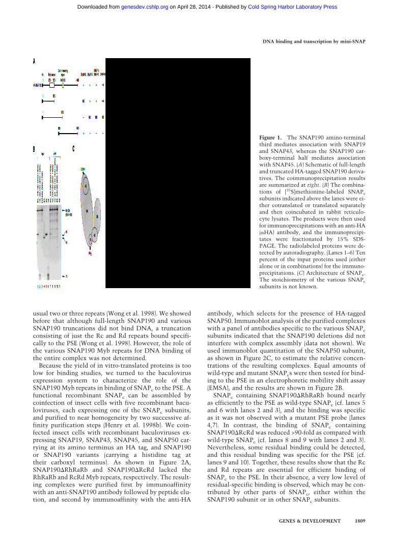
SNAPc is a five-subunit complex consisting of SNAP190,SNAP50, SNAP45, SNAP43, and SNAP19 (Henry et al.1998b). To understand the structural and functional as-pects of the complex, it is important to determine itsarchitecture. Our previous coimmunoprecipitation stud-ies have shown that SNAP190 does not associate withSNAP50 but associates directly with each SNAP19 andSNAP45, and that it associates efficiently with SNAP43in the presence, but not in the absence, of SNAP19(Henry et al. 1998a; Wong et al. 1998). To determinewhich regions of SNAP190 are responsible for these as-sociations, we tested the abilities of in vitro translatedfull-length HA-tagged SNAP190 and the HA-taggedSNAP190 truncations shown in Figure 1A to associatewith (1) SNAP45, (2) SNAP19, (3) SNAP43, and (4)SNAP19 and SNAP43 together, in a coimmunoprecipi-tation assay performed with anti-HA tag monoclonal an-
tibodies (mAb) (Niman et al. 1983). The results of theseexperiments are summarized in Figure 1A; as an exampleof the data, coimmunoprecipitations with the HA–SNAP190(800–1469) truncation are shown in Figure 1B.
With full-length HA–SNAP190, we observed, consis-tent with our previous results (Henry et al. 1998a; Wonget al. 1998), efficient coimmunoprecipitation of SNAP45as well as SNAP19, and efficient coimmunoprecipitationof SNAP43 in the presence, but not in the absence, ofSNAP19 (Fig. 1A; data not shown). With the HA–SNAP190(1–505) truncation, which contains the amino-terminal sequences of SNAP190 to amino acid 505, wealso observed efficient coimmunoprecipitation of SNAP19and efficient coimmunoprecipitation of SNAP43 in thepresence, but not in the absence, of SNAP19. It is worthnoting, however, that we could observe a low level ofSNAP43 coimmunoprecipitate with both HA–SNAP190and the HA–SNAP190(1–505) truncation even in the ab-sence of SNAP19 (see Fig. 1A), which was not detectedin our previous experiments (Henry et al. 1998b). Wedid not test whether the HA–SNAP190(1–505) trunca-tion coimmunoprecipitated with SNAP45, but the largertruncation SNAP190(1–900) did not coimmunoprecipitatewith SNAP45. Thus, SNAP19 and SNAP43, but notSNAP45, associate with the amino-terminal part ofSNAP190.
When we tested the HA–SNAP190(800–1469) trunca-tion, which contains the carboxy-terminal half of theprotein, we obtained different results, as shown in Figure1B. With this SNAP190 derivative, we did not observecoimmunoprecipitation of SNAP19, SNAP43 togetherwith SNAP19, or SNAP43 alone (lanes 8–10), but we didobserve coimmunoprecipitation of SNAP45 (lane 7). Asexpected, the anti-HA antibody did not cross-react de-tectably with untagged SNAP45 (lane 11). Thus,SNAP19 and SNAP43 associate only with the amino-terminal part of SNAP190, and SNAP45 associates onlywith the carboxy-terminal part of SNAP190.
Figure 1C shows a schematic of SNAPc indicating theprotein–protein interactions between SNAPc subunitsrobust enough to be detected in coimmunoprecipitationassays of in vitro-translated proteins. As shown above,SNAP45 interacts directly with the carboxy-terminalhalf of SNAP190, whereas SNAP19 interacts directlywith the amino-terminal third of SNAP190. SNAP43joins the complex through interactions with bothSNAP19 and SNAP190. SNAP50 can only be coimmu-noprecipitated with SNAP43, suggesting that it joins thecomplex through direct interaction with SNAP43 (Henryet al. 1998a). The stoichiometry of the individual sub-units in SNAPc is not known.
The Rc and Rd repeats of SNAP190 are essentialfor efficient binding of SNAPc to the PSE
As shown in Figure 2A, the largest subunit of SNAPc,SNAP190, contains an unusual Myb domain consistingof four repeats designated Ra, Rb, Rc, and Rd, precededby a half repeat designated Rh, rather than the more
Mittal et al.
1808 GENES & DEVELOPMENT
Cold Spring Harbor Laboratory Press on April 28, 2014 - Published by genesdev.cshlp.orgDownloaded from
usual two or three repeats (Wong et al. 1998). We showedbefore that although full-length SNAP190 and variousSNAP190 truncations did not bind DNA, a truncationconsisting of just the Rc and Rd repeats bound specifi-cally to the PSE (Wong et al. 1998). However, the role ofthe various SNAP190 Myb repeats for DNA binding ofthe entire complex was not determined.
Because the yield of in vitro-translated proteins is toolow for binding studies, we turned to the baculovirusexpression system to characterize the role of theSNAP190 Myb repeats in binding of SNAPc to the PSE. Afunctional recombinant SNAPc can be assembled bycoinfection of insect cells with five recombinant bacu-loviruses, each expressing one of the SNAPc subunits,and purified to near homogeneity by two successive af-finity purification steps (Henry et al. 1998b). We coin-fected insect cells with recombinant baculoviruses ex-pressing SNAP19, SNAP43, SNAP45, and SNAP50 car-rying at its amino terminus an HA tag, and SNAP190or SNAP190 variants (carrying a histidine tag attheir carboxyl terminus). As shown in Figure 2A,SNAP190DRhRaRb and SNAP190DRcRd lacked theRhRaRb and RcRd Myb repeats, respectively. The result-ing complexes were purified first by immunoaffinitywith an anti-SNAP190 antibody followed by peptide elu-tion, and second by immunoaffinity with the anti-HA
antibody, which selects for the presence of HA-taggedSNAP50. Immunoblot analysis of the purified complexeswith a panel of antibodies specific to the various SNAPc
subunits indicated that the SNAP190 deletions did notinterfere with complex assembly (data not shown). Weused immunoblot quantitation of the SNAP50 subunit,as shown in Figure 2C, to estimate the relative concen-trations of the resulting complexes. Equal amounts ofwild-type and mutant SNAPcs were then tested for bind-ing to the PSE in an electrophoretic mobility shift assay(EMSA), and the results are shown in Figure 2B.
SNAPc containing SNAP190DRhRaRb bound nearlyas efficiently to the PSE as wild-type SNAPc (cf. lanes 5and 6 with lanes 2 and 3), and the binding was specificas it was not observed with a mutant PSE probe (lanes4,7). In contrast, the binding of SNAPc containingSNAP190DRcRd was reduced >90-fold as compared withwild-type SNAPc (cf. lanes 8 and 9 with lanes 2 and 3).Nevertheless, some residual binding could be detected,and this residual binding was specific for the PSE (cf.lanes 9 and 10). Together, these results show that the Rcand Rd repeats are essential for efficient binding ofSNAPc to the PSE. In their absence, a very low level ofresidual-specific binding is observed, which may be con-tributed by other parts of SNAPc, either within theSNAP190 subunit or in other SNAPc subunits.
Figure 1. The SNAP190 amino-terminalthird mediates association with SNAP19and SNAP43, whereas the SNAP190 car-boxy-terminal half mediates associationwith SNAP45. (A) Schematic of full-lengthand truncated HA-tagged SNAP190 deriva-tives. The coimmunoprecipitation resultsare summarized at right. (B) The combina-tions of [35S]methionine-labeled SNAPc
subunits indicated above the lanes were ei-ther cotranslated or translated separatelyand then coincubated in rabbit reticulo-cyte lysates. The products were then usedfor immunoprecipitations with an anti-HA(aHA) antibody, and the immunoprecipi-tates were fractionated by 15% SDS-PAGE. The radiolabeled proteins were de-tected by autoradiography. (Lanes 1–6) Tenpercent of the input proteins used (eitheralone or in combinations) for the immuno-precipitations. (C) Architecture of SNAPc.The stoichiometry of the various SNAPc
subunits is not known.
DNA binding and transcription by mini-SNAP
GENES & DEVELOPMENT 1809
Cold Spring Harbor Laboratory Press on April 28, 2014 - Published by genesdev.cshlp.orgDownloaded from

Partial SNAPcs missing SNAP45, SNAP19, or bothSNAP19 and SNAP45 are capable of bindingto the PSE
Having established the crucial role of SNAP190, andmore specifically the Rc and Rd repeats of SNAP190, forspecific binding to the PSE, we turned our attention tothe other SNAPc subunits. We coinfected insect cellswith viruses expressing all possible combinations of fourof the five SNAPc subunits, as well as one combinationof three SNAPc subunits. SNAP190 carried a histidinetag at its carboxyl terminus and SNAP50 carried an HAtag at its amino terminus. We devised purificationschemes that select for the various subcomplexes andeliminate unassociated subunits. These, as well as thestructures of the purified complexes, are summarized inFigure 3A. For each infection, all the SNAPc subunitspresent in the insect cells are shown, with the subunitsthat assembled into the purified complexes in gray, andthose that were not incorporated into the complexes inwhite. All complexes except those assembled in the ab-sence of SNAP190 were purified first on nickel agarosebeads, which select for the presence of the His-taggedSNAP190. The full complex and complexes assembled inthe absence of SNAP45, or SNAP19, or both SNAP45and SNAP19, were further purified on anti-HA mAbbeads, which select for the presence of HA-taggedSNAP50. Complexes assembled in the absence ofSNAP43 or SNAP50 were not expected to containSNAP50 (see Fig. 1C and 3A) and were, therefore, notpurified further. Complexes assembled in the absence ofSNAP190 were purified over anti-HA mAb beads andcontained, therefore, HA-tagged SNAP50 and the associ-ated SNAP43. The composition of all complexes wasthen confirmed by immunoblots with antibodies di-rected against each of the SNAPc subunits, and the re-sults obtained with the anti-SNAP50 antibody areshown in Figure 3C: Complete SNAPc and complexesassembled in the absence of SNAP45, SNAP19, bothSNAP45 and SNAP19, and SNAP190, all containedSNAP50, whereas complexes assembled in the absenceof SNAP43 or SNAP50 did not, as expected.
Equal amounts of purified complete SNAPc and partialSNAPcs as determined, where possible, by immunoblotquantitation of SNAP50 (see Fig. 3C) or, in the case ofpartial complexes lacking SNAP50, by quantitation ofSNAP190 (data not shown), were tested by EMSA fortheir ability to bind to the PSE, and the results are shownin Figure 3B. Complete SNAPc bound efficiently to thePSE, as expected (lanes 1,2). Partial SNAPcs assembled inthe absence of SNAP43, SNAP50, or SNAP190 did notdisplay any detectable DNA-binding activity (lanes9–11). In contrast, a partial SNAPc assembled in the ab-sence of SNAP45 formed a weak diffuse complex migrat-ing faster than the complete SNAPc–PSE complex (lanes3,4). Remarkably, a partial SNAPc assembled in the ab-sence of SNAP19 also formed a weak complex migratingat an intermediate position between the completeSNAPc–PSE and the SNAPc–45–PSE complexes, consis-tent with an intermediate molecular weight for this
Figure 2. The SNAP190 Rc and Rd Myb repeats are requiredfor efficient SNAPc binding to the PSE. (A) Schematic structureof SNAP190, SNAP190DRhRaRb, and SNAP190DRcRd. The lo-cation of the Myb domain is indicated, as well as the location ofthe region that interacts with the Oct-1 POU domain (Ford et al.1998). The regions deleted in SNAP190DRhRaRb andSNAP190DRcRd are indicated. (B) EMSA performed withSNAPc or the SNAPc derivatives shown above the lanes. Theprobe contained either a wild-type (Wt) or mutant (Mu) mouseU6 PSE, as indicated above the lanes. We used equivalentamounts of SNAPc or derivatives as determined by immunoblot(see Fig. 2C) in the binding reactions, specifically: (lanes 2–4) 1,2, and 2 µl of SNAPc; (lanes 5–7), 3, 6, and 6 µl of SNAPc con-taining the SNAP190DRhRaRb subunit; (lanes 8–10) 3, 6, and 6µl of SNAPc containing the SNAP190DRcRd subunit. (C) Im-munoblot analysis of SNAPc and the SNAPc derivatives indi-cated above the lanes with an anti-SNAP50 antibody. (Lane 1)uninfected Sf9 cell lysate; (lane 2) lysate from Sf9 cells infectedwith an HA–SNAP50-expressing recombinant baculovirus;(lanes 3–5) 0.17, 0.5, and 1 µl of complete SNAPc; and (lanes 6–8)0.3, 1, and 3 µl of SNAPc containing the SNAP190DRhRaRbsubunit; (lanes 9–11) or the SNAP190DRcRd subunit. SNAPc
and the SNAPc derivatives were purified by sequential anti-SNAP190 and anti-HA immunoaffinity steps.
Mittal et al.
1810 GENES & DEVELOPMENT
Cold Spring Harbor Laboratory Press on April 28, 2014 - Published by genesdev.cshlp.orgDownloaded from

complex lacking just SNAP19 (lanes 5,6). A complex as-sembled in the absence of both SNAP45 and SNAP19formed a weak, diffuse complex migrating, as expectedfaster than the SNAPc–45–PSE complex (lanes 7,8). Noneof the protein complexes bound to a probe containing amutated PSE (data not shown).
Together, these results indicate that complexes miss-ing SNAP19, SNAP45, or both, are still capable of bind-ing, albeit weakly, to the PSE. In contrast, complexesmissing SNAP43, SNAP50, or SNAP190 did not binddetectably to the PSE. Thus, consistent with our previ-ous observations that none of the full-length SNAPc sub-units can bind to DNA on its own, a combination ofat least three of the five SNAPc subunits, namely
SNAP190, SNAP50, and SNAP43, is necessary to gener-ate detectable DNA-binding ability. Because binding ofthe complexes assembled in the absence of SNAP19 and/or SNAP45 was not as efficient as binding of completeSNAPc, these data also suggest that SNAP19 andSNAP45 contribute, directly or indirectly, to efficientbinding of SNAPc to the PSE.
The observation that we could assemble a complexmissing SNAP19 in baculovirus-infected cells was sur-prising because SNAP19 is required for efficient associa-tion of SNAP43 and SNAP190 in a coimmunoprecipita-tion assay of in vitro-translated proteins (Fig. 1; Henry etal. 1998b). We therefore checked, and could confirm, thesubunit composition of the complex assembled in the
Figure 3. DNA-binding properties of partial SNAPcs (A) Structure of the various partial SNAPcs used for binding studies. Insect cellswere coinfected with four or, in one case, three baculoviruses, each expressing a different SNAPc subunit. In each panel, the subunitsmissing in the infection as well as the steps used to purify the complex are listed (lower left). All subunits present in the insect cellsare illustrated at right, with the subunits present in the purified complex shaded. The SNAP190 subunit carries a His tag (HT) at itscarboxyl terminus, and the SNAP50 subunit carries an HA tag (HA) at its amino terminus. (B) EMSA performed with either completeSNAPc or SNAPcs assembled in the absence of the subunits indicated above the lanes and a probe carrying the high affinity mouse U6PSE. The structures of the various SNAPcs are illustrated in A. We used equivalent amounts of the various complexes as determinedby immunoblot (see Fig. 3C) in the binding reactions. (Lanes 1,2) 1 and 2 µl of complete SNAPc; (lanes 3,4) 4 and 8 µl of SNAPc–SNAP45; (lanes 5,6) SNAPc–SNAP19; (lanes 7,8) SNAPc–SNAP19–SNAP45; (lane 9) 2 µl each of SNAPc–SNAP43; (lane 10) SNAPc–SNAP50; and (lane 11) SNAPc–SNAP190. (C) Immunoblot analysis of complete SNAPc and the partial SNAPcs indicated above thelanes with an anti-SNAP50 antibody. The samples shown are as follows: (Lane 1) uninfected Sf9 cell lysate; (lane 2) lysate from Sf9cells infected with an HA–SNAP50-expressing recombinant baculovirus; (lanes 3–5) 0.17, 0.5, and 1 µl of complete SNAPc; (lanes 6–8)1, 5, and 10 µl of SNAPc–SNAP45; (lanes 9–11) SNAPc–SNAP19; or (lanes 12–14) SNAPc–SNAP19–SNAP45; and (lane 15) 5 µl ofSNAPc–SNAP43; (lane 16) SNAPc–SNAP50; or (lane 17) SNAPc–SNAP190.
DNA binding and transcription by mini-SNAP
GENES & DEVELOPMENT 1811
Cold Spring Harbor Laboratory Press on April 28, 2014 - Published by genesdev.cshlp.orgDownloaded from

absence of SNAP19 by antibody supershift experiments(data not shown). These results suggest that at highSNAPc subunit concentration, as is likely to be the casein baculovirus-infected cells, a SNAPc missing justSNAP19 can be assembled. As described above, we doobserve a low level of association between in-vitro trans-lated SNAP43 and SNAP190 or the amino-terminal thirdof SNAP190, even in the absence of SNAP19. It is alsopossible, however, that an insect homolog of SNAP19that is not recognized by our antibodies gets incorpo-rated into these complexes. We do not consider thislikely, however, because the SNAPc–SNAP19 complexmigrates significantly faster in the EMSA than theSNAPc complex, suggesting that it is missing a subunit.
SNAP45 relieves a binding inhibition conferredby the carboxy-terminal region of SNAP190
SNAPcs assembled in the absence of SNAP45 formedweak and diffuse complexes with the PSE in an EMSA,
suggesting that these SNAPcs did not bind efficiently tothe PSE. To address the role of SNAP45 in SNAPc bind-ing, we tested the effect of adding increasing amounts ofrecombinant SNAP45 expressed in Escherichia coli to apartial SNAPc assembled in the absence of SNAP45. Asillustrated in Figure 4A, addition of recombinantSNAP45 had no effect on the complete SNAPc–PSE com-plex (cf. lanes 7 and 8). In contrast, however, addition ofincreasing amounts of SNAP45 to the complex as-sembled in the absence of SNAP45 resulted in a moreintense and more discrete band, which comigrated withthe complete SNAPc–PSE complex (lanes 4–6). This re-sult suggests that exogenous SNAP45 produced in E. coliwas incorporated into the partial SNAPc lackingSNAP45 and stabilized binding to the PSE.
To investigate the mechanism through whichSNAP45 stabilizes binding to the PSE, we compared theDNA-binding abilities of equivalent amounts (as deter-mined by quantitation of the SNAP50 subunit by immu-noblot; data not shown) of SNAPcs assembled in the ab-sence of SNAP45 and containing either an intact
Figure 4. In the absence of SNAP45, the carboxy-terminal region of SNAP190 inhibits binding ofSNAPc to the PSE. (A) EMSA performed with aprobe carrying the wild-type mouse U6 PSE andeither no proteins (lane 1), increasing amounts of E.coli-expressed SNAP45 (lanes 2,3), SNAPc–SNAP45 alone (lane 4), SNAPc–SNAP45 and in-creasing amounts of E. coli-expressed SNAP45(lanes 5,6), SNAPc alone (lane 7), or SNAPc and anamount of E. coli expressed SNAP45 equivalent tothat used in lane 5. (B) EMSA performed withprobes carrying either a wild-type (lanes 1–3,5,6) ora mutant (lanes 4,7) mouse U6 PSE and either noprotein (lane 1), or equivalent amounts, as deter-mined by immunoblot of the SNAP50 subunit (notshown) of SNAPc–SNAP45 containing either a full-length SNAP190 subunit (lanes 2–4) or a SNAP190subunit missing the carboxy-terminal two thirds ofthe protein (lanes 5–7). (C) Structure of full-lengthSNAP190 and SNAP190DC. SNAP190DC is miss-ing amino acids 515–1469 of SNAP190.
Mittal et al.
1812 GENES & DEVELOPMENT
Cold Spring Harbor Laboratory Press on April 28, 2014 - Published by genesdev.cshlp.orgDownloaded from

SNAP190 or a SNAP190 missing the carboxy-terminaltwo-thirds of the protein, as illustrated in Figure 4C. Theresults are shown in Figure 4B. As before, a SNAPc as-sembled in the absence of SNAP45 gave rise to a weakand diffuse complex that bound specifically to the PSE(lanes 2–4). In stark contrast, a SNAPc assembled in theabsence of SNAP45 and containing only the amino-ter-minal third of SNAP190 gave rise to a prominent anddiscrete complex that also bound specifically to the PSE(lanes 5–7). These results show that a complex missingthe carboxy-terminal region of SNAP190 and its associ-ated subunit, SNAP45, binds efficiently to the PSE. Theyare consistent with the idea that in the SNAPc–SNAP45complex, the carboxy-terminal region of SNAPc adopts aflexible conformation that diminishes the ability of thecomplex to bind to DNA. Addition of SNAP45 engagesthe carboxy-terminal region of SNAP190 in protein–pro-tein interactions, thus giving it a fixed structure and re-lieving the inhibition.
A mini-SNAPc consisting of the amino-terminal thirdof SNAP190, SNAP43, and SNAP50 is capableof specific and efficient binding to the PSE
We have shown above that in the absence of the carboxy-terminal region of SNAP190, SNAP45 is dispensable forefficient binding to the PSE (Fig. 4), and that a PSE-bind-ing SNAPc can be assembled in the absence of SNAP19(Fig. 3). In an attempt to define the minimal number ofSNAPc subunits required for binding, we asked whetherwe could assemble a mini-SNAPc missing SNAP45,SNAP19, and the carboxy-terminal two-thirds ofSNAP190. We coinfected insect cells with recombinantviruses encoding the amino-terminal third of SNAP190carrying a histidine tag, SNAP43, and HA-taggedSNAP50, and purified the complex first by either immu-noaffinity with an anti-SNAP190 antibody or on a nickelagarose column, and second by immunoaffinity with ananti-HA antibody. The subunit composition of the puri-fied complex was determined by fractionation on a SDS–polyacrylamide gel and silver staining, and is shown inFigure 5A, lanes 5 and 6. Lanes 1–4 correspond to anautoradiogram of marker lanes in the same gel loadedwith a mixture of in vitro-translated SNAP43, HA-tagged SNAP50, and an HA-tagged amino-terminal trun-cation of SNAP190 (lane 1), or each of these in vitro-translated proteins separately (lanes 2–4). Only threemain bands are visible in the mini-SNAPc preparation,even when the gel is overloaded (lane 6), of which theone corresponding to HA-tagged SNAP50 comigratesprecisely with the in vitro-translated protein. In con-trast, the in vitro-translated SNAP190 truncation and invitro-translated SNAP43 both migrate slightly moreslowly than the proteins expressed in insect cells. Thismay be due to differences in protein modifications or, inthe case of SNAP190, to the different tags and theslightly different lengths of the constructs.
To determine whether mini-SNAPc was able to bindspecifically to the PSE and confirm its subunit compo-sition, we performed the EMSA shown in Figure 5B. To
facilitate comparison of the two complexes, the amountsof SNAPc and mini-SNAPc were adjusted so as to obtainequivalent amounts of PSE-binding activity. The mobil-ity of the SNAPc–PSE complex was retarded by additionof antibodies directed against peptides from the amino-and carboxy-terminal regions of SNAP190, SNAP50,SNAP45, SNAP43, and SNAP19, as expected (lanes 4–9).In contrast, the mobility of the mini-SNAPc–PSE com-plex was affected by antibodies directed against a peptidefrom the amino-terminal region of SNAP190, SNAP50,and SNAP43, but not by antibodies directed against apeptide from the carboxy-terminal region of SNAP190,SNAP45, and SNAP19 (lanes 12–17). Thus, a mini-SNAPc containing the amino-terminal third of SNAP190,SNAP43, and SNAP50, is capable of binding specificallyand efficiently to the PSE.
Mini-SNAPc binds cooperatively with full-lengthhuman TBP on the U6 promoter
We have shown previously that SNAPc and human TBP(hTBP) bind cooperatively to their respective bindingsites on the U6 promoter, and that this effect is depen-dent on the nonconserved amino-terminal domain ofhTBP (Mittal and Hernandez 1997). On a probe contain-ing a high affinity PSE such as the mouse U6 PSE, coop-erative binding results, in effect, in SNAPc recruitinghTBP to the TATA box (Mittal and Hernandez 1997).Which subunits of SNAPc are required for this effect hasnot been determined. Therefore, we were interested indetermining whether mini-SNAPc, like SNAPc, is ca-pable of recruiting hTBP to the U6 TATA box. For thispurpose, we used a DNase I footprinting assay, and theresults are shown in Figure 6. Mini-SNAPc, on its own,efficiently protected the PSE from DNase I digestion(lane 2), and the footprint is very similar to that obtainedwith SNAPc (Mittal and Hernandez 1997). In contrast tothe efficient PSE protection obtained with mini-SNAPc,hTBP on its own protected the TATA box only at thehigher protein concentration, and the protection wasweak (lanes 3,4). Thus, as observed previously (Mittaland Hernandez 1997), under these conditions hTBP didnot bind efficiently to the TATA box. When both mini-SNAPc and hTBP were incubated with the probe, how-ever, TBP was recruited to the TATA box much moreefficiently as evidenced by a partial TATA box protec-tion (lanes 5,6) even at the lower concentration of TBP(lane 5). The efficiency of TBP recruitment was similarto that observed with SNAPc (data not shown). Thus,like SNAPc, mini-SNAPc is capable of recruiting TBP tothe TATA box, indicating that the carboxy-terminal twothirds of SNAP190, SNAP45, and SNAP19 are all dis-pensable for this effect.
Mini-SNAPc is functional for basal transcriptionby both RNA Pol II and RNA Pol III
We have shown before that SNAPc is capable of directingtranscription from both RNA Pol II and Pol III snRNA
DNA binding and transcription by mini-SNAP
GENES & DEVELOPMENT 1813
Cold Spring Harbor Laboratory Press on April 28, 2014 - Published by genesdev.cshlp.orgDownloaded from

promoters (Henry et al. 1998b). Because mini-SNAPc iscapable of binding specifically to the PSE and recruitingTBP to the TATA box, we tested whether it might befunctional for basal transcription. For this purpose, wetreated HeLa cell extracts with protein A agarose beadscross-linked to preimmune antibodies or to three anti-bodies directed against the SNAP19, SNAP45, andSNAP190 subunits of SNAPc, respectively. Such a treat-ment was expected to deplete endogenous SNAPc as wellas any free SNAP19, SNAP45, or SNAP190 subunit thatmight be present in the extracts. As shown in Figure 7B,both SNAP45 and SNAP19, which were directly recog-nized by the antibodies, and SNAP50, which was recog-nized by virtue of its association with other SNAPc sub-units, were efficiently depleted. We then tested the abili-ties of equivalent amounts of SNAPc and mini-SNAPc,as determined by immunoblot quantitation of both the
SNAP190 and SNAP50 subunits (data not shown), to re-store transcription activity to these extracts, and the re-sults are shown in Figure 7A. As templates, we usedRNA Pol II and Pol III snRNA promoters that carry thehigh affinity mouse U6 PSE and lack an octamer se-quence upstream, and thus score basal transcription. Ex-tracts depleted with preimmune antibodies directedtranscription from both an RNA Pol III and an RNA PolII snRNA promoter, whereas extracts depleted with theanti-SNAPc antibodies had little or no activity (cf. lanes1 and 2). Addition of increasing amounts of recombinantSNAPc restored both RNA Pol III and Pol II transcription(lanes 3–5), as observed before (Henry et al. 1998b). Strik-ingly, addition of equivalent increasing amounts of mini-SNAPc also restored both RNA Pol III and Pol II tran-scription (lanes 6–8). In contrast, complexes assembledin the absence of SNAP43 or SNAP50 were inactive
Figure 5. The amino-terminal parts of SNAP190 (amino acids 1–514), SNAP50, and SNAP43 are sufficient for assembly of a mini-SNAPc. (A) Composition of mini-SNAPc. A mixture of in vitro-translated HA-tagged SNAP190 (1–505), HA-tagged SNAP50, andSNAP43 (lane 1) or SNAP43 (lane 2), HA-tagged SNAP50 (lane 3), and HA-tagged SNAP190 (1–505) (lane 4) alone, was loaded alongsideincreasing amounts of purified mini-SNAPc (lanes 5,6). The gel was stained with silver, dried, and photographed (lanes 5,6). The driedgel was then autoradiographed to reveal the location of the radiolabeled in vitro-translated proteins (lanes 1–4). Mini-SNAPc waspurified by chromatography over nickel–agarose beads, which selects for the presence of the His-tagged SNAP190 subunit, followedby chromatography over anti-HA antibody beads, which selects for the presence of the HA-tagged SNAP50 subunit. (B) EMSAperformed with a probe carrying either the wild-type (lanes 1,2,4–10,12–17) or mutant (lanes 3,11) mouse U6 PSE and either no protein(lane 1) or equivalent DNA-binding units of complete SNAPc (lanes 2–9) or mini-SNAPc (lanes 10–17). (Lanes 4–9,12–13,15–17),anti-peptide polyclonal antibodies directed either against the amino-terminal region of SNAP190 (aSNAP190 N-ter, CS696), thecarboxy-terminal region of SNAP190 (aSNAP190 C-ter, CS402) or other SNAPc subunits as indicated above the lanes were added tothe binding reactions. (Lane 14) The mAb 12CA5 directed against the HA tag on SNAP50 was used. The locations of the SNAPc–PSE,SNAPc–Ab–PSE, mini-SNAPc–PSE, and mini-SNAPc–Ab–PSE complexes are indicated. SNAPc and mini-SNAPc were purified first overnickel–agarose beads and anti-SNAP190 antibody beads, respectively, which selects for the presence of the His-tagged SNAP190subunit. Both were then purified over anti-HA antibody beads, which selects for the presence of the HA-tagged SNAP50 subunit.
Mittal et al.
1814 GENES & DEVELOPMENT
Cold Spring Harbor Laboratory Press on April 28, 2014 - Published by genesdev.cshlp.orgDownloaded from

(lanes 9–12). These data indicate that on naked DNAtemplates, mini-SNAPc is capable of nucleating the as-sembly of RNA Pol II and Pol III transcription initiationcomplexes and that SNAP45, SNAP19, and the carboxy-terminal two-thirds of SNAP190 are all dispensable forthese functions.
Mini-SNAPc binds efficiently to the PSE independentof the Oct-1 POU domain
We and others have shown previously that on probescontaining both a PSE and an octamer sequence, SNAPc
and Oct-1 POU bind cooperatively, such that the Oct-1POU domain in effect recruits SNAPc to the PSE (Mur-phy et al. 1992; Mittal et al. 1996). Cooperative bindingresults, at least in part, from a direct protein–proteininteraction involving the Oct-1 POUs domain as well asa small region within the carboxy-terminal part ofSNAP190 (Mittal et al. 1996; Ford et al. 1998). Becausemini-SNAPc is missing the entire carboxy-terminal two-
thirds of SNAP190, it seemed likely that mini-SNAPc
would be unable to bind cooperatively with Oct-1 POU.To test this directly, we used a probe containing the H2Boctamer, a high affinity Oct-1-binding site, upstream ofthe human U6 PSE, a weak affinity binding site forSNAPc (Mittal et al. 1996).
Figure 7. Mini-SNAPc is capable of directing basal transcrip-tion by both RNA Pol II and Pol III. (A) HeLa cell extracts weredepleted with either rabbit preimmune antibody beads (lane 1),or a mixture of beads carrying anti-peptide antibodies directedagainst SNAP19, SNAP45, and SNAP190 (lanes 2–12) and testedfor their ability to support RNA Pol III transcription from a U6promoter carrying the mouse U6 PSE (top) and RNA Pol II tran-scription from a U1 promoter carrying the mouse U6 PSE (bot-tom; Sadowski et al. 1993). The transcription reactions weresupplemented with 1, 3, and 6 µl of SNAPc (lanes 3–5), equiva-lent increasing amounts of mini-SNAPc (lanes 6–8), and 2 µl ofSNAPc (lanes 9,10), or mini-SNAPc (lanes 11,12) assembled inthe absence of the SNAP43 or SNAP50 subunit, as indicatedabove the lanes. The purification of SNAPc and mini-SNAPc issummarized in the legend to Fig. 5B, and the amounts of thesecomplexes were equalized by immunoblot quantitation of theSNAP190 and SNAP50 subunits (not shown). Complexes miss-ing either the SNAP43 or SNAP50 subunits were purified on ananti-SNAP190 antibody column, and their amounts were equal-ized by immunoblot quantitation of the SNAP190 subunit. (B)Immunoblot analysis of the extracts depleted with either pre-immune (lane 1) or anti-SNAP19, anti-SNAP45, and anti-SNAP190 beads (lane 2) used in A. The top, middle, and bottompanels were probed with antibodies directed against SNAP50,SNAP45, and SNAP19, respectively. The positions of SNAP50,SNAP45, and SNAP19 are indicated at left.
Figure 6. Mini-SNAPc can recruit TBP to the TATA box. DN-ase I footprinting experiment performed with a probe containingthe mouse U6 PSE and the human U6 TATA box. The bindingreactions contained probe alone (lane 1), mini-SNAPc alone(lane 2), 40 and 80 ng of full-length human TBP (hTBP) alone(lanes 3,4), and 40 and 80 ng of hTBP together with mini-SNAPc
(lanes 5,6). The positions of the PSE and the TATA box areindicated at right.
DNA binding and transcription by mini-SNAP
GENES & DEVELOPMENT 1815
Cold Spring Harbor Laboratory Press on April 28, 2014 - Published by genesdev.cshlp.orgDownloaded from
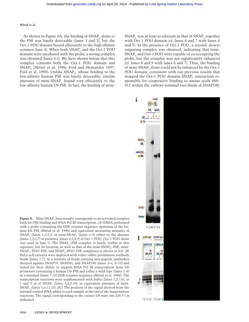
As shown in Figure 8A, the binding of SNAPc alone tothe PSE was barely detectable (lanes 1 and 2), but theOct-1 POU domain bound efficiently to the high affinityoctamer (lane 3). When both SNAPc and the Oct-1 POUdomain were incubated with the probe, a strong complexwas obtained (lanes 4,5). We have shown before that thiscomplex contains both the Oct-1 POU domain andSNAPc (Mittal et al. 1996; Ford and Hernandez 1997;Ford et al. 1998). Unlike SNAPc, whose binding to thelow-affinity human PSE was barely detectable, similaramounts of mini-SNAPc bound very efficiently to thelow-affinity human U6 PSE. In fact, the binding of mini-
SNAPc was at least as efficient as that of SNAPc togetherwith Oct-1 POU domain (cf. lanes 6 and 7 with lanes 4and 5). In the presence of Oct-1 POU, a second, slowermigrating complex was obtained, indicating that mini-SNAPc and Oct-1 POU were capable of co-occupying theprobe, but the complex was not significantly enhanced(cf. lanes 8 and 9 with lanes 6 and 7). Thus, the bindingof mini-SNAPc alone could not be enhanced by the Oct-1POU domain, consistent with our previous results thatmapped the Oct-1 POU domain–SNAPc interaction re-sponsible for cooperative binding to amino acids 869–912 within the carboxy-terminal two-thirds of SNAP190,
Figure 8. Mini-SNAPc functionally corresponds to an activated complexboth for PSE-binding and RNA Pol III transcription. (A) EMSA performedwith a probe containing the H2B octamer sequence upstream of the hu-man U6 PSE (Mittal et al. 1996) and equivalent increasing amounts ofSNAPc (lanes 1,2,4,5) or mini-SNAPc (lanes 6–9) either in the absence(lanes 1,2,6,7) or presence (lanes 4,5,8,9) of Oct-1 POU. Oct-1 POU alonewas used in lane 3. The SNAPc–PSE complex is barely visible at thisexposure, but its location, as well as that of the mini-SNAPc–PSE, mini-SNAPc–POU–PSE, and SNAPc–POU–PSE complexes is shown at left. (B)HeLa cell extracts were depleted with either rabbit preimmune antibodybeads (lanes 1,7), or a mixture of beads carrying anti-peptide antibodiesdirected against SNAP19, SNAP45, and SNAP190 (lanes 2–6, 8–12) andtested for their ability to support RNA Pol III transcription from U6promoters containing a human U6 PSE and either a wild-type (lanes 1–6)or a mutated (lanes 7–12) H2B octamer sequence (Mittal et al. 1996). Thetranscription reactions were supplemented with buffer (lanes 1,2,7,8), or1 and 3 µl of SNAPc (lanes 3,4,9,10), or equivalent amounts of mini-SNAPc (lanes 5,6,11,12). (IC) The position of the signal derived from theinternal control RNA added to each sample at the end of the transcriptionreactions. The signal corresponding to the correct U6 start site (U6 58) isindicated.
Mittal et al.
1816 GENES & DEVELOPMENT
Cold Spring Harbor Laboratory Press on April 28, 2014 - Published by genesdev.cshlp.orgDownloaded from

which are absent in mini-SNAPc. The striking result,however, is that mini-SNAPc bound as efficiently to thePSE as SNAPc in the presence of the Oct-1 POU domain.This suggests that the carboxy-terminal domain ofSNAP190 down-regulates binding of the complex to thePSE. This down-regulation is counteracted through in-teraction of the carboxy-terminal domain of SNAP190with the Oct-1 POU domain.
Mini-SNAPc is activated for RNA Pol III snRNAgene transcription
The observation that mini-SNAPc bound as efficiently tothe PSE as SNAPc in the presence of Oct-1 POUprompted us to compare the efficiency of transcriptiondirected by these two complexes, either in the absence(basal transcription) or presence (activated transcription)of an octamer sequence. For this purpose, we depleted anuclear extract of endogenous SNAPc as above, with an-tibodies directed against SNAP19, SNAP45, andSNAP190. We then used as a template an RNA Pol IIIsnRNA promoter containing either a wild-type(OCTAH2B–PSEWt) or a mutated (OCTAmutant–PSEWt)octamer site upstream of the low-affinity human U6PSE. We also included in the transcription reactions anRNA fragment to serve as an internal control (IC) forRNA handling and recovery. As shown in Figure 8B, thetemplate containing a wild-type H2B octamer was moreactive than that containing a mutant octamer sequence(cf. lane 1 with lane 7), indicating that as in vivo (seeLobo and Hernandez 1994), and as observed before (Mur-phy et al. 1992; Mittal et al. 1996), the octamer activatestranscription in this assay. This activation of transcrip-tion is dependent on the Oct-1 POU domain (Murphy etal. 1992; Mittal et al. 1996). When the extract was de-pleted of endogenous SNAPc, both activated and basaltranscription were reduced (lanes 2,8), albeit in the caseof basal transcription, this decrease was barely detect-able, mainly because the starting level of basal transcrip-tion was very low. On addition of increasing amounts ofSNAPc, the template with a wild-type octamer directedhigher levels of transcription than the template with amutated octamer, indicating that in the presence of theoctamer sequence, transcription was activated (cf. lanes3 and 4 with lanes 9 and 10). In sharp contrast, on addi-tion of increasing amounts of mini-SNAPc, both tem-plates directed similar levels of transcription (cf. lanes 5and 6 with lanes 11 and 12), and strikingly, these levelswere as high as those obtained with SNAPc on the tem-plate containing a wild-type octamer (cf. lanes 5,6,11,and 12 with lanes 3 and 4). Thus, mini-SNAPc function-ally corresponds to an activated SNAPc for RNA Pol IIIsnRNA gene transcription in vitro.
Discussion
We have dissected the role of the various SNAPc sub-units for a number of SNAPc functions including specificbinding to the PSE, cooperative binding with TBP and
with the Oct-1 POU domain, and basal and activatedtranscription. Our results show that a mini-SNAPc con-sisting of SNAP43, SNAP50, and the amino-terminalthird of SNAP190 is capable of binding to the PSE, ofrecruiting TBP to the TATA box, and of directing RNAPol II and Pol III snRNA gene transcription. Mini-SNAPc
binds to the PSE as efficiently as SNAPc together withthe Oct-1 POU domain. Consistent with this observa-tion, mini-SNAPc constitutively directs activated levelsof RNA Pol III transcription in vitro.
Architecture of SNAPc
We find that SNAP45 interacts with the carboxy-termi-nal, and SNAP43 and SNAP19 with the amino-terminalregion of SNAP190. We had shown before that SNAP50interacts only with SNAP43 (Henry et al. 1996, 1998a;Wong et al. 1998). Consistent with these observations,we could assemble partial SNAPcs lacking SNAP45 orSNAP50. More surprisingly, we could also assemble acomplex missing just SNAP19, even though in our pre-vious coimmunoprecipitation experiments with in vitrotranslated proteins, SNAP19 was required for associationof SNAP43 and SNAP190 (Henry et al. 1998b). However,we show here that we can detect a weak association ofSNAP43 and the amino-terminal region of SNAP190even in the absence of SNAP19, suggesting that at higherprotein concentrations than can be achieved in cotrans-lations, SNAP43 is able to associate reasonably effi-ciently with SNAP190 even in the absence of SNAP19.The SNAPc–SNAP19 complex may be less stable thanSNAPc, because it displayed reduced binding to the PSErelative to SNAPc. Alternatively, the reduced bindingmay reflect a direct role of SNAP19 in contacting theDNA.
The SNAPc complex missing just the SNAP45 subunitbound much less efficiently than SNAPc to the PSE, andin an EMSA, the resulting protein–DNA complex wasstrikingly diffuse. We do not think that this reflects dis-sociation of the protein–DNA complex during electro-phoresis, because this would result in a diffuse signalmigrating faster than the expected position of theSNAPc–45–SNAP–PSE complex. Instead, much of thediffuse signal migrated more slowly, suggesting a heter-ogenous population of SNAPc–45–SNAP–PSE com-plexes. Because SNAP45 interacts with the carboxy-ter-minal region of SNAP190, this in turn suggests that inthe absence of SNAP45, the carboxy-terminal region ofSNAP190 assumes more than one conformation, therebygenerating a heterogenous population of SNAPcs. Someof these complexes may not be able to bind DNA, thusthe overall weaker binding observed with the SNAPc–SNAP45 as compared with SNAPc. Addition of exog-enous SNAP45 probably results in incorporation ofSNAP45 into many of the SNAPc–SNAP45 complexes,stabilization of the SNAP190 carboxy-terminal domainconformation, and thus conversion of many of the com-plexes formerly unable to bind DNA into complexes ca-pable of binding. Cleavage of the carboxy-terminal two
DNA binding and transcription by mini-SNAP
GENES & DEVELOPMENT 1817
Cold Spring Harbor Laboratory Press on April 28, 2014 - Published by genesdev.cshlp.orgDownloaded from

thirds of SNAP190, which bypasses the requirement forSNAP45 altogether, also resulted in an increase andtightening of the SNAPc–PSE complex.
Requirements for binding of SNAPc to the PSE
The Myb domains of the c-Myb, A-Myb, and B-Myb pro-teins consist of three repeats called the R1, R2, and R3repeats, of which the R2 and R3 repeats are sufficient forDNA binding (Nomura et al. 1988; Luscher and Eisen-man 1990). SNAP190 has an unusual Myb domain con-sisting of four and a half repeats. We have been able toassemble mutant SNAPcs lacking either the RhRaRb orthe RcRd SNAP190 repeats. In both cases, the complexcould be assembled, indicating that the Myb repeats donot mediate essential protein–protein interactions withother SNAPc subunits. However, only the complex miss-ing the RhRaRb repeats was still able to bind efficientlyto the PSE. The complex missing the RcRd repeatsbound with much reduced efficiency. This is consistentwith our previous observations that (1) a small trunca-tion of SNAP190 consisting of just the RcRd repeatscould bind to the PSE, and (2) of the four SNAP190 re-peats, the Rc and Rd repeats are the most similar to theR2 repeat with 38% identities each, and the Rd repeat isthe most similar to the R3 repeat with 30% identities(Wong et al. 1998). Together, these results emphasize thecrucial role of the Rc and Rd repeats for SNAPc DNAbinding.
It is likely, however, that parts of SNAPc other thanthe SNAP190 Rc and Rd repeats also contribute to DNAbinding. Although a small SNAP190 truncation consist-ing of just the Rc and Rd repeats was able to bind to thePSE, we have been unable to demonstrate binding by anylonger SNAP190 truncation (Wong et al. 1998). Whenusing either full-length SNAP190 or the amino-terminal514 amino acids of SNAP190, we find that the smallestcomplex that still binds DNA contains SNAP43 andSNAP50 in addition to SNAP190. This suggests thatSNAP50 and/or SNAP43 confer to SNAP190, and morespecifically to the amino-terminal region of SNAP190, astructure compatible with DNA binding. In addition, be-cause SNAP50 can be cross-linked to the PSE (Henry etal. 1996), it may provide additional contacts with theDNA.
A mini-SNAPc active for both basal RNA Pol IIand Pol III snRNA gene transcription
The smallest complex we have generated so far that isstill capable of binding to the PSE contains the amino-terminal region of SNAP190, SNAP43, and SNAP50. Be-cause this minicomplex is assembled in insect cells, it isdifficult to exclude that it contains an insect cell sub-unit. We consider this unlikely, however, because silverstaining of purified mini-SNAPc does not reveal any stoi-chiometric component beside the three expected sub-units. This mini-SNAPc was still able to recruit TBP tothe TATA box of an RNA Pol III snRNA promoter. We
have shown previously that recruitment of TBP bySNAPc is dependent on the amino-terminal domain ofTBP, and have speculated that it may occur through adirect protein–protein contact involving the amino-ter-minal domain of TBP and SNAPc (Mittal and Hernandez1997). If this is the case, the target contacted by theamino-terminal domain of TBP must reside within mini-SNAPc.
Strikingly, mini-SNAPc is capable of directing bothbasal RNA Pol II and Pol III snRNA gene transcription.Thus, just two and one-third subunits of SNAPc containall of the required information for assembly of an RNAPol II initiation complex, and together with TBP boundto the TATA box, an RNA Pol III initiation complex.Mini-SNAPc was, however, consistently less efficientthan SNAPc in directing basal RNA Pol II transcription,suggesting that it may be less efficient at recruiting RNAPol II-specific transcription factors such as TFIIA andTFIIB, which are required for RNA polymerase II snRNAgene transcription (Kuhlman et al. 1999). But the mainrole of the SNAPc proteins not present in mini-SNAPc islikely to be regulatory. The carboxy-terminal region ofSNAP190 mediates activation by the Oct-1 POU do-main; other SNAPc proteins may be involved in the re-sponse to the Oct-1 activation domains, which is notscored in our assays. And importantly, we used nakedDNA templates for the in vitro transcription assays.Complete SNAPc may be required for transcription fromchromatin templates.
The carboxy-terminal region of SNAP190down-regulates binding of SNAPc to the PSE
SNAPc binds cooperatively with the Oct-1 POU domainto DNA, and this results in transcription activation(Murphy et al. 1992; Mittal et al. 1996; Ford et al. 1998).Thus, Oct-1 activates transcription through at least twomechanisms. One involves the Oct-1 POU domain, con-sists in recruitment of SNAPc through a direct protein–protein contact (Ford et al. 1998), and is scored in vitro;the other involves the Oct-1 activation domains (Tanakaet al. 1992), functions through another mechanism thanSNAPc recruitment to histone-free DNA (Ford and Her-nandez 1997), and is not scored in our in vitro transcrip-tion system. We find that mini-SNAPc binds more effi-ciently to the PSE than SNAPc, as well in fact as SNAPc
in the presence of Oct-1 POU. Accordingly, mini-SNAPc
directs levels of RNA Pol III snRNA gene transcriptionin vitro that correspond to the activated levels obtainedwith SNAPc. This observation confirms that the Oct-1POU domain activates transcription by recruitment ofSNAPc. It also reveals that SNAPc contains a built-inmechanism to down-regulate its binding to DNA, whichis counteracted by a direct protein–protein contact withthe Oct-1 POU domain.
The ability of SNAPc to down-regulate its own bindingis reminiscent of our previous observations with TBP(Mittal and Hernandez 1997). In this case, full-length hu-man TBP binds much less efficiently to TATA boxes
Mittal et al.
1818 GENES & DEVELOPMENT
Cold Spring Harbor Laboratory Press on April 28, 2014 - Published by genesdev.cshlp.orgDownloaded from
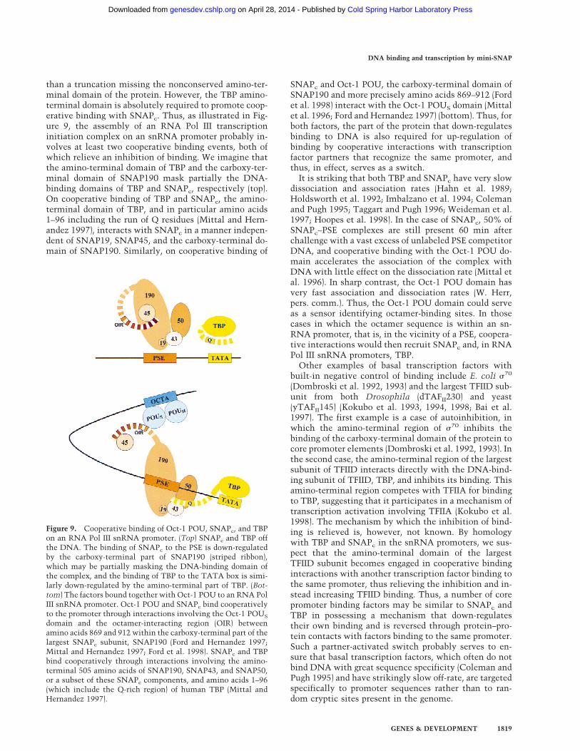
than a truncation missing the nonconserved amino-ter-minal domain of the protein. However, the TBP amino-terminal domain is absolutely required to promote coop-erative binding with SNAPc. Thus, as illustrated in Fig-ure 9, the assembly of an RNA Pol III transcriptioninitiation complex on an snRNA promoter probably in-volves at least two cooperative binding events, both ofwhich relieve an inhibition of binding. We imagine thatthe amino-terminal domain of TBP and the carboxy-ter-minal domain of SNAP190 mask partially the DNA-binding domains of TBP and SNAPc, respectively (top).On cooperative binding of TBP and SNAPc, the amino-terminal domain of TBP, and in particular amino acids1–96 including the run of Q residues (Mittal and Hern-andez 1997), interacts with SNAPc in a manner indepen-dent of SNAP19, SNAP45, and the carboxy-terminal do-main of SNAP190. Similarly, on cooperative binding of
SNAPc and Oct-1 POU, the carboxy-terminal domain ofSNAP190 and more precisely amino acids 869–912 (Fordet al. 1998) interact with the Oct-1 POUS domain (Mittalet al. 1996; Ford and Hernandez 1997) (bottom). Thus, forboth factors, the part of the protein that down-regulatesbinding to DNA is also required for up-regulation ofbinding by cooperative interactions with transcriptionfactor partners that recognize the same promoter, andthus, in effect, serves as a switch.
It is striking that both TBP and SNAPc have very slowdissociation and association rates (Hahn et al. 1989;Holdsworth et al. 1992; Imbalzano et al. 1994; Colemanand Pugh 1995; Taggart and Pugh 1996; Weideman et al.1997; Hoopes et al. 1998). In the case of SNAPc, 50% ofSNAPc–PSE complexes are still present 60 min afterchallenge with a vast excess of unlabeled PSE competitorDNA, and cooperative binding with the Oct-1 POU do-main accelerates the association of the complex withDNA with little effect on the dissociation rate (Mittal etal. 1996). In sharp contrast, the Oct-1 POU domain hasvery fast association and dissociation rates (W. Herr,pers. comm.). Thus, the Oct-1 POU domain could serveas a sensor identifying octamer-binding sites. In thosecases in which the octamer sequence is within an sn-RNA promoter, that is, in the vicinity of a PSE, coopera-tive interactions would then recruit SNAPc and, in RNAPol III snRNA promoters, TBP.
Other examples of basal transcription factors withbuilt-in negative control of binding include E. coli s70
(Dombroski et al. 1992, 1993) and the largest TFIID sub-unit from both Drosophila (dTAFII230) and yeast(yTAFII145) (Kokubo et al. 1993, 1994, 1998; Bai et al.1997). The first example is a case of autoinhibition, inwhich the amino-terminal region of s70 inhibits thebinding of the carboxy-terminal domain of the protein tocore promoter elements (Dombroski et al. 1992, 1993). Inthe second case, the amino-terminal region of the largestsubunit of TFIID interacts directly with the DNA-bind-ing subunit of TFIID, TBP, and inhibits its binding. Thisamino-terminal region competes with TFIIA for bindingto TBP, suggesting that it participates in a mechanism oftranscription activation involving TFIIA (Kokubo et al.1998). The mechanism by which the inhibition of bind-ing is relieved is, however, not known. By homologywith TBP and SNAPc in the snRNA promoters, we sus-pect that the amino-terminal domain of the largestTFIID subunit becomes engaged in cooperative bindinginteractions with another transcription factor binding tothe same promoter, thus relieving the inhibition and in-stead increasing TFIID binding. Thus, a number of corepromoter binding factors may be similar to SNAPc andTBP in possessing a mechanism that down-regulatestheir own binding and is reversed through protein–pro-tein contacts with factors binding to the same promoter.Such a partner-activated switch probably serves to en-sure that basal transcription factors, which often do notbind DNA with great sequence specificity (Coleman andPugh 1995) and have strikingly slow off-rate, are targetedspecifically to promoter sequences rather than to ran-dom cryptic sites present in the genome.
Figure 9. Cooperative binding of Oct-1 POU, SNAPc, and TBPon an RNA Pol III snRNA promoter. (Top) SNAPc and TBP offthe DNA. The binding of SNAPc to the PSE is down-regulatedby the carboxy-terminal part of SNAP190 (striped ribbon),which may be partially masking the DNA-binding domain ofthe complex, and the binding of TBP to the TATA box is simi-larly down-regulated by the amino-terminal part of TBP. (Bot-tom) The factors bound together with Oct-1 POU to an RNA PolIII snRNA promoter. Oct-1 POU and SNAPc bind cooperativelyto the promoter through interactions involving the Oct-1 POUS
domain and the octamer-interacting region (OIR) betweenamino acids 869 and 912 within the carboxy-terminal part of thelargest SNAPc subunit, SNAP190 (Ford and Hernandez 1997;Mittal and Hernandez 1997; Ford et al. 1998). SNAPc and TBPbind cooperatively through interactions involving the amino-terminal 505 amino acids of SNAP190, SNAP43, and SNAP50,or a subset of these SNAPc components, and amino acids 1–96(which include the Q-rich region) of human TBP (Mittal andHernandez 1997).
DNA binding and transcription by mini-SNAP
GENES & DEVELOPMENT 1819
Cold Spring Harbor Laboratory Press on April 28, 2014 - Published by genesdev.cshlp.orgDownloaded from

Materials and methods
Expression of proteins in E. coli
The wild-type GST–Oct-1 POU and GST–SNAP45 proteinswere expressed in E. coli BL21 (DE3) cells with the T7 expres-sion system, as described before (Mittal et al. 1996). The pro-teins were purified by binding to glutathione–agarose beads andelution with thrombin, which removed the GST moiety of thefusion proteins. Protein purity was assessed by Coomassiestaining of a 15% SDS–polyacrylamide gel.
Assembly and purification of SNAPc and partial SNAPcs
SNAPc or partial SNAPcs containing SNAP190 with a His tag atits carboxyl terminus and SNAP50 with an HA tag at its aminoterminus were assembled and purified as described before(Henry et al. 1998b). Mini-SNAPc was purified first over proteinA–agarose beads (Boehringer Mannheim) covalently coupled toan anti-SNAP190 antibody (CS696). Bound proteins were elutedwith a buffer containing 0.5 mg/ml of the peptide against whichthe antibody was raised in 20 mM HEPES (pH 7.9), 5 mM MgCl2,0.1% Tween 20, 15% glycerol, 100 mM KCl, 1 mM dithiothreitol(DTT), and the following protease inhibitors: 0.5 mM PMSF, 1mM benzamidine, 2 µg/ml aprotonin, 1 µg/ml leupeptin, 1 mM
sodium bisulfite, 0.5 µM pepstatin A, and 2 mM pefablock (Boe-hringer-Mannheim). Alternatively, mini-SNAPc was purifiedfirst over nickel agarose beads as described previously forSNAPc (Henry et al. 1998b). In both cases, the eluates werefurther purified over protein G–agarose beads coupled to theanti-HA mAb 12CA5 (Niman et al. 1983). The bound proteinswere eluted with the same buffer as above containing 0.7 mg/mlHA peptide. The composition of the complexes was checked byimmunoblots with the following antibodies: anti-SNAP19(CS543, anti-p19Cpep; Henry et al. 1998b), anti-SNAP43 (CS49,anti-CSH375; Henry et al. 1995), anti-SNAP45 (CS234,a-CSH467; Sadowski et al. 1996), anti-SNAP50 (CS303,a-CSH482; Henry et al. 1996), anti-SNAP190 (CS402, Ab402;Wong et al. 1998). The composition of mini-SNAPc was alsochecked by SDS-PAGE followed by silver staining.
EMSAs
The EMSAs involving SNAPc or SNAPc and Oct-1 POU wereperformed as described (Mittal et al. 1996), and those involvingSNAPc and TBP were performed as described (Mittal and Her-nandez 1997). The antibodies used in Figure 5B were as follows:anti-SNAP190 N-ter (CS696), anti-SNAP190 C-ter (CS402,Ab402; Wong et al. 1998), anti-SNAP50 (CS303, a a-CSH482;Henry et al. 1996), anti-SNAP45 (CS234, a-CSH467; Sadowskiet al. 1996), anti-SNAP43 (CS49, anti-CSH375; Henry et al.1995); anti-SNAP19 (CS543, anti-p19Cpep; Henry et al. 1998b).
DNase I footprinting
The probes for DNase I footprinting were prepared by PCR withtwo primers, one of which was 58 end-labeled with [g-32P] ATP.The binding reaction was performed at 30°C for 30 min in atotal volume of 50 µl and contained 20 mM HEPES (pH 7.9), 100mM KCl, 5 mM MgCl2, 0.2 mM EDTA, 10% glycerol, 1 mM DTT,0.2 µg each of poly[d(G-C)–(G-C)] and pUC118, 2% polyvinylalcohol, and 10,000 cpm of radiolabeled probe. DNase I diges-tion was carried out as described (Schmidt et al. 1989), and thereaction products were analyzed on a 7% polyacrylamide–ureagel.
In vitro transcription
The U1 and U6 constructs used in Figure 7 have been described
in Sadowski et al. (1993), and the reporter U6 construct used inFigure 8B has been described in Mittal et al. (1996). HeLa cellextracts were depleted of SNAPc with an equal volume of amixture of protein A–agarose beads cross-linked to anti-SNAP19 (CS543, anti-p19Cpep; Henry et al. 1998b), anti-SNAP45 (CS234, a-CSH467; Sadowski et al. 1996), and anti-SNAP190 (CS696) antibodies. Control extracts were depletedsimilarly, but with beads cross-linked to preimmune antibodies.The transcription reactions were performed as described before(Sadowski et al. 1993).
Acknowledgments
We thank W.P. Tansey for comments on the manuscript, W.Herr for discussion, S. Sepehri for a constant supply of U6 RNAprobe and HeLa cell extract, and M. Ockler, J. Duffy, and P.Renna for artwork and photography. This work was funded inpart by National Institutes of Health grant GM38810. We aresupported by the Howard Hughes Medical Institute.
The publication costs of this article were defrayed in part bypayment of page charges. This article must therefore be herebymarked ‘advertisement’ in accordance with 18 USC section1734 solely to indicate this fact.
References
Bai, Y., G.M. Perez, J.M. Beechem and P.A. Weil. 1997. Struc-ture-function analysis of TAF130: Identification and charac-terization of a high-affinity TATA-binding protein interac-tion domain in the N terminus of yeast TAF(II)130. Mol.Cell. Biol. 17: 3081–3093.
Chen, J.-L., L.D. Attardi, P.C. Vrreijzer, K. Yokomori, and R.Tjian. 1994. Assembly of recombinant TFIID reveals differ-ential coactivator requirements for distinct transcriptionalactivators. Cell 79: 93–105.
Coleman, R.A. and B.F. Pugh. 1995. Evidence for functionalbinding and stable sliding of the TATA binding protein onnonspecific DNA. J. Biol. Chem. 270: 13850–13859.
Dombroski, A.J., W.A. Walter, M.T.J. Record, D.A. Siegele, andC.A. Gross. 1992. Polypeptides containing highyl conservedregions of transcription initiation factor s70 exhibit specific-ity of binding to promoter DNA. Cell 70: 501–512.
Dombroski, A.J., W.A. Walter, and C.A. Gross. 1993. Amino-terminal amino acids modulate s-factor DNA-binding activ-ity. Genes & Dev. 7: 2446–2455.
Ford, E. and N. Hernandez. 1997. Characterization of a trimericcomplex containing Oct-1, SNAPc, and DNA. J. Biol. Chem.272: 16048–16055.
Ford, E., M. Strubin, and N. Hernandez. 1998. The Oct-1 POUdomain activates snRNA gene transcription by contacting aregion in the SNAPc largest subunit that bears sequencesimilarities with the Oct-1 coactivator OBF-1. Genes & Dev.12: 3528–3540.
Hahn, S., S. Buratowski, P.A. Sharp, and L. Guarente. 1989.Yeast TATA-binding protein TFIID binds to TATA elementswith both consensus and nonconsensus DNA sequences.Proc. Natl. Acad. Sci. 86: 5718–5722.
Henry, R.W., C.L. Sadowski, R. Kobayashi, and N. Hernandez.1995. A TBP-TAF complex required for transcription of hu-man snRNA genes by RNA polymerases II and III. Nature374: 653–657.
Henry, R.W., B. Ma, C.L. Sadowski, R. Kobayashi, and N. Her-nandez. 1996. Cloning and characterization of SNAP50, asubunit of the snRNA activating protein complex SNAPc.
Mittal et al.
1820 GENES & DEVELOPMENT
Cold Spring Harbor Laboratory Press on April 28, 2014 - Published by genesdev.cshlp.orgDownloaded from

EMBO J 15: 7129–7136.Henry, R.W., E. Ford, R. Mital, V. Mittal, and N. Hernandez.
1998a. Crossing the line between RNA polymerases: Tran-scription of human snRNA genes by RNA polymerases IIand III. Cold Spring Harbor Symp. Quant. Biol.Volume 63,pp. 111–120.
Henry, R.W., V. Mittal, B. Ma, R. Kobayashi and N. Hernandez.1998b. Assembly of a functional, core promoter complex(SNAPc) shared by RNA polymerase II and III. Genes & Dev.12: 2664–2672.
Herr, W., R.A. Sturm, R.G. Clerc, L.M. Corcoran, D. Baltimore,P.A. Sharp, H.A. Ingraham, M.G. Rosenfeld, M. Finney, G.Ruvkun, and H.R. Horvitz. 1988. The POU domain: A largeconserved region in the mammalian pit-1, oct-1, oct-2, andCaenorhabditis elegans unc-86 gene products. Genes &Dev. 2: 1513–1516.
Holdsworth, M.J., C. Grierson, W. Schuch, and M. Bevan. 1992.DNA-binding properties of cloned TATA-binding proteinfrom potato tubers. Plant Mol. Biol. 19: 455–464.
Hoopes, B.C., J.F. LeBlanc, and D.K. Hawley. 1998. Contribu-tions of the TATA box sequence to rate-limiting steps intranscription initiation by RNA polymerase II. J. Mol. Biol.277: 1015–1031.
Imbalzano, A., K.S. Zaret, and R.E. Kingston. 1994. Transcrip-tion factor (TF) IIB and TFIIA can independently increase theaffinity of the TATA-binding protein for DNA. J. Biol.Chem. 269: 8280–8286.
Kokubo, T., D.-W. Gong, S. Yamashita, M. Horikoshi, R.G.Roeder, and Y. Nakatani. 1993. Drosophila 230-kD TFIIDsubunit, a functional homolog of the human cell cycle geneproduct, negatively regulates DNA binding of the TATAbox-binding subunit of TFIID. Genes & Dev. 7: 1033–1046.
Kokubo, T., S. Yamashita, M. Horikoshi, R.G. Roeder, and Y.Nakatani. 1994. Interaction between the N-terminal domainof the 230-kDa subunit and the TATA box-binding subunitof TFIID negatively regulates TATA-box binding. Proc. Natl.Acad. Sci. 91: 3520–3524.
Kokubo, T., M.J. Swanson, J.I. Nishikawa, A.G. Hinnebusch,and Y. Nakatani. 1998. The yeast TAF145 inhibitory domainand TFIIA competitively bind to TATA- binding protein.Mol. Cell. Biol. 18: 1003–1012.
Kuhlman, T.C., H. Cho, D. Reinberg, and N. Hernandez. 1999.The general transcription factors IIA, IIB, IIF, and IIE arerequired for RNA polymerase II transcription from the hu-man U1 snRNA promoter. Mol. Cell. Biol. 19: 2130–2141.
Lobo, S.M. and N. Hernandez. 1994. Transcription of snRNAgenes by RNA polymerases II and III. Transcription, mecha-nisms and regulation (ed. R.C. Conaway and J.W. Conaway),pp. 127–159. Raven Press, New York, NY.
Luscher, B. and R.N. Eisenman. 1990. New light on Myc andMyb. Part II. Myb. Genes & Dev. 4: 2235–2241.
Mittal, V. and N. Hernandez. 1997. Role for the amino-terminalregion of human TBP in U6 snRNA transcription. Science275: 1136–1140.
Mittal, V., M.A. Cleary, W. Herr, and N. Hernandez. 1996. TheOct-1 POU-specific domain can stimulate small nuclearRNA gene transcription by stabilizing the basal transcrip-tion complex SNAPc. Mol. Cell. Biol. 16: 1955–1965.
Murphy, S., J.-B. Yoon, T. Gerster, and R.G. Roeder. 1992. Oct-1and Oct-2 potentiate functional interactions of a transcrip-tion factor with the proximal sequence element of smallnuclear RNA genes. Mol. Cell. Biol. 12: 3247–3261.
Niman, H.L., R.A. Houghten, L.E. Walker, R.A. Reisfeld, I.A.Wilson, J.M. Hogle, and R.A. Lerner. 1983. Generation ofprotein-reactive antibodies by short peptides is an event ofhigh frequency: Implications for the structural basis of im-
mune recognition. Proc. Natl. Acad. Sci. 80: 4949–4953.Nomura, N., M. Takahashi, M. Matsui, S. Ishii, T. Date, S.
Sasamoto, and R. Ishizaki. 1988. Isolation of human cDNAclones of myb-related genes, A-myb and B-myb. Nucleic Ac-ids Res. 16: 11075–11089.
Sadowski, C.L., R.W. Henry, S.M. Lobo, and N. Hernandez.1993. Targeting TBP to a non-TATA box cis-regulatory ele-ment: A TBP-containing complex activates transcriptionfrom snRNA promoters through the PSE. Genes & Dev.7: 1535–1548.
Sadowski, C.L., R.W. Henry, R. Kobayashi, and N. Hernandez.1996. The SNAP45 subunit of the small nuclear RNA (sn-RNA) activating protein complex is required for RNA poly-merase II and III snRNA gene transcription and interactswith the TATA box binding protein. Proc. Natl. Acad. Sci.93: 4289–4293.
Schmidt, M.C., Q. Zhou, and A.J. Berk. 1989. Sp1 activates tran-scription without enhancing DNA-binding activity of theTATA box factor. Mol. Cell. Biol. 8: 3299–3307.
Taggart, A.K. and B.F. Pugh. 1996. Dimerization of TFIID whennot bound to DNA. Science 272: 1331–1333.
Tanaka, M., J.-S. Lai, and W. Herr. 1992. Promoter-selectiveactivation domains in Oct-1 and Oct-2 direct differential ac-tivation of an snRNA and mRNA promoter. Cell 68: 755–767.
Tansey, W.P. and W. Herr. 1997. TAFs: Guilt by association?Cell 88: 729–732.
Weideman, C.A., R.C. Netter, L.R. Benjamin, J.J. McAllister,L.A. Schmiedekamp, R.A. Coleman, and B.F. Pugh. 1997.Dynamic interplay of TFIIA, TBP and TATA DNA. J. Mol.Biol. 271: 61–75.
Wong, M.W., R.W. Henry, B. Ma, R. Kobayashi, N. Klages, P.Matthias, M. Strubin, and N. Hernandez. 1998. The largesubunit of basal transcription factor SNAPc is a Myb domainprotein that interacts with Oct-1. Mol. Cell. Biol. 18: 368–377.
DNA binding and transcription by mini-SNAP
GENES & DEVELOPMENT 1821
Cold Spring Harbor Laboratory Press on April 28, 2014 - Published by genesdev.cshlp.orgDownloaded from