sodium hexametaphosphate in the important role of · sodium hexametaphosphate in the important role...
TRANSCRIPT
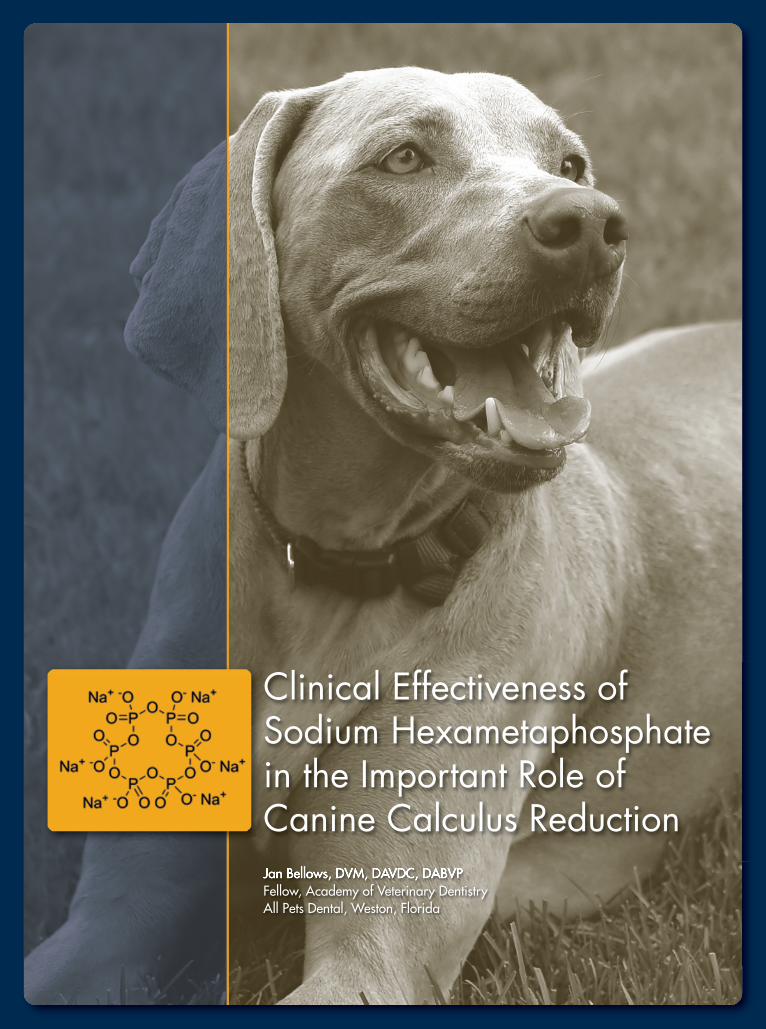
Jan Bellows, DVM, DAVDC, DABVPFellow, Academy of Veterinary DentistryAll Pets Dental, Weston, Florida
Clinical Effectiveness ofSodium Hexametaphosphatein the Important Role ofCanine Calculus Reduction
— 2—
anine dental disease is the most common malady in dogs.
Most pet owners consider “dog breath” an expected
part of dog ownership and not a treatable or preventable illness.
In a landmark study of over 30,000 dogs in numerous companion
animal practices, the number-one and -two abnormalities reported on
veterinarian-conducted physical examinations were dental calculus
and gingivitis, respectively.1 If left untreated, gingivitis often progresses
to painful inflammation and periodontal disease.
Further, thousands of dogs and cats are
placed under general anesthesia daily for
veterinary removal of accumulated plaque and
calculus above and below the gumline to help
treat halitosis and delay the progression of
periodontal disease. Unless the owner initiates
a plaque and calculus control program, calculus,
gingivitis, and halitosis soon return.
C
Plaque and Calculus FormationWithin minutes a!er professional plaque and calculusremoval under general anesthesia, salivary proteinsactively adhere to the exposed tooth surfaces, creatinga conditioning "lm (the acquired pellicle).#is "lmacts as a connective layer between the enamel andexposed dentin tooth surfaces and a bacterial plaque,which colonizes within hours.
Supragingival plaque forms on the coronal toothsurfaces. Subgingival plaque occurs a!ermicroorganismspenetrate and colonize the gingival sulcus. Supragingivaland subgingival bacteriaform microenvironmentsof bacterial colonies calledbio!lms, which are separatedfrom the junctional epithe-lium by a wall of neutrophils.Gram-positive, nonmotile,aerobic cocci bacteria natu-rally occupy the area betweenthe tooth and gingiva (sulcus).As periodontal infectionprogresses, the number of bacteria increases at thegingival margin, decreasing the subgingival oxygen.#ese anaerobic conditions allow gram-negative,motile, anaerobic rods and spirochetes to predominate.Toxins produced by these bacteria cause prostaglandinstimulation and lysosome release, which can damagethe neutrophil wall, allowing invasion of the junctionalepithelium. Supragingival calculus is composed ofplaque, food debris, calcium, and phosphate oncoronal surfaces. Over time, calcium and phosphorus,
the principal precipitated salt is calcium carbonate(calcite form).#is may be due to the less acidiccondition and low phosphate concentration in caninesaliva compared with human saliva.3
Subgingival calculus is preceded by supragingivalplaque. With the increased buildup of plaque on thesurface of the subgingival calculus, the combinationhas the potential to extend the radius of destructionand the rate of displacement of the adjacent junctionalepithelium, which loosens the seal between the toothand gingiva, creating periodontal disease.
Calculus is always covered with bacteria, playinga role in maintaining and accelerating periodontaldisease by keeping plaque in close contact with gingivaltissue.#is decreases the potential for repair and newattachment.
Consequences of Plaque and Calculus FormationPlaque initiates and promotes continued in$amma-tion of the gingiva as well as the progression towardperiodontal disease through the presence of toxicstimulators (cytokines and prostaglandins) of boneresorption. Calculus covered with plaque producesmechanical irritation and physical interference withnormal plaque removal during mastication. Onceformed, calculus can only readily be removed throughtime-consuming scaling, o!en in areas that are di%cultto visualize and/or access.#e rough surface andporosity of calculus compared with hard enamelencourages the accumulation of more plaque.#ecombination of plaque and calculus is more irritatingto the gingiva than either one alone.#e early colo-nizing bacteria are not pathogenic.#ey cannot causeperiodontitis but can cause gingivitis. As the plaquebio"lm continues to grow, periodontal pathogenscolonize and become the predominant species.#e establishment of periodontal disease depends on
the complex regulatory interaction between bacteriaand immune modulators of the host’s response. Many
Clinical Effectiveness of Sodium Hexametaphosphate in the Important Role of Canine Calculus Reduction
— 3—
Toy (smaller) canine breeds are prone to developingperiodontal disease because small dogs have short toothroots, allowing bacteria by-products to destroy a greaterpercentage of the tooth support than in larger dogs.
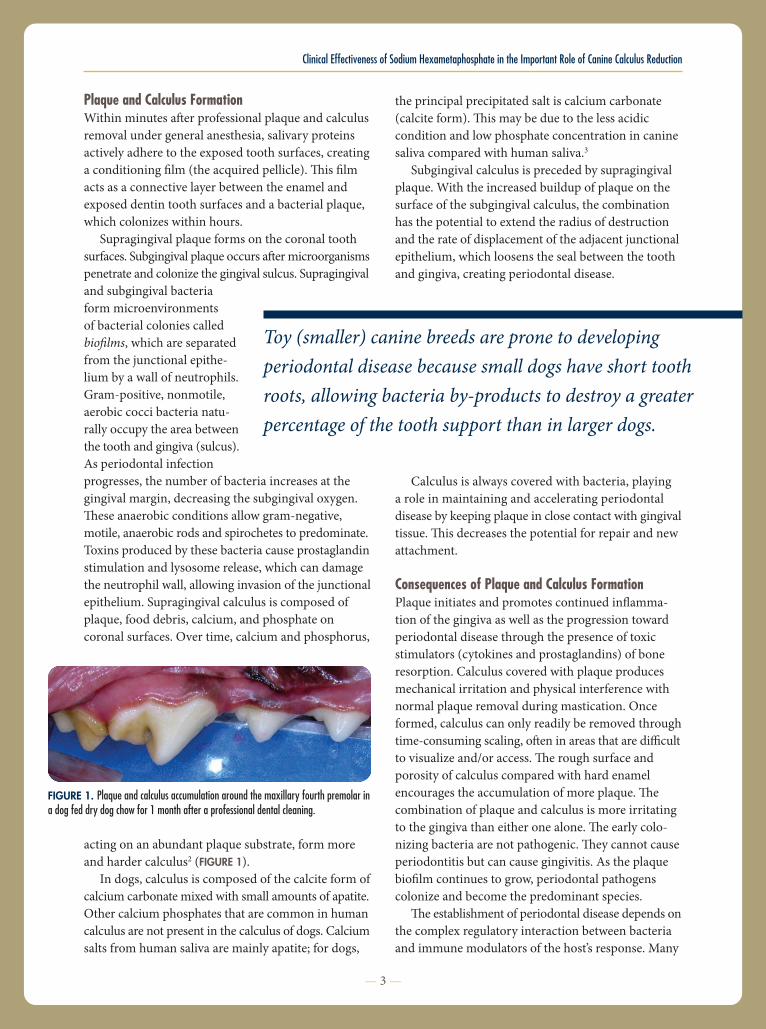
acting on an abundant plaque substrate, form moreand harder calculus2 (FIGURE 1).
In dogs, calculus is composed of the calcite form ofcalcium carbonate mixed with small amounts of apatite.Other calcium phosphates that are common in humancalculus are not present in the calculus of dogs. Calciumsalts from human saliva are mainly apatite; for dogs,
FIGURE 1. Plaque and calculus accumulation around the maxillary fourth premolar ina dog fed dry dog chow for 1 month after a professional dental cleaning.
variables in$uence why some animals develop diseaseand others do not. Animals with compromised healtho!en cannot "ght periodontal pathogens. Diseases andconditions that predispose dogs and cats to periodontaldisease include diabetes, hypothyroidism, hyperadreno-corticism, pemphigus, lupus, FIV infection, and FeLVinfection.
Toy (smaller) canine breeds are prone to developingperiodontal disease because small dogs have shorttooth roots, allowing bacteria by-products to destroya greater percentage of the tooth support than in largerdogs. Additionally, small dogs tend to live longer thanlarger dogs.#e longer an animal lives, the more timeperiodontal disease has to cause damage. Small dogsare also more prone to dental malocclusions. Crowdingabnormalities decrease the normal dental self-cleaningprocess, predisposing these dogs to periodontal disease.
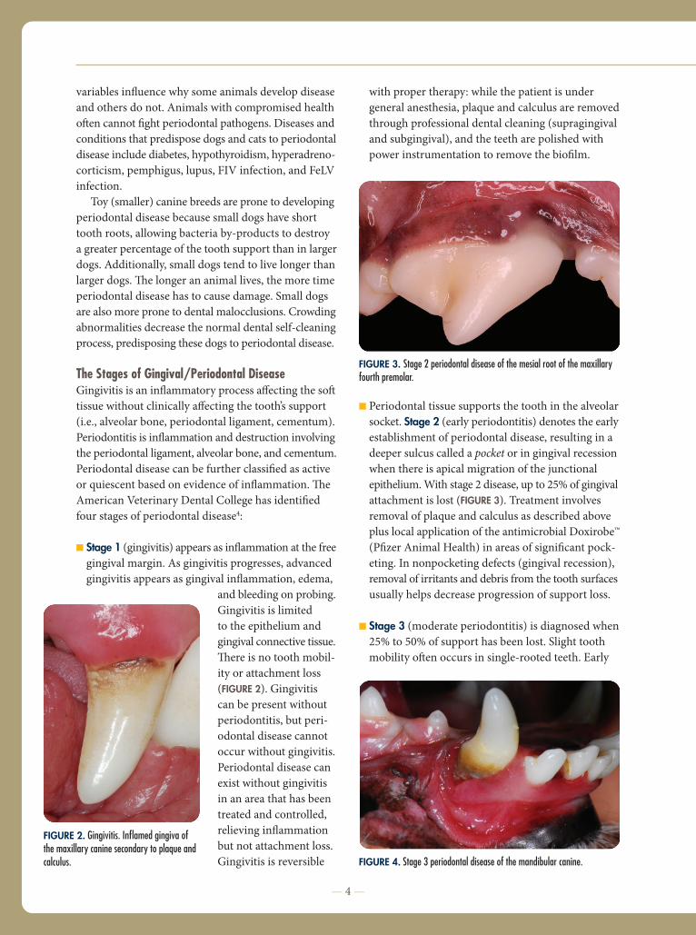
The Stages of Gingival/Periodontal DiseaseGingivitis is an in$ammatory process a&ecting the so!tissue without clinically a&ecting the tooth’s support(i.e., alveolar bone, periodontal ligament, cementum).Periodontitis is in$ammation and destruction involvingthe periodontal ligament, alveolar bone, and cementum.Periodontal disease can be further classi"ed as activeor quiescent based on evidence of in$ammation.#eAmerican Veterinary Dental College has identi"edfour stages of periodontal disease4:
! Stage 1 (gingivitis) appears as in$ammation at the freegingival margin. As gingivitis progresses, advancedgingivitis appears as gingival in$ammation, edema,
and bleeding on probing.Gingivitis is limitedto the epithelium andgingival connective tissue.#ere is no tooth mobil-ity or attachment loss(FIGURE 2). Gingivitiscan be present withoutperiodontitis, but peri-odontal disease cannotoccur without gingivitis.Periodontal disease canexist without gingivitisin an area that has beentreated and controlled,relieving in$ammationbut not attachment loss.Gingivitis is reversible
with proper therapy: while the patient is undergeneral anesthesia, plaque and calculus are removedthrough professional dental cleaning (supragingivaland subgingival), and the teeth are polished withpower instrumentation to remove the bio"lm.
! Periodontal tissue supports the tooth in the alveolarsocket. Stage 2 (early periodontitis) denotes the earlyestablishment of periodontal disease, resulting in adeeper sulcus called a pocket or in gingival recessionwhen there is apical migration of the junctionalepithelium.With stage 2 disease, up to 25% of gingivalattachment is lost (FIGURE 3). Treatment involvesremoval of plaque and calculus as described aboveplus local application of the antimicrobial Doxirobe™(P"zer Animal Health) in areas of signi"cant pock-eting. In nonpocketing defects (gingival recession),removal of irritants and debris from the tooth surfacesusually helps decrease progression of support loss.
! Stage 3 (moderate periodontitis) is diagnosed when25% to 50% of support has been lost. Slight toothmobility o!en occurs in single-rooted teeth. Early
— 4 —
FIGURE 3. Stage 2 periodontal disease of the mesial root of the maxillaryfourth premolar.
FIGURE 4. Stage 3 periodontal disease of the mandibular canine.
FIGURE 2. Gingivitis. Inflamed gingiva ofthe maxillary canine secondary to plaque andcalculus.
furcation exposure and/or gingival recession mayexist (FIGURE 4). Unless the client (and patient) iscommitted to stringent daily plaque prevention, thebest choice is o!en tooth extraction. Dogs generallydo better without teeth compared with living with apainful dentition.
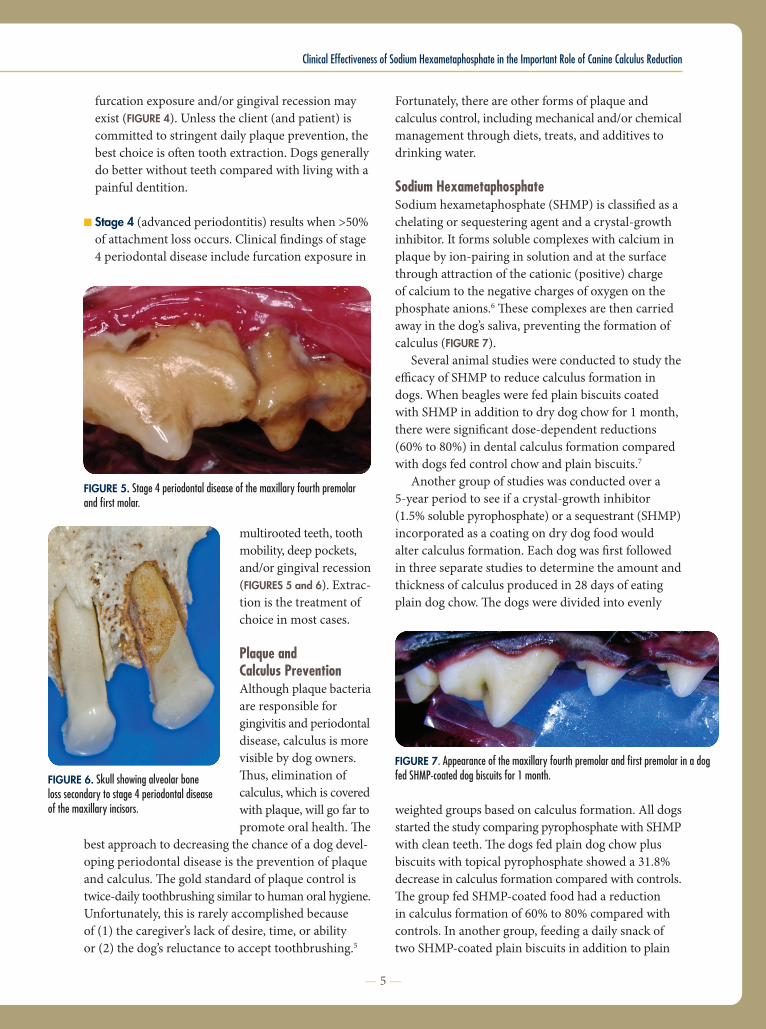
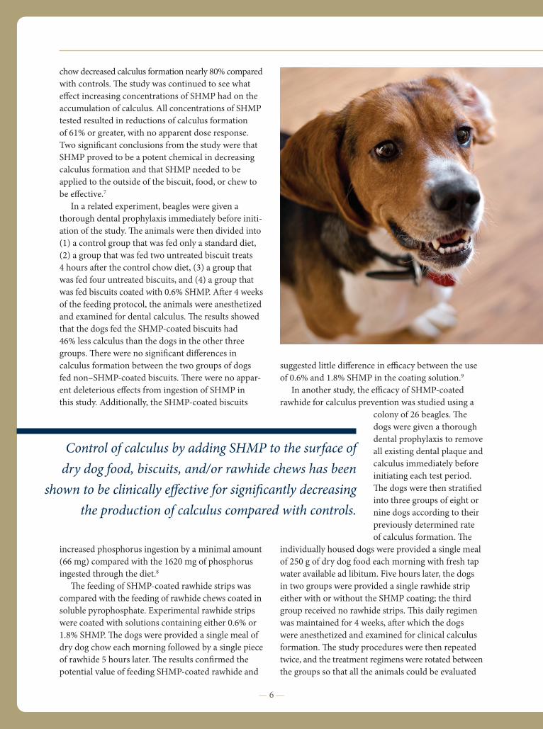
! Stage 4 (advanced periodontitis) results when >50%of attachment loss occurs. Clinical "ndings of stage4 periodontal disease include furcation exposure in
multirooted teeth, toothmobility, deep pockets,and/or gingival recession(FIGURES 5 and 6). Extrac-tion is the treatment ofchoice in most cases.
Plaque andCalculus PreventionAlthough plaque bacteriaare responsible forgingivitis and periodontaldisease, calculus is morevisible by dog owners.#us, elimination ofcalculus, which is coveredwith plaque, will go far topromote oral health.#e
best approach to decreasing the chance of a dog devel-oping periodontal disease is the prevention of plaqueand calculus.#e gold standard of plaque control istwice-daily toothbrushing similar to human oral hygiene.Unfortunately, this is rarely accomplished becauseof (1) the caregiver’s lack of desire, time, or abilityor (2) the dog’s reluctance to accept toothbrushing.5
Fortunately, there are other forms of plaque andcalculus control, including mechanical and/or chemicalmanagement through diets, treats, and additives todrinking water.
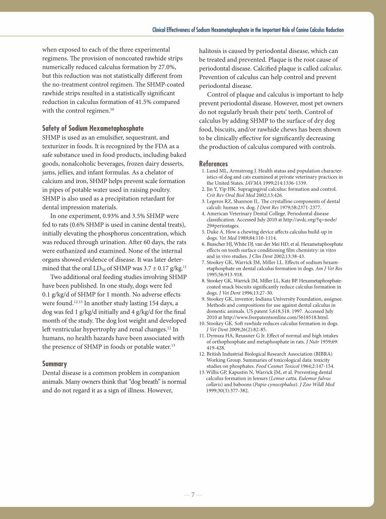
Sodium HexametaphosphateSodium hexametaphosphate (SHMP) is classi"ed as achelating or sequestering agent and a crystal-growthinhibitor. It forms soluble complexes with calcium inplaque by ion-pairing in solution and at the surfacethrough attraction of the cationic (positive) chargeof calcium to the negative charges of oxygen on thephosphate anions.6#ese complexes are then carriedaway in the dog’s saliva, preventing the formation ofcalculus (FIGURE 7).
Several animal studies were conducted to study thee%cacy of SHMP to reduce calculus formation indogs. When beagles were fed plain biscuits coatedwith SHMP in addition to dry dog chow for 1 month,there were signi"cant dose-dependent reductions(60% to 80%) in dental calculus formation comparedwith dogs fed control chow and plain biscuits.7
Another group of studies was conducted over a5-year period to see if a crystal-growth inhibitor(1.5% soluble pyrophosphate) or a sequestrant (SHMP)incorporated as a coating on dry dog food wouldalter calculus formation. Each dog was "rst followedin three separate studies to determine the amount andthickness of calculus produced in 28 days of eatingplain dog chow.#e dogs were divided into evenly
weighted groups based on calculus formation. All dogsstarted the study comparing pyrophosphate with SHMPwith clean teeth.#e dogs fed plain dog chow plusbiscuits with topical pyrophosphate showed a 31.8%decrease in calculus formation compared with controls.#e group fed SHMP-coated food had a reductionin calculus formation of 60% to 80% compared withcontrols. In another group, feeding a daily snack oftwo SHMP-coated plain biscuits in addition to plain
Clinical Effectiveness of Sodium Hexametaphosphate in the Important Role of Canine Calculus Reduction
— 5—
FIGURE 6. Skull showing alveolar boneloss secondary to stage 4 periodontal diseaseof the maxillary incisors.
FIGURE 5. Stage 4 periodontal disease of the maxillary fourth premolarand first molar.
FIGURE 7. Appearance of the maxillary fourth premolar and first premolar in a dogfed SHMP-coated dog biscuits for 1 month.
chow decreased calculus formation nearly 80% comparedwith controls.#e study was continued to see whate&ect increasing concentrations of SHMP had on theaccumulation of calculus. All concentrations of SHMPtested resulted in reductions of calculus formationof 61% or greater, with no apparent dose response.Two signi"cant conclusions from the study were thatSHMP proved to be a potent chemical in decreasingcalculus formation and that SHMP needed to beapplied to the outside of the biscuit, food, or chew tobe e&ective.7
In a related experiment, beagles were given athorough dental prophylaxis immediately before initi-ation of the study.#e animals were then divided into(1) a control group that was fed only a standard diet,(2) a group that was fed two untreated biscuit treats4 hours a!er the control chow diet, (3) a group thatwas fed four untreated biscuits, and (4) a group thatwas fed biscuits coated with 0.6% SHMP. A!er 4 weeksof the feeding protocol, the animals were anesthetizedand examined for dental calculus.#e results showedthat the dogs fed the SHMP-coated biscuits had46% less calculus than the dogs in the other threegroups.#ere were no signi"cant di&erences incalculus formation between the two groups of dogsfed non–SHMP-coated biscuits.#ere were no appar-ent deleterious e&ects from ingestion of SHMP inthis study. Additionally, the SHMP-coated biscuits
increased phosphorus ingestion by a minimal amount(66 mg) compared with the 1620 mg of phosphorusingested through the diet.8#e feeding of SHMP-coated rawhide strips was
compared with the feeding of rawhide chews coated insoluble pyrophosphate. Experimental rawhide stripswere coated with solutions containing either 0.6% or1.8% SHMP.#e dogs were provided a single meal ofdry dog chow each morning followed by a single pieceof rawhide 5 hours later.#e results con"rmed thepotential value of feeding SHMP-coated rawhide and
suggested little di&erence in e%cacy between the useof 0.6% and 1.8% SHMP in the coating solution.9
In another study, the e%cacy of SHMP-coatedrawhide for calculus prevention was studied using a
colony of 26 beagles.#edogs were given a thoroughdental prophylaxis to removeall existing dental plaque andcalculus immediately beforeinitiating each test period.#e dogs were then strati"edinto three groups of eight ornine dogs according to theirpreviously determined rateof calculus formation.#e
individually housed dogs were provided a single mealof 250 g of dry dog food each morning with fresh tapwater available ad libitum. Five hours later, the dogsin two groups were provided a single rawhide stripeither with or without the SHMP coating; the thirdgroup received no rawhide strips.#is daily regimenwas maintained for 4 weeks, a!er which the dogswere anesthetized and examined for clinical calculusformation.#e study procedures were then repeatedtwice, and the treatment regimens were rotated betweenthe groups so that all the animals could be evaluated
— 6 —
Control of calculus by adding SHMP to the surface ofdry dog food, biscuits, and/or rawhide chews has been
shown to be clinically e"ective for signi!cantly decreasingthe production of calculus compared with controls.
Clinical Effectiveness of Sodium Hexametaphosphate in the Important Role of Canine Calculus Reduction
when exposed to each of the three experimentalregimens.#e provision of noncoated rawhide stripsnumerically reduced calculus formation by 27.0%,but this reduction was not statistically di&erent fromthe no-treatment control regimen.#e SHMP-coatedrawhide strips resulted in a statistically signi"cantreduction in calculus formation of 41.5% comparedwith the control regimen.10
Safety of Sodium HexametaphosphateSHMP is used as an emulsi"er, sequestrant, andtexturizer in foods. It is recognized by the FDA as asafe substance used in food products, including bakedgoods, nonalcoholic beverages, frozen dairy desserts,jams, jellies, and infant formulas. As a chelator ofcalcium and iron, SHMP helps prevent scale formationin pipes of potable water used in raising poultry.SHMP is also used as a precipitation retardant fordental impression materials.
In one experiment, 0.93% and 3.5% SHMP werefed to rats (0.6% SHMP is used in canine dental treats),initially elevating the phosphorus concentration, whichwas reduced through urination. A!er 60 days, the ratswere euthanized and examined. None of the internalorgans showed evidence of disease. It was later deter-mined that the oral LD50 of SHMPwas 3.7 ± 0.17 g/kg.11
Two additional oral feeding studies involving SHMPhave been published. In one study, dogs were fed0.1 g/kg/d of SHMP for 1 month. No adverse e&ectswere found.12,13 In another study lasting 154 days, adog was fed 1 g/kg/d initially and 4 g/kg/d for the "nalmonth of the study.#e dog lost weight and developedle! ventricular hypertrophy and renal changes.12 Inhumans, no health hazards have been associated withthe presence of SHMP in foods or potable water.13
SummaryDental disease is a common problem in companionanimals. Many owners think that “dog breath” is normaland do not regard it as a sign of illness. However,
halitosis is caused by periodontal disease, which canbe treated and prevented. Plaque is the root cause ofperiodontal disease. Calci"ed plaque is called calculus.Prevention of calculus can help control and preventperiodontal disease.
Control of plaque and calculus is important to helpprevent periodontal disease. However, most pet ownersdo not regularly brush their pets’ teeth. Control ofcalculus by adding SHMP to the surface of dry dogfood, biscuits, and/or rawhide chews has been shownto be clinically e&ective for signi"cantly decreasingthe production of calculus compared with controls.
References1. Lund ML, Armstrong J. Health status and population character-istics of dog and cats examined at private veterinary practices inthe United States. JAVMA 1999;214:1336-1339.
2. Jin Y, Yip HK. Supragingival calculus: formation and control.Crit Rev Oral Biol Med 2002;13:426.
3. Legeros RZ, Shannon IL.#e crystalline components of dentalcalculi: human vs. dog. J Dent Res 1979;58:2371-2377.
4. American Veterinary Dental College. Periodontal diseaseclassi"cation. Accessed July 2010 at http://avdc.org/?q=node/29#periostages.
5. Duke A. How a chewing device a&ects calculus build-up indogs. Vet Med 1989;84:110-1114.
6. Busscher HJ,White DJ, van derMei HD, et al. Hexametaphosphatee&ects on tooth surface conditioning "lm chemistry: in vitroand in vivo studies. J Clin Dent 2002;13:38-43.
7. Stookey GK, Warrick JM, Miller LL. E&ects of sodium hexam-etaphosphate on dental calculus formation in dogs. Am J Vet Res1995;56:913-918.
8. Stookey GK,Warrick JM,Miller LL, Katz BP. Hexametaphosphate-coated snack biscuits signi"cantly reduce calculus formation indogs. J Vet Dent 1996;13:27-30.
9. Stookey GK, inventor; Indiana University Foundation, assignee.Methods and compositions for use against dental calculus indomestic animals. US patent 5,618,518. 1997. Accessed July2010 at http://www.freepatentsonline.com/5618518.html.
10. Stookey GK. So! rawhide reduces calculus formation in dogs.J Vet Dent 2009;26(2):82-85.
11. Dymsza HA, Reussner G Jr. E&ect of normal and high intakesof orthophosphate and metaphosphate in rats. J Nutr 1959;69:419-428.
12. British Industrial Biological Research Association (BIBRA)Working Group. Summaries of toxicological data: toxicitystudies on phosphates. Food Cosmet Toxicol 1964;2:147-154.
13.Willis GP, Kapustin N, Warrick JM, et al. Preventing dentalcalculus formation in lemurs (Lemur catta, Eulemur fulvuscollaris) and baboons (Papio cynocephalus). J Zoo Wildl Med1999;30(3):377-382.
— 7 —

Dr. Bellows reports that he has received honoraria from The Hartz Mountain Corporation.
Trademarks used herein are not trademarks of the publisher and the publisher claims no interest therein.
Photo credits: cover—Shutterstock/Dwight Lyman; page 2—Shutterstock/Eric Isselée; pages 3–5—Courtesy of The Hartz Mountain Corporation; page 6 – Shutterstock/ARENA Creative.
© 2010 The Hartz Mountain Corporation, Secaucus, New Jersey 7/10 VT10044