soft tissue tumors - ucsd musculoskeletal radiologybonepit.com/lectures/soft tissue tumors hannah...
TRANSCRIPT

Soft Tissue Tumors
Hannah Koh
5/21/15

• Introduction
• Utility of MR in differentiating benign vs malignant masses
• MR protocol and reporting
• WHO classification of soft tissue tumors
• Imaging features of soft tissue masses with distinct characteristics

• Imaging provides limited ability to reliably distinguish between benign and malignant soft tissue lesions
• Primary goal of imaging is to confirm presence of mass and assess extent in preparation of possible treatment
• In some cases, clinical and imaging information can narrow differential diagnosis – Clinical history
– Lesion location
– Mineralization on radiograph
– Signal intensity characteristics on MR

Spectrum of Soft Tissue Lesions
• Histological classified on basis of soft-tissue component that comprise the lesion
– Fat
– Skeletal muscle
– Peripheral nerves
– Blood vessels
– Fibrous tissue

Clinical History
• Age • History of trauma – hematoma, myositis ossificans • Anticoagulants • Pain – inflammatory process • Change in size – rapid growth from malignancy or
hemorrhage of benign mass • Fluctuation in size – engorged with blood or fluid
(hemangioma, ganglia) • h/o malignancy – soft tissue metastasis or radiation
induced sarcoma • Number of lesions – metastatic disease, syndromes
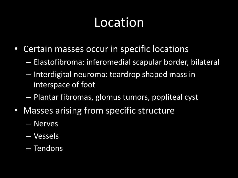
Location
• Certain masses occur in specific locations
– Elastofibroma: inferomedial scapular border, bilateral
– Interdigital neuroma: teardrop shaped mass in interspace of foot
– Plantar fibromas, glomus tumors, popliteal cyst
• Masses arising from specific structure
– Nerves
– Vessels
– Tendons

Radiographs
• Distortion of soft tissue planes • Radiolucent masses • Indolent or aggressive remodeling of bone • Foreign bodies • Soft tissue calcifications or ossifications
– Mature ossification (can look like aggressive sarcoma on MR)
– Hazy calcification, gouty tophus – Nonspecific dystrophic calcifications in lower
extremity in young adult, synovial sarcoma

MR
• Most lesions show nonspecific signal characteristics
• Correct histologic diagnosis reached in only 25-50% of cases
• Some diagnoses can be made based on basis of lesion signal intensity, pattern of growth, location, and associated signs and findings
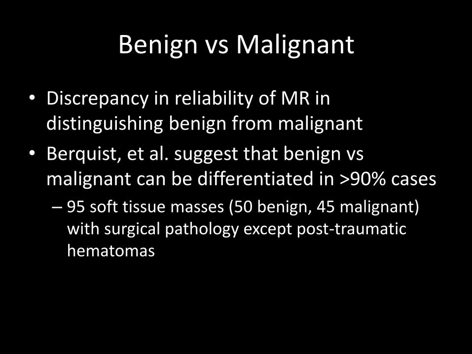
Benign vs Malignant
• Discrepancy in reliability of MR in distinguishing benign from malignant
• Berquist, et al. suggest that benign vs malignant can be differentiated in >90% cases
– 95 soft tissue masses (50 benign, 45 malignant) with surgical pathology except post-traumatic hematomas


*involvement of neurovascular structures, hemorrhage, and/or edema around lesions, bone involvement
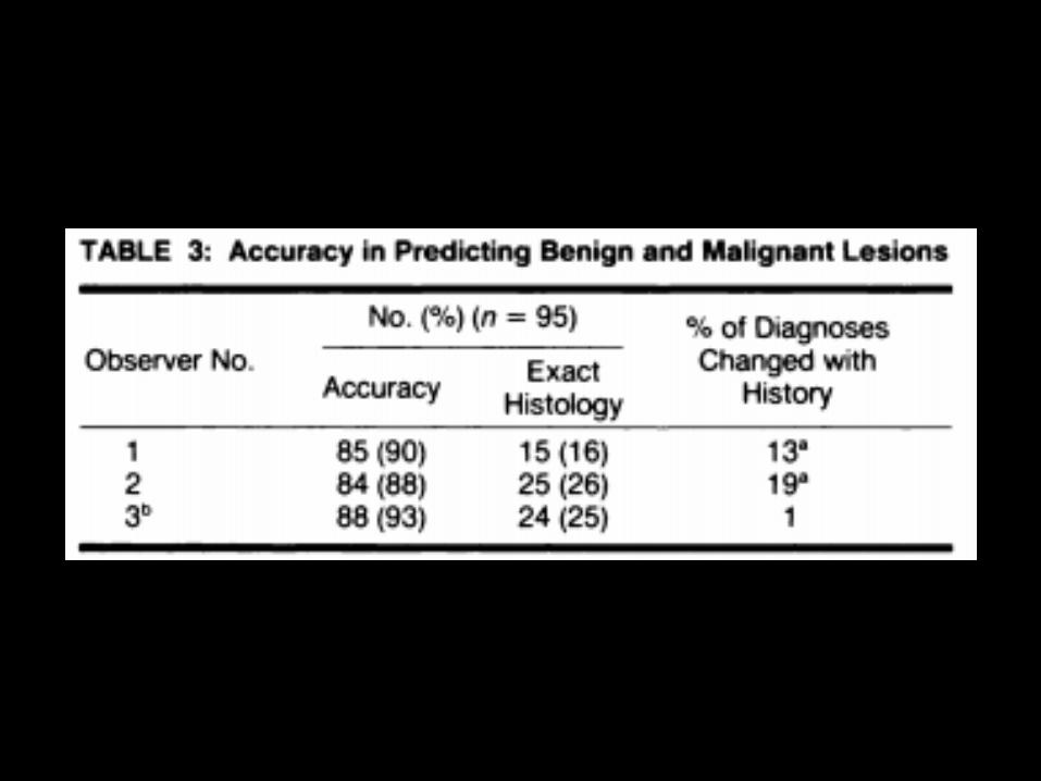


• Desmoid tumors and necrotic benign neoplasms most commonly classified incorrectly as malignant
• Synovial sarcoma was malignant lesion most commonly misclassified as benign
• Many benign lesions (ganglion cyst, lipoma, hemangioma, neuroma, hematomas) accurately diagnosed on basis of imaging findings alone

• Benign
– Well marginated
– Homogeneous signal intensity
– Do not encase neurovascular structures
• Malignant
– Irregular margins
– Inhomogeneous signal intensity
– More often encase neurovascular structures

• Crim, et al: 83 masses (49 benign and 34 malignant)
• Mean sensitivity 50% for benign lesions, 80% for malignant lesions

• Tumor margin, signal intensity homogeneity, size, peritumoral high signal intensity, neurovascular bundle encasement, and bone invasion not reliable to differentiate benign vs. malignant

• Factors that might explain differences in results in different studies…
– Differences in patient population
– Expertise of radiologist
– Study samples not appropriate for lesion prevalence and differences in characterization and differentiation of malignant vs benign lesions

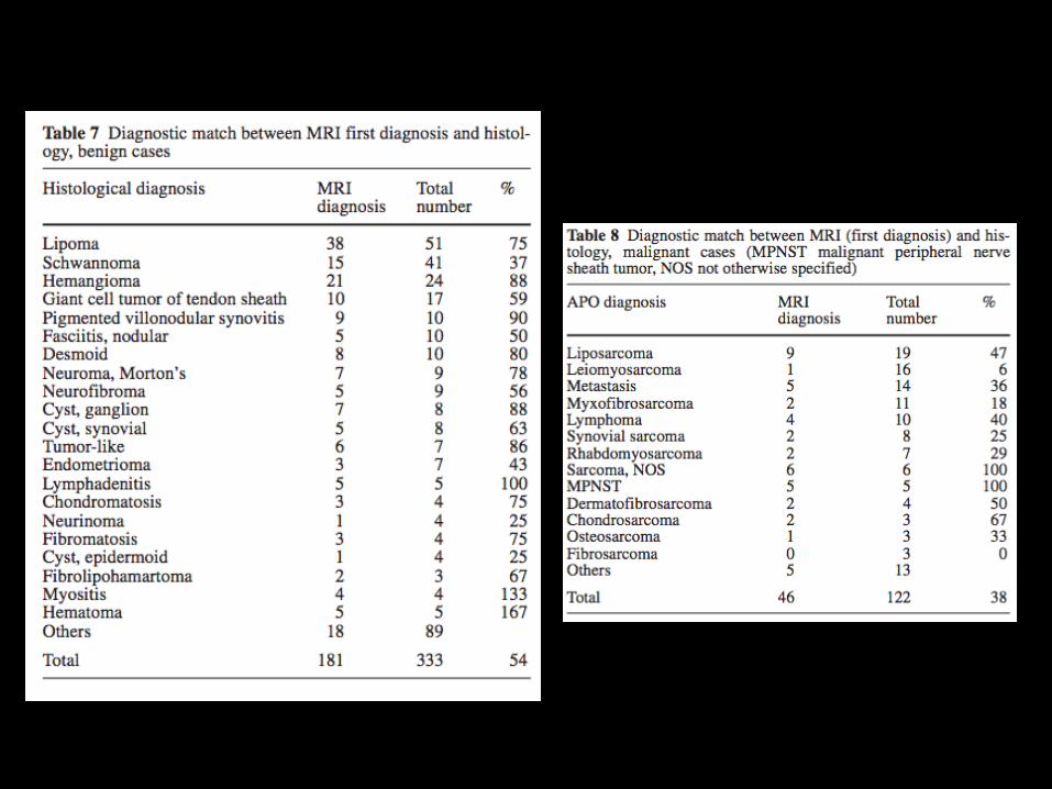
• Gielen et al • 548 untreated soft tissue tumors from 58 MRI centers • Images prospectively reviewed by 2 experienced
radiologists (12 and 15 yrs experience) • Threshold to differentiate b/t benign and malignant based
on… – Origin, size, shape, margins, SI, signal homogeneity, grade and
pattern of enhancement, low SI septations, peritumoral edema, distribution, fluid-fluid levels, signal voids, intra-tumoral necrosis
• Reference standard was histology by biopsy or resection (455) or follow-up in 6 months without clinical or MRI evolution of benign tumors (93)

• 123 malignant STT, 425 benign STT
• MRI reliability in identifying malignancy
– Sensitivity: 93%, NPV: 98%
– Specificity: 82%; PPV: 60%
• Exact histology predicted in 50%
– 38% of malignant cases
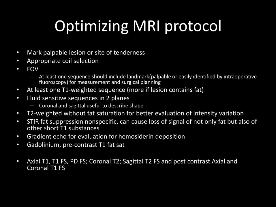
Optimizing MRI protocol
• Mark palpable lesion or site of tenderness • Appropriate coil selection • FOV
– At least one sequence should include landmark(palpable or easily identified by intraoperative fluoroscopy) for measurement and surgical planning
• At least one T1-weighted sequence (more if lesion contains fat) • Fluid sensitive sequences in 2 planes
– Coronal and sagittal useful to describe shape
• T2-weighted without fat saturation for better evaluation of intensity variation • STIR fat suppression nonspecific, can cause loss of signal of not only fat but also of
other short T1 substances • Gradient echo for evaluation for hemosiderin deposition • Gadolinium, pre-contrast T1 fat sat
• Axial T1, T1 FS, PD FS; Coronal T2; Sagittal T2 FS and post contrast Axial and
Coronal T1 FS
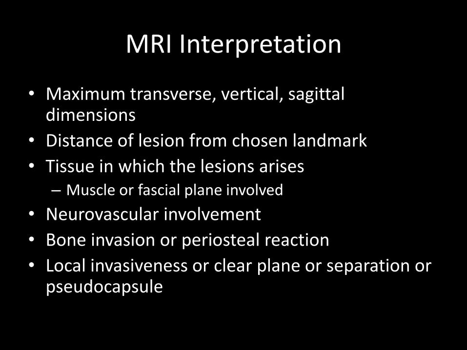
MRI Interpretation
• Maximum transverse, vertical, sagittal dimensions
• Distance of lesion from chosen landmark
• Tissue in which the lesions arises – Muscle or fascial plane involved
• Neurovascular involvement
• Bone invasion or periosteal reaction
• Local invasiveness or clear plane or separation or pseudocapsule

MRI Interpretation
• Describe intrinsic appearance • SI on T1-weighted imaging related to muscle • Hyperintense signal on fluid-sensitive images evaluated for
homogeneity • Specific features
– Fluid levels, focal fluid collections, lobularity, leaking of fluid, prominent feeding vessels
• Describe pattern and degree of contrast enhancement – Degree of necrosis
• Biopsy site – Confer with surgeon, biopsy track resected along with lesion – Most aggressive site, avoid areas of necrosis, hemorrhage, or
dystrophic calcifications
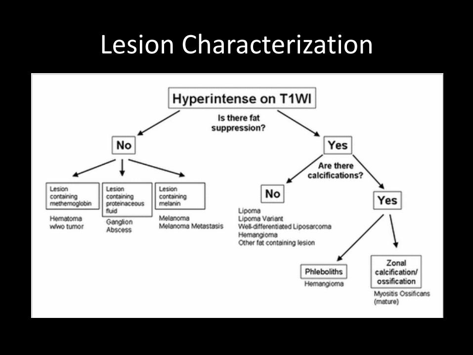
Lesion Characterization


WHO Classification of Soft Tissue Tumors -2013
• 12 categories
• Each category divided into 4 biological behavior subgroups
– Benign
– Intermediate (locally aggressive)
– Intermediate (rarely metastasizing)
– Malignant

WHO Classification of Soft Tissue Tumors
• Adipocytic tumors • Fibroblastic/myofibroblastic tumors • So-called Fibrohistiocytic tumors • Smooth muscle tumors • Pericytic (perivascular) tumors • Skeletal muscle tumors • Vascular tumors • Chondro-osseous tumors • Gastrointestinal stromal tumors • Nerve sheath tumors • Tumors of uncertain differentiation • Undifferentiated/unclassified sarcomas
*not included: ganglia/cyst, hematoma/abscess, granuloma, Morton neuroma, anatomical variants

Soft Tissue Tumors with Specific Characterization
• Group 1- Lipomatous tumors
• Group 2- Fibromatosi/Elastofibroma dorsi/Myositis Ossificans
• Groups 3- PVNS, GCTTS
• Group 5- Glomus
• Group 7- Hemangioma
• Group 9- Neurogenic tumors
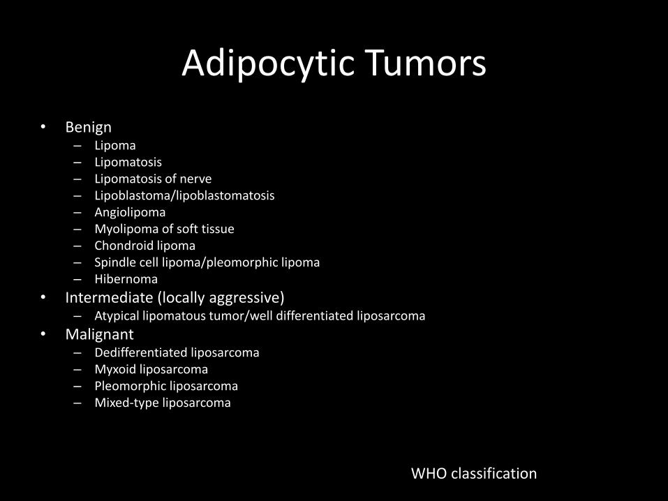
Adipocytic Tumors
• Benign – Lipoma – Lipomatosis – Lipomatosis of nerve – Lipoblastoma/lipoblastomatosis – Angiolipoma – Myolipoma of soft tissue – Chondroid lipoma – Spindle cell lipoma/pleomorphic lipoma – Hibernoma
• Intermediate (locally aggressive) – Atypical lipomatous tumor/well differentiated liposarcoma
• Malignant – Dedifferentiated liposarcoma – Myxoid liposarcoma – Pleomorphic liposarcoma – Mixed-type liposarcoma
WHO classification

Lipomas and Lipomatous Lesions
• Most common soft tissue tumor
• 2.1 in 100 individuals
• Histologically identical to adipose fat
• Classic lipoma: entirely fat without nodularity or thickened septations
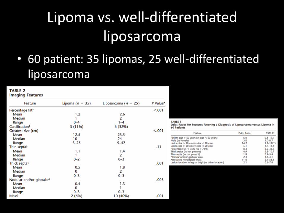
Lipoma vs. well-differentiated liposarcoma
• 60 patient: 35 lipomas, 25 well-differentiated liposarcoma

• 18 lesions had areas of increased signal intensity on fluid sensitive MR, 7 (39%) were benign
48 y/o M with lipoma in posterior compartment of thigh

Lipoma
• 11 of 35 lipomas (31%) had significant nonadipose content
• Nonadipose content typically fat necrosis with associated calcification, fibrosis, inflammation, and areas of myxoid change
• 8 contrast enhanced studies available – 1 lipoma showed no enhancement while 3 showed
mild linear enhancement
– 4 liposarcomas showed mild to moderate enhancement
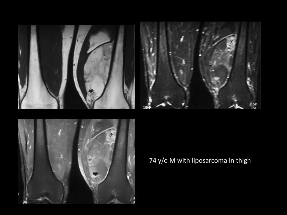
74 y/o M with liposarcoma in thigh

184 palpable subcutaneous fatty masses evaluated on MR -all masses localized with skin markers on images, MR reported stated that patient examined by radiologists and location of mass palpable, or exact location of recorded in medical record -85 (46%) classified as partially or completely encapsulated -99 (54%) classified as nonencapsulated -no histologic analysis to determine if nonencapsulated masses differ from normal fat (fatty hypertrophy, asymmetric fatty deposition, or areas of fat surrounded by fibrosis) -report as "nonencapsulated lipoma" instead of normal to avoid additional imaging
Hemangiomas
• Benign vascular lesions composed of various vessels, 7% of all benign soft tissue tumors
• Can be found in any organ
• Common in infancy and childhood but can occur in any age group
• Can manifest as bluish skin discoloration and history of size fluctuation
• Pain may occur following exercise owing to shunting of blood away from surrounding tissue

• Phleboliths on radiographs in 20-67% patients
• On MR, may be well-circumscribed or have poorly defined margins with varying amounts of T1 signal owing to either reactive fat overgrowth or hemorrhage
• Contain serpentine vessels, fat, smooth muscles, hemosiderin, and phleboliths
• Areas of slow flow have high T2 signal, rapid flow demonstrate flow void
Soft Tissue Hemangioma vs. Malignant Soft Tissue Masses
• Teo et al • 22 peripheral hemangiomas and 22 primary malignant soft tissue
masses (MFH, rhabdomyosarcoma, neurofibrosarcoma, primitive neuroectodermal tumors, 6 others)
• T1-weighted imaging – No reliable distinguishing feature
• T2-weighted imaging – Lobulation, septation, central low-intensity dots more frequently seen
in hemangiomas, masses with all three were exclusively hemangiomas • Central low intensity dots represent fibrofatty septa seen in cross section,
thrombosed vascular channels, smooth muscle components, fast flow, calcification or ossification
– Higher T2 signal intensities
• Post-contrast T1-weighted imaging – Hemangiomas enhance markedly
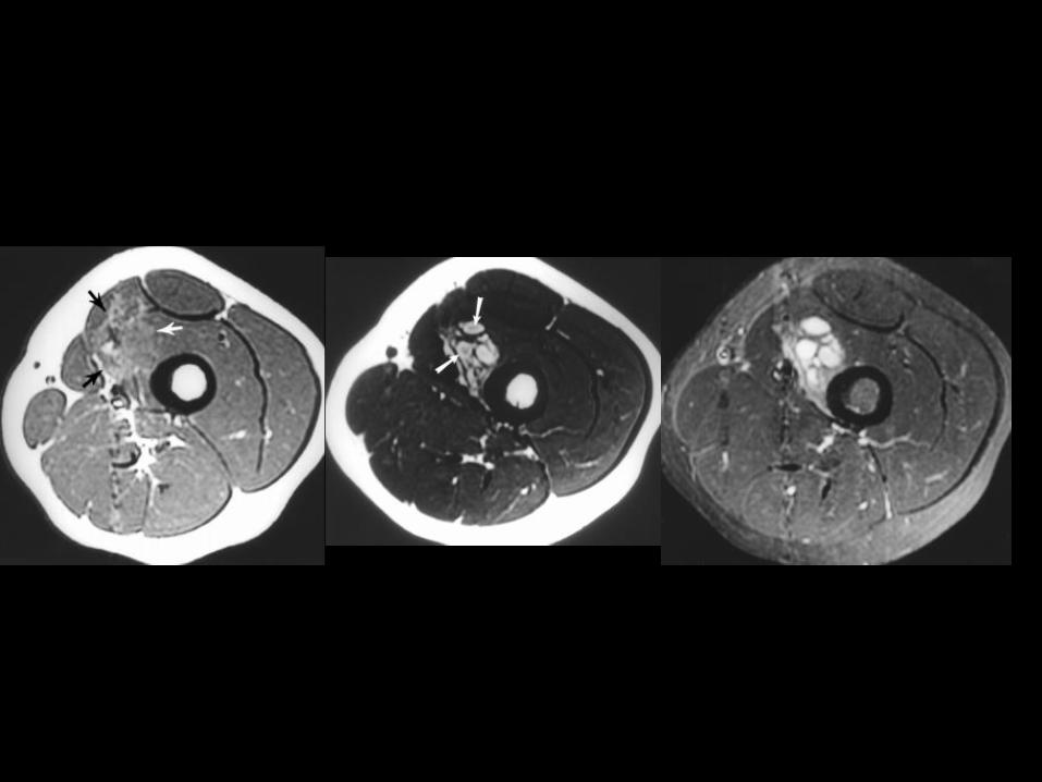

• Regional bone changes adjacent to soft tissue hemangiomas, exact mechanism unknown
• Radiographs and MR of 35 patients with pathologically proven hemangiomas reviewed
• 14/35 patients had osseous changes – periosteal (23%), cortical (31%), medullary (29%)
• No correlation between presence of symptoms and presence of osseous change
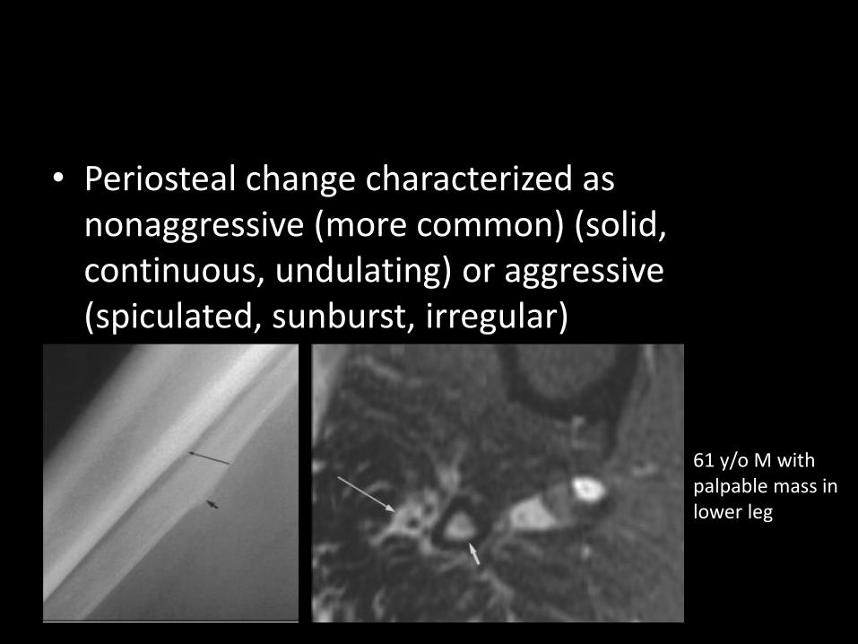
• Periosteal change characterized as nonaggressive (more common) (solid, continuous, undulating) or aggressive (spiculated, sunburst, irregular)
61 y/o M with palpable mass in lower leg


• Cortical findings:
– Thickening, erosion, tunneling (pseudopermeative cortex,
osteoporosis, radiation therapy), osteopenia

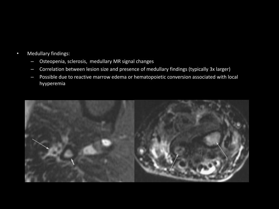
• Medullary findings:
– Osteopenia, sclerosis, medullary MR signal changes
– Correlation between lesion size and presence of medullary findings (typically 3x larger)
– Possible due to reactive marrow edema or hematopoietic conversion associated with local hyyperemia

• Proximity of hemangioma correlates with all three categories of osseous change
• Hemangioma contacts bone in nearly all cases

Glomus Tumor
• Hamartoma arising from glomus body, an arteriovenous shunt within dermis that contributes to temperature regulation
• Each glomus body is 300 μm long
• Nail beds of fingers and toes contain 93-501 glomus bodies per square centimeter

Glomus tumor
Epidemiology • Rare, <2% of soft tissue tumors • Multiple lesions in 10% • Malignant in <10% • No sex predilection except in subungal- F>M Site of involvement • Majority occur in distal extremities
– Subungal, hand, wrist, foot
• Reported in every location – Stomach, penis, mediastinum, nerve, bone, lung
• Almost always in skin or superficial soft tissue • Malignant tumors usually deeply seated

Glomus Tumor
Clinical features • Typically small, <1 cm • Red-blue nodules a/w long history of pain, particularly with
exposure to cold or minor tactile stimulation • Deeply seated tumors are asymptomatic or have pain
referable to involved organ • Hildreth sign: disappearance of pain after tourniquet
application, diagnostic • Treatment
– Surgical resection leads to immediate pain relief – Recurrence 12-24%

Glomus Tumor

• Most tumors surrounded by capsule, as secondary reaction of surrounding tissue – Dark rim on T2 weighted images
• T2 hyperintense • Variable T1 signal intensity, low signal to
moderate high signal – Increased T1 signal due to hemorrhage or vascularity
• Intense enhancement • Bone erosion, 15-65%
– Smooth bony expansion


Glomus Tumor
• Differential diagnosis – Mucous cysts
• Commonly seen dorsal aspect DIP • Fluid signal • No enhancement
– Epidermoid inclusion cyst • Can be similar to glomus tumor in signal intensity • Bone expansion uncommon • Unlikely to be centered at nail bed • History of penetrating trauma • Painless
– Giant cell tumor of tendon sheath • Proximity to tendon sheath • Lower T2 signal intensity • Hemosiderin

Giant Cell Tumor of Tendon Sheath
• Histologically identical to intra-articular pigmented villonodular synovitis
• Most commonly seen in the hand, adjacent to an interphalangeal joint
• Manifest as small slow-growing mass with or without pain
• Radiographs normal or reveal nonaggressive remodeling of bone
Giant Cell Tumor of Tendon Sheath
• Sites of involvement – Second most common soft tissue tumor in hand after
ganglion cyst – 85% occur in fingers – Infrequently erode or infiltrate bone – Other sites: wrist, ankle/foot, knee, hip
• Clinical features – Benign lesion with local recurrence 9-44% after
recurrence – 30-50 y/o – Most commonly present as painless swelling

GSTTS
MR features • Well marginated • Isointense or hypointense to muscle on T1- and T2-
weighted MR owing to abundant collagen and hemosiderin – Some lesions don’t contain enough hemosiderin to be T1 and T2
hypointense
• Inhomogeneous signal intensities with nodular, linear, or peripheral low signal areas
• Strong enhancement • Difficult to differentiate from fibroma of tendon sheath
– Fibromas occur in slightly younger population and more common in men


Sonographic Features of GCTTS
• Hypoechoic • Homogeneous (rarely heterogeneous) • Posterior acoustic enhancement occasionally
seen • No cystic elements or calcifications • Vascularity, both central and peripheral • Circumferential contact with tendon on short axis
ranged from 30 to 360 degrees • Tumors do not move with affected digit flexed or
extended


Myositis Ossificans
• Benign, solitary, self limiting, ossifying soft tissue mass occurring within skeletal muscle
• Often no history of trauma
• No association with primary inflammation of muscle
• Clinical features – Frequently present with pain and tenderness and soft
tissue mass
– May be incidental finding
– 80% arise in large muscles of extremities

Myositis Ossificans
• Imaging features dependent on age of lesion • Full course of growth 7-8 weeks from inception • 30% demonstrate spontaneous regression

• Early lesion – Radiographs: normal
– T2: iso-to hyperintense to muscle
– T1: iso- to hyperintense
– +/- T2 hypointense rim
– Heterogeneous with surrounding soft tissue edema
– Poorly marginated and may be recognized only secondarily due to mass effect and displacement of fascial planes
– Marked enhancement
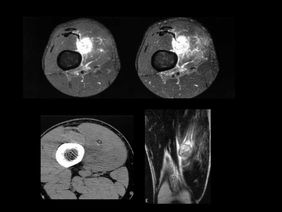
• Intermediate stage – Radiographs: continuous/noncontinuous
peripheral calcification with central lucent core, faint irregular calcifications within lesion
– MRI • Inhomogeneous, variable signal at center on T2
• Well defined, decreased signal rim of varying thickness on all sequences
• Varying, nonspecific enhancement
• Decreased perilesional abnormal signal

• Late stage
– Radiographs: heavily calcified lesion with trabecular bone formation, may merge with adjacent bone
– MRI:
• Overall low signal due to ossification, fibrosis, hemosiderin
• Areas of signal identical to normal bone marrow corresponding to fatty marrow formation
• Resolution of perilesional edema


Sonographic Features of Myositis Ossificans
• Early – Thin echo-poor zone I in surrounding muscle – Broad, reflective zone II – Amorphous, echo free zone III
• IntermediateMature – Zone II more reflective due to increased mineralization

• Most soft tissue tumors have nonspecific imaging characteristics
• Identify benign lesions to avoid unnecessary intervention
• Any indeterminate lesions should be biopsied

References
1 Berquist, Thomas H., et al. "Value of MR imaging in differentiating benign from malignant soft-tissue masses: study of 95 lesions." AJR. American journal of roentgenology 155.6 (1990): 1251-1255.
2 Chhabra, Avneesh, and Theodoros Soldatos. "Soft-tissue lesions: when can we exclude sarcoma?." American Journal of Roentgenology 199.6 (2012): 1345-1357.
3Crim, J. R., et al. "Diagnosis of soft-tissue masses with MR imaging: can benign masses be differentiated from malignant ones?." Radiology 185.2 (1992): 581-586.
4Drape, Jean-Luc, et al. "Subungual glomus tumors: evaluation with MR imaging." Radiology 195.2 (1995): 507-515.
5Gielen, Jan LMA, et al. "Accuracy of MRI in characterization of soft tissue tumors and tumor-like lesions. A prospective study in 548 patients." European radiology 14.12 (2004): 2320-2330.
6Kitagawa, Yasuyuki, et al. "MR imaging for preoperative diagnosis and assessment of local tumor extent on localized giant cell tumor of tendon sheath." Skeletal radiology 32.11 (2003): 633-638.
7Kransdorf, Mark J., and Mark D. Murphey. "Radiologic evaluation of soft-tissue masses: a current perspective." American Journal of Roentgenology 175.3 (2000): 575-587.
8Kransdorf, Mark J., et al. "Imaging of Fatty Tumors: Distinction of Lipoma and Well-differentiated Liposarcoma 1." Radiology 224.1 (2002): 99-104.
9Lacout, Alexis, et al. "Myositis ossificans imaging: keys to successful diagnosis." The Indian journal of radiology & imaging 22.1 (2012): 35.
10Ly, Justin Q., et al. "Osseous change adjacent to soft-tissue hemangiomas of the extremities: correlation with lesion size and proximity to bone." American Journal of Roentgenology 180.6 (2003): 1695-1700.

11Manaster, B. J. "Soft-tissue masses: optimal imaging protocol and reporting."American Journal of Roentgenology 201.3 (2013): 505-514.
12Middleton, William D., et al. "Giant cell tumors of the tendon sheath: analysis of sonographic findings." American Journal of Roentgenology 183.2 (2004): 337-339.
13Teo, Eu-Leong HJ, Peter J. Strouse, and Ramiro J. Hernandez. "MR imaging differentiation of soft-tissue hemangiomas from malignant soft-tissue masses."American Journal of Roentgenology 174.6 (2000): 1623-1628.
14Thomas, E. A_, V. N. Cassar-Pullicino, and I. W. McCall. "The role of ultrasound in the early diagnosis and management of heterotopic bone formation." Clinical radiology 43.3 (1991): 190-196.
15Wang, X. L., et al. "Pictorial essay. Myositis ossificans circumscripta." JBR-BTR: organe de la Societe royale belge de radiologie (SRBR)orgaan van de Koninklijke Belgische Vereniging voor Radiologie (KBVR) 86.5 (2002): 278-285.
16Wu, Jim S., and Mary G. Hochman. "Soft-Tissue Tumors and Tumorlike Lesions: A Systematic Imaging Approach 1." Radiology 253.2 (2009): 297-316.