soluble human leukocyte antigen -g during pregnancy and
TRANSCRIPT

RESEARCH ARTICLE
Soluble human leukocyte antigen -G during
pregnancy and infancy in Benin: Mother/child
resemblance and association with the risk of
malaria infection and low birth weight
Tania C. d’Almeida1,2☯*, Ibrahim Sadissou2,3,4,5☯, Jacqueline Milet2,6, Gilles Cottrell2,6,
Amandine Mondière7, Euripide Avokpaho8, Laure Gineau2, Audrey Sabbagh2,
Achille Massougbodji3,4, Kabirou Moutairou4, Eduardo A. Donadi5, Benoit Favier9,10,
Edgardo Carosella9,10, Philippe Moreau9,10‡, Nathalie Rouas-Freiss9,10‡, David Courtin2,6‡,
Andre Garcia1,2,3‡
1 Universite Pierre et Marie Curie, Paris, France, 2 UMR 216-MERIT, Institut de Recherche pour le
Developpement, Faculte de Pharmacie - Universite Paris Descartes, Sorbonne Paris-Cite, Paris, France,
3 Centre d’Etude et de Recherche sur le Paludisme Associe à la Grossesse et à l’Enfance, Faculte des
Sciences de la Sante, Cotonou, Benin, 4 Universite d’Abomey-Calavi, Cotonou, Benin, 5 Division of Clinical
Immunology, School of Medicine of Ribeirão Preto, University of São Paulo, Brazil, 6 Universite Paris
Descartes, Sorbonne Paris Cite, Paris, France, 7 UMR 216-MERIT, Institut de Recherche pour le
Developpement, Campus de la Faculte des Sciences de la Sante (FSS) et de l’Institut des Sciences
Biomedicales Appliquees (ISBA), Cotonou, Benin, 8 Ecole Pasteur – CNAM de sante publique, Paris,
France, 9 CEA, Institut des Maladies Emergentes et des Therapies Innovantes (IMETI), Service de
Recherches en Hemato-Immunologie (SRHI), Hopital Saint-Louis, IUH, Paris, France, 10 Universite Paris
Diderot, Sorbonne Paris Cite, IUH, Hopital Saint-Louis, UMR_E5, IUH, Paris, France
☯ These authors contributed equally to this work.
‡ These authors also contributed equally to this work.
Abstract
Human leukocyte antigen (HLA) G is a tolerogenic molecule involved in the maternal-fetal
immune tolerance phenomenon. Its expression during some infectious diseases leading to
immune evasion has been established. A first study conducted in Benin has shown that the
production of soluble HLA-G (sHLA-G) during the first months of life is strongly correlated
with the maternal level at delivery and associated with low birth weight and malaria. How-
ever sHLA-G measurements during pregnancy were not available for mothers and further-
more, to date the evolution of sHLA-G in pregnancy is not documented in African
populations. To extend these previous findings, between January 2010 and June 2013, 400
pregnant women of a malaria preventive trial and their newborns were followed up in Benin
until the age of 2 years. Soluble HLA-G was measured 3 times during pregnancy and
repeatedly during the 2 years follow-up to explore how sHLA-G evolved and the factors
associated. During pregnancy, plasma levels of sHLA-G remained stable and increased
significantly at delivery (p<0.001). Multigravid women seemed to have the highest levels
(p = 0.039). In infants, the level was highest in cord blood and decreased before stabilizing
after 18 months (p<0.001). For children, a high level of sHLA-G was associated with malaria
infection during the follow-up (p = 0.02) and low birth weight (p = 0.06). The mean level of
sHLA-G during infancy was strongly correlated with the mother’s level during pregnancy
PLOS ONE | DOI:10.1371/journal.pone.0171117 February 6, 2017 1 / 17
a1111111111
a1111111111
a1111111111
a1111111111
a1111111111
OPENACCESS
Citation: d’Almeida TC, Sadissou I, Milet J, Cottrell
G, Mondière A, Avokpaho E, et al. (2017) Soluble
human leukocyte antigen -G during pregnancy and
infancy in Benin: Mother/child resemblance and
association with the risk of malaria infection and
low birth weight. PLoS ONE 12(2): e0171117.
doi:10.1371/journal.pone.0171117
Editor: Sylvie Bisser, Universite de Limoges,
FRANCE
Received: August 11, 2016
Accepted: January 4, 2017
Published: February 6, 2017
Copyright: © 2017 d’Almeida et al. This is an open
access article distributed under the terms of the
Creative Commons Attribution License, which
permits unrestricted use, distribution, and
reproduction in any medium, provided the original
author and source are credited.
Data Availability Statement: All relevant data are
within the paper file.
Funding: This study was supported by the Agence
Nationale de la Recherche (ANR) (project 2006/
040/001), Programa Ciênca sem Fronteiras «
Pesquisador Visitante Especial » (grant 406594/
2013-9) and iDEX Sorbonne Paris Cite « mobilite
internationale Bresil 2015 ». Tania C d’Almeida was
partly supported by grants from « Fondation pour
la Recherche Medicale » (Grant

(<0.001), and not only at delivery. Moreover, mothers with placental malaria infection had a
higher probability of giving birth to a child with a high level of sHLA-g (p = 0.006). High sHLA-
G levels during pregnancy might be associated with immune tolerance related to placental
malaria. Further studies are needed but this study provides a first insight concerning the
potential role of sHLA-G as a biomarker of weakness for newborns and infants.
Introduction
Human leukocyte antigen-G (HLA-G) is a nonclassical HLA class I antigen that differs from
the others HLA class I molecules in its limited polymorphism, its restricted tissue distribution
and its particular expression [1]. Alternative splicing encodes four membrane-bound and
three soluble isoforms. The main isoforms present in the plasma are soluble HLA-G1 and -G5
[2]. HLA-G also differs from the HLA class I molecule by exerting inhibitory functions on
immune responses [3–5].
The expression of HLA-G during pregnancy and its importance in maternal—fetal toler-
ance is well established [6, 7]. Indeed, the major physiological role of HLA-G is the protection
of the fetal semi-allograft against lysis by maternal NK cells. Moreover, interaction between
HLA-G and CD158d in NK cells seems to promote vascularization in maternal decidua during
early pregnancy which is important for the development of the fetus [8, 9]. The levels of soluble
HLA-G (sHLA-G) are significantly higher in pregnant than in nonpregnant women [10–12].
sHLA-G has been used as a reliable marker for following in vitro fertilization [13, 14] and was
significantly lower in preeclampsia than in normal pregnancy [15]. In newborns, studies have
shown that the levels in cord blood are lower than in mothers [12, 16].
The origin of the variability of sHLA-G blood levels is complex and not fully known, involv-
ing genetic mechanisms [17]. Until recently, African populations have been underrepresented
in studies despite the greater genetic diversity and heterogeneity observed across Africa [18].
There are currently no data on how sHLA-G evolves during pregnancy in African populations
except one study with data available only at delivery for mothers. A strong positive correlation
between sHLA-G levels in mothers at delivery and children has been highlighted, and high lev-
els of sHLA-G in children appeared to increase the malaria risk and are associated with low
birth weight (LBW) [19], which is highly correlated with neonatal morbidity and mortality
[20, 21]. Unfortunately, gestational information was not available in this first study and it
seemed important to account for the course of pregnancy.
In case of malaria infection of the placenta an immune tolerance phenomenon has been
described as responsible for the higher susceptibility of newborns to malaria infection during
the first months of life [22–24]. Due to the important role of HLA-G in immune regulation
and immune tolerance and to its strong expression during pregnancy, the potential association
between sHLA-G during the whole pregnancy, and not only at delivery and malaria needs to
be explored.
In the current study involving pregnant women, our aim was to explore the mother/child
resemblance and to confirm or not the association between malaria, LBW and sHLA-G level.
Materials and methods
Study design and follow-up
The present follow-up is part of a study concerning 1,183 human immunodeficiency virus
(HIV)-negative pregnant women participating in Malaria in Pregnancy Preventive
Soluble HLA-G during pregnancy and infancy in Benin
PLOS ONE | DOI:10.1371/journal.pone.0171117 February 6, 2017 2 / 17
FDM20130727043) and by Institut de Recherche
pour le Developpement (IRD). The clinical trial in
which the study was nested was funded by the
European Developing Countries Clinical Trials
Partnership (EDCTP; IP.2007.31080.002), the
Malaria in Pregnancy Consortium.
Competing Interests: The authors have declared
that no competing interests exist.

Alternative Drugs (http://clinicaltrials.gov/ct2/show/NCT00811421), a randomized trial of
intermittent preventive treatment (IPTp) with either sulfadoxine-pyrimethamine or meflo-
quine. The first 400 infants born to these women were enrolled from January 2010 to June
2011 and followed throughout the first 2 years of life [25, 26]. Twin pregnancies, stillbirth,
or fetal abnormalities were excluded (<1%). In the same way, women with renal, hepatic,
neurologic and psychiatric active diseases were not selected. Women were included before
the end of 28 gestational weeks (GW) and two doses of IPTp were administered at antenatal
visits (ANVs). Between ANVs, women had to attend the health centre for all health com-
plaints. Women were examined and a questionnaire completed. At inclusion, sociodemo-
graphic information, socioeconomic characteristics, reproductive and medical history were
collected.
Three blood samples were collected during scheduled ANVs (before IPTp) for plasma
sHLA-G measurement and malaria diagnosis. After delivery, a placental blood smear was used
to assess placental malaria. Malaria infection was defined as the presence of asexual Plasmo-dium falciparum parasites in a blood smear.
Newborns were followed from birth to 24 months. At 6, 9, 12, 18 and 24 months, children
were visited and clinically examined, and anthropometric data were collected. Children were
regularly visited, a medical questionnaire was filled out and a systematic thick blood smear
(TBS) was performed to search for asymptomatic malaria. Mothers were invited to visit health
centres with their child if there was any health problem and in case of fever or history of fever,
a rapid diagnosis test (RDT) was performed. A malaria attack was defined as an axilary tem-
perature greater or equal to 37.5 (or history of fever) and a positive RDT. Cord blood was sam-
pled at birth, peripheral blood at 6, 9, 12, 18 and 24 months, and the same tests as in mothers
were performed. All the medications prescribed were free of charge.
Soluble HLA-G quantification
Soluble HLA-G was quantified using MEM-G/9 [27], which recognizes the most abundant sol-
uble isoforms (sHLA-G1, -G5) and anti-human β2-microglobulin as capture and detection
antibodies, respectively [28]. All incubation steps were performed at room temperature and
followed by four washes using washing buffer (H2O, PBS 1X, 0.1% Tween20). The plates were
incubated for 30 min with the substrate (Tetramethyl benzidine, Sigma Aldrich, USA) and
absorbance was measured at 450 nm after adding HCL (1 N). Total sHLA-G levels were deter-
mined from a five-point standard curve (12.5–200 ng/ml) using dilutions of calibrated
HLA-G5 purified from M8-HLA-G5 cell line culture supernatant, and the results were
expressed as ng/ml. The limit of detection is ~1 ng/ml. The methodology to measure HLA-G
using ELISA has been validated [27].
Ethics
The study protocol and informed consent were approved by the Comite Consultatif de
Deontologie et d’Ethique (CCDE) of the Institut de Recherche pour le Developpement (IRD,
France) and the Ethics Committee of the Faculte des Sciences de la Sante de Cotonou
(Benin), the local ethic review Committee. Before inclusion, the study was explained in the
local language. A verbal agreement and a written consent were obtained: a copy of this docu-
ment was given to each participant of the study. In the case that the woman could not read,
an impartial witness was involved in the process. In addition to the assent of minors, consent
was obtained from the parents or legal guardians. Women were free to interrupt their partic-
ipation at any time.
Soluble HLA-G during pregnancy and infancy in Benin
PLOS ONE | DOI:10.1371/journal.pone.0171117 February 6, 2017 3 / 17

Definition of variables
The outcome variable was the level of sHLA-G in mothers and infants, used as a quantitative
variable. To study mother/child resemblance, the mean level of sHLA-G was calculated
throughout the pregnancy for women and during follow-up for infants. The cord blood level
was not included in the mean calculation. Every woman/infant pair was classified as belonging
to one of the four quartiles of the mean distribution depending on their mean level (ng/ml).
The quartiles allowed us to define the sHLA-G profile for each individual. For mothers the
profiles (P) were: very low (P1�1.19), low (1.19<P2�9.11), medium high (9.11<P3�19.05)
and high (19.05<P4); for children: very low (P1�1.34), low (1.34<P2�7.21), medium high
(7.21<P3�18.43) and high (18.43<P4).
The following covariates were used:
• For mothers: women’s age, ethnicity (Aïzo, Fon, and others), newborn’s gender and birth
weight, parity, GW, placental and peripheral malaria infection during follow-up;
• For children: birth weight, prematurity (�37 GW), age, gender, height, ethnicity, fever, CRP
(C-reactive protein), maternal age, parity, maternal malaria (placental or peripheral) and
malaria infection of children.
Statistical analysis
Data were analyzed with Stata1 Software, Version 12 (StatCorp LP, College Station, TX,
USA). Mothers and children were analyzed separately.
For mothers, the three sHLA-G measurements were not considered as classical repeated
measurements (IPTp administration for the first two ANVs and delivery at the third visit). Dif-
ferent arguments could explain this choice. Pregnancy could be considered as a special period
for the mother, related to the presence of the fetus. The first and second antenatal visits
(ANV1 and 2) occurred at different times for the women and were characterized by many
important interventions of the care team. Therefore, the effects of covariates on the sHLA-G
level at each visit were explored separately using a Tobit regression model, a censored regres-
sion model, to account for the truncated values generated by the sHLA-G detection limit [29,
30]. The evolving sHLA-G level at each gestational month was compared using a Kruskal-Wal-
lis test with Bonferroni correction. For children, systematic scheduled visits are not indepen-
dent and the longitudinal data collected along the first two years of children presented a
hierarchical two-level structure, where sHLA-G measurements (level 1) were clustered within
children (level 2). Hierarchical mixed Tobit regression models were used for univariate and
multivariate analyses with an independent variance-covariance structure of the random effects
which is the default option [31]. Lastly, mother/child resemblance was explored using the
sHLA-G profiles as outcome. Since the profiles were ordered from “very low” to “very high”,
an ordinal logistic regression model was used.
All factors with a p-value<0.20 during univariate analyses and other interesting factors
were included in the multivariate step. Statistical significance was set at p<0.05.
Results
The women’s mean age was 25.9 years (95% confidence interval [25.4–26.4]; range, 18–42).
The first ANV occurred before 29 GW and 15.7% were primigravid. Throughout the follow-
up, 27.7% of women developed peripheral malaria and 10.9% had placental malaria at delivery.
In children, the mean birthweight was 3033.92 g (IC95%: [2992.50–3075.35]), 9.05% of
them had a LBW, and 14 newborns were preterm. During the study, 12.16% of children devel-
oped a malaria infection between 6 and 24 months. There is no infection at birth in newborns.
Soluble HLA-G during pregnancy and infancy in Benin
PLOS ONE | DOI:10.1371/journal.pone.0171117 February 6, 2017 4 / 17

The general characteristics of the study population are presented in Table 1.
Evolution of soluble HLA-G in pregnancy
Blood was sampled in 85% of mothers for the three ANVs, and 98% were sampled at least
twice. sHLA-G was described by month of pregnancy after grouping the first 3 months (Fig 1).
The sHLA-G level remained stable during pregnancy and increased significantly at delivery
(p<0.001).
At the first ANV, univariate analysis showed that multigravid women had a higher sHLA-G
level (p = 0.036) than primigravid women. The sHLA-G level seemed lower in women who
took mefloquine for IPTp (p = 0.05) and those with placental malaria (p = 0.04). In contrast,
there is no effect of age, peripheral malaria, foetus gender and LBW. In multivariate analysis,
gravidity and IPTp remained significant (p = 0.039 and p = 0.05, respectively), but the associa-
tion of sHLA-G with placental malaria became marginally significant (p = 0.07).
At the second ANV, previous associations were no longer significant. However, the oldest
women (>25 years) had a higher sHLA-G level than the youngest (p = 0.009) (Table 2).
At delivery, there was no significant association. Women giving birth to a LBW newborn
had a higher sHLA-G level and women with placental malaria had lower levels, but the differ-
ences were marginally significant (p = 0.086 and p = 0.07, respectively) (Table 2).
Table 1. Characteristics of women and newborns in Allada, 2007–2010.
Mothers (n = 400) Newborns (n = 400)
Age 25.92 years Birth weight 3033.92 g
SD: 5.45 SD: 420.37
Schooling 29.75% (117) Low birthweight 9.05% (36)
Ethnicity Aïzo 69.50% (278) Prematurity 10.00% (40)
Fon 20.75% (83) Gender Female 53.00% (212)
Others 9.75% (39) Male 47.00% (188)
Previous pregnancies 0 15.75% (63) HLA-G (ng/ml) (c) Birth 20.16, SD: 28.13
1 20.75% (83) 6 months 18.17, SD: 33.76
2 14.75% (59) 9 months 15.60, SD: 35.87
3 17.25% (69) 12 months 11.23, SD: 19.84
4 10.00% (40) 18 months 8.38, SD: 15.43
�5 21.50% (86) 24 months 9.66, SD: 23.85
Married 97.75% (391)
Primigravidae 15.75% (63)
IPTp (a) SP 34.50% (138)
MQFD 35.50% (142)
MFSD 30.00% (120)
Placental malaria 10.75% (43)
Health center Attogon 24.00% (96)
Sekou 76.00% (304)
HLA-G (ng/ml) (b) ANV1 10.08, SD: 13.60
ANV2 10.57, SD: 14.05
Delivery 17.25, SD: 34.55
(a) Two molecules were used for IPTp according to the protocol of the MIPPAD study: Sulfadoxine-pyrimethamine (SP: 1500/75 mg) and Mefloquine (MQ:
15 mg/kg), which is given once as a full dose (MQFD) or split over 2 days (MQSD).(b): mean level of soluble HLA-G in the population at each ANV and at delivery.(c): mean level of soluble HLA-G in children at birth and at each visit.
doi:10.1371/journal.pone.0171117.t001
Soluble HLA-G during pregnancy and infancy in Benin
PLOS ONE | DOI:10.1371/journal.pone.0171117 February 6, 2017 5 / 17

Soluble HLA-G during infancy
Blood was collected for 373 newborns, 311 with at least three samples. The level was highest in
cord blood, and decreased until 18 months (p<0.001) (Fig 2).
There was no significant association between gender, birth weight and sHLA-G level.
Mothers’ malaria infection (placental or peripheral) was not associated with the children’s
sHLA-G level. In contrast, children infected by malaria at least once during the follow-up had
a higher sHLA-G level (p = 0.02) (Table 3). We identified an interaction between age and LBW
showing that children with LBW had higher sHLA-G levels and a different evolution during
the first 24 months of life (p = 0.06) (Fig 3).
Mother/child resemblance
As described above, the sHLA-G level in children is correlated with their mother’s level. There
were a strong correlation (p<0.001) between each mother’s cross-sectional measurement and
her child’s levels throughout the follow-up (Table 4) and an association between the mother’s
profile and her child’s mean sHLA-G level during the follow-up (p<0.001). Children born to a
mother with very low profile had the lowest level, whereas children born to a mother with very
high profile had the highest mean sHLA-G level.
Fig 1. Evolution of the mean level (and 95 confidence intervals) of soluble HLA-G during pregnancy. (*) indicates that the
level of sHLA-G is significantly higher at delivery (p<0.001; Kruskal-Wallis test). (�3 months: n = 26, SD: 12.39, 4 months: n = 65,
SD: 13.92, 5 months: n = 177, SD: 17.01, 6 months: n = 254, SD: 11.48, 7 months: n = 131, SD: 15.20, 8 months: n = 115, SD:
12.09, 9 months: n = 360, SD: 35.11).
doi:10.1371/journal.pone.0171117.g001
Soluble HLA-G during pregnancy and infancy in Benin
PLOS ONE | DOI:10.1371/journal.pone.0171117 February 6, 2017 6 / 17

Using multinomial ordinal models, a mother belonging to a certain profile had a signifi-
cantly higher probability of giving birth to a child belonging to the same profile (Fig 4). Fur-
thermore, the odds ratio for giving birth to a child with a “very high” profile versus all others
was strongly associated with the increase in the mother’s profile (p<0.001). For an increase in
the mother’s profile from “very low” to “low”, this OR was 3.05 (CI95 = [1.75–5.31]). The OR
was 17.59 (CI95 = [9.54–32.41]) for an increase from “very low” to “very high” (Table 5). The
OR for giving birth to a child with a very high profile for a mother with placental malaria,
adjusted on the mother’s profile, was 2.39 [1.29–4.45] compared to a mother without placental
infection (Table 5).
Discussion
The present study is the first conducted in an African population describing the physiological
evolution of the sHLA-G level during pregnancy. This work complements a study conducted
in Benin that found a strong correlation between maternal sHLA-G level at delivery and chil-
dren’s levels during the first years of life as well as an association between the infant’s level and
LBW and malaria [19]. However, in this study mothers were included at delivery and preg-
nancy data were not available. Associations between both malaria and LBW and the infant’s
sHLA-G level were confirmed here. Furthermore, the correlation between the mother’s and
the child’s sHLA-G levels was reinforced since we showed that the child’s level throughout the
follow-up was correlated with its mother’s level at each measurement. Finally, mothers with
placental malaria gave birth to children with high sHLA-G profile during infancy. This last
result is consistent with the fact that the immune tolerance described in case of placental
malaria [22–24] could be correlated with sHLA-G levels and that placental malaria might be
considered as a marker of a more complex phenomenon involving the tolerogenic molecule
HLA-G.
Table 2. Factors associated with sHLA-G level in pregnancy: Regression of Tobit (univariate and multivariate).
Covariates ANV 1 (IPT1) ANV 2 (IPT2) Delivery
β [95% CI] Adjus-
ted β[95% CI] β [95% CI] β [95% CI] Adjus-
ted ß
[95% CI]
(p) (p) (p) (p) (p)
Age (years) � 25 0 [-0.79, 7.34] 0 [1.70, 10.01] 0 [-9.93, 8.04]
> 25 3.27 (0.11) 5.86 (0.006) −0.95 (0.84)
Ethnicity Fon 0 [-5.81, 4.07] 0 [-6.33, 3.83] 0 [-11.36, 10.71]
Others −0.87 (0.73) −1.25 (0.63) −0.32 (0.95)
Gender Male 0 [-5.16, 2.96] 0 [-5.90, 2.44] 0 [-11.12, 6.81]
Female −1.09 (0.59) −1.73 (0.41) −2.15 (0.64)
Primigravidity No 0 [-11.94, -0.41] 0 [-11.79, -0.30] 0 [-8.98, 2.65] 0 [-13.96, 11.12]
Yes −6.17 (0.036) −6.04 (0.039) −3.17 (0.28) −1.42 (0.82)
IPT SP + MQFD 0 [-8.87, 0.09] 0 [-8.75, 0.18] 0 [-1.38, 1.75] 0 [-6.49, 13.02] (
MQSD −4.38 (0.05) −4.4 (0.05) −2.81 (0.23) 3.26 0.511)
Placental infection No 0 [-13.65, -0.18] 0 [-12.93, 0.61] 0 [-11.59, 2.02] 0 [-27.59, 1.66] 0 [-28.12, 1.14]
Yes −6.92 (0.04) −6.16 (0.07) −4.78 (0.17) −13.0 (0.08) −13.49 (0.07)
Peripheral malaria No 0 [-4.26, 6.76] 0 0 [-12.17, 8.32] 0 [-22.65, 6.18]
Yes 1.25 (0.67) 2.87 −1.92 (0.71) −8.23 (0.26)
LBW No 0 [-1.17, 9.36] 0 0 [-12.60, 2.44] 0 [-2.52, 28.42] 0 [-1.94, 29.02]
Yes 4.09 (0.13) −4.25 −5.08 (0.18) 12.95 (0.10) 13.54 (0.086)
doi:10.1371/journal.pone.0171117.t002
Soluble HLA-G during pregnancy and infancy in Benin
PLOS ONE | DOI:10.1371/journal.pone.0171117 February 6, 2017 7 / 17

The analysis of how the sHLA-G level evolved showed that it remained stable but increased
considerably at delivery. In different populations, Hunt et al. and Klitkou et al. found that the
sHLA-G level increased in early pregnancy and decreased later [12, 16]. In the first study, the
majority of samples were collected during the first trimester and differences in levels during
the first, second and third trimesters were not significant [12]. Moreover, only 22 women out
of 129 were sampled more than once. In the second study, sHLA-G levels were significantly
higher at 20 GW than at term, after 37 GW, without indicating the time between the last sam-
pling and delivery [16]. Another study concluded that sHLA-G level is constant with a peak in
the third month [10]. However, in this study the sHLA-G levels of pregnant women were com-
pared to non-pregnant women. Nevertheless, the main difference between these previous anal-
yses and our results comes from the population and its behaviour. It has been described that in
rural Africa pregnant women reach the maternity clinic late, and most often labour is already
advanced [32]. Yet, Knafel et al. showed that the progression of labour is associated with a con-
tinuous and significant increase in the sHLA-G plasma level [33]. In 2009, Rizzo et al evaluated
the effect of labour on the plasma sHLA-G levels by measuring sHLA-G5 in 43 women at 3
times: during the 3rd trimester, at labour and at 2 years postpartum. They found that sHLA-G
was significantly increased during labour in comparison with the others times [34]. The
Fig 2. Evolution of the mean level of soluble HLA-G in the first two years of life (p<0.001 for the overall
evolution; mixed Tobit model). (*) The mean level of soluble HLA-G in infants at 18 and 24 months are not
significantly different (p = 0.77).
doi:10.1371/journal.pone.0171117.g002
Soluble HLA-G during pregnancy and infancy in Benin
PLOS ONE | DOI:10.1371/journal.pone.0171117 February 6, 2017 8 / 17

elevation of the protein could be associated with the stress induced by labour and be part of a
cascade reaction leading to foetal expulsion. Another important difference is the genetic origin
of the population. It is now accepted that African populations differ from Caucasian popula-
tions in their greater genetic diversity and heterogeneity [18], and these differences could
influence the variability of sHLA-G levels [17, 35, 36]. The particular role of HLA-G in immu-
nity and the competing needs to maintain both maternal and foetal immune tolerance and an
efficient host immune response make this gene a potential target for selection. This phenome-
non has been recently demonstrated in African populations including a Beninese population,
Table 3. Factors associated with sHLA-G level in infancy: Regression of Tobit (univariate and multivariate mixed models).
Covariates β [95% CI] Adjusted β(a) [95% CI]
(p) (p)
Gender M 0 [-4.05, 8.17]
F 2.06 (0.51)
LBW No 0 [-5.22, 17.23] 0 [-29.72, 7.88]
Yes 6.001 (0.29) −10.92 (0.25)
Fever No 0 [-6.07, 5.91]
Yes −0.08 (0.98)
Malaria infection No 0 [0.76, 14.14] 0 [1.28, 14.68]
Yes 7.45 (0.03) 7.92 (0.02)
Positive CRP No 0 [-2.86, 7.39]
Yes 2.26 (0.39)
Maternal age � 25 years 0 [-2.90, 9.36]
> 25 years 3.23 (0.31)
Primigravidity No 0 [-11.93, 4.98]
Yes −3.47 (0.42)
Ethnicity Fon 0 [-7.64, 7.23]
Others −0.2 (0.96)
Placental malaria No 0 [-7.48, 11.83]
Yes 2.17 (0.66)
Age (months) 0 0 0
6 −8.67 [-16.85, -3.48] −10.16 [-16.85, -3.48]
9 −11.69 [-20.71, -7.53] −14.12 [-20.71, -7.53]
12 −17.65 [-26.86, -13.64] −20.25 [-26.86, -13.64]
18 −23.07 [-33.03, -19.11] −26.07 [-33.03, -19.11]
24 −22.27 [-34.83, -20.68] −27.75 [-34.83, -20.68]
(<0.001) (<0.001)
LBW*Age (months) (interaction) 0 0
6 7.6 [-17.48, 32.70]
9 12.36 [-11.73, 36.45]
12 19.04 [-4.60, 42.68]
18 31.39 [5.44, 57.34]
24 34.68 [9.40, 59.97]
(0.06)(b)
(a) The final multivariate model contains LBW, malaria infection, age and interaction LBW*Age.(b) For the interaction LBW*Age, p = 0.06 represents the p global, and shows that the evolution of soluble HLA-G level is variable according to the birth
weight: overall, the mean level of soluble HLA-G is higher in case of LBW, while the mean level in newborns with normal weight seems lower. This
difference is more significant at 18 (p = 0.05) and 24 months (p = 0.03).
doi:10.1371/journal.pone.0171117.t003
Soluble HLA-G during pregnancy and infancy in Benin
PLOS ONE | DOI:10.1371/journal.pone.0171117 February 6, 2017 9 / 17

highly exposed to multiple infectious agents [37]. This multiplicity of exposure to various
infections represents another potential difference between our population and Caucasian
groups.
The three sHLA-G measurements were not considered as classical repeated measurements.
The following arguments explain this choice. We are facing a special population, constituted
by pregnant women. Pregnancy is a special period for the women, with important physiologi-
cal changes related to the presence of the fetus, and these changes are variable according to the
gestational age and from a woman to another one. In this study, the first and second antenatal
visits (ANV1 and 2) occurred at different times for women (several week of lag as shown in Fig
1). Secondly, these ANV were characterized by many important interventions of the care team
such as administration of several medicines (intermittent preventive treatment against malaria,
antihelminthics, iron and folic acid supplements. . .). Finally, delivery represents a very partic-
ular moment with a possible great variability in women. For these reasons, we have chosen not
to consider the measurements performed during pregnancy mothers as classical repeated data
and we decided to make transversal analyses.
At the beginning of pregnancy, multigravid women have higher sHLA-G levels than primi-
gravid women and sHLA-G increases gradually with the number of pregnancies (data not
shown). It could be that multigravid subjects produce more HLA-G and have a better aptitude
for acceptance of semiallogenic foetal tissue. In other studies, a gravidity effect was not found
[10, 16], but here again Caucasian and African populations differ since in our study approxi-
mately 50% of the women had had at least three pregnancies before the study. The low level of
sHLA-G in primigravid women is consistent with the fact that preeclampsia, more prevalent
in first-time pregnancies, has been found frequently associated with a decreased level of soluble
HLA-G [38]. The difference observed between primi and multigravid mothers was only pres-
ent at the beginning of pregnancy. Later during pregnancy this association was no more
Fig 3. Predicted evolution of sHLA-G from birth to 24 months for low birth weight versus normal birth
weight children.
doi:10.1371/journal.pone.0171117.g003
Table 4. Correlation between mothers’ and children’s levels of soluble HLA-G (Spearman’s rho).
Spearman’s rho (p) Cord blood 6 months 9 months 12 months 18 months 24 months
ANV1 0.34
(<0.001)
0.44
(<0.001)
0.40
(<0.001)
0.48
(<0.001)
0.46
(<0.001)
0.48
(<0.001)
ANV2 0.37
(<0.001)
0.36
(<0.001)
0.38
(<0.001)
0.41
(<0.001)
0.39
(<0.001)
0.50
(<0.001)
Delivery 0.39
(<0.001)
0.35
(<0.001)
0.36
(<0.001)
0.39
(<0.001)
0.39
(<0.001)
0.39
(<0.001)
doi:10.1371/journal.pone.0171117.t004
Soluble HLA-G during pregnancy and infancy in Benin
PLOS ONE | DOI:10.1371/journal.pone.0171117 February 6, 2017 10 / 17
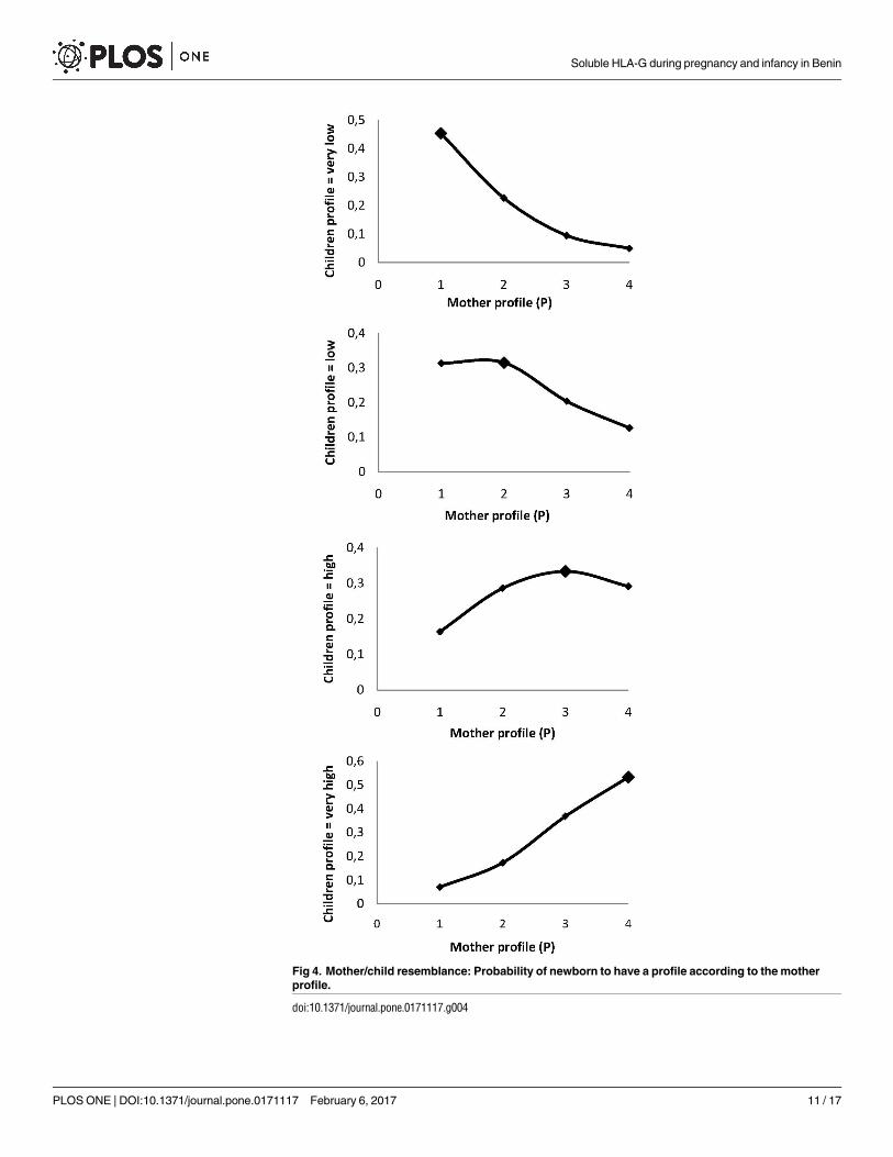
Fig 4. Mother/child resemblance: Probability of newborn to have a profile according to the mother
profile.
doi:10.1371/journal.pone.0171117.g004
Soluble HLA-G during pregnancy and infancy in Benin
PLOS ONE | DOI:10.1371/journal.pone.0171117 February 6, 2017 11 / 17

significant. Age and gravidity are classically correlated and the association between sHLA-G
and age at the second ANV could be related to this association. Indeed the effect of gravidity
could be more important at the beginning of pregnancy to allow the correct implantation of
the embryo.
The source of sHLA-G in newborns is not precisely known and documented to date. We
hypothesize that this HLA-G could be produced by the organs of newborns himself such as
thymus or umbilical cells [39] or immune cells, but also resulted mainly for placental produc-
tion after a transfer at the end of pregnancy. The protein could stay in the newborn serum dur-
ing the first three months, and disappear gradually until six months. To date, there is no data
about this production of HLA-G in newborns to our knowledge.
Malaria infection (placental or peripheral) in mothers has no effect on the sHLA-G level
throughout pregnancy. This might be due to the substantial increase of sHLA-G in pregnancy,
which could result in not detecting a malaria effect. It is noteworthy that pregnant women
received two supervised doses of Intermittent Preventive Treatment against malaria, as recom-
mended by WHO in all endemic areas, and that both the incidence of clinical malaria and
parasitemia were low, for all IPT groups [26]. Independently of this parameter, this result may
seem surprising since during pregnancy the presence of HLA-G would be associated with a
reduction in the cytotoxicity of NK cells and should result in a decrease in the immunity
responsible for placental infection such as in the case of human cytomegalo-virus infection
[40]. The difference between these infections could be due to the production of proinflamma-
tory cytokines which could induce interruption of HLA-G cycle more in human CMV infec-
tion than in malaria infection. However, in infancy, the association between high levels of
sHLA-G and malaria infection was confirmed. It has recently been shown that not only high
levels of sHLA-G could increase the risk of developing a malaria attack, but also that the pres-
ence of malaria infection at sampling is associated with high sHLA-G levels [19]. During HIV
or hepatitis infections, patients have higher sHLA-G levels, and it has been suggested that
these levels contribute toimmune evasion of virus by inducing immune tolerance [41, 42]. We
could hypothesize that a similar phenomenon occurs during malaria. P. falciparum could
upregulate the HLA-G secretion by stimulating cytokines such as IL-10 and IFN-γ, which are
well-known HLA-G inducers. HLA-G inhibits the function of T, NK, dendritic cells, neutro-
phils and B cells through direct interaction with ILT2 and/or ILT4 receptors [40, 43–47]. Pre-
vious reports indicate that HLA-G may have dual role on NK cells depending on its
interaction with either KIR2DL4 or ILT2. In this context, the interaction of HLA-G with ILT2
would favour the inhibition NK cell cytolytic function while its interaction with KIR2DL4 may
promote vascularization processes [8, 9]. Therefore, the impact of HLA-G on NK cell functions
should be considered at two levels since it may have a role in immune responses against
malaria but also in vascularization processes induced during pregnancy. Besides its dual role
Table 5. Maternal factors associated with the probability to belong to a high sHLA-G profile in infancy: Ordinal multinomial regression.
Variable OR(a) 95 confidence interval p
Mother’s sHLA-G profile Very low 1
Low 3.05 [1.75, 5.31] <0.001
High 8.28 [4.68, 14.67] <0.001
Very high 17.59 [9.54, 32.41] <0.001
Placental malaria infection No 1
Yes 2.39 [1.29, 4.45] 0.006
(a) Odds ratio of giving birth to a child with very high profile for a mother with low, high and very high profile compared to a mother with a very low profile.
doi:10.1371/journal.pone.0171117.t005
Soluble HLA-G during pregnancy and infancy in Benin
PLOS ONE | DOI:10.1371/journal.pone.0171117 February 6, 2017 12 / 17

on NK cells, sHLA-G is also a negative regulator of B cells leading to antibody secretion inhibi-
tion in a mouse model [48]. It has long been recognized that antibodies play a pivotal role in
anti-malarial protection [49]. Consequently, the inhibition of IgG specifically directed against
P. falciparum mediated by HLA-G may allow the parasite to escape the immune system and be
responsible for higher susceptibility to infection. We have shown recently that high level of
sHLA-G is associated with a higher probability to develop a malaria attack in the following
weeks but also that the presence of malaria infection can increase sHLA-G that could lead to
immune evasion [19, 50]. Overall, the results underline the complexity of the association
between HLA-G and malaria and need further experimental exploration. Furthermore, it is
important to notify that in this study, all women included and their children are virus (HIV)-
negative. The high level of soluble HLA-G observed in a part of them, could not be due to HIV
infection. According to hepatitis, we do not evaluate the incidence of this infection in the
study population but mothers who had an evolutive hepatic disease were not included. More-
over, as recommended by the world health organization, all the newborns of the program
received different doses of the vaccine against hepatitis B infection, the most prevalent in this
region, and they are normally protected. We did not performed virological or bacterial diagno-
sis tests neither during pregnancy nor during the follow-up of the children. HLA-G has been
associated with these viral infections [41, 42] and this could influence our results. However, all
the included individuals can be considered as comparable concerning the exposure to viral
infections and we do not consider that this risk may introduce a systematic bias.
The association between HLA-G and LBW shows that the evolution of sHLA-G during the
first 2 years of life strongly differs between LBW children and others. LBW is correlated with
infants’ morbidity and mortality [20, 21] and this result is consistent with our previous finding
that some children could have particular trajectory of sHLA-G during infancy [50]. The pres-
ent result strengthens the hypothesis that the susceptibility of LBW children to infections
could be associated with high sHLA-G levels [19], and that infants with LBW could have a par-
ticular mechanism of HLA-G regulation.
Our results also confirmed the strong correlation between the mother’s sHLA-G levels at
delivery and the cord blood level [19]. Comparable findings were described by Klitkou et al.,even though the sHLA-G level in cord blood was lower than at term [16]. However, since
repeated measurements were available for both women and infants, mother/child resemblance
was explored more precisely. It was shown that not only does a mother/child correlation exist
at delivery, but also that each mother’s measurement was correlated with each measurement in
her child. Moreover, it was also demonstrated that a mother harbouring a sHLA-G profile dur-
ing pregnancy has a higher probability of giving birth to a child with a similar profile. This
kind of resemblance could obviously be due to a shared family environment and behaviour.
However, a genetic or epigenetic origin must also be discussed. Indeed, even though the
genetic control of sHLA-G secretion is complex [17, 35], it has recently been shown that there
is an association between combined feto-maternal HLA-G genotypes and the sHLA-G level
[51].
Mother/child resemblance could have consequences from a public health point of view. If it
is confirmed that high levels of sHLA-G are associated with LBW and a higher risk for malaria
(or more generally for infections), the mother’s sHLA-G level during pregnancy could be a
useful biomarker of frailty in children before delivery.
It is accepted that children born from placental malaria are more susceptible to malaria dur-
ing infancy, suggesting that this could stem from immune tolerance [23, 52, 53]. Such children
are not only more susceptible to malaria but also to non-malaria fevers [54], emphasizing the
fact that immune tolerance could imply immunity in a more general manner besides specific
immunity to malaria. In that sense, placental malaria could just be a biomarker of this more
Soluble HLA-G during pregnancy and infancy in Benin
PLOS ONE | DOI:10.1371/journal.pone.0171117 February 6, 2017 13 / 17

complex phenomenon. These results, showing that mothers with placental malaria have a sig-
nificant probability of giving birth to a newborn with high levels of sHLA-G during infancy,
possibly making the child more susceptible to infections, strongly support this hypothesis and
could have important consequences from a public health point of view.
Acknowledgments
We are grateful to all the women who participated in the study. We thank village community
agents who participated actively in the medical and follow-up surveys. We are also very grate-
ful to the technicians, nurses, drivers, and students from the program and from the two dis-
pensaries where the study took place.
Author contributions
Conceptualization: PM NRF DC AG.
Data curation: TCdA JM DC.
Formal analysis: TCdA GC JM AS LG EA.
Funding acquisition: AG DC NRF PM.
Investigation: DC A. Mondière GC JM IS TCdA.
Methodology: AG GC DC NRF JM LG AS BF A. Mondière IS TCdA.
Resources: PM NRF BF DC AG.
Software: TCdA JM.
Supervision: DC GC AG.
Validation: DC NRF AG MK A. Massougbodji EAD PM.
Visualization: TCdA IS AG DC GC NRF PM EAD.
Writing – original draft: TCdA IS JM GC.
Writing – review & editing: AG DC NRF BF PM LG AS CE MK A. Massougbodji EAD.
References1. Carosella ED, Rouas-Freiss N, Roux DT, Moreau P, LeMaoult J. HLA-G: An Immune Checkpoint Mole-
cule. Advances in immunology. 2015; 127:33–144. doi: 10.1016/bs.ai.2015.04.001 PMID: 26073983
2. Carosella ED, Gregori S, Rouas-Freiss N, LeMaoult J, Menier C, Favier B. The role of HLA-G in immu-
nity and hematopoiesis. Cellular and molecular life sciences: CMLS. 2011; 68(3):353–68. doi: 10.1007/
s00018-010-0579-0 PMID: 21116680
3. Bahri R, Hirsch F, Josse A, Rouas-Freiss N, Bidere N, Vasquez A, et al. Soluble HLA-G inhibits cell
cycle progression in human alloreactive T lymphocytes. Journal of Immunology. 2006; 176(3):1331–9.
4. Baudhuin J, Migraine J, Faivre V, Loumagne L, Lukaszewicz AC, Payen D, et al. Exocytosis acts as a
modulator of the ILT4-mediated inhibition of neutrophil functions. P Natl Acad Sci USA. 2013; 110
(44):17957–62.
5. Lesport E, Baudhuin J, LeMaoult J, Sousa S, Doliger C, Carosella ED, et al. Human melanoma cell
secreting human leukocyte antigen-G5 inhibit natural killer cell cytotoxicity by impairing lytic granules
polarization toward target cell. Human immunology. 2009; 70(12):1000–5. PMID: 19654030
6. Moreau P, Paul P, Rouas-Freiss N, Kirszenbaum M, Dausset J, Carosella ED. Molecular and immuno-
logic aspects of the nonclassical HLA class I antigen HLA-G: evidence for an important role in the mater-
nal tolerance of the fetal allograft. Am J Reprod Immunol. 1998; 40(3):136–44. PMID: 9764357
Soluble HLA-G during pregnancy and infancy in Benin
PLOS ONE | DOI:10.1371/journal.pone.0171117 February 6, 2017 14 / 17

7. Rouas-Freiss N, Goncalves RM, Menier C, Dausset J, Carosella ED. Direct evidence to support the
role of HLA-G in protecting the fetus from maternal uterine natural killer cytolysis. Proceedings of the
National Academy of Sciences of the United States of America. 1997; 94(21):11520–5. PMID: 9326642
8. Rajagopalan S, Bryceson YT, Kuppusamy SP, Geraghty DE, van der Meer A, Joosten I, et al. Activation
of NK cells by an endocytosed receptor for soluble HLA-G. PLoS biology. 2006; 4(1):e9. doi: 10.1371/
journal.pbio.0040009 PMID: 16366734
9. Rajagopalan S, Long EO. KIR2DL4 (CD158d): An activation receptor for HLA-G. Frontiers in immunol-
ogy. 2012; 3:258. doi: 10.3389/fimmu.2012.00258 PMID: 22934097
10. Alegre E, Diaz-Lagares A, Lemaoult J, Lopez-Moratalla N, Carosella ED, Gonzalez A. Maternal antigen
presenting cells are a source of plasmatic HLA-G during pregnancy: longitudinal study during preg-
nancy. Human immunology. 2007; 68(8):661–7. PMID: 17678720
11. Yie SM, Taylor RN, Librach C. Low plasma HLA-G protein concentrations in early gestation indicate the
development of preeclampsia later in pregnancy. American journal of obstetrics and gynecology. 2005;
193(1):204–8. doi: 10.1016/j.ajog.2004.11.062 PMID: 16021080
12. Hunt JS, Jadhav L, Chu W, Geraghty DE, Ober C. Soluble HLA-G circulates in maternal blood during
pregnancy. American journal of obstetrics and gynecology. 2000; 183(3):682–8. doi: 10.1067/mob.
2000.106762 PMID: 10992193
13. Rebmann V, Switala M, Eue I, Schwahn E, Merzenich M, Grosse-Wilde H. Rapid evaluation of soluble
HLA-G levels in supernatants of in vitro fertilized embryos. Hum Immunol. 2007; 68(4):251–8. doi: 10.
1016/j.humimm.2006.11.003 PMID: 17400060
14. Rizzo R, Fuzzi B, Stignani M, Criscuoli L, Melchiorri L, Dabizzi S, et al. Soluble HLA-G molecules in fol-
licular fluid: a tool for oocyte selection in IVF? Journal of reproductive immunology. 2007; 74(1–2):133–
42. doi: 10.1016/j.jri.2007.02.005 PMID: 17399800
15. He Y, Chen S, Huang H, Chen Q. Association between decreased plasma levels of soluble human leu-
kocyte antigen-G and severe pre-eclampsia. J Perinat Med. 2015.
16. Klitkou L, Dahl M, Hviid TVF, Djurisic S, Piosik ZM, Skovbo P, et al. Human leukocyte antigen (HLA)-G
during pregnancy part I: Correlations between maternal soluble HLA-G at midterm, at term, and umbili-
cal cord blood soluble HLA-G at term. Human immunology. 2015; 76(4):254–9. PMID: 25636573
17. Donadi EA, Castelli EC, Arnaiz-Villena A, Roger M, Rey D, Moreau P. Implications of the polymorphism
of HLA-G on its function, regulation, evolution and disease association. Cell Mol Life Sci. 2011; 68
(3):369–95. doi: 10.1007/s00018-010-0580-7 PMID: 21107637
18. Campbell MC, Tishkoff SA. African genetic diversity: implications for human demographic history, mod-
ern human origins, and complex disease mapping. Annual review of genomics and human genetics.
2008; 9:403–33. doi: 10.1146/annurev.genom.9.081307.164258 PMID: 18593304
19. Sadissou I, d’Almeida T, Cottrell G, Luty A, Krawice-Radanne I, Massougbodji A, et al. High plasma lev-
els of HLA-G are associated with low birth weight and with an increased risk of malaria in infancy.
Malaria J. 2014; 13.
20. Ntuli ST, Malangu N, Alberts M. Causes of deaths in children under-five years old at a tertiary hospital in
Limpopo province of South Africa. Glob J Health Sci. 2013; 5(3):95–100. doi: 10.5539/gjhs.v5n3p95
PMID: 23618479
21. Lawn JE, Cousens S, Zupan J. 4 million neonatal deaths: when? Where? Why? Lancet. 2005; 365
(9462):891–900. doi: 10.1016/S0140-6736(05)71048-5 PMID: 15752534
22. Le Hesran JY, Cot M, Personne P, Fievet N, Dubois B, Beyeme M, et al. Maternal placental infection
with Plasmodium falciparum and malaria morbidity during the first 2 years of life. American journal of
epidemiology. 1997; 146(10):826–31. PMID: 9384203
23. Le Port A, Watier L, Cottrell G, Ouedraogo S, Dechavanne C, Pierrat C, et al. Infections in infants during
the first 12 months of life: role of placental malaria and environmental factors. PloS one. 2011; 6(11):
e27516. doi: 10.1371/journal.pone.0027516 PMID: 22096588
24. Mutabingwa TK, Bolla MC, Li JL, Domingo GJ, Li X, Fried M, et al. Maternal malaria and gravidity inter-
act to modify infant susceptibility to malaria. PLoS medicine. 2005; 2(12):e407. doi: 10.1371/journal.
pmed.0020407 PMID: 16259531
25. Ouedraogo S, Koura GK, Bodeau-Livinec F, Accrombessi MM, Massougbodji A, Cot M. Maternal ane-
mia in pregnancy: assessing the effect of routine preventive measures in a malaria-endemic area. The
American journal of tropical medicine and hygiene. 2013; 88(2):292–300. doi: 10.4269/ajtmh.12-0195
PMID: 23296448
26. Gonzalez R, Mombo-Ngoma G, Ouedraogo S, Kakolwa MA, Abdulla S, Accrombessi M, et al. Intermit-
tent preventive treatment of malaria in pregnancy with mefloquine in HIV-negative women: a multicentre
randomized controlled trial. Plos Med. 2014; 11(9):e1001733. doi: 10.1371/journal.pmed.1001733
PMID: 25247709
Soluble HLA-G during pregnancy and infancy in Benin
PLOS ONE | DOI:10.1371/journal.pone.0171117 February 6, 2017 15 / 17

27. Rebmann V, LeMaoult J, Rouas-Freiss N, Carosella ED, Grosse-Wilde H. Report of the Wet Workshop
for Quantification of soluble HLA-G in Essen, 2004. Hum Immunol. 2005; 66(8):853–63. doi: 10.1016/j.
humimm.2005.05.003 PMID: 16216668
28. Menier C, Saez B, Horejsi V, Martinozzi S, Krawice-Radanne I, Bruel S, et al. Characterization of mono-
clonal antibodies recognizing HLA-G or HLA-E: New tools to analyze the expression of nonclassical
HLA class I molecules. Hum Immunol. 2003; 64(3):315–26. PMID: 12590976
29. Amemiya T. Regression analysis when the dependent variable is truncated normal. Econometrica.
1973; 41:997–1016.
30. Wang W, Griswold ME. Natural interpretations in Tobit regression models using marginal estimation
methods. Statistical methods in medical research. 2015.
31. McCulloch CE, Conference Board of the Mathematical Sciences., National Science Foundation (U.S.).
Generalized linear mixed models. Beachwood, Ohio. Alexandria, Va.: Institute of Mathematical Statis-
tics; American Statistical Association; 2003. viii, 84 pp.
32. Thaddeus S, Maine D. Too Far to Walk—Maternal Mortality in Context. Soc Sci Med. 1994; 38
(8):1091–110. PMID: 8042057
33. Knafel A, Basta P, Pitynski K, Mach P, Bednarek W, Klimek M, et al. Soluble HLA-G changes in mater-
nal blood serum during the progression of labor. Neuroendocrinol Lett. 2009; 30(1):67–73. PMID:
19300382
34. Rizzo R, Stignani M, Amoudruz P, Nilsson C, Melchiorri L, Baricordi O, et al. Allergic women have
reduced sHLA-G plasma levels at delivery. Am J Reprod Immunol. 2009; 61(5):368–76. doi: 10.1111/j.
1600-0897.2009.00703.x PMID: 19341387
35. Porto IO, Mendes-Junior CT, Felicio LP, Georg RC, Moreau P, Donadi EA, et al. MicroRNAs targeting
the immunomodulatory HLA-G gene: a new survey searching for microRNAs with potential to regulate
HLA-G. Molecular immunology. 2015; 65(2):230–41. doi: 10.1016/j.molimm.2015.01.030 PMID:
25700346
36. Martelli-Palomino G, Pancotto JA, Muniz YC, Mendes-Junior CT, Castelli EC, Massaro JD, et al. Poly-
morphic sites at the 3’ untranslated region of the HLA-G gene are associated with differential hla-g solu-
ble levels in the Brazilian and French population. PloS one. 2013; 8(10):e71742. doi: 10.1371/journal.
pone.0071742 PMID: 24204558
37. Gineau L, Luisi P, Castelli EC, Milet J, Courtin D, Cagnin N, et al. Balancing immunity and tolerance:
genetic footprint of natural selection in the transcriptional regulatory region of HLA-G. Genes Immun.
2014; 16(1):57–60. doi: 10.1038/gene.2014.63 PMID: 25393930
38. He YD, Chen S, Huang H, Chen Q. Association between decreased plasma levels of soluble human
leukocyte antigen-G and severe pre-eclampsia. J Perinat Med. 2016; 44(3):283–90. doi: 10.1515/jpm-
2015-0062 PMID: 26352061
39. Buzzi M, Alviano F, Campioni D, Stignani M, Melchiorri L, Rotola A, et al. Umbilical cord blood CD34(+)
cell-derived progeny produces human leukocyte antigen-G molecules with immuno-modulatory func-
tions. Human immunology. 2012; 73(2):150–5.
40. Tilburgs T, Evans JH, Crespo AC, Strominger JL. The HLA-G cycle provides for both NK tolerance and
immunity at the maternal-fetal interface. P Natl Acad Sci USA. 2015; 112(43):13312–7.
41. Weng PJ, Fu YM, Ding SX, Xu DP, Lin A, Yan WH. Elevation of plasma soluble human leukocyte anti-
gen-G in patients with chronic hepatitis C virus infection. Human immunology. 2011; 72(5):406–11.
PMID: 21377504
42. Celsi F, Catamo E, Kleiner G, Tricarico PM, Vuch J, Crovella S. HLA-G/C, miRNAs, and their role in
HIV infection and replication. Biomed Res Int. 2013; 2013:693643. doi: 10.1155/2013/693643 PMID:
23841087
43. Ristich V, Liang S, Zhang W, Wu J, Horuzsko A. Tolerization of dendritic cells by HLA-G. Eur J Immunol.
2005; 35(4):1133–42. doi: 10.1002/eji.200425741 PMID: 15770701
44. Baudhuin J, Lesport E, Sousa S, Migraine J, Vigneron J, Lemaoult J, et al. HLA-G inhibition of NK-cell
cytolytic function is uncoupled from tumor cell lipid raft reorganization. Eur J Immunol. 2012; 42(3):700–
9. doi: 10.1002/eji.201141930 PMID: 22144141
45. Baudhuin J, Migraine J, Faivre V, Loumagne L, Lukaszewicz AC, Payen D, et al. Exocytosis acts as a
modulator of the ILT4-mediated inhibition of neutrophil functions. P Natl Acad Sci USA. 2013; 110
(44):17957–62.
46. Favier B. Regulation of neutrophil functions through inhibitory receptors: an emerging paradigm in
health and disease. Immunological reviews. 2016; 273(1):140–55. doi: 10.1111/imr.12457 PMID:
27558333
Soluble HLA-G during pregnancy and infancy in Benin
PLOS ONE | DOI:10.1371/journal.pone.0171117 February 6, 2017 16 / 17

47. Naji A, Menier C, Morandi F, Agaugue S, Maki G, Ferretti E, et al. Binding of HLA-G to ITIM-bearing Ig-
like transcript 2 receptor suppresses B cell responses. J Immunol. 2014; 192(4):1536–46. doi: 10.4049/
jimmunol.1300438 PMID: 24453251
48. Naji A, Menier C, Morandi F, Agaugue S, Maki G, Ferretti E, et al. Binding of HLA-G to ITIM-Bearing Ig-
like Transcript 2 Receptor Suppresses B Cell Responses. J Immunol. 2014; 192(4):1536–46. doi: 10.
4049/jimmunol.1300438 PMID: 24453251
49. Bouharountayoun H, Attanath P, Sabchareon A, Chongsuphajaisiddhi T, Druilhe P. Antibodies That
Protect Humans against Plasmodium-Falciparum Blood Stages Do Not on Their Own Inhibit Parasite
Growth and Invasion Invitro, but Act in Cooperation with Monocytes. J Exp Med. 1990; 172(6):1633–41.
PMID: 2258697
50. d’Almeida TC, Sadissou I, Cottrell G, Tahar R, Moreau P, Favier B, et al. Evolution of the levels of
human leukocyte antigen G (HLA-G) in Beninese infant during the first year of life in a malaria endemic
area: using latent class analysis. Malaria J. 2016; 15.
51. Dahl M, Klitkou L, Christiansen OB, Djurisic S, Piosik ZM, Skovbo P, et al. Human leukocyte antigen
(HLA)-G during pregnancy part II: associations between maternal and fetal HLA-G genotypes and solu-
ble HLA-G. Hum Immunol. 2015; 76(4):260–71. PMID: 25637667
52. Schwarz NG, Adegnika AA, Breitling LP, Gabor J, Agnandji ST, Newman RD, et al. Placental malaria
increases malaria risk in the first 30 months of life. Clinical Infectious Diseases. 2008; 47(8):1017–25.
doi: 10.1086/591968 PMID: 18781874
53. Mutabingwa TK, Bolla MC, Li JL, Domingo GJ, Li XH, Fried M, et al. Maternal malaria and gravidity
interact to modify infant susceptibility to malaria. PLoS medicine. 2005; 2(12):1260–8.
54. Rachas A, Le Port A, Cottrell G, Guerra J, Choudat I, Bouscaillou J, et al. Placental Malaria is Associ-
ated With Increased Risk of Nonmalaria Infection During the First 18 Months of Life in a Beninese Popu-
lation. Clinical Infectious Diseases. 2012; 55(5):672–8. doi: 10.1093/cid/cis490 PMID: 22610927
Soluble HLA-G during pregnancy and infancy in Benin
PLOS ONE | DOI:10.1371/journal.pone.0171117 February 6, 2017 17 / 17