solution structure of the dna-binding domain of human telomeric protein, htrf1
TRANSCRIPT
Solution structure of the DNA-binding domain of humantelomeric protein, hTRF1Tadateru Nishikawa1, Aritaka Nagadoi1, Shoko Yoshimura2, Saburo Aimoto2
and Yoshifumi Nishimura1*
Background: Mammalian telomeres consist of long tandem arrays of the double-stranded TTAGGG sequence motif packaged by a telomere repeat bindingfactor, TRF1. The DNA-binding domain of TRF1 shows sequence homology toeach of three tandem repeats of the DNA-binding domain of the transcriptionalactivator c-Myb. The isolated c-Myb-like domain of human TRF1 (hTRF1) bindsspecifically to telomeric DNA as a monomer, in a similar manner to that ofhomeodomains. So far, the only three-dimensional structure of a telomeric proteinto be determined is that of a yeast telomeric protein, Rap1p. The DNA-bindingdomain of Rap1p contains two subdomains that are structurally closely related toc-Myb repeats. We set out to determine the solution structure of the DNA-bindingdomain of hTRF1 in order to establish its mode of DNA binding.
Results: The solution structure of the DNA-binding domain of hTRF1 has beendetermined and shown to comprise three helices. The architecture of the threehelices is very similar to that of each Rap1p subdomain and also to that of eachc-Myb repeat. The second and third helix form a helix-turn-helix (HTH) variant.The length of the third helix of hTRF1 is similar to that of the second subdomainof Rap1p.
Conclusions: The hTRF1 DNA-binding domain is likely to bind to DNA in asimilar manner to that of the second subdomain of Rap1p. On the basis of the Rap1p–DNA complex, a model of the hTRF1 DNA-binding domain in complexwith human telomeric DNA was constructed. In addition to DNA recognition bythe HTH variant, a flexible N-terminal arm of hTRF1 is likely to interact with DNA.
IntroductionTelomeres are the protein–DNA complexes that protectthe ends of eukaryotic linear chromosomes from degrada-tion and fusion [1]. Mammalian telomeres are composedof long tandem arrays of the double-stranded telomericrepeat, TTAGGG [1–5], packaged by the telomeric protein,telomeric repeat binding factor (TRF1) [6–11]. The humanTRF1 (hTRF1) consists of 439 amino acids comprisingthree presumed functional domains: an N-terminal acidicdomain, a central TRF-specific dimerization domain, anda C-terminal DNA-binding domain [7,10,11]. The DNA-binding domain of hTRF1 consists of a 53 amino acidsequence that shows sequence homology to each of threetandem repeats of the c-Myb DNA-binding domain [12,13].The c-Myb protein is a transcriptional activator that bindsto the consensus sequence TAACNG [14–18]. Each ofthe three repeats in c-Myb has a very similar tertiarystructure containing three helices [19–21]; the second andthird helix in each repeat form a helix-turn-helix (HTH)variant motif [22–24].
The length of telomeres seems to be related to cell divi-sion number; after cell development in somatic cells the
length of telomeres will be shortened during the course ofcell division, whereas in oncogenic cells the length oftelomeres remains constant [25–29]. Recently, it wasfound that the length of telomeres in oncogenic cells ismaintained by an enzyme — the telomerase that addstelomeric DNA sequences [30,31]. The access of thetelomerase to DNA seems to be controlled by TRF1 [32].To understand the mechanism of chromosome stabilityand cell senescence it will be essential to know the tertiarystructure of telomeres.
So far, only the structure of the yeast Rap1p DNA-bindingdomain in complex with telomeric DNA has been deter-mined [33]. Rap1p is a constituent of the telomeres of thebudding yeast Saccharomyces cerevisiae, which consist of300–450 base pairs of an irregularly repeated sequencemotif, C2–3A(CA)1–6, in contrast to the vertebrate regularlyrepeated sequence [34–37]. The DNA-binding domain ofRap1p contains two subdomains that bind DNA in a tandemorientation. The structure shows that despite lacking anysignificant sequence similarity, the Rap1p subdomains arestructurally closely related to the c-Myb DNA-bindingrepeats [33]. In contrast, the hTRF1 DNA-binding domain
Addresses: 1Graduate School of IntegratedScience, Yokohama City University, 22-2 Seto,Kanazawa-ku, Yokohama 236-0027, Japan and2Protein Research Institute, Osaka University, Suita,Osaka 565-0871, Japan.
*Corresponding author.E-mail: [email protected]
Key words: c-Myb repeats, DNA-binding domain,NMR, telomere, TRF1
Received: 24 March 1998Revisions requested: 17 April 1998Revisions received: 9 June 1998Accepted: 6 July 1998
Structure 15 August 1998, 6:1057–1065http://biomednet.com/elecref/0969212600601057
© Current Biology Publications ISSN 0969-2126
Research Article 1057
contains only a single c-Myb repeat homologous domain.Recently it has been shown that the isolated c-Myb-likedomain of hTRF1 binds specifically, and with significantaffinity, to telomeric DNA as a monomer [38]. It has beenshown that the isolated c-Myb-like domain recognizes abinding site centered on the sequence GGGTTA, andthat its DNA-binding mode is similar to that of each of theRap1p subdomains [38].
We report here the solution structure of the c-Myb-likedomain of hTRF1 determined by nuclear magnetic reso-nance (NMR) spectroscopy. The structure consists ofthree helices. The architecture of the three helices is verysimilar to that of each Rap1p subdomain and also to that ofeach c-Myb repeat. The length of the third helix of thehTRF1 DNA-binding domain is most similar to that ofthe second subdomain of Rap1p. The hTRF1 DNA-binding domain is quite likely to bind to DNA in a similarmanner to the second subdomain of Rap1p.
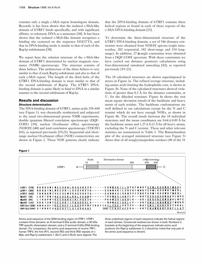
Results and discussionStructure determinationThe DNA-binding domain of hTRF1, amino acids 378–430(see Figure 1), was chemically synthesized and subjectedto the usual two-dimensional proton NMR experiments,double quantum filtered correlation spectroscopy (DQF-COSY) [39], nuclear Overhauser effect spectroscopy(NOESY) [40] and total correlation spectroscopy (TOCSY)[41], as reported previously [19,21]. Sequential and short-range nuclear Overhauser effect (NOE) connectivities areshown in Figure 2. These NOE patterns clearly indicate
that the DNA-binding domain of hTRF1 contains threehelical regions as found in each of three repeats of thec-Myb DNA-binding domain [21].
To determine the three-dimensional structure of thehTRF1 DNA-binding domain, a set of 546 distance con-straints were obtained from NOESY spectra (eight intra-residue, 202 sequential, 182 short-range and 154 long-range). In addition, 27 φ angle constraints were obtainedfrom a DQF-COSY spectrum. With these constraints wehave carried out distance geometry calculations usingfour-dimensional simulated annealing [42], as reportedpreviously [19–21].
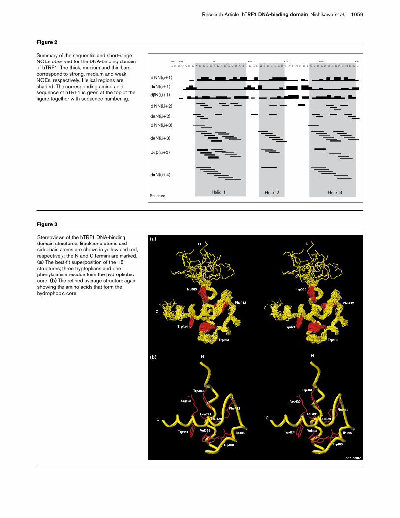
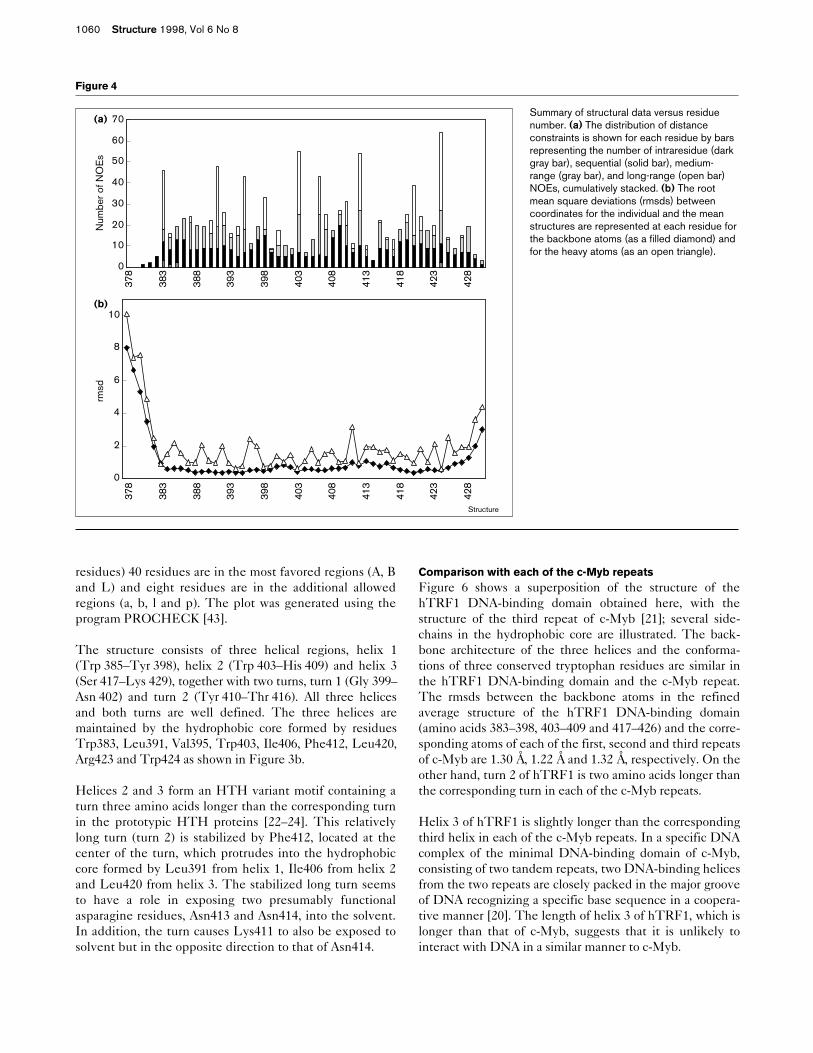
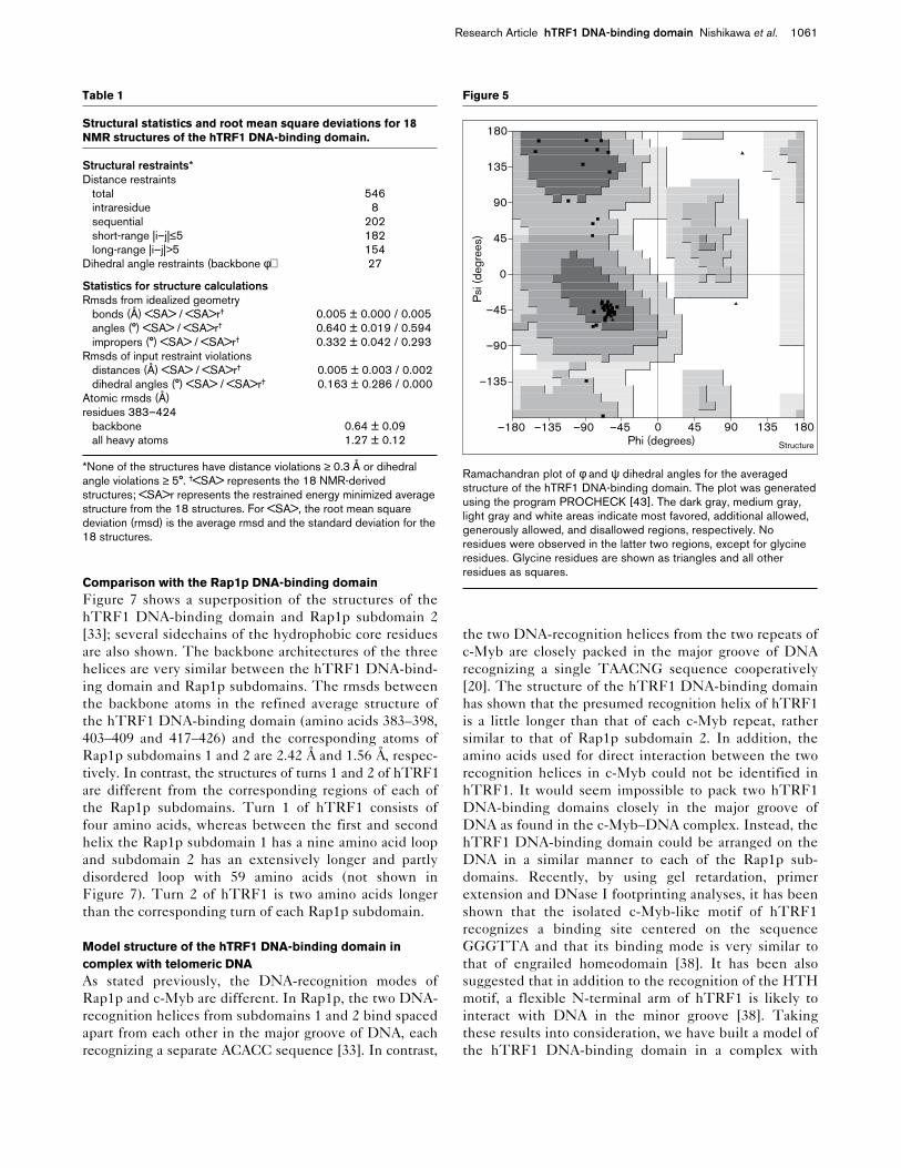
The 18 calculated structures are shown superimposed instereo in Figure 3a. The refined average structure, includ-ing amino acids forming the hydrophobic core, is shown inFigure 3b. None of the calculated structures showed viola-tions of greater than 0.3 Å, for the distance constraints, or5°, for the dihedral restraints. Figure 4a shows the rootmean square deviation (rmsd) of the backbone and heavyatoms of each residue. The backbone conformations arewell defined in our calculations except for the N and Ctermini which do not have enough NOEs, as shown inFigure 4b. The overall rmsds between the 18 individualstructures and the mean coordinates are 0.64 ± 0.09 Å forthe backbone atoms and 1.27 ± 0.12 Å for all heavy atoms,excluding the N and C termini. These and other relevantstatistics are summarized in Table 1. The Ramachandranplot of the averaged minimized structure (see Figure 5)shows that of all nonglycine/proline residues (48 of the 51
1058 Structure 1998, Vol 6 No 8
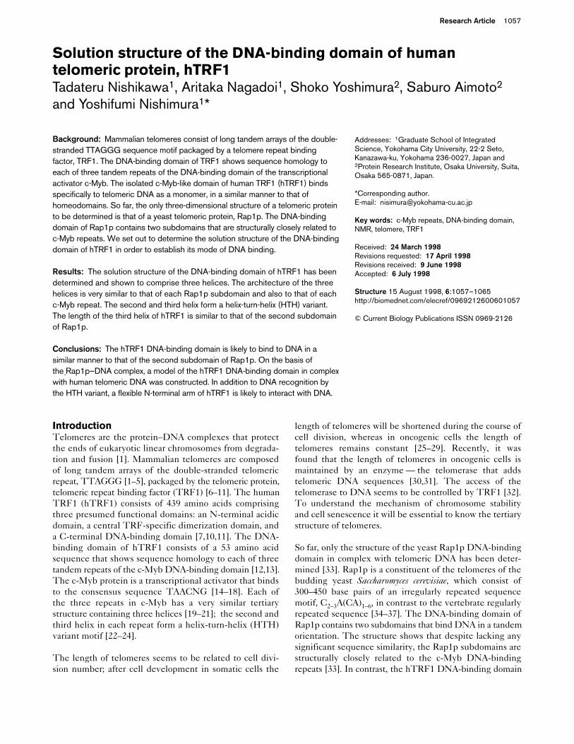
Figure 1
NhTRF1 C
19 71 265 378 430 Acidic Dimerization domain c-Myb homology
Structure
Amino acid sequence of the DNA-binding region of hTRF1. hTRF1contains three domains: an N-terminal 6 kDa acidic domain; a 30 kDaTRF-specific dimerization domain; and a C-terminal 6 kDa DNA-bindingdomain. For comparison, the amino acid sequences of murine TRF1,human TRF2, the first (R1), second (R2) and third (R3) repeats of c-Myb, and Rap1p subdomains 1 (Sc1) and 2 (Sc2) were aligned. The
three underlined regions of each sequence indicate the helical regionsof each domain. Conserved residues are shown in bold. Numbers inbrackets at the beginning of the sequences indicate amino acidpositions (for Rap1p subdomain 2, it should be noted that only part ofthe amino acid sequence is shown).
Research Article hTRF1 DNA-binding domain Nishikawa et al. 1059
Figure 2
Summary of the sequential and short-rangeNOEs observed for the DNA-binding domainof hTRF1. The thick, medium and thin barscorrespond to strong, medium and weakNOEs, respectively. Helical regions areshaded. The corresponding amino acidsequence of hTRF1 is given at the top of thefigure together with sequence numbering.
R K R Q A W L W E E D K N L R S G V R K Y G E G N W S K I L L H Y K F N N R T S V M L K D R W R T M K K L378 380 390 400 410 420
d NN(i,i+1)
dαN(i,i+1)
dβN(i,i+1)
d NN(i,i+2)
dαN(i,i+2)
d NN(i,i+3)
dαN(i,i+3)
dαβ(i,i+3)
dαN(i,i+4)
Helix 1 Helix 2 Helix 3
430
Structure
Figure 3
Stereoviews of the hTRF1 DNA-bindingdomain structures. Backbone atoms andsidechain atoms are shown in yellow and red,respectively; the N and C termini are marked.(a) The best-fit superposition of the 18structures; three tryptophans and onephenylalanine residue form the hydrophobiccore. (b) The refined average structure againshowing the amino acids that form thehydrophobic core.
residues) 40 residues are in the most favored regions (A, Band L) and eight residues are in the additional allowedregions (a, b, l and p). The plot was generated using theprogram PROCHECK [43].
The structure consists of three helical regions, helix 1(Trp 385–Tyr 398), helix 2 (Trp 403–His 409) and helix 3(Ser 417–Lys 429), together with two turns, turn 1 (Gly 399–Asn 402) and turn 2 (Tyr 410–Thr 416). All three helicesand both turns are well defined. The three helices aremaintained by the hydrophobic core formed by residuesTrp383, Leu391, Val395, Trp403, Ile406, Phe412, Leu420,Arg423 and Trp424 as shown in Figure 3b.
Helices 2 and 3 form an HTH variant motif containing aturn three amino acids longer than the corresponding turnin the prototypic HTH proteins [22–24]. This relativelylong turn (turn 2) is stabilized by Phe412, located at thecenter of the turn, which protrudes into the hydrophobiccore formed by Leu391 from helix 1, Ile406 from helix 2and Leu420 from helix 3. The stabilized long turn seemsto have a role in exposing two presumably functionalasparagine residues, Asn413 and Asn414, into the solvent.In addition, the turn causes Lys411 to also be exposed tosolvent but in the opposite direction to that of Asn414.
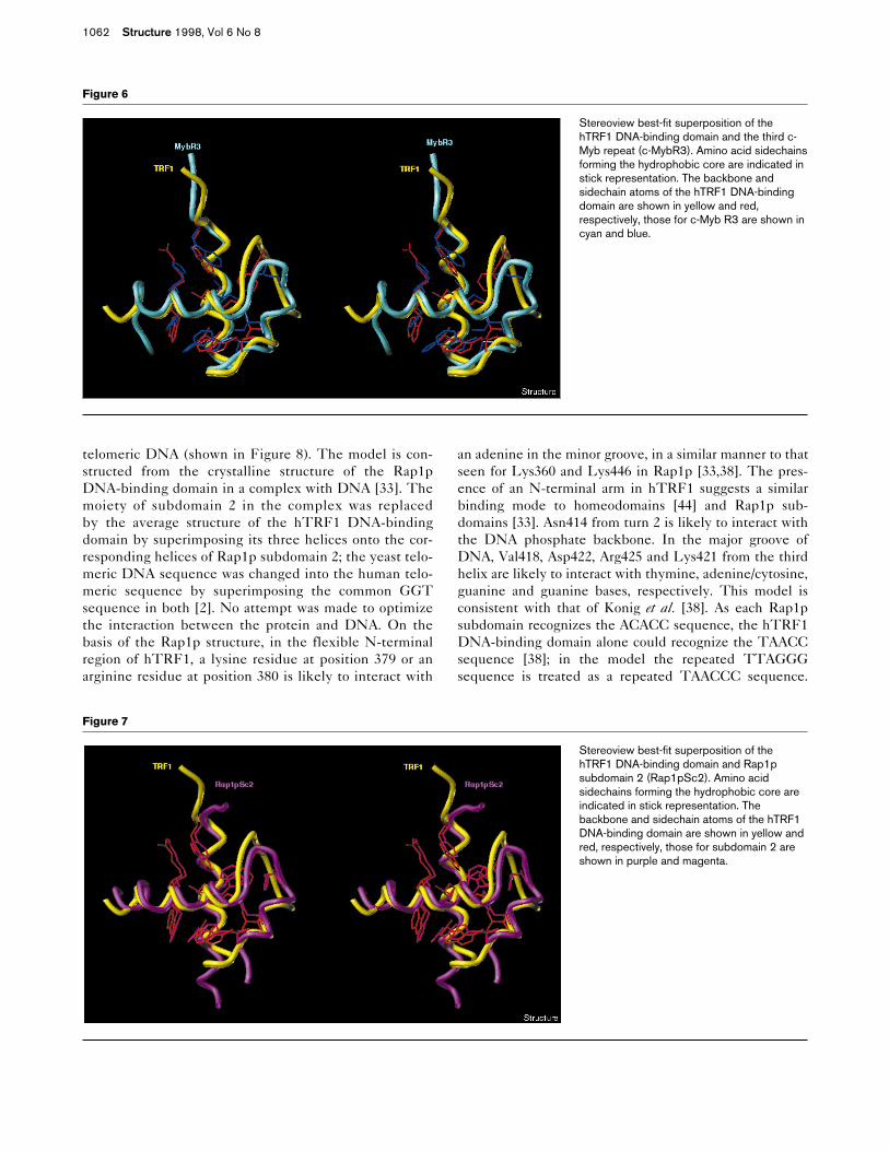
Comparison with each of the c-Myb repeatsFigure 6 shows a superposition of the structure of thehTRF1 DNA-binding domain obtained here, with thestructure of the third repeat of c-Myb [21]; several side-chains in the hydrophobic core are illustrated. The back-bone architecture of the three helices and the conforma-tions of three conserved tryptophan residues are similar inthe hTRF1 DNA-binding domain and the c-Myb repeat.The rmsds between the backbone atoms in the refinedaverage structure of the hTRF1 DNA-binding domain(amino acids 383–398, 403–409 and 417–426) and the corre-sponding atoms of each of the first, second and third repeatsof c-Myb are 1.30 Å, 1.22 Å and 1.32 Å, respectively. On theother hand, turn 2 of hTRF1 is two amino acids longer thanthe corresponding turn in each of the c-Myb repeats.
Helix 3 of hTRF1 is slightly longer than the correspondingthird helix in each of the c-Myb repeats. In a specific DNAcomplex of the minimal DNA-binding domain of c-Myb,consisting of two tandem repeats, two DNA-binding helicesfrom the two repeats are closely packed in the major grooveof DNA recognizing a specific base sequence in a coopera-tive manner [20]. The length of helix 3 of hTRF1, which islonger than that of c-Myb, suggests that it is unlikely tointeract with DNA in a similar manner to c-Myb.
1060 Structure 1998, Vol 6 No 8
Figure 4
Summary of structural data versus residuenumber. (a) The distribution of distanceconstraints is shown for each residue by barsrepresenting the number of intraresidue (darkgray bar), sequential (solid bar), medium-range (gray bar), and long-range (open bar)NOEs, cumulatively stacked. (b) The rootmean square deviations (rmsds) betweencoordinates for the individual and the meanstructures are represented at each residue forthe backbone atoms (as a filled diamond) andfor the heavy atoms (as an open triangle).
0
10
20
30
40
50
60
7037
8
383
388
393
398
403
408
413
418
423
428
378
383
388
393
398
403
408
413
418
423
428
Num
ber o
f NO
Es
rmsd
0
2
4
6
8
10
(a)
(b)
Structure
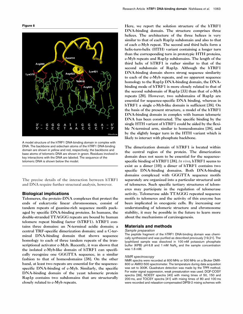
Comparison with the Rap1p DNA-binding domainFigure 7 shows a superposition of the structures of thehTRF1 DNA-binding domain and Rap1p subdomain 2[33]; several sidechains of the hydrophobic core residuesare also shown. The backbone architectures of the threehelices are very similar between the hTRF1 DNA-bind-ing domain and Rap1p subdomains. The rmsds betweenthe backbone atoms in the refined average structure ofthe hTRF1 DNA-binding domain (amino acids 383–398,403–409 and 417–426) and the corresponding atoms ofRap1p subdomains 1 and 2 are 2.42 Å and 1.56 Å, respec-tively. In contrast, the structures of turns 1 and 2 of hTRF1are different from the corresponding regions of each ofthe Rap1p subdomains. Turn 1 of hTRF1 consists offour amino acids, whereas between the first and secondhelix the Rap1p subdomain 1 has a nine amino acid loopand subdomain 2 has an extensively longer and partlydisordered loop with 59 amino acids (not shown inFigure 7). Turn 2 of hTRF1 is two amino acids longerthan the corresponding turn of each Rap1p subdomain.
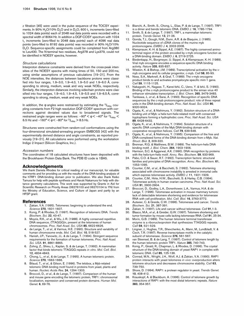
Model structure of the hTRF1 DNA-binding domain incomplex with telomeric DNAAs stated previously, the DNA-recognition modes ofRap1p and c-Myb are different. In Rap1p, the two DNA-recognition helices from subdomains 1 and 2 bind spacedapart from each other in the major groove of DNA, eachrecognizing a separate ACACC sequence [33]. In contrast,
the two DNA-recognition helices from the two repeats ofc-Myb are closely packed in the major groove of DNArecognizing a single TAACNG sequence cooperatively[20]. The structure of the hTRF1 DNA-binding domainhas shown that the presumed recognition helix of hTRF1is a little longer than that of each c-Myb repeat, rathersimilar to that of Rap1p subdomain 2. In addition, theamino acids used for direct interaction between the tworecognition helices in c-Myb could not be identified inhTRF1. It would seem impossible to pack two hTRF1DNA-binding domains closely in the major groove ofDNA as found in the c-Myb–DNA complex. Instead, thehTRF1 DNA-binding domain could be arranged on theDNA in a similar manner to each of the Rap1p sub-domains. Recently, by using gel retardation, primerextension and DNase I footprinting analyses, it has beenshown that the isolated c-Myb-like motif of hTRF1recognizes a binding site centered on the sequenceGGGTTA and that its binding mode is very similar tothat of engrailed homeodomain [38]. It has been alsosuggested that in addition to the recognition of the HTHmotif, a flexible N-terminal arm of hTRF1 is likely tointeract with DNA in the minor groove [38]. Takingthese results into consideration, we have built a model ofthe hTRF1 DNA-binding domain in a complex with
Research Article hTRF1 DNA-binding domain Nishikawa et al. 1061
Table 1
Structural statistics and root mean square deviations for 18NMR structures of the hTRF1 DNA-binding domain.
Structural restraints*Distance restraints
total 546intraresidue 8sequential 202short-range |i–j|≤5 182long-range |i–j|>5 154
Dihedral angle restraints (backbone φ) 27
Statistics for structure calculationsRmsds from idealized geometry
bonds (Å) <SA> / <SA>r† 0.005 ± 0.000 / 0.005angles (°) <SA> / <SA>r† 0.640 ± 0.019 / 0.594impropers (°) <SA> / <SA>r† 0.332 ± 0.042 / 0.293
Rmsds of input restraint violationsdistances (Å) <SA> / <SA>r† 0.005 ± 0.003 / 0.002dihedral angles (°) <SA> / <SA>r† 0.163 ± 0.286 / 0.000
Atomic rmsds (Å)residues 383–424
backbone 0.64 ± 0.09all heavy atoms 1.27 ± 0.12
*None of the structures have distance violations ≥ 0.3 Å or dihedralangle violations ≥ 5°. †<SA> represents the 18 NMR-derivedstructures; <SA>r represents the restrained energy minimized averagestructure from the 18 structures. For <SA>, the root mean squaredeviation (rmsd) is the average rmsd and the standard deviation for the18 structures.
Figure 5
Ramachandran plot of φ and ψ dihedral angles for the averagedstructure of the hTRF1 DNA-binding domain. The plot was generatedusing the program PROCHECK [43]. The dark gray, medium gray,light gray and white areas indicate most favored, additional allowed,generously allowed, and disallowed regions, respectively. Noresidues were observed in the latter two regions, except for glycineresidues. Glycine residues are shown as triangles and all otherresidues as squares.
–180 –135 –90 –45 0 45 90 135 180
–135
–90
–45
0
45
90
135
180
Phi (degrees)
Psi
(deg
rees
)
Structure
telomeric DNA (shown in Figure 8). The model is con-structed from the crystalline structure of the Rap1pDNA-binding domain in a complex with DNA [33]. Themoiety of subdomain 2 in the complex was replacedby the average structure of the hTRF1 DNA-bindingdomain by superimposing its three helices onto the cor-responding helices of Rap1p subdomain 2; the yeast telo-meric DNA sequence was changed into the human telo-meric sequence by superimposing the common GGTsequence in both [2]. No attempt was made to optimizethe interaction between the protein and DNA. On thebasis of the Rap1p structure, in the flexible N-terminalregion of hTRF1, a lysine residue at position 379 or anarginine residue at position 380 is likely to interact with
an adenine in the minor groove, in a similar manner to thatseen for Lys360 and Lys446 in Rap1p [33,38]. The pres-ence of an N-terminal arm in hTRF1 suggests a similarbinding mode to homeodomains [44] and Rap1p sub-domains [33]. Asn414 from turn 2 is likely to interact withthe DNA phosphate backbone. In the major groove ofDNA, Val418, Asp422, Arg425 and Lys421 from the thirdhelix are likely to interact with thymine, adenine/cytosine,guanine and guanine bases, respectively. This model isconsistent with that of Konig et al. [38]. As each Rap1psubdomain recognizes the ACACC sequence, the hTRF1DNA-binding domain alone could recognize the TAACCsequence [38]; in the model the repeated TTAGGGsequence is treated as a repeated TAACCC sequence.
1062 Structure 1998, Vol 6 No 8
Figure 7
Stereoview best-fit superposition of thehTRF1 DNA-binding domain and Rap1psubdomain 2 (Rap1pSc2). Amino acidsidechains forming the hydrophobic core areindicated in stick representation. Thebackbone and sidechain atoms of the hTRF1DNA-binding domain are shown in yellow andred, respectively, those for subdomain 2 areshown in purple and magenta.
Figure 6
Stereoview best-fit superposition of thehTRF1 DNA-binding domain and the third c-Myb repeat (c-MybR3). Amino acid sidechainsforming the hydrophobic core are indicated instick representation. The backbone andsidechain atoms of the hTRF1 DNA-bindingdomain are shown in yellow and red,respectively, those for c-Myb R3 are shown incyan and blue.
The precise details of the interaction between hTRF1and DNA require further structural analysis, however.
Biological implicationsTelomeres, the protein–DNA complexes that protect theends of eukaryotic linear chromosomes, consist oftandem repeats of guanine-rich sequence motifs pack-aged by specific DNA-binding proteins. In humans, thedouble-stranded TTAGGG repeats are bound by humantelomere repeat binding factor (hTRF1). hTRF1 con-tains three domains: an N-terminal acidic domain; acentral TRF-specific dimerization domain; and a C-ter-minal DNA-binding domain that shows sequencehomology to each of three tandem repeats of the tran-scriptional activator c-Myb. Recently, it was shown thatthe isolated c-Myb-like domain of hTRF1 can specifi-cally recognize one GGGTTA sequence, in a similarfashion to that of homeodomains [38]. On the otherhand, at least two repeats are essential for the sequence-specific DNA-binding of c-Myb. Similarly, the specificDNA-binding domain of the yeast telomeric proteinRap1p contains two subdomains that are structurallyclosely related to c-Myb repeats.
Here, we report the solution structure of the hTRF1DNA-binding domain. The structure comprises threehelices. The architecture of the three helices is verysimilar to that of each Rap1p subdomain and also to thatof each c-Myb repeat. The second and third helix form ahelix-turn-helix (HTH) variant containing a longer turnthan the corresponding turn in prototypic HTH proteins,c-Myb repeats and Rap1p subdomains. The length of thethird helix of hTRF1 is rather similar to that of thesecond subdomain of Rap1p. Although the hTRF1DNA-binding domain shows strong sequence similarityto each of the c-Myb repeats, and no apparent sequencehomology to the Rap1p DNA-binding domain, the DNA-binding mode of hTRF1 is more closely related to that ofthe second subdomain of Rap1p [33] than that of c-Mybrepeats [20]. However, two subdomains of Rap1p areessential for sequence-specific DNA binding, whereas inhTRF1 a single c-Myb-like domain is sufficient [38]. Onthe basis of the present structure, a model of the hTRF1DNA-binding domain in complex with human telomericDNA has been constructed. The specific binding by thesingle HTH variant of hTRF1 could be aided by the flexi-ble N-terminal arm, similar to homeodomains [38], andby the slightly longer turn in the HTH variant which islikely to interact with phosphate backbone.
The dimerization domain of hTRF1 is located withinthe central region of the protein. The dimerizationdomain does not seem to be essential for the sequence-specific binding of hTRF1 [38]. In vivo, hTRF1 seems toexist as a dimer [10]; a dimer of hTRF1 contains twospecific DNA-binding domains. Both DNA-bindingdomains complexed with GGGTTA sequence motifsseparately are organized into a particular structural unitof telomeres. Such specific tertiary structures of telom-eres may participate in the regulation of telomeraseactivity. Telomerase adds TTAGGG repeated sequencemotifs to telomeres and the activity of this enzyme hasbeen implicated in oncogenic cells. By increasing ourunderstanding of telomeric structure and chromosomestability, it may be possible in the future to learn moreabout the mechanisms of carcinogenesis.
Materials and methodsSample preparationThe peptide fragment of the hTRF1 DNA-binding domain was chemi-cally synthesized and was purified as described previously [19,21]. Thelyophilized sample was dissolved in 100 mM potassium phosphatebuffer (KPB) pH 6.8 and 1 mM NaN3 and the sample concentrationwas 1.6 mM.
NMR spectroscopyNMR spectra were recorded at 600 MHz or 500 MHz on a Bruker DMX-600 or AMX2-500 spectrometer. The temperature during data acquisitionwas set to 300K. Quadrature detection was made by the TPPI method.For water signal suppression, weak presaturation was used. DQF-COSYspectra [39], NOESY spectra [40] with mixing times of 50, 150 and200 ms, and TOCSY spectra [41] with mixing times of 80 and 100 mswere recorded and relaxation-compensated DIPSI-2 mixing schemes with
Research Article hTRF1 DNA-binding domain Nishikawa et al. 1063
Figure 8
A model structure of the hTRF1 DNA-binding domain in complex withDNA. The backbone and sidechain atoms of the hTRF1 DNA-bindingdomain are shown in yellow and red, respectively; the backbone andbase atoms of telomeric DNA are shown in green. Residues involved inkey interactions with the DNA are labeled. The sequence of thetelomeric DNA is shown below the model.
z filtration [45] were used in the pulse sequence of the TOCSY experi-ments. In 90% H2O/10% D2O and in D2O, 600 t1 increments (zero-filledto 1024 data points) each of 2048 real data points were recorded with aspectral width of 8064 Hz. In addition a DQF-COSY spectrum with 1024t1 increments (zero-filled to 2048 data points) each of 4096 real datapoints (zero-filled to 8192 data points) was recorded in 90% H2O/10%D2O. Sequence-specific assignments could be completed from Arg380to Leu430. The N-terminal two residues, Arg378 and Lys379, could notbe identified in NOESY spectra, however.
Structure calculationsInterproton distance constraints were derived from the cross-peak inten-sities of the NOESY spectra with mixing times of 50, 150 and 200 ms,using similar assumptions of previous calculations [19–21]. From theNOE intensities, the distances between backbone protons were classi-fied into four ranges, 1.9–3.0, 1.9–4.0, 1.9–5.0 and 1.9–6.0 Å, corre-sponding to strong, medium, weak and very weak NOEs, respectively.Similarly, the interproton distances involving sidechain protons were clas-sified into four ranges, 1.9–4.0, 1.9–4.5, 1.9–5.5 and 1.9–6.5 Å, corre-sponding to strong, medium, weak and very weak NOEs.
In addition, the φ angles were restrained by estimating the 3JHNα cou-pling constants from F2 high resolution DQF-COSY spectrum with cor-rections against deviated values due to broadened signals. Therestrained angle ranges were as follows: –90° < φ < –40° for 3JHNα <6.5 Hz and –160° < φ < –80° for 3JHNα > 8.5 Hz.
Structures were constructed from random-coil conformations using thefour-dimensional simulated-annealing program EMBOSS [42] with theexperimentally derived distance and angle constraints, as reported pre-viously [19–21]. All calculations were performed using the workstationIndigo 2 Impact (Silicon Graphics, Inc.).
Accession numbersThe coordinates of 18 calculated structures have been deposited withthe Brookhaven Protein Data Bank. The PDB ID code is 1BA5.
AcknowledgementsWe thank Daniela Rhodes for critical reading of our manuscript, for valuablecomments and for providing us with the results of the DNA-binding analysis ofthe hTRF1 DNA-binding domain prior to publication. We also thank KeikoTamura for help with peptide synthesis and Haruki Nakamura for help with dis-tance geometry calculations. This work was supported by Grants in Aid forScientific Research on Priority Areas (06276103 and 06270104 to YN) fromthe Ministry of Education, Science, and Culture of Japan and partly by anHFSP grant.
References1. Zakian, V.A. (1995). Telomeres: beginning to understand the end.
Science 270, 1601-1607.2. Konig, P. & Rhodes, D. (1997). Recognition of telomeric DNA. Trends
Biochem. Sci. 22, 43-47.3. Moyzis, R.K., et al., & Wu, J.-R. (1988). A highly conserved repetitive
DNA sequence, (TTAGGG)n, present at the telomeres of humanchromosomes. Proc. Natl Acad. Sci. USA 85, 6622-6626.
4. de Lange, T., et al., & Varmus, H.E. (1990). Structure and variability ofhuman chromosome ends. Mol. Cell. Biol. 10, 518-527.
5. Hanish, J.P., Yanowitz, J.L. & de Lange, T. (1994). Stringent sequencerequirements for the formation of human telomeres. Proc. Natl Acad.Sci. USA. 91, 8861-8865.
6. Zohng, Z., Shieu, L., Kaplan, S. & de Lange, T. (1992). A mammalianfactor that binds telomeric TTAGGG repeats in vitro. Mol. Cell. Biol.12, 4834-4843.
7. Chong, L., et al., & de Lange, T. (1995). A human telomeric protein.Science 270, 1663-1666.
8. Bilaud, T., et al., & Gilson, E. (1996). The telobox, a Myb-relatedtelomeric DNA binding motif found in proteins from yeast, plants andhuman. Nucleic Acids Res. 24, 1294-1303.
9. Broccoli, D., et al., & de Lange, T. (1997). Comparison of the humanand mouse gene encoding the telomeric protein, TRF1: chromosomallocalization, expression and conserved protein domains. Human Mol.Genet. 6, 69-76.
10. Bianchi, A., Smith, S., Chong, L., Elias, P. & de Lange, T. (1997). TRF1is a dimer and bends telomeric DNA. EMBO J. 16, 1785-1794.
11. Smith, S. & de Lange, T. (1997). TRF1, a mammalian telomericprotein. Trends Genet. 13, 21-26.
12. Gonda, T.J., Gough, N.M., Dunn, A.R. & de Biaquire, J. (1985).Nucleotide sequence of cDNA clones of the murine mybprotooncogene. EMBO J. 4, 2003-2008.
13. Klempnauer, K.-H. & Sippel, A.E. (1987). The highly conserved amino-terminal region of the protein encoded by v-myb oncogene function asa DNA-binding domain. EMBO J. 6, 2719-2725.
14. Biedenkapp, H., Borgmeyer, U. Sippel, A. & Klempnauer, K.-H. (1989).Viral myb oncogene encodes a sequence-specific DNA bindingactivity. Nature 335, 835-837.
15. Weston, K. & Bishop, J.M. (1989). Transcriptional activation by the v-myb oncogene and its cellular progenitor, c-myb. Cell 58, 85-93.
16. Ness, S.A., Marknell, A. & Graf, T. (1989). The v-myb oncogeneproduct binds to and activates promyelocyte specific mim-1 gene.Cell 59, 1115-1125.
17. Nakagoshi, H., Nagase, T., Kanei-Ishii, C., Ueno, Y. & Ishii, S. (1990).Binding of the c-myb protooncogene product to the simian virus 40enhancer stimulates transcription. J. Biol. Chem. 265, 3479-3483.
18. Tanikawa, J., et al., & Sarai, A. (1993). Recognition of specific DNAsequences by the c-myb protooncogene product: role of three repeatunits in the DNA-binding domain. Proc. Natl Acad. Sci. USA 90,9320-9324.
19. Ogata, K., et al., & Nishimura, Y. (1992). Solution structure of a DNA-binding unit of Myb: a helix-turn-helix-related motif with conservedtryptophans forming a hydrophobic core. Proc. Natl Acad. Sci. USA89, 6428-6432.
20. Ogata, K., et al., & Nishimura, Y. (1994). Solution structure of aspecific DNA complex of the Myb DNA-binding domain withcooperative recognition helices. Cell 79, 639-648.
21. Ogata, K., et al., & Nishimura, Y. (1995). Comparison of the free andDNA-complexed forms of the DNA-binding domain from c-Myb. Nat.Struct. Biol. 2, 309-320.
22. Brennan, R.G. & Matthews, B.W. (1989). The helix-turn-helix DNAbinding motif. J. Biol. Chem. 264, 1903-1906.
23. Harrison, S.C. & Aggarwal, A.K. (1990). DNA recognition by proteinswith the helix-turn-helix motif. Annu. Rev. Biochem. 59, 933-969.
24. Pabo, C.O. & Sauer, R.T. (1992). Transcription factors: structuralfamilies and principles of DNA recognition. Annu. Rev. Biochem. 61,1053-1095.
25. Counter, C.M., et al., & Bacchetti, S. (1992). Telomere shorteningassociated with chromosome instability is arrested in immortal cellswhich express telomerase activity. EMBO J. 11, 1921-1929.
26. Counter, C.M., Hirte, H.W., Bacchetti, S. & Harley, C.B. (1994).Telomerase activity in human ovarian carcinoma. Proc. Natl Acad. Sci.USA 91, 2900-2904.
27. Broccori, D., Godley, L.A., Donenhower, L.A., Varmus, H.A. & deLange, T. (1996). Telomerase activation in mouse mammary tumors:lack of detectable telomere shortening and evidence for telomeraseRNA with cell proliferation. Mol. Cell. Biol. 16, 3763-3772.
28. Autexier, C. & Greide, C.W. (1996). Telomerase and cancer. TrendsBiochem. Sci. 21, 387-391.
29. Zakian, V. (1997). Life and cancer without telomerase. Cell 91,1-3.30. Blasco, M.A., et al., & Greider, G.W. (1997). Telomere shortening and
tumor formation by mouse cells lacking telomerase RNA. Cell 91, 25-34.30. Morin, G.B. (1989). The human telomere terminal transferase
enzyme is a ribonucleoprotein that synthesizes TTAGGG repeats.Cell 59, 521-529.
31. Lingner, J., Hughes, T.R., Shevchenko, A., Mann, M., Lundbladt, V. &Cech, T.R. (1997). Reverse transcriptase motifs in the catalyticsubunit of telomerase. Science 276, 561-567.
32. van Steensel, B. & de Lang, T. (1997). Control of telomere length bythe human telomeric protein TRF1. Nature 385, 740-743.
33. Konig, P., Girald, R., Chapman, L. & Rhodes, D. (1996). The crystalstructure of the DNA-binding domain of yeast RAP1 in complex withtelomeric DNA. Cell 85, 125-136.
34. Conrad, M.N., Wright, J.H., Wolf, A.J. & Zakian, V.A. (1990). RAP1protein interacts with yeast telomeres in vivo: overproduction alterstelomere structure and decreases chromosome stability. Cell 63,739-750.
35. Shore, D. (1994). RAP1: a protean regulator in yeast. Trends Genet.10, 408-412.
36. Krauskopf, A. & Blackburn, A. (1996). Control of telomere growth byinteractions of RAP1 with the most distal telomeric repeats. Nature383, 354-357.
1064 Structure 1998, Vol 6 No 8
37. Marcand, S. & Gilson, E. & Shore, D. (1997). A protein-countingmechanism for telomere length regulation in yeast. Science 275, 986-990.
38. Konig, P., Fairall, L. & Rhodes, R. (1998). Sequence specific DNArecognition by the Myb-like domain of the human telomere bindingprotein TRF1: a model for the protein/DNA complex. Nucleic AcidsRes. 1731-1740.
39. Rance, M., et al., & Wuthrich, K. (1983). Improved spectral resolutionin COSY 1H NMR spectra of proteins via double quantum filtering.Biochim. Biophys. Res. Commun. 171, 479-485.
40. Macura, S. & Ernst, R.R. (1980). Elucidation of cross relaxation inliquids by two-dimensional NMR spectroscopy. Mol. Phys. 41, 95-117.
41. Bax, A. & Davis, D.C. (1985). MLEV-17 based two dimensionalhomonuclear magnetization transfer spectroscopy. J. Magn. Reson.65, 355-360.
42. Nakai, T., Kidera, A. & Nakamura, H. (1993). Intrinsic nature of thethree dimensional structure of proteins as determined by distancegeometry with good sampling properties. J. Biomol. NMR 3, 19-40.
43. Lagkowski, R.A., Rullmann, J.A.C., MacArthur, M.W., Kaptein, R. &Thornton, J.M. (1996). AQUA and PROCHECK-NMR: programs forchecking the quality of protein structure solved by NMR. J. Biomol.NMR 8, 477-486.
44. Kissinger, C.R., Liu, B., Martin-Blanco, E., Kornberg, T.B. & Pabo, C.O.(1990). Crystal structure of an engrailed homeodomain–DNA complexat 2.8 Å resolution. Cell 63, 579-590.
45. Griesinger, C., Otting, G., Wuthrich, K. & Ernst, R.R. (1988). CleanTOCSY for 1H spin system identification in macromolecules. J. Am.Chem. Soc. 110, 7870-7872.
Research Article hTRF1 DNA-binding domain Nishikawa et al. 1065