somatic embryogenesis in forest plants - intech -...
TRANSCRIPT
20
Somatic Embryogenesis in Forest Plants
Katarzyna Nawrot-Chorabik Department of Forest Pathology, Faculty of Forestry,
University of Agriculture in Kraków, Kraków Poland
1. Introduction
Somatic embryogenesis has become an increasingly applied in vitro method in plant
breeding in many laboratories across the world. It provides potentially high
micropropagation efficiency and other possibilities, such as: cryopreservation of plant
material (Hargreaves & Menzies 2007, Misson et al. 2006), obtaining secondary metabolites
(Mulabagal & Tsay 2004), obtaining valuable and selected planting stock in short time
(Rodriguez et al. 2007), obtaining transformed plants (Walters et al. 2005) and conducting
studies on pathogenicity on embryonic level (Hendry et al. 1993; Nawrot-Chorabik et al.
2011). Hence, there is considerable interest in application of this vegetative
micropropagation method in many tree species.
When defining the method of somatic embryogenesis, it needs to be stressed, that this is an
in vitro morphogenesis, in which adventive embryos, which are not the product of gametic
fusion, are formed from plant somatic cells. This method is based on a theory of plant cell
totipotency, according to which there is unlimited ability of living cells to divide and to
reproduce the whole organism.
The first research on somatic embryogenesis was carried out in the 50s of the 20th century,
when the first somatic embryos were obtained in carrot (Daucus carota) (Steward 1958).
Seven years later embryos of a deciduous species – sandalwood (Santalum album) were
obtained (Rao 1965). Pioneering studies on somatic embryogenesis in conifers were
conducted in Canada in the period of 1968–1980 (Durzan & Steward 1968, Chalupa &
Durzan 1973, Durzan & Chalupa 1976). In 1985 Hakman et al. (1985) and Chalupa (1995)
initiated somatic embryogenesis in European spruce. In recent years in vitro studies of
woody plants have become more important, since they created new perspectives for
development of many industries, such as: pharmacy (e.g. obtaining Taxol from European
yew) (Cusidó et al. 1999), cosmetology (e.g. obtaining Juglone from walnut and saponins
from Conker tree) (Wilkinson and Brown 1999) or production of Christmas trees and
decorations (establishment of plantation areas of Nordmann fir) (Misson et al. 2006).
Intensive research on improving and on the potential of somatic embryogenesis of
economically important tree species are carried out on the following genera: Abies (Nawrot-
Chorabik 2008; 2009; Salaj & Salaj 2003/4), Picea (Klimaszewska et al. 2010; Mihaljević &
Jelaska 2005), Pinus (Lelu-Walter et al. 2008; Klimaszewska et al. 2001), Taxus (Nhut et al.
www.intechopen.com
Embryogenesis
424
2007), Acer (Ďurkovič & Mišalová 2008), Castanea (Corredoira et al. 2003), Quercus (Toribo et
al. 2005), Salix (Naujoks 2007) and Ulmus (Ďurkovič & Mišalová 2008; Mala et al. 2007).
Before entering the material from in vitro cultures into a commercial scale, it is required to
conduct long-term observation of growth and development of large amount of somatic
seedlings, that represent significant number of genotypes. This allows valuable species of
trees to be produced in vitro in commercial tissue culture laboratories around the world, i.e.
in United States of America (Plant Tissue Cultures Lab – West Lafayette) and in Canada,
where Park et al. (2001) included somatic seedlings of white spruce (Picea glauca) – the most
widespread species in this country - into the program of forest tree breeding and selection.
Moreover, somatic seedlings are produced in Great Britain (Date Palm Developments),
Israel (Ginosa Tissue Culture Nurseries Ltd.), France – planting stock of Pinus pinaster for
establishment of forest cultivation (Cyr & Klimaszewska 2002) and in Poland (Tissue culture
laboratory Vitroflora in Łochowo), in Italy (Department of Plant Production Di.Pro.Ve.) and
in many other countries.
The purpose of this chapter is to present information on somatic embryogenesis of trees in a
concise manner, which should introduce the reader into the most important aspects related
to this topic. Various stages of the method will be explained indicating the difficulties
encountered during in vitro culture of trees. Additionally, external factors affecting the
breeding success will be discussed. Finally, a short history of research on somatic
embryogenesis will be presented.
Concisely presented information on somatic embryogenesis of trees will introduce the
reader to key concepts of this topic, briefly present the history of the research on this
method, it will also explain each stage of the process indicating the difficulties encountered
during in vitro cultures of trees with somatic embryogenesis and discuss external factors
affecting the success of cultures.
All these aspects will help to identify the most appropriate future research directions in in vitro cultures of trees and to introduce the importance of somatic embryogenesis as an alternative method for vegetative reproduction and its contribution to the plant biotechnology development.
2. Material and experimental procedures
2.1 Primary explants used for initiation of in vitro cultures with somatic embryogenesis
The term “primary explant” refers to the initial plant material inoculated on a medium, i.e. a fragment of a plant from which the in vitro culture was initiated (other plant fragments are called secondary explants). The following types of primary explants may be used in the somatic embryogenesis method: mature zygotic embryos isolated from mature seeds of trees, megagametophytes - immature seeds collected from immature cones with embryo and endosperm, buds, needles or leaves of trees and progenitor cells. Next, the following aspects need to be considered when choosing the primary explant: age of tissues and organs of the parent plants. Most preferably, young organs should be collected, because they have grater potential for development. The minimum storage time, particularly for megagametophytes and seeds of coniferous trees, needs to be reduced. The highest frequency of embryogenesis
www.intechopen.com
Somatic Embryogenesis in Forest Plants
425
is obtained from “fresh” seeds and megagametophytes. The location within the plant is also important – in the case of buds – initial, developed leaf buds should be collected. Moreover, the location of primary explant on the medium is significant too – zygotic embryos should be placed on solidified media in a horizontal position, since there is variation in the explants’ development on media, which results from natural polarity of plant fragments (Tab. 2). The Author’s own research showed, that mature zygotic embryos of silver fir, which did not adhere strictly to the medium, did not produce callus or the initiated callus tissue quickly decays. Another significant factor is the date of explant collection. Generally: in a temperate climate buds need to be collected in the early spring, megagametophytes should be collected from closed cones in June, while mature zygotic embryos should be isolated from non-stored seeds, acquired immediately after physiological maturity. This phenomenon is associated with the natural biological rhythm of parent plants, which affects the effect of embryogenesis. From a physiological point of view this is associated with the period of intensity of most metabolic and enzymatic activity of cells, which is the most intensive in spring. For example, the ability of the explants of white poplar (Populus alba) to form callus is maintained at high levels from spring to autumn, while it decreases in winter.
2.2 Disinfection of plant material
In the case of forest trees it is difficult to optimize the method of explants’ disinfection due to large contamination of most plant organs with bacteria and endophytic fungi (Kowalski & Kehr 1992). The disinfection procedure should be optimized for a specific tree species, and even for the type of primary explant, for which the chemical agent will be effective against microorganisms. Disinfection of forest trees’ explants needs to be conducted in several stages. Disinfection time is sometimes quite long (up to 2-day). Based on many experiments, the Author recommends that during disinfection the seeds of coniferous trees should be kept at 4°C for 24 hours in sterile water with the addition of ascorbic acid or PVP (Polyvinylpyrrolidone), which act as antioxidants. This promotes easier isolation of zygotic embryos, but also embryos inoculated onto media produce smaller amount of phenolic glycosides, which disrupt the process of callogenesis (Pict. 1d). Disinfecting solutions should be supplied with substances that reduce surface tension and facilitate the penetration of the surface of plant material, e.g. Tween 80. In specific cases, plant material is additionally disinfected with solutions of fungicides or antibiotics, which may sometimes have negative impact on reducing the frequency of callus initiation.
Explants’ disinfection stages of forest trees can be presented in the following way:
- initial disinfection: explants should be rinsed with running water in temperature about 18ºC for 30 minutes (by the end with the addition of Tween 80) to get rid of the resin which in varying degrees covers the ligneous plant material. In some cases it is recommended to use brushes and sometimes even 2-second flaming is recommended to remove the epidermal products.
- proper disinfection: it is performed under sterile conditions in a laminar air flow chamber Biohazard, in 70% solution of ethyl alcohol, followed by the selected disinfectant, e.g. sodium hypochlorite - NaOCl, calcium hypochlorite - Ca(OCl)2, hydrogen peroxide – H2O2 or mercuric chloride (sublimate) – HgCl2 (Tab. 2). Finally, the explants should be 3-5 times rinsed with sterile deionized water. Seeds should be tightly closed in a beaker with sterile deionized water with the addition of ascorbic acid
www.intechopen.com
Embryogenesis
426
or PVP and placed at 4°C. After 24 hours in sterile conditions the primary explants are inoculated on the medium for initiation.
2.3 Chemical composition of media
Choosing the right type of culture medium, that contains the optimum concentrations of
growth regulators for the species, is a key factor to achieve the desired effects in in vitro
cultures of trees. In almost every step of somatic embryogenesis the composition of basic
media needs to be modernized and plant hormone concentrations need to be adjusted.
Culture media applied for woody species are rich in macro- and microelements, vitamins,
carbon source and growth and development regulators, and sometimes a source of amino
acids (enzymatic digest of casein) (Tab. 1).. Activated charcoal– AC is used in some stages of
somatic embryogeniesis, usually during the change in the medium composition between the
successive stages of embryogenesis. The consistency of media is usually solid and in
bioreactors media are liquid. The pH of media is within the range of 5.6 – 5.8. Macro-and
micronutrients and vitamins are prepared in a concentrated form of so-called stock
solutions, that can be portioned in 10 or 100 dm3 and stored at minus 20ºC in plastic bags.
Macronutrients (N, K, P, Ca, Mg, S) are added in concentrations up to 3000mg×dm-3 in the form of inorganic salts. For induction of somatic embryogenesis it is necessary to maintain balance between cations – NH4+ and anions – NO3-. Macronutrients are necessary for synthesis of proteins, nucleic acids and for proper functioning of the water balance of plant cells. They ensure appropriate cytoplasmic membrane permeability and are involved in the synthesis of chlorophyll.
Micronutrients (Fe, Cu, Zn, Mn, B, Mo, I, Al) are added to media in concentrations from 0.03
to 100 mg×dm-3, in the form of inorganic salts. However, too low concentration of
micronutrients in a medium may inhibit proliferation of embryogenic callus or its dieback.
The Author’s own research indicates that frequency and quality of embryogenic callus of
trees may be increased by adding higher concentrations of zinc (Zn) in hydrated form into
the media: ZnSO4×7H2O. Micronutrients are essential for the synthesis of chlorophyll, they
are involved in the functioning of chloroplasts, and assimilation of atmospheric nitrogen.
Vitamins such as thiamine (vitamin B1), nicotinic acid (vitamin B3), pyridoxine (vitamin B6), folic acid (vitamin B9) and myo-inositol (isomeric form of vitamin B8, precursor of vitamins) and biotin (vitamin H) aim to improve the physiological condition of cells, they are also necessary for the proliferation of fir (Abies) and pine (Pinus) callus.
Disaccharides, mainly sucrose and sometimes maltose, are the carbon source in the media, necessary for synthesis of organic compounds. Carbohydrates act also as osmotic balance stabilizers of the media, which affect the absorbance of substances influencing the embryogenic cells’ development (Tab. 1).
Substances that solidify media for forest trees are most frequently Phytagel and less frequently agar – natural extract from red algae. The concentration of these substances in the medium is important for the correct development of callus. Too high concentration hinders diffusion, and hence reduces the availability of nutrients for cells, while too low concentration favors the occurrence of "vitreous" explants i.e. callus is too hydrated, it is characterized by anatomical and physiological anomalies. Genus Pinus is an exception
www.intechopen.com

Somatic Embryogenesis in Forest Plants
427
among coniferous trees, for which the medium should be less solidified. Based on the own research, the Author recommends application of Phytogel (Sigma-Aldrich) for the genus Pinus in the concentration of 3.8 g x dm-3.
Growth regulators, so called phytohormones - auxins, cytokines, gibberellins and inhibitors affect callus growth and development through regulation of gene expression. An auxin to cytokine ratio is of particular importance in the early stages of embryogenesis. The presence of auxins: 2,4–D (2,4-dichlorophenoxy acetic acid), IBA (indolyl-3-butyric acid), picloram (4-amino-3,5,6-trichloro-2-pyridinecarboxylic acid) is essential for the induction of somatic embryogenesis and rooting of somatic embryos in the cotyledonary stage. The role of auxins is to stimulate the differentiation of primary explants, which leads to unleashing the embryogenic potential of cells. Such cells rapidly divide and form clusters of embryonic cells. Cytokines such as BA (benzylaminopurine), KIN (kinetin), TDZ (thidiazuron) promote the proliferation of callus and somatic embryo formation in the globular stage. Cytokines stimulate the biosynthesis of nucleic acids, structural proteins and enzymes, inhibit the activity of ribonuclease and protease, and accelerate cell division.
On the other hand, inhibitors such as ABA (abscisic acid) are used in the further stages of somatic embryogenesis. They cause the maturation of somatic embryos through globular, heart-shaped, torpedo and cotyledonary stages (Fig. 1), and in the final stage - their development into the seedling. Moreover, ABA increases the resistance of cells to stress conditions.
3. Stages of somatic embryogenesis of trees
The method of somatic embryogenesis is a multi-stage process. It consists of 5 basic phases
(Fig. 1). For a planned outcome of the subsequent stages of embryogenesis, the media need
to be selected and optimized for each phase separately. The selected medium, whose
composition depends on the species of a tree, must be enriched with optimized
concentration of growth regulators to enforce specific organogenetic changes in the plant
material. Furthermore, one needs to determine what environmental conditions should be
adjusted for the success of in vitro culture for each morphogenetic level.
3.1 Embryogenic callus initiation
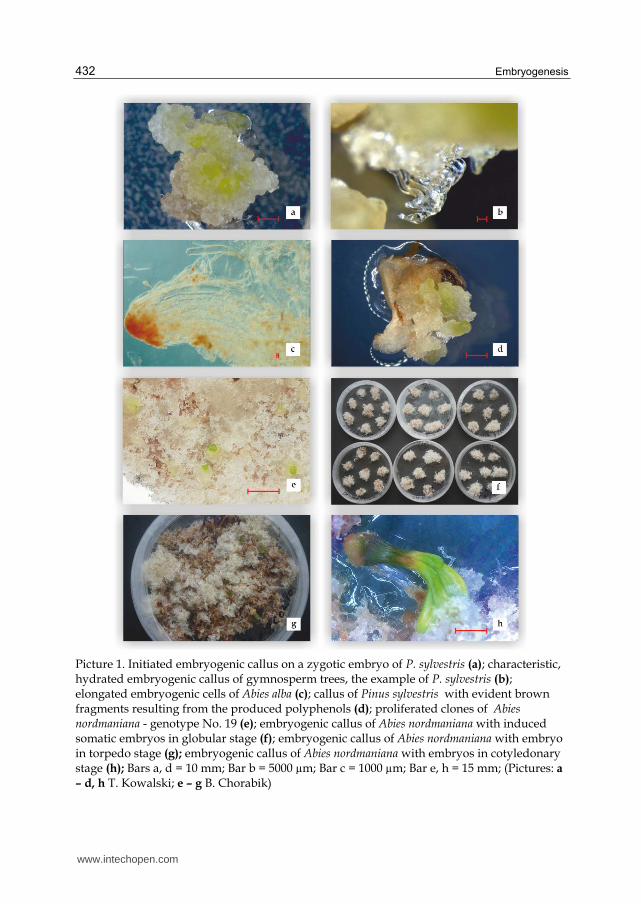
Initiation, also called indution, is a process of unleashing the embryogenic potential of a single cell or a group of cells. Embryogenic callus, in trees having the form of floculent, usually transparent or white, often viscous, well-hydrated mass, originates on the initial explant (Pict. 1a). The first formed embryogenic cells are called proembryogenic masses (PEM). For about 2-3 weeks the initiated embryogenic callus grows on the initiation medium and then embryogenic cell mass is formed, otherwise known as embryogenic suspensor mass (ESM) (Pict. 1b). It creates a shapeless mass of rapidly dividing cells differentiated in size and shape (from izometric cells to loosely bound, large cells). Callus initiated on a single explant is called a line, which is a single genotype with single set of chromosomes. In this phase, it must be determined whether the callus is embryogenic, by staining with acetocarmine and microscopic observations of proembryos (Gupta & Durzan 1987), (Pict. 1c). In the callus of gymnosperm trees, zones of embryogenic masses may be distinguished – small cells whose nuclei stain red and long, colorless cells with small nuclei and large vacuoles. In some cases, one can find
www.intechopen.com
Embryogenesis
428
polyploid cells in the callus, originating from selective effect of cytokines, which favors the development of micro-seedlings with unfavorable characteristics.
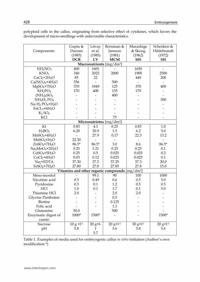
Components:
Gupta & Durzan (1985) DCR
Litvayet al.
(1985)LV
Bornman & Jansson (1981) MCM
Murashige& Skoog
(1962) MS
Schenken & Hildebrandt
(1972) SH
Macronutrients [mg/dm3]NH4NO3
KNO3
CaCl2×2H2O Ca(NO3)2×4H2O
MgSO4×7H2O KH2PO4
(NH4)2SO4
NH4H2 PO4
Na H2 PO4×H2O FeCL3×6H2O
K2 SO4
KCl
400340 85
556 370 170
- - - - - -
16012022 22 -
1849 408
- - - - - -
-2000
500 125 135 400
- - - -
75
16501900 440
- 370 170
- - - - - -
- 2500 200
- 400
- -
300 - - - -
Micronutrietns [mg/dm3]KI
H3BO3
MnSO4×4H2O MnSO4×H2O ZnSO4×7H2O
Na2MoO4×2H2O
CuSO4×5H2O CoCl2×6H2O Na2×EDTA
FeSO4×7H2O
0.836.20
- 22.30 86.5* 0.25 0.25 0.03
37.30 27.80
4.130.9 27.9
- 86.5* 1.21 0.5 0.12 37.3 27.8
0.251.5 0.17
- 3.0 0.25
0.025 0.025 37.25 27.85
0.836.2 22.3
- 8.6 0.25
0.025 0.025 37.3 27.8
1.0 5.0 13.2
- 86.5* 0.1 0.2 0.1 20.0 15.0
Vitamins and other organic compounds [mg/dm3]Meso-inositolNicotinic acid
Pyridoxine HCl
Thiamine HCl Glycine Panthoten
Biotine Folic acid Glutamine
Enzymatic digest of casein
-0.5 0.5 1.0 2.0 - - -
50.0 1000*
99.10.49 0.1 0.1 - - - -
1500*
900.6 1.2 1.7 2.0 0.5
0.125 1.1 500
-
1000.5 0.5 0.1 2.0 - - - - -
1000 5.0 0.5 5.0 - - - - -
1500*
Sucrose pH
10 g ×l-1
5.8 20 g×l-
1 5.7
20 g×l-1
5.6 30 g×l-1
5.8 20 g×l-1
5.6
Table 1. Examples of media used for embryogenic callus in vitro initiation (Author’s own modification *)
www.intechopen.com
Somatic Embryogenesis in Forest Plants
429
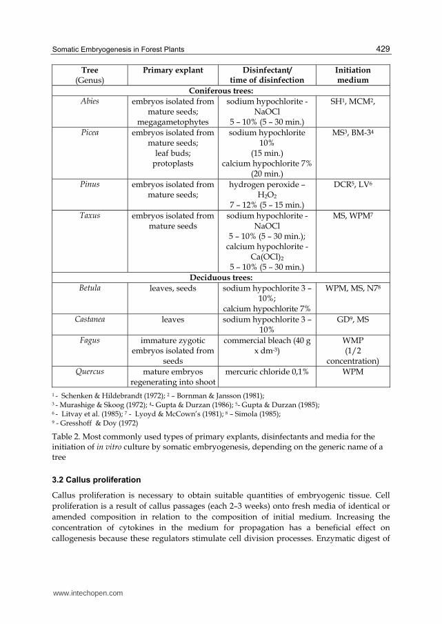
Tree (Genus)
Primary explant Disinfectant/ time of disinfection
Initiation medium
Coniferous trees:
Abies embryos isolated from mature seeds;
megagametophytes
sodium hypochlorite - NaOCl
5 – 10% (5 – 30 min.)
SH1, MCM2,
Picea embryos isolated from mature seeds;
leaf buds; protoplasts
sodium hypochlorite 10%
(15 min.) calcium hypochlorite 7%
(20 min.)
MS3, BM-34
Pinus embryos isolated from mature seeds;
hydrogen peroxide – H2O2
7 – 12% (5 – 15 min.)
DCR5, LV6
Taxus embryos isolated from mature seeds
sodium hypochlorite - NaOCl
5 – 10% (5 – 30 min.); calcium hypochlorite -
Ca(OCl)2
5 – 10% (5 – 30 min.)
MS, WPM7
Deciduous trees:
Betula leaves, seeds sodium hypochlorite 3 – 10%;
calcium hypochlorite 7%
WPM, MS, N78
Castanea leaves sodium hypochlorite 3 – 10%
GD9, MS
Fagus immature zygotic embryos isolated from
seeds
commercial bleach (40 g x dm-3)
WMP (1/2
concentration) Quercus mature embryos
regenerating into shootmercuric chloride 0,1% WPM
1 - Schenken & Hildebrandt (1972); 2 – Bornman & Jansson (1981); 3 - Murashige & Skoog (1972); 4- Gupta & Durzan (1986); 5- Gupta & Durzan (1985); 6 - Litvay et al. (1985); 7 - Lyoyd & McCown’s (1981); 8 – Simola (1985); 9 - Gresshoff & Doy (1972)
Table 2. Most commonly used types of primary explants, disinfectants and media for the initiation of in vitro culture by somatic embryogenesis, depending on the generic name of a tree
3.2 Callus proliferation
Callus proliferation is necessary to obtain suitable quantities of embryogenic tissue. Cell
proliferation is a result of callus passages (each 2–3 weeks) onto fresh media of identical or
amended composition in relation to the composition of initial medium. Increasing the
concentration of cytokines in the medium for propagation has a beneficial effect on
callogenesis because these regulators stimulate cell division processes. Enzymatic digest of
www.intechopen.com
Embryogenesis
430
casein added to the medium in quantities of 1000 – 1500 mg x dm3, particularly in the case of
gymnosperm trees, often has a beneficial effect on proliferation of callus (Nawrot-Chorabik
2008), (Fig. 1).
Not all callus lines are embryogenic and are capable of intensive proliferation. Only certain genotypes are characterized by high frequency of embryogenesis (Pict. 1e). The origin of plant material, particularly seeds, has a significant impact on embryogenic capacity of callus. First somatic embryos are formed in a globular stage. This is the induction of somatic embryos (Pict. 1f). Also the explant itself, e.g. a leaf, has a particular meaning in the process of embryo induction on a proliferated callus. Younger – the innermost leaf tissues (constituting the primary explant) produce callus with large quantities of somatic embryos, while further - more external - parts of leaves may produce smaller callus or the embryos are induced directly on this leaf fragment (Trigiano & Gray 2011).
3.3 Conversion of somatic embryos, gene expresion
Globally speaking, the phenomenon of conversion is understood in two ways – as the
development of somatic embryos into plants of identical genotype as initial explant capable
of ex vitro growth and development, morphologically developed - with a root, apical bud
and first assimilation organs (Becwar et al. 1989) - and more rarely as survivability of
seedlings regenerated after inoculation and adaptation to ex vitro conditions. For somatic
embryogenesis of forest trees conversion also refers to the successive stages of somatic
embryos’ development. The following stages may be distinguished: globular, heart-shaped,
torpedo, early-cotyledonary and cotyledonary (Fig. 1, Pict. 1 g, h). Subsequent stage of
somatic embryos development (maturation) has gained importance due to its aftermath.
With proper embryonic morphogenesis in the process of micropropagation one can obtain
valuable plants with large capacities for uniform and rapid germination, with normal
growth in the broadly understood range of environmental factors. Properly developed
seedlings in terms of physiology are formed not only from embryos, that have the
appropriate morphology, but also that gathered the necessary amount of reserve material.
Otherwise, often only a rootless shoot is developed. In woody plants the embryo
development starts from small clusters of embryogenic cells called proembryogenic masses
(PEM I), composed of cells with dense cytoplasm, adjacent to a single vacuolated cell
showing tendency to elongation. After about three days further elongated cells develop
from a group of cells with dense cytoplasm and form PEM II. Then, after about two weeks
large aggregates of cells are formed, classified as PEM III (von Arnold & Clapham 2008).
Reducing the amount of auxins and cytokines sometimes stimulates the differentiation of
somatic embryos, but the rule is that the addition of abscisic acid (ABA) to the medium is
necessary to obtain a cotyledonary embryo. The first visible response of somatic embryos to
abscisic acid is their change to colorless. From this point the embryo begins to elongate and
form cotyledons.
During the conversion of somatic embryos gene expression occurs – which, if started at the
right time, ensures proper construction and development of the embryo. So far the
following types of genes have been identified: genes responsible for cell cycle and cell wall
synthesis, genes responsive to hormones and transcription process associated with somatic
embryogenesis. Cell division and growth requires a strict control in time and space.
www.intechopen.com

Somatic Embryogenesis in Forest Plants
431
Expression of genes responsible for cell-cycle process is therefore important for the further
development of the embryo. Genes responsible for cell wall synthesis are also important, as
somatic embryogenesis depends on proper formation of cell wall components. Among
others, the following genes are included in the group responsible for cell-cycle and cell wall
synthesis: cdc2M, CEM6, SERP, AGP. The expression of these genes at the right time ensures
proper construction of the embryo (Yiang & Zhang 2011). Induction and growth of somatic
cells can be stimulated by appropriate hormones that affect hormone-sensitive genes, among
which one can distinguish genes responsive to abscisic acid (ABA) e.g. LEA, but also genes
responsive to auxins (indolyl-3-acetic acid – IAA and picloram - PIC). These genes include
GH3, PIN, ARF, SAUR. The proper course of somatic embryogenesis requires genes that
regulate the individual stages and the entire process. These include transcription factors
associated with somatic embryogenesis, such as LEC, BBM, WUC, AGL15. All these factors
ensure the proper development of somatic embryo (Yiang & Zhang 2011).
3.4 In vitro seedling rhizogenesis
Only somatic embryos in cotyledonary stage with clearly developed cotyledons, hypocotyl
and primordial root, with morphology similar or identical to zygotic embryo, will germinate
and develop into somatic seedlings. Such embryos are transferred onto germination
medium. The majority of these are media poor in macro- and micronutrients and sugar,
often without growth regulators. These media should be supplemented with auxin, which
acts as root inducer - indole-3-butyric acid (IBA). Some species, particularly gymnosperm
trees, require additional treatments during the rhizogenesis stage, such as drying of
embryos (so called desiccation) under conditions of high humidity (ca. 95%) or higher
concentrations of solidifiers in the culture media. These treatments cause that the developed
embryos in the cotyledonary stage have proper turgidity, which enhances their ability to
germinate. According to the literature, the germination ability of somatic embryos obtained
by somatic embryogenesis is relatively low and the average is around 15% (Cornu &
Geoffrion 1990; Salajova et al. 1995).
3.5 Acclimation to environmental conditions
Adaptation of developed forest tree seedlings to ex vitro conditions is difficult due to physiological determinants of young tree seedlings. Their slenderness due to lack of woody tissue and the covering tissue – cuticle, poorly developed root system and assimilation apparatus and significant hydration of the tissues causes instability of seedlings in a new medium – cellulose-peat pots. The cultivated seedlings are transferred from media to pots with a volume of 0.065 liters distributed under different names, e.g. Fetlipots, Finnpots, Jiffypots. The substrate in pots should be watered with basic medium diluted in a 1:1 ratio. Due to the above-mentioned physiological determinants of seedlings, the following treatments should be used to facilitate acclimation: undercooling (important for conifer species) that prevent growth interruption, increasing the light intensity and the use of fogging and variable conditions of light (photoperiod) and temperature in computer-controlled greenhouses. One can also introduce LED (Light Emitting Diode) illumination. For better growth and development of seedlings of gymnosperm species, the Author recommends white LED light with color temperature of 5000 – 6500 K.
www.intechopen.com
Embryogenesis
432
Picture 1. Initiated embryogenic callus on a zygotic embryo of P. sylvestris (a); characteristic, hydrated embryogenic callus of gymnosperm trees, the example of P. sylvestris (b); elongated embryogenic cells of Abies alba (c); callus of Pinus sylvestris with evident brown fragments resulting from the produced polyphenols (d); proliferated clones of Abies nordmaniana - genotype No. 19 (e); embryogenic callus of Abies nordmaniana with induced somatic embryos in globular stage (f); embryogenic callus of Abies nordmaniana with embryo in torpedo stage (g); embryogenic callus of Abies nordmaniana with embryos in cotyledonary stage (h); Bars a, d = 10 mm; Bar b = 5000 µm; Bar c = 1000 µm; Bar e, h = 15 mm; (Pictures: a
– d, h T. Kowalski; e – g B. Chorabik)
www.intechopen.com

Somatic Embryogenesis in Forest Plants
433
Fig. 1. Development phases of somatic embryogenesis methods
www.intechopen.com
Embryogenesis
434
4. Physical conditions of in vitro cultures
The physical conditions affecting the state of in vitro culture during plant tissue morphogenesis are the abiotic factors such as temperature, light, relative humidity, pH, oxygen and carbon dioxide concentration. These factors must be closely coordinated, but also they must be coordinated with other chemical factors such as e.g. composition of media. For the somatic embryogenesis culture to be successful, usually a constant temperature is maintained in vitro both during the day and night. Only in rare cases it is necessary to apply proper temperature variation. Most frequently the temperature in a phytotron chamber varies from 23 to 25°C. The optimum temperature, however, should be determined experimentally, depending on the tested species and primary explant. Each stage of the culture may require different temperature. Furthermore, it should be noted that the temperature inside the room with cultures is a few degrees lower than inside the vessel with explants. Therefore, tree explants respond better to the temperature slightly decreased in relation to its optimum temperature than to increased temperature. The light impacts the morphogenetic changes, that often are induced by this important factor. The first two stages of somatic embryogenesis in most species of trees progress without light, due to the fact that development process of embryogenic callus does not require intensive photosynthesis. Biotechnological Laboratory of in vitro Cultures in the Department of Forest Pathology, University of Agriculture in Cracow, carries out research on the impact of light wavelength and light intensity on the morphogenetic changes in embryogenic callus with somatic embryos of basic, forest-forming gymnosperm tree species of Poland (fir, spruce, pine). It was found that white, diffused, low intensity LED light, which is a mixture of various wavelengths (380–780 nm), in 12-hours’ photoperiod is needed only during the conversion of embryos. White light is the most favorable due to the similarity to the prevailing natural conditions. It matches the range of photosynthetically active light (Photosynthetically Active Radiation – PAR) with a wavelength of 400 – 700 nm. It affects the induction of chlorophyll synthesis, chloroplast development and formation of adventitious organs from callus cells. In the beginning of later stages, namely during rhizogenesis, darkness is required (similarly to initiation and proliferation of callus). Only after 10-14 days, the seedlings need to be transferred to white LED light (about 10 times lower intensity than in natural conditions). During the acclimation of somatic seedlings the plants should be placed in a higher intensity white LED light – of intensity similar to natural conditions, optimal for each species. Moreover, it was experimentally demonstrated that blue LED light in the wavelength of 440-490 nm, used for the 12-hour photoperiod has beneficial effect on callus with forming embryos. Embryogenic callus passaged onto activated charcoal medium without plant hormones for the period of 10 - 14 days, kept in blue light, is easier to purify from growth regulators obtained from proliferation medium. Thanks to blue light in the later stage, i.e. conversion, morphogenetic processes are launched more rapidly and the matured somatic embryos are correctly transformed into somatic seedlings in cotyledonary stage (Nawrot-Chorabik, unpublished data). The mechanism of biochemical processes occurring in embryogenic callus with developing embryos exposed to light of different wavelengths is not fully understood, therefore this issue should be carefully investigated. Other light wavelengths, i.e. green light (490-560 nm) and yellow light (560-590 nm) sometimes stimulates formation of adventitious buds from hypocotyl fragments. Conducting in vitro culture with different wavelengths argues for the introduction of light parameters (wavelength, light intensity and exposure time) control in phytotrons for better plant growth. Relative humidity - RH determines the content of water
www.intechopen.com
Somatic Embryogenesis in Forest Plants
435
vapor in the gas phase of the vessel in which the culture is conducted (above the medium surface). RH depends among others on the temperature, chemical composition of a medium, size of explants and vessels. Once the medium and the interior of e.g. a Petri dish have the same temperature, and the vessel is sealed, then the relative humidity should theoretically be 98-99%. However, glass and plastic materials used in vitro are not sufficiently leakproof and water vapor gradually escapes on the outside. Therefore, phytotrons with humidity settings should be used, which in the case of micropropagation of trees should oscillate between 50 and 70%. A pH of media has significant impact on in vitro cultures of trees. Callus and somatic embryos of woody species are formed in acidic pH, i.e. within the range of pH 5.6 – 5.8. pH of a medium may change during the culture. Such changes may be observed particularly in liquid media, and in solid media they may result from too rarely conducted passages. Oxygen (product of photosynthesis) and carbon dioxide (product of respiration) are two gasses, components of air occurring at a concentration of 21% and 0.036%. However, in Petri dishes or in Erlenmeyer’s flasks, in which the culture is carried out, the concentration of these gasses depends among others on: the size of explant, and thus the intensity of photosynthesis, respiration or transpiration and the composition of media (mainly carbohydrate content), light, temperature and the size and shape of the vessel. It is most preferred to maintain oxygen concentration in the vessel at a level higher than its concentration in the air. It was demonstrated that under such conditions, i.e. at the concentration of oxygen within the range of 60-70%, intensive cell divisions occur and the amount of callus, adventitious shoots and somatic embryos increases. Lower than in the air oxygen concentration generally inhibits the plant development. Even roots, which naturally grow in conditions of oxygen deprivation, develop more intensively on the medium in sufficient oxygen supply. Characteristic and frequently observed growth of roots over the level of the medium suggests a lack of oxygen. Sometimes, however, low level of oxygen (7.8%) induces formation of lower amounts of callus, but with the majority of embryogenesis-competent cells. Proper CO2 concentration primarily determines the proper course of photosynthesis. It has been shown that concentration of this gas higher than in the air (approximately 1-5%) stimulates the explants. It accelerates the intensity of photosynthesis, the cell proliferation intensity in suspension and callus proliferation (Woźny & Przybył 2004).
5. Application of bioreactors
In large production in vitro laboratories somatic embryogenesis method is used for reproduction of selected tree species by using bioreactors. In the early stages of micropropagation, embryogenesis is induced on solid media, and after a certain time embryogenic tissue is transferred to liquid media, where it is propagated in the cell suspension in bioreactors. Bioreactors are constructed to enable conducting cell cultures under conditions appropriate to minimize or completely eliminate the possibility of infection. They allow not only the commercial tissue culture, but also the production of somatic embryos. They also allow to obtain embryogenic cells from non-embryogenic ones and to carry out microbiological and enzymatic processes. Bioreactors contain a number of control and measurement sensors, that measure and continuously maintain the following parameters: speed of mixing and aeration, concentration of dissolved oxygen, concentration of dissolved carbon dioxide, the amount of foam, overpressure in the tank, but also oxygen and carbon dioxide concentration in exhaust gasses. This is ensured by appropriate technological parameters and modern design solutions of bioreactors by applying
www.intechopen.com
Embryogenesis
436
specialized computer software. Bioreactors allow to precisely control the metabolism of plants and processes of proliferation and development of callus. The basic requirements for bioreactors designed for in vitro culture are: high efficiency of oxygen exchange and discharge of secreted heat. Air lift bioreactors are now the largest group, since they are very versatile. On the other hand, balloon-type bubble bioreactors are practical because of their shape which prevents media foaming. Bioreactors without forced mixing seem to be particularly useful in cell, tissue and plant organ cultures, as they do not generate the stress of mechanical or pneumatic agitation. The current review of bioreactors used in research and practice was presented in the papers by: Paek & Chakrabarty (2003), and Ziv (2005).
6. Cryopreservation as a method for long-term storage of material obtained by in vitro cultures
Cryopreservation is considered the best method for the long-term storage of plant tissues
cultured in vitro at the temperature of liquid nitrogen (-196°C). When plant material is kept
at such low temperature, cell divisions and metabolic processes are stopped for an indefinite
time. Additional advantages of this method is low storage space and relatively low costs.
Cryopreservation is a method of storage of callus, somatic embryos, pollen, buds and tree
seeds in Dewar flasks. As a result of biotechnology development, genetic resources in gene
banks have been supplemented with new, valuable genotypes of endangered, economically
and ecologically important tree species. The success of cryopreservation depends on
increasing tissue tolerance to dehydration stress and the stress caused by rehydration after
thawing. Physical state that ensures cell survival in the process of dehydration and freezing
is non-crystalline state i.e. vitrification. Under the stress of dehydration, the protein-lipid
cytoplasmic membranes are the most vulnerable to damage, since polynusaturated fatty
acids of membrane phospholipids are easily peroxidized during desiccation. Moreover,
during freezing and thawing of plant material, spontaneous mutations as well as
biochemical and structural changes at the cellular level may occur. Therefore, before and
after freezing, the plant material should be analyzed using molecular biology techniques,
e.g. by checking the somaclonal variation of embryogenic callus (Nawrot-Chorabik 2009).
Cryopreservation process may be carried out in different ways from placing the cryo-tubes
in a Mr. Frosty vessel (NALGENETM W USA), which ensure a slow temperature decrease by
1ºC to the use of computerized cryobath equipment (CryolLogic) with freeze control system.
7. Short review of economically important coniferous and deciduous trees micropropagated with somatic embryogenesis including difficulties encountered during in vitro cultures
Pioneering studies on somatic embryogenesis of coniferous trees were carried out in 1968-1980, when the development and metabolism of callus and suspended cells was studied (Durzan & Steward 1968, Chalupa & Durzan 1973, Durzan & Chalupa 1976). Thorpe & Biondi (1984), Dunstan (1988) and Becwar et al. (1988) paid attention to the potential of a method, which could be used for vegetative proliferation of gymnosperm species. They conducted the in vitro culture of selected species of conifers. However, the first studies on deciduous trees originate from the forties, when reports on the in vitro regeneration of adventitious buds from the callus tissue of field elm (Ulmus campestris) were published (Gautheret 1940). Experiments
www.intechopen.com
Somatic Embryogenesis in Forest Plants
437
on in vitro organogenesis of field elm were also conducted in 1949 by Jacquiot (1949). Results of his study were similar to the results obtained nine years earlier by Gautheret (1940) (Szczygieł 2005 after Gautheret 1940). It should be noted that field elm was cultivated in the forties as park and avenue tree. Currently this species is endangered due to its susceptibility to Dutch elm disease. Jacquiot (1949) simultaneously conducted research on organogenesis of silver birch (Betula verrucosa). Apart from adventitious buds Jacquiot (1949) obtained the beginnings of roots, however, he did not manage to grow fully developed plants (Szczygieł 2005). The first complete plant, which was obtained during this intensive research, was common aspen (Populus tremula) regenerated from a leaf in 1970 by Winton (1970). This achievement initiated greater interest in the method of vegetative in vitro propagation of trees.
Currently over 300 plant species, including 120 species of deciduous trees, are propagated by somatic embryogenesis (Bajaj 1995). Some of them are forest trees, such as: European beech (Fagus sylvatica), English oak (Quercus robur), ash (Fraxinus spp.), small-leaved lime (Tilia cordata), walnut tree (Juglans spp.), poplar (Populus spp.) and locust tree (Robinia). Among all in vitro propagated plants there are 50 coniferous trees, e.g.: European silver fir (Abies alba), European larch (Larix decidua), larch hybrids, Norway spruce (Picea abies), white spruce (Picea glauca), black spruce (Picea mariana), sugar cone pine (Pinus lambertiana), Caribbean pine (Pinus caribea), Loblolly pine (Pinus taeda) and others.
As indicated in Chapter 2.1, in order to initiate embryogenic tissue of coniferous trees,
mature and immature zygotic embryos are mainly used as primary explants. The cotyledons
of 7-day germinated embryos were used for initiation of embryogenic callus of Picea abies
(Krogstrup 1986, Lelu et al. 1990), and 12-day cotyledons were used for initiation of Picea
glauca and Picea mariana (Lelu & Bornman 1990). Other researchers applied also cotyledons
of 12-30-day seedlings of Picea glauca and Picea mariana (Attree et al. 1990). Ruaud et al.
(1992) used hypocotyls and cotyledons of 1-month somatic and zygotic seedlings and
needles of 14-months somatic seedlings cultured in a greenhouse, as well as needles of 7-56 –
day somatic and zygotic seedlings for initiation of embryogenic callus of Picea abies (Ruaud
1993). Harvengt et al. (2001) obtained embryogenic tissue on 3-year needles of somatic
seedlings of Picea abies. These needles are the oldest spruce explant, from which
embryogenic tissue was obtained (Szczygieł 2005). Nagmani & Bonga (1985) in Larix decidua
and von Aderkas et al. (1990) in L. decidua and L. leptolepis used megagametophytes with
removed immature embryos for initiation of haploid embryogenic tissue and
megagametophytes with immature embryos to initiate diploid embryogenic callus. For
initiation of somatic embryogenesis also protoplasts were used (Attree et al. 1987,
Klimaszewska 1989, von Aderkas et al. 1990). Attree et al. (1987) regenerated somatic
embryos from protoplasts isolated from embryogenic tissue of Picea glauca and
Klimaszewska (1989) cultured seedlings of Larix decidua x L. leptolepis hybrid also using
protoplasts from embryogenic tissue as explants in somatic embryogenesis. Similarly, von
Aderkas et al. (1990) regenerated seedlings from protoplasts isolated from haploid callus of
Larix decidua. Higher frequency of embryogenic callus initiation was obtained using
immature emryos as explants. However, more practical is the use of mature zygotic
embryos, isolated from seeds stored in cold rooms. Embryogenic tissue on mature zygotic
embryos of European silver fir (Abies alba) was initiated by Hristoforoglu et al. (1995) in 40%,
Szczygieł (2005) after Braumüller et al. (2001) in 52% and Nawrot – Chorabik (2008) in 6%.
Currently, other sources of explants (needles, cotyledons, hypocotyls) and new, synthetic
www.intechopen.com
Embryogenesis
438
growth regulators that stimulate the process of somatic embryogenesis initiation are
searched for (Szczygieł 2005). The first reports on somatic embryogenesis of Scots pine were
focused predominantly on initiation from immature seeds and on studying the reactions of
cut zygotic embryos at several developmental stages on various culture media (Lelu et al.
1999). The efforts during the regeneration of a small amount of somatic seedlings and
young trees were not aimed at the development of mature somatic embryos, but the
creation of a protocol for the efficient production of large amounts of plant clones. In
another case, crossbreeding was conducted among selected parent trees in order to assess
the impact of genotype of parents on somatic embryogenesis (Niskanen et al. 2004).
During initiation, maternal effect was clearly visible, while paternal effect was
predominantly invisible. A similar conclusion was reached during somatic embryogenesis
studies in Pinus taeda (MacKay et al. 2006). In other conducted experiments the impact of
several factors on the maturation of somatic embryos was investigated. These factors are:
age of the culture, abscisic acid and sucrose concentration in the medium (Lelu-Walter et
al. 2008). The most current research on somatic embryogenesis in genus Pinus has
concentrated on examining the impact of parental genotypes and initiation that origin
from controlled crossing between maternal trees, that had previously been tested for their
response to initiation (Lelu et al. 1999).
The research on somatic embryogenesis in deciduous trees has shown that during in vitro
culture many difficulties may be encountered. Based on the vine, it was found that although
somatic and zygotic embryos are nearly identical in structural and functional characteristics,
the ontogeny of somatic embryos tends to be more variable. Somatic embryos of most
species, including grapevine (Vitis spp.), tend to exhibit several typical morphological
abnormalities such as variation in shape, size, and number of cotyledons. In most species,
somatic embryos are larger than zygotic embryos, and their regeneration rates are lower.
For instance, in Vitis rupestris Scheele, only 3% of somatic embryos were capable of
developing into complete plants (Jayasankar et al. 2003). This kind of abnormalities may
also be found in forest tree species.
Penduculate oak (Quercus robur) is an endangered species due to the periodic dieback of oak stands in ecosystems of Troncais forest in France, but also in Austria, Hungary, Romania, Northern Germany, Russia and Ukraine. Oaks die also in Poland in The Krotoszyn Plateau and within the area of Mediterranean Basin. Oaks reproduce in nature by acorns, which are very sensitive to drying during storage, therefore their ability to germinate drops significantly. For this reason, this species should definitely be rescued by micropropagation in in vitro laboratories, although the oak is less susceptible to tissue cultures. Such attempt was undertaken in Slovakia, where the method of somatic embryogenesis was used for micropropagation of oak (Querkus spp.). Callus was initiated on mature oak seeds. Disinfection of seeds was slightly different than in the case of conifers, i.e.: the seeds were washed in 70% ethanol, extending the duration of the alcohol to 10 minutes and then they were treated for 15–20 with 0.1% solution of mercuric chloride. Embryogenic axes were aseptically isolated from the surrounding cotyledons with preservation of cotyledonary nodes and plumule. Isolated explants were treated with 100 mg x dm-3 solution of ascorbic acid to prevent oxidation for 30 min. For the cultivation of embryogenic axes in Quercus spp. WPM medium (Lyoyd & McCown 1980) with 20 g x dm-3 sucrose and 6 g x dm-3 Difco-Bacto Agar, supplemented with 1 mg x dm-3 BA and 0.01 mg x dm-3 NAA was used. In all
www.intechopen.com
Somatic Embryogenesis in Forest Plants
439
experiments the medium pH was adjusted to 5.5 – 5.7. The culture required less frequent passages than in the case of conifers, i.e. in 4-5 week intervals. The number of shoots per explant formed during the subculture was recorded (Ostrolucká et al. 2007).
Silver birch (Betula pendula) is one of the most important birch species. It naturally occurs throughout Europe and in central parts of Asia. Silver birch is economically significant because of its medicinal properties. Birch leaves, that contain saponins, flavonoids and terpene compounds are used as medicinal substances. Chaga mushroom was used in folk medicine as an anticancer agent. Its therapeutic properties have been confirmed by research conducted in Poland and Russia. Forest yield can be enhanced significantly by large-scale multiplication of selected genotypes with improved growth rates, valuable quality of wood and high stress and disease tolerance. Vegetative propagation is an important tool of preserving unique characteristics of some of the selected trees of Silver birch (Chalupa 1995). In 1990 Kurtén et al. (1990) induced somatic embryogenesis of B. pendula using eight different families, obtained by crossing parent plants with different herbivore resistance. Embryogenic culture was initiated from seeds, seedlings and leaves from 1-year-old plants. Sodium hypochlorite was used for explant disinfection (leaves for 10 min., seeds for 30 min.), and N7 basal medium was used as the medium for initiation (Simola 1985). Nuutila et al. (1991) studied effect of different sugar and inorganic nitrogen concentrations in in vitro cultures of birch. Embryogenic callus was initiated on N7 medium, on which somatic embryos were obtained in a later stage (Simola 1985). The first stage of in vitro culture (callus initiation) and maturation of somatic embryos was carried out on solidified medium, while the second stage (proliferation) was carried out in liquid medium. Embryogenic tissue of birch in Chalupa’s cultures (1987) was characterized by regions of meristematic cells, which were small, thin-walled and highly cytoplasmic. Globular somatic embryos developed at the periphery of the meristematic regions and consisted of densely cytoplasmic cells. At later stages, embryos were surrounded by epidermic cells. Late heart-shaped embryos developed only in small part of callus initiated from explants. Somatic embryos matured after 4 – 6 weeks (Chalupa 1987).
Sweet chestnut (Castanea sativa) is the tree valued mostly because of its fruit (chestnuts). This species of Fagaceae family occurs naturally in the Mediterranean Basin, Asia Minor and the Caucasus. A major limitation of the embryogenic systems used in chestnut is the maitenance of embryogenic competance and the low conversion rate of somatic embryos into plants. Experiments were performed to determine the influence of proliferation medium on the maintenance of embryogenic competence and on repetitive embryogenesis in C. sativa somatic embryos derived from leaf explants. Somatic embryo proliferation was carried out by both direct secondary embryogenesis and by the culture of nodular callus tissue originated from cotyledons of somatic embryos. Both systems led to the production of cotyledonary somatic embryos on MS proliferation medium supplemented with 0.1 mg x dm-3 BA and 0.1 mg x dm-3 NAA. A total of 39% of embryos eventually produced plants either through conversion to plantlets or indirectly through rooting of shoots. Shoots formed by somatic embryos could be excised, multiplied and rooted following the micropropagation procedures (Corredoira et al. 2003).
European beech (Fagus sylvatica) occurs almost in the whole Europe. Its wood is hard,
compact, with no heartwood, which causes the beech to be the most commonly used in the
art and practice of utility (high calorific value). Therefore, the trees selected for desirable
www.intechopen.com
Embryogenesis
440
genotypes can be cloned by in vitro cultures developing an efficient method of
micropropagation. Until now, very few researchers dealt with this economically significant
species. Among them were Vieitez et al. (1992) and Naujoks (2001). Vieitez et al. (1992)
isolated immature embryos from seeds disinfected in commercial bleach (40 g x dm-3). The
culture was established on WPM medium with addition of 2,4-D (0.45; 2.36; 4.52 µM x dm-3)
and BA (2.2 µM x dm-3). Embryogenic cell suspensions of 5 genotypes were successfully
established in LM medium containing 2,4-D, on which the secondary explants were
passaged. The earliest stage, identified as embryogenic, consisted of single cells, undergoing
a first asymmetric division leading to the formation of two daughter cells of unequal size, a
small cell with dense cytoplasm and strong affinity for acetocarmine and a larger cell with
only slight affinity for acetocarmine. Subsequently, either the smaller cells underwent
further polarized divisions to form pro-embryos or both daughter cells underwent a series
of anarchical divisions leading to the formation of cell aggregates or proembryogenic
masses, from which multiple embryos developed by cleavage polyembryony. Finally, 10%
of embryos (63 plantlets from 638 embryos) obtained by somatic embryogenesis had
developed roots, shoots and well-formed leaves (Vieitez et al. 1992). On the other hand, the
research by Naujoks (2001) showed, that on WPM medium, the somatic embryos
regenerated on 4 callus lines among 33 lines analyzed (7.9%). Somatic embryos of beech
matured on a medium 50% WPM (with 2,4-D and BA) followed by rooting and acclimation
in the ground. However, based on the experience and results gained, it was concluded that
the long-term storage of embryogenic lines and somatic embryos should be carried out
(Naujoks 2001).
The above examples show that the method of somatic embryogenesis is intensively tested in in vitro cultures. However, despite conducting numerous experiments on obtaining micro-seedlings of tree species, some of the issues related to this method remain still unresolved. More attention should be paid to the optimization methods, so that the plant material obtained in vitro (somatic embryos, callus tissue and properly developed seedlings) was useful for selection, breeding and molecular biology of forest trees and to be useful in other business sectors.
8. Acknowledgements
The author would like to express their special thanks to Prof. dr hab. Tadeusz Kowalski for the given valuable suggestions and doing microscopic photos.
9. References
Attree, S.M.; Bekkaoui, F.; Dunstan, D.L. & Fowke, L.C. (1987). Regeneration of somatic embryos from protoplasts isolated from an embryogenic suspension culture of white spruce (Picea glauca). Plant Cell Reports, Vol. 6, pp. 480-483.
Attree, S.M.; Budimir S. & Fowke, L.C. (1990). Somatic embryogenesis and plantlet regeneration from cultured shoots and cotyledons of seedlings from stored seeds of black and white spruce (Picea mariana and. P. glauca). Canadian Journal of Botany, Vol. 68, pp. 30-34.
www.intechopen.com
Somatic Embryogenesis in Forest Plants
441
Bajaj, Y.P.S. (1995). Somatic embryogenesis and its applications for crop improvement. In: Bajaj Y.P.S. (Ed.). Biotechnology in Agriculture and Forestry. Vol. 30, Somatic embryogenesis and synthetic seeds I, pp. 221-233. Springer-Verlag,
Becwar, M.R.; Noland T.J. & Wyckoff, J.L. (1989). Maturation, germination and conversion of Norway spruce (Picea abies L.) somatic embryos to plants. In Vitro Cellular and Developmental Biology – Plant, Vol. 25, pp. 575-580.
Becwar, M.R.; Wann, S.R.; Johnson, M.A.; Verhagen, S.A.; Feirer R. & Nagmani, R. (1988). Development and characterization of in vitro embryogenic systems in conifers. In: Ahuja M.R. (Ed.). Somatic cell genetics and woody plants. Kluwer Academic Publishers, Dordrecht. pp. 1-18.
Bornman, C.H. & Jansson, E. (1981). Regeneration of plants from conifer leaf, with special reference to Picea abies and Pinus sylvestris. Colloque International sur la Culture In Vitro des Essances Forestieres. Fontaineblau, AFOCEL. Nangis. pp. 41-53.
Braumüller, S.; Ross, H.; Rahmad, A. & Zoglauer, K. (2001). Somatic embryogenesis in silver fir (Abies alba Mill.). International Conference on Wood, Breeding, Biotechnology and Industrial Expectations. Abstract, Bordeaux, France, pp. 62.
Chalupa, V. & Durzan, D.J. (1973). Growth and development of resting buds of conifers in vitro. Canadian Journal of Forest Research, Vol. 3, pp. 196-208.
Chalupa, V. (1985). Somatic embryogenesis and plantlet regeneration from cultured immature and mature embryos of Picea abies (L.) Karst. Communication Institute of Forest Czech Republik, Vol. 14, pp. 57-63.
Chalupa, V. (1987). Somatic embryogenesis and plant regeneration in Picea, Quercus, Betula, Tilia, Robinia, Fagus and Aesculus. Communication Institute of Forest Czech Republik, Vol. 15, pp. 133-148.
Chalupa, V. (1995). Somatic embryogenesis in birch (Betula pendula Roth.). In: Jain, S.M., Gupta, P.K. & Newton, R.J. (Ed(s).). (1995). Vol. 2: 207-220. Somatic Embryogenesis in Woody Plants. Kluwer Academic Publishers, Dordrecht.
Chen, Z.; Yao, Y. & Zhang, L. (1988). Studies on embryogenesis of woody plants in China. In: Ahuja M.R. (Ed(s).). (1988). Somatic Cell Genetics of Woody Plants. Kluwer Academic Publishers, Dordrecht. pp. 19-25.
Cornu, D. & Geoffrion, C. (1990). Aspects de l’embryogenese somatique chez le meleze. Bulletin de la Société Botanique de France, Paris. Vol. 137, pp. 25-34.
Corredoira, E.; Balleste, A. & Vieitez, A.M. (2003). Proliferation, Maturation and Germination of Castanea sativa Mill. Somatic Embryos Orginated from Leaf Explants. Annals of Botany, Vol. 92, pp. 129-136.
Cusidó, R.M.; Palazón, J.; Navia-Osorio, A.; Mallol, A.; Bonfill, M.; Morales, C. & Piñol, M.T. (1999). Production of Taxol and baccatin III by a selected Taxus baccata callus line and its derived cell suspension culture. Plant Science, Vol. 146, pp. 101-107.
Cyr, D.R. & Klimaszewska K. (2002). Conifer somatic embryogenesis: II Applications. Dendrobiology, Vol. 48, pp. 41-49.
Dunstan, D.I. (1988). Prospects and progress in conifer biotechnology. Canadian Journal of Forest Research, Vol. 18, No. 12, pp. 1497-1506.
Ďurkovič, J. & Mišalová, A. (2008). Micropropagation of Temperate Noble Hardwoods: An Overview. Functional Plant Science and Biotechnology, Vol. 2, No. 1, pp. 1-9.
www.intechopen.com
Embryogenesis
442
Durzan, D.J. & Chalupa, V. (1976). Growth and metabolism of cells and tissue of jack pine (Pinus banksiana). 3. Growth of cells in liquid suspension cultures in light and darkness. Canadian Journal of Botany, Vol. 54, pp. 456-467.
Durzan, D.J. & Steward, F.C. (1968). Cell and tissue culture of white spruce and jack pine. Bi-monthly research notes / Canadian Forestry Service, Vol. 24, pp. 30.
Gautheret, R.J. (1940). Nouvelles recherches sur le bourgeonnernent du tissu cambial d'Ulmus campestris cultive in vitro. C. R. Academy of Science Paris, 210: 744-746.
Gresshoff, P.M. & Doy, C.H. (1972). Development and differentiation of haploid Lycopersicon esculentum. Planta, Vol. 107, pp. 161-170.
Gupta, P.K. & Durzan, D.J. (1985). Shoot multiplication from mature trees of Douglas – fir (Pseudotsuga menziesii) and sugar pine (Pinus lambertiana). Plant Cell Reports, Vol. 4, pp. 177-179.
Gupta, P.K. & Durzan, D.J. (1987). Biotechnology of somatic polyembryogenesis and plantlet regeneration in loblolly pine. Bio/Technology, Vol. 5, pp. 147-151.
Gupta, P.K., Durzan, D.J. (1986). Plantlet regeneration via somatic embryogenesis from subcultured callus of mature embryos of Picea abies (Norway spruce). In Vitro Cellular and Developmental Biology Plant, Vol. 22, pp. 685-688.
Hakman, I.; Fowke, L.C; von Arnold, S. & Eriksson, T. (1985). The development of somatic embryos in tissue cultures initiated from immature embryos of Picea abies (Norway spruce). Plant Science, Vol. 38, pp. 53-59.
Hargreaves, C. & Menzies, M. (2007). Organogenesis and cryopreservation of juvenile radiata pine. Vol. 6, pp. 51-66. In: Jain, S.M. & Häggman H. (Ed(s).). (2007). Protocols for Micropropagation of Woody Trees and Fruits, Springer-Verlag.
Harvengt, L.; Trontin, J.F.; Reymond, L.; Canlet, F. & Paques, M. (2001). Molecular evidence of true-to-type propagation of 3-year old Norway spruce through somatic embryogenesis. Planta, Vol. 213, No. 5, pp. 828-832.
Hendry, S.J.; Boddy, L. & Lonsdale, D. (1993). Interactions between callus cultures of European beech, indigenous ascomycetes and derived fungal extracts. New Phytologist, Vol. 123, pp. 421-428.
Hristoforoglu, K.; Schmidt, J. & Bolhar-Nordenkampf, H. (1995). Development and germination of Abies alba somatic embryos. Plant Cell Tissue and Organ Culture, Vol. 40, pp. 277-284.
Jacquiot, C. (1949). Observations sur la neoformation de burgeons chez le tissu cambial d’Ulmus campestris cultive in vitro. C. R. Academy of Science Paris, Vol. 229, pp. 529-530.
Jayasankar, S.; Bondada, B.R., Li Z. & Gray, D. J. (2003). Comparative anatomy and morphology of Vitis vinifera (Vitaceae) somatic embryos from solid- and liquid-culture-derived proembryogenic masses. American Journal of Botany, Vol. 90, pp. 973-979.
Klimaszewska, K. (1989). Recovery of somatic embryos and plantlets from protoplast cultures of Larix x eurolepsis. Plant Cell Reports, Vol. 8, pp. 440-444.
Klimaszewska, K.; Overton C.; Stewart D. & Rutledge R.G. (2010). Initiation of somatic embryos and regeneration of plants from primordial shoots of 10-year-old somatic white spruce and expression profiles of 11 genes followed during the tissue culture process. Planta, Vol. 233, No. 3, pp. 635-647.
www.intechopen.com
Somatic Embryogenesis in Forest Plants
443
Klimaszewska, K.; Park, Y.-S.; Overton, C.; Maceacheron, I. & Bonga, J.M. (2001). Optimized somatic embryogenesis in Pinus strobus L. In Vitro Cellular and Developmental Biology Plantarum, Vol. 37, pp. 392-399.
Kowalski, T. & Kehr, R.D. (1992). Endophytic fungal colonization of branch bases in several forest tree species. Sydowia, Vol. 44, pp. 137-168.
Krogstrup, P. (1986). Embryo-like structures from cotyledons and ripe embryos of Norway spruce (Picea abies). Canadian Journal of Forest Research, 16: 664-668.
Kúrten, U.; Nuutila, A.-M.; Kauppinen, V. & Rousi, M. (1990). Somatic embryogenesis in cell cultures of birch (Betula pendula Roth). Plant Cell Tissue and Organ Cultures, Vol. 23, pp. 101-105.
Lelu, M.A. & Bornman, C.H. (1990). Induction of somatic embryogenesis in excised cotyledons of Picea glauca and Picea mariana. Plant Physiology and Biochemistry, Vol. 28, No. 6, pp. 785-791.
Lelu, M.A.; Bastein, K.; Drugeault, A. & Klimaszewska, K. (1999). Somatic embryogenesis and plantles development in Pinus sylvestris and Pinus pinaster on medium with and without growth regulators. Physiologia Plantarum, Vol. 105, pp. 719-728.
Lelu, M.A.; Boulay, M.P. & Bornman, C.H. (1990). Somatic embryogenesis in cotyledons of Picea abies is enhanced by an adventitious bud-inducing treatment. New Forests, Vol. 4, pp. 125-135.
Lelu-Walter, M.A.; Bernier-Cardou, M. & Klimaszewska, K. (2008). Clonal plant production from self- and cross-pollinated seed families of Pinus sylvestris (L.) through somatic embryogenesis. Plant Cell Tissue and Organ Cultures, Vol. 92, pp. 31-45.
Litvay, J.D.; Verma, D.C. & Johnson, M.A. (1985). Influence of loblolly pine (Pinus taeda L.) culture medium and its components on growth and somatic embryogenesis of wild carrot (Daucus carota L.). Plant Cell Reports, Vol. 4, pp. 325-328.
Lloyd, G. & McCown, B. (1981). Commercially-feasible micropropagation of Mountain laurel, Kalmia latifolia , by use of shoot tip culture. International Plant Propagators Society, Vol. 30, pp. 421-427.
MacKay, J.J.; Becwar, M.R. & Park, Y.S. (2006). Genetic control of somatic embryogenesis initiation in loblolly pine and implications for breeding. Tree Genetics and Genomes, Vol. 2, pp. 1-9.
Malá, J.; Cvikrová, M. & Chalupa, V. (2007). Micropropagation of mature trees of Ulmus glabra, Ulmus minor and Ulmus laevis. In: Jain S.M. & Häggman H. (Ed(s).). Protocols for Micropropagation of Woody Trees and Fruits, Vol. 22, pp. 237-248. Springer-Verlag.
Mihaljevic, S. & Jelaska, S. (2005). Omorica spruce (Picea omorica). In: Jain, S.M. & Gupta P. (Ed(s).). Protocol for somatic embryogenesis in woody plants. Forestry Sciences, 77. Vol. 4, pp. 35-46. Springer-Verlag.
Misson, J.-P.; Druart, P.; Panis, B. & Watillon, B. (2006). Contribution to the study of the maintenance of somatic embryos of Abies nardmaniana Lk: culture media and cryopreservation method. Propagation of Ornamental Plants, Vol. 6, No. 1, pp. 17-23.
Mulabagal, V. & Tsay, H.-S. (2004). Plant Cell Cultures – An Alternative and efficient source for the production of biologically important secondary metabolites. International Journal of Engineering Science, Vol. 2, No. 1, pp. 29-48.
Murashige, T. & Skoog, F. (1962). A revised medium for rapid growth and bioassays with tobacco tissue cultures. Physiologia Plantarum, Vol. 15, pp. 473-494.
www.intechopen.com
Embryogenesis
444
Nagmani, R. & Bonga, J.M. (1985). Embryogenesis in subcultured callus of Larix decidua. Canadian Forest Research, Vol. 15, pp. 108-1091.
Naujoks, G. (2001). Investigations on somatic embryogenesis of beech (Fagus sylvatica L.). Poster at the Xth International Conference on Plant Embryology -"From Gametes to Embryos", 5. – 8. September 2001, Nitra, Slovak Republik.
Naujoks, G. (2007). Micropropagation of Salix caprea L. In: Jain S.M. & Häggman, H. (Ed(s).). Protocols for Micropropagation of Woody Trees and Fruits, Vol. 20, pp. 213-220. Springer-Verlag.
Nawrot – Chorabik, K. (2008). Embryogenic callus induction and differentiation in silver fir (Abies alba Mill.) tissue culture. Dendrobiology, Vol. 59, pp. 31-40.
Nawrot – Chorabik, K. (2009). Somaclonal variation in embryogenic cultures of silver fir (Abies alba Mill.). Plant Biosystems, Vol. 143, pp. 377-385.
Nawrot – Chorabik, K.; Jankowiak, R. & Grad, B. (2011). Growth of two blue-stain fungi associated with Tetropium beetles in the presence of callus cultures of Picea abies. Dendrobiology, Vol. 66, pp. 41-47.
Nhut, D.T.; Hien, N.T.T.; Don, N.T. & Khiem, D.V. (2007). In vitro shoot development of Taxus wallichiana Zucc., a valuable medicinal plant. In: Jain, S.M. & Häggman, H. (Ed(s).). Protocols for Micropropagation of Woody Trees and Fruits, Vol. 10, pp. 107-116. Springer-Verlag.
Niskanen, A.M.; Lu, J.; Seitz, S.; Keinonen, K.; Weissenberg, K. & Pappinen, A. (2004). Effect of parent genotype on somatic embryogenesis in Scots pine (Pinus sylvestris). Tree Physiology, 24, pp. 1259-1265.
Nuutila, A.M.; Kúrten, U. & Kauppinen, V. (1991). Optimization of sucrose and inorganic nitrogen concentration for somatic embryogenesis of birch (Betula pandula Roth.) callus cultures: A statistical approach. Plant Cell, Tissue and Organ Culture, Vol. 24, pp. 73-77.
Ostrolucká, M.G.; Gajdošová, A. & Libiaková, G. (2007). Protocol for micropropagation of Quercus spp. In: Jain S.M. & Häggman H. (Ed(s).). Protocols for Micropropagation of Woody Trees and Fruits, Vol. 8, pp. 85–92. Springer-Verlag.
Paek, K.Y. & Chakrabarty, D. (2003). Micropropagation of woody plants using bioreactor. In: Jain, S.M. & Ishii, K. (Ed(s).). (2003) Micropropagation of woody trees and fruits. Dordrecht, The Netherlands: Kluwer Academic Publishers, pp. 735-756.
Park, Y.S. (2001). Implementation of somatic embryogenesis in clonal forestry: technical requirements and deployment strategies. International Conference on: Wood, Breeding, Biotechnology and Industrial expectations, Abstract, Bordeaux, France, pp. 106.
Rao, P.S. (1965). In vitro induction of embryonal proliferation in Santalum album L. Phytomorphology, Vol. 15, pp. 175-179.
Rodríguez, R.; Valledor, L.; Sánchez, P.; Fraga, M.F.; Berdasco, M.; Hasbún, R.; Rodríguez, J.L.; Pacheco, J.C.; García, I.; Uribe, M.M.; Ríos, D.; Sánchez-, M.; Materán, M.E.; Walter, C. & Cañal, M.J. (2007). Propagation of selected Pinus genotypes regardless of age. In: Jain, S.M. & Häggman, H. (Ed(s).). Protocols for Micropropagation of Woody Trees and Fruits, Vol. 13, pp. 137-146. Springer-Verlag.
Ruaud, J.N. (1993). Maturation and conversion into plantlets of somatic embryos derived from needles and cotyledons of 7-56-day old Picea abies. Plant Sciences, Vol. 92, pp. 213-220.
www.intechopen.com
Somatic Embryogenesis in Forest Plants
445
Ruaud, J.N.; Bercetche, J. & Paques, M. (1992). First evidence of somatic embryogenesis from needles of 1-yearold Picea abies plants. Plant Cell Reports, Vol. 11, pp. 563-566.
Salaj, T. & Salaj, J. (2003/4). Somatic embryo formation on mature Abies alba × Abies cephalonica zygotic embryo explants. Biologia Plantarum, Vol. 47, pp. 7– 11.
Salajova, T.; Salaj, J., Jasik, J. & Kormutak, A. (1995). Somatic embryogenesis in Pinus nigra Arn. In: Jain, S.M.; Gupta, P.K. & Newton, R.J. (Ed(s).). Somatic Embryogenesis in Woody Plants. Kluwer Academic Publishers, Dordrecht. pp. 207-220.
Schenck, R.U. & Hildebrandt, A.C. (1972). Medium and techniques for induction and growth of monocotyledonous and dicotyledonous plant cell cultures. Canadian Journal of Botany, Vol. 50, pp. 199-204.
Simola, L.K. (1985). Propagation of plantlets from leaf callus Betula pendula f. purpurea. Scientia Horticulare, Vol. 26, pp. 77-85.
Steward, F.C. (1958). Growth and development of cultivated cells. III. Interpretations of the growth from free cell of carrot. American Journal of Botany, Vol. 45, pp. 709-713.
Szczygieł, K. (2005). Somatyczna embriogeneza – alternatywny sposób uzyskiwania wyslekcjonowanego materiału sadzeniowego gatunków drzew iglastych. Leśne Prace Badawcze, 3: 71-92. [Somatic embryogenesis – alternative way of obtaining selected planting stock of coniferous tree species]. Forest Research Papers, Vol. 3, pp. 71-92.
Thorpe, T.A. & Biondi, S. (1984). Conifers. Handbook of plant cell culture. In: Sharp, W.R.; Evans, A.; Ammirato, P.V. & Yamada, Y. (Ed(s).). MacMillian Publishing Company of New York, pp. 435-470.
Toribo, M.; Celestino, C. & Molinas, M. (2005). Cork oak, Quercus suber L. In: Jain, SM. & Gupta, P. (Ed(s).). Protocol for somatic embryogenesis in woody plants. Forestry Sciences 77, Vol. 35: 445-458. Springer-Verlag.
Trigiano, R.N. & Gray, D.J. (Ed(s).). (2011). Plant Development and Biotechnology. Section IV Propagation and development concepts. CRC PRESS.
Troch, V.; Werbrouck, S.; Geelen D. & Van Labeke, M-Ch. (2009). Optimization of horse chestnut (Aesculus hippocastanum L.) somatic embryo conversion. Plant Cell, Tissue and Organ Culture, Vol. 98, No. 1, pp. 115-123.
Vieitez, J.F.; Ballester, A. & Vieitez, A.M. (1992). Somatic embryogenesis and plantlet regeneration from cell suspension cultures of Fagus sylvatica L. Plant Cell Reports, Vol. 11, pp. 609-613.
von Aderkas, P.; Klimaszewska, K. & Bonga, J. (1990). Diploid and haploid embryogenesis in Larix letlolepis, L. decidua, and their reciprocal hybrids. Canadian Forest Research, Vol. 20, pp. 9-14.
von Arnold, S. & Clapham, D. (2008). Spruce embryogenesis. In: Suárez M.F., & Bozhkov P.V.) (Ed(s).). Methods in molecular biology, Plant embryogenesis. Vol. 427, pp. 31-45. Humana Press, Inc. in Totowa, NJ, US (United States).
Walters, C.; Find, J.I. & Grace, L.J. (2005). Somatic embryogenesis and genetic transformation in Pinus radiata. In: Jain S.M. & Gupta P. (Ed(s).). Protocol for somatic embryogenesis in woody plants. Forestry Sciences 77, Vol. 2, pp. 11-24. Springer-Verlag.
Wilkinson, J.A. & Brown, A.M.G. (1999). Horse Chestnut - Aesculus hippocastanum: Potential applications in cosmetic Skin-care products. International Journal of Cosmetic Science, Vol. 21, No. 6, pp. 437-447.
www.intechopen.com

Embryogenesis
446
Winton, L.L. (1970). Shoot and tree production from aspen tissue cultures. American Journal of Botany, Vol. 57, pp. 904-909.
Woźny, A. & Przybył, K. (Ed(s).). (2004). Komórki roślinne w warunkach stresu. Tom II Komórki in vitro. [Plant cells under stress conditions]. Vol. 2. In vitro cells. Adam Mickiewicz University. pp. 72-91.
Yang, X. & Zhang, X. (2011). Developmental and molecular aspects of nonzygotic (somatic) embryogenesis In: Trigiano R.N. & Gray D.J. (Ed(s).). (2011). Plant tissue culture, development, and biotechnology, CRC Press. pp. 307-326.
Ziv, M. (2005). Simple bioreactors for mass propagation of plants. Plant Cell, Tissue and Organ Culture, Vol. 81, pp. 277-285.
www.intechopen.com
EmbryogenesisEdited by Dr. Ken-Ichi Sato
ISBN 978-953-51-0466-7Hard cover, 652 pagesPublisher InTechPublished online 20, April, 2012Published in print edition April, 2012
InTech EuropeUniversity Campus STeP Ri Slavka Krautzeka 83/A 51000 Rijeka, Croatia Phone: +385 (51) 770 447 Fax: +385 (51) 686 166www.intechopen.com
InTech ChinaUnit 405, Office Block, Hotel Equatorial Shanghai No.65, Yan An Road (West), Shanghai, 200040, China
Phone: +86-21-62489820 Fax: +86-21-62489821
The book "Embryogenesis" is a compilation of cutting edge views of current trends in modern developmentalbiology, focusing on gametogenesis, fertilization, early and/or late embryogenesis in animals, plants, and someother small organisms. Each of 27 chapters contributed from the authorships of world-wide 20 countriesprovides an introduction as well as an in-depth review to classical as well as contemporary problems thatchallenge to understand how living organisms are born, grow, and reproduce at the levels from molecule andcell to individual.
How to referenceIn order to correctly reference this scholarly work, feel free to copy and paste the following:
Katarzyna Nawrot - Chorabik (2012). Somatic Embryogenesis in Forest Plants, Embryogenesis, Dr. Ken-IchiSato (Ed.), ISBN: 978-953-51-0466-7, InTech, Available from:http://www.intechopen.com/books/embryogenesis/somatic-embryogenesis-in-woody-plants

© 2012 The Author(s). Licensee IntechOpen. This is an open access articledistributed under the terms of the Creative Commons Attribution 3.0License, which permits unrestricted use, distribution, and reproduction inany medium, provided the original work is properly cited.