some clinico-histopathologic aspects of kaposi’s sarcoma
TRANSCRIPT

r
TITLEt SOME CLINICO-HISTOPATHOLOGIC ASPECTS OF KAPOSI’S SARCOMA IN PATIENTS WITH AND WITHOUT THE ACQUIRED IMMUNE DEFICIENCY SYNDROME
A Dissertation presented in part fulfillment for the degree of
Master of Medicine in Pathology of the University of Nairobi.
By: Dr. Paresh A. Dave MBchB (Nairobi).
MEDICAL LIBRARY
UNIVERSITY OF NAIROBI
P. O. Box 19o/6
NAIROBIUniversity of NAIROBI Library
i l l

DECLARATION
I certify that this is my original work and has not been
presented for a degree in any other university.
xThis dissertation has been submitted for examination with my
approval as University supervisor.
Prof. A. Kungu M.B; chB, F.R.C. Path(U.K)
2
d e d i c a t i o n
I dedicate this book in loving memory of my late mother.
ji
3
LIST OF CONTENTS
page
Title 1Declaration 2Dedication 3
List of Contents 4
List of figures 5
List of tables 6List of appendices 7
List of abbreviation 8Acknowledgement 9
Summary 10Introduction and literature review 11Objectives 23
Materials and methods 23
Results 28
Discussion 43
Conclusion 48
Recommendations, Constraints 50
Photomicrographs 51
References 60
Appendices 66
4

1,1ST FIGURES
Fig Is
Fig 2s
Fig 3s
Fig 4 5
No. of cases per year of Kaposi's sarcoma and other
common malignancies.
No of cases of KS. in relation to Age.
No. of cases in relation to tribe.
Histological subtypes of AIDS and Non AIDS KS.
I
- 5 - I
I

Ijjrr OF TABLES
Table Is
Table II:
Table Ills
Table IV:
Table V:
Table VI:
Biopsy sites in KS lesions
Major symptoms of HIV positive KS.
Major symptoms of, HIV negative KS.
Duration of symptoms (months)
Distribution of lesions in HIV positive
Distribution of lesion in HIV negatives
KS.
KS.
6
j-TST o f a p p e n d i c e s
Appendix A:
Appendix B:
Appendix C:
Appendix D:I
Appendix E:
Proforma sheet
Tabulation of histological feature
Haematoxylin and Eosin staining/
Peris Prussian blue staining method for haemosiderin.
Approval by ethical committee (KNH-ERC)
- 7 -
1

oP* a b b r e v i a t i o n sLIST
a id s Acquired Immune Deficiency Syndrome
KS Kaposi's sarcoma
KNH✓Kenyatta National Hospital
M:F Male:Female ratio
CMV Cytomegalo Virus
HIV Human Immunodeficiency Virus
HTLV III Human T-Lymphocyte Virus
HPF High Power Field
HLA Human Leucocyte Antigen
ELISA Enzyme Linked Immunoabsorbent Assay
- 8 -
>

acknovoedgement
I am indebted to the following without whose assistance this
dissertation would not have been possible.
1. My most sincere gratitude to my supervisor Prof. A. Kungu
for his invaluable assistance and guidance.
2. Prof. D.G. Gatei for his review and opinion on some of the
difficult KS lesions on histology.
3. The technical staff and especially Mr. P. Shuja, Mrs
Wangechi, Susan and John for the preparation of the slides
and special stains.
4. My family for their understanding and patience during this
per i od.
5. My fellow colleagues for their moral support.
6. Miss Peres Ongewe for her secretarial; computer services.
7. All those who may have helped in the completion of this
dissertation and who I have regrettably not mentioned.
- 9 -
*

SUMMARY
This is a five year retrospective study of Kaposi's Sarcoma as
seen at Kenyatta National Hospital. A dramatic change in
epidemiology and clinical presentation is observed. This include
a lower M:F ratio of 4.5:1, a younger age group, changes in
ethnic distribution and an increase in the lymphadenopathic form
of the disease.
There is a diversity of clinical presentation in AIDS-Kaposi' s
Sarcoma patients with the neoplasm being widely disseminated,
however limb involvement is not uncommon. All forms of Kaposi’s
Sarcoma were seen during this period. Histologically slit
forming type predominated in AIDS-Kaposi's Sarcoma lesions.
Other significant differences on histology include the degree of
inflammatory cell infiltrate (p<0.05), presence of sclerosis
(p<0.01) and the presence of necrosis or ulceration (p<0.05).
- 10 -
1

KAPOSI'S SARCOMA
INTRODUCTION!
Kaposi’s sarcoma by definition is a multicentric malignant
neoplastic process characterized by a proliferation of spindle
cells and vascular structures. This proliferation typically
occurs in multiple sites most frequently in the dermis and
results in the production of nodules or plaques possessing a
characteristic violaceous appearance(1,2).
Kaposi's sarcoma was first described in 1872 by Moritz Kaposi in
a paper entitled "Idiopathic multiple pigmented sarcoma of the
s k i n" ( 3) .
Kaposi's sarcoma is considered to be an obscure tumour with an
interesting epidemiology and an unknown histogenesis. KS has
recently come to the forefront because of its frequent occurrence
in patients with the Acquired Immune Deficiency Syndrome (AIDS).
Since its original description by Moritz Kaposi, four forms of
KS are recognised.(4) These are:-
1) The CLASSIC or European form: This was first described in
1872 and was endemic to older men of Eastern European or Mediterranean descent.
2) The AFRICAN form: This is an endemic form of KS recognised
- 11 -
from Central Africa, occurring in a younger age group and
occasionally associated with generalized involvement of
lymph nodes.
3) KS associated with RENAL TRANSPLANT: This occurs in
transplant recipients undergoing immunosuppressive therapy.
/
4) Epidemic form of Kaposi’s sarcoma associated with AIDS:
This is the most recent form of the disease described
initially in male homosexauals and found in varying
proportions of AIDS patients.
REVIEW OF LITERATURE
In 1897, Decamicis described 12 patients seen in Northern Italy.
This high frequency in people of Italian extraction was confirmed
by later reports. Dorfell reported 15 cases and reviewed
existing European literature where it was noted that there was
a higher incidence among Ashkenazit Jews and Italians. By
contrast fewer cases were reported in the United Kingdom,
Scandinavia and parts of Northern and Western Europe. In all
regions the disease occurred predominantly in elderly males. In
the United States, KS occurred sporadically and accounted for
only 0.02% of all malignancies. The disease was seen in both
American Jews and blacks and occurred predominantly in elderly
men. In Asia, KS occurred sporadically and the incidence was
extremely low despite similar etiological conditions as sub-
Saharan Africa. Sporadic cases have also been reported from
- 12 -

h America and Austra1ia(5).Sout
In Africa although cancer in general was thoughtto be rare, a
large number of cases of Kaposi's sarcoma were described
especially from Central and South Africa.(6).
In Tanzania KS occurred frequently and accounted for 4% of all
malignancies. The M : F ratio was 12:1 with the disease occurring
in all ages but most frequently in the 4th to 7th decade.(7)
In Uganda KS was the 7th commonest tumour with an annual
inc idence of 0.7/100,000 in Kampala and a M:F ratio of 9: 1. (8)
In Malawi KS formed 4 . 2% of all malignant tumours.(9)
In 1980 a symposium on KS was held in Makerere Medical College,
Kampala Uganda. At this symposium the experience of KS had been
enhanced by the reporting of over 1000 new cases. The highest
proportional rates were found in North Eastern and Eastern Zaire,
Rwanda and Burundi followed by French Equatorial Africa, Uganda,
Malawi, Tanzania, Zimbabwe and Kenya. There was also a localized
distribution within some of there countries. The high incidence
however was limited to sub-Saharan Africa.(10)
In all endemic areas the tumour occurs mainly in males but the
M : F ratio increases with age from 1.7:1 in the first decade to
15:1 in those over 60 years of age.(5)
The earliest indication associating KS with cellular
13

immunosuppression came from its sporadic occurrence in patients
who had undergone renal transplantation. Hardwood et al
described 4 renal transplant recipients developing KS on the
lower limbs from 3 months to 4 years after transplant. In their
review of literature the age of the patients ranged from 24 years
to 61 years with a mean age of 43 years and a M-.F ratio of 2:1.
4 cases of KS in renal transplant recipients were of similar
ethnic background (Jewish or Mediterranean ancestry). These
patients had been immunosuppressed with corticosteroids and
azathioprine and regression of the lesions were not uncommon
following discontinuation of immunosuppressive therapy.(11)
In the two year before January 1981, an epidemic involving 73
homosexual men with a disseminated type of Kaposi's sarcoma had
been reported to the Centre of Disease Control Atlanta, Georgia.
These patients had been seen primarily in New York and
California. The clinical features, natural history and mortality
of the disease in homosexual men was similar to the
lymphadenopathic form of Kaposi's sarcoma seen in Africa and in
immunosuppressed renal transplant pat lents.(12)
Epidemiologic and Immunologic data later suggested that these
patients with KS were part of a wider spectrum of AIDS that
includes patients which present clinically with various other
opportunistic infections.(13)
KS is an epiphenomenon of AIDS, with a varying distribution among
the risk groups. Homosexual men have a 46% incidence of KS at
- 14 -
>

their initial diagnoses of AIDS, whiVe the incidence amongst
heterosexual intravenous drug abusers i^ 3.8%.(14) The incidence
of KS is AIDS varies from region to region. In United Kingdom,
Kg is the presenting feature in 21% of AIDS pat 1ents.(15) The
centre of Disease Control estimate that nearly 34% of AIDS
patients develop KS.(16)
In Uganda this figure is found to be 10% while the incidence of
KS in AIDS patients was found to be 10% at Kenyatta National
Hospital. In Zaire, KS was seen in 16% of AIDS
patients.(17,18,19)
As a result of this high incidence KS remains a diagnostic
presentation of AIDS in high risk population.(16)
The clinical features of endemic and European KS are variable but
can be divided into three patterns.(1)
1) Nodular disease: This presents subcutaneous nodules on the
lower limbs usually around the ankles. The nodules are in
the dermis and may be accompanied by oedema. The prognosis
is good with survival from 8-10 years or even longer.
2) Locally aggressive disease which presents as lobulated
solitary lesions. There may arise de novo or more commonly
from pre-existing lesions. The tumour ulcerates and may
erode local structures.
- 15 -
*
3) rionftra 1 ized disease: The disease may be widespread from
the beginning or it may present as nodular disease in which
the tempo of the disease accelerates after a period of slow
progression. This form also includes the lymphadenopathic
form of KS seen in African children in which there is
involvement of lymph nodes without cutaneous manifestation.
It also includes patients who have nodal, visceral and/
cutaneous involvement with rapid development of widespread
oedema and fleshy nodules of short duration.
In AIDS patients there exists a diversity of clinical
presentation. In AIDS, the disease manifests as a multicentric
neoplastic process that manifests with a single or more
frequently multiple pink, red or violet macules, papules or
nodules on the skin or oral cavity. The lesions are detected
frequently on the trunk, arms, head and neck. The tip of the
nose is a common and unique location. Lesions of the legs are
less common than in the classical form of KS. Involvement of the
upper and lower gastrointestinal tract is infrequently reported.
The visceral lesions are usually asymptomatic although gastro
intestinal lesions may occasionally result in haemorrhage and
malabsorption. Pulmonary involvement is ominous and may present
with insidious dyspnoea or haemoptysis. The cutaneous lesions
are non blanching, pink to red, non tender and generally nonpruritic. (12,16,20,21,37)
Post-mortem studies on AIDS patients have revealed widespread
dissemination of KS lesions involving all internal tissues except
- 16 -

the brain. Furthermore KS may be disseminated without cutaneous
involvement in approximately 5% of AIDS patients.(22,23,24 )
At Kenyatta National Hospital, C. Mwangi found 20% of AIDS
patients had cutaneous involvement while 80% had visceral
involvement without cutaneous involvement at post mortem
examination. The lesions of KS were found in the lungs, trachea,/
oesophagus and spleen.(25)
The aetiology and pathogenesis of Kaposi's sarcoma is unknown.
Several mechanisms have been suggested and include the effects
of an oncogenic virus, an immunosuppressed state resulting in
impaired immune tolerance or a combination of both.(26,27) The
role of immunosuppression is supported by the high incidence of
KS in renal transplant recipients and in patients receiving
corticosteroids or cytotoxic drugs . (11,28 ) The evidence of a
viral ecology includes the high prevalence of CMV antibodies
among patients with KS.(29) Other possible aetiologies under
investigation, due to the clinical importance of AIDS related KS,
include the use of potentially carcinogenic recreational drugs,
a direct association with and cellular transformation by human
immune deficiency virus (HIV) and the presence of circulating
factor(s ) responsible for endothelial proliferation.( 30,31,32 )
The diagnosis of KS depends on the recognition of the two
elements, the spindle cell and the vascular component. In the
majority of cases these components are in equal proportions. The
characteristic element is the small clear space surrounded by the
17

sees of a single endothelial like cell.(33)proce
The spindle cell resembles a fibroblast with the nucleus
centrally placed and elongated with rounded ends and staining
darkly- The mitotic rate is low. In a few tumours the cells are
plumper with more rounded nuclei, prominent nucleoli and many
mitoses. The vascular component of the tumour is usually well
marked. Mature vessels of homogenises and lymphatic type are
seen at the periphery of the nodule and are though to be reactive
in nature. Red cells are found in both the vascular slits and
between the spindle cells. Inflammatory cells are usually found
in and around the tumour nodules.(20)
Apart from the mixed cell type described above, there are two
other histological variants. The monomorphic tumour pattern is
diagnosed when a single cell type predominates. However the
spindle cells are shorter and plumper with a higher mitotic index than that of mixed cellularity.
The other variant exhibits a rather pleomorphic or anaplastic
appearance. There is a high degree of mitoses and marked
pleomorphism. Diagnosis may be difficult as the tumour resembles
haemangioendothelioma and some pleomorphic rhabdomyosarcomas.
A relationship has been observed between histological type and
clinical characteristics. Nodular disease usually shows mixed
cell appearance. Generalized Kaposi's sarcoma also shows a
similar pattern while the aggressive form show the monomorphic
- 18 -

or anaplastic types.(34)
Kungu and Gatei reviewed 357 cases in Kenya and classified the
histological patterns into 3 types.(35) These are as follows:-
(a ) si.IT forming type:- Featuring mainly slits between and
within spindle cells with little pieomorphism, low mitotic
index and lymphocyte infiltration.
(b) SOLID type:- featuring marked pleomorphism and absence of
slits. Numerous mitotic figures were observed with mainly
a plasma cell infiltration.
(c) MIXED type:- where both slit forming and solid types are
seen in different areas of the tissue. However one pattern
usually predominates.
Kaposi's sarcoma in AIDS patients is generally thoughT^to be
histologically no different from the classical form.(24,36)
However patients with AIDS and Kaposi’s sarcoma tend to have a
more angiomatous appearance with a more progressive disease and
multiple organ involvement. Histological appearances of autopsy
specimens show the lesions as having taken a more aggressive
angiosarcomatous pattern with spindle cell proliferation.(14,37)
* D - Serwadda et al described 4 AIDS pat ients with KS havinggenera 1i zed lymphadenopathy oral lesions and v i scera1involvement. Histologically the di sease was of the mixed
- 19 -

ceiiuiarity type except in one case where monomorphic KS was seen
in a lymph node, thus indicating a difference in pathology.(38)
McNutt et al found the light microscopic appearance of AIDS and Non-AIDS KS to be similar except that necrotic endothelial cells were seen more in the AIDS KS. The KS lesions from male
homosexuals showed abundant entrapped erythrocytes with small
amounts of haemosiderin present. Few clusters of plasma cell
were seen within the lesions. Mitoses were sparse with only 0-1
per 10 high power field and in only 2 cases were there upto 2 mitoses per 10 HPF.(39)
C. Mwangi found KS lesions in AIDS patients at postmortem to be
of the Slit forming type.(26)
The question relating to histopathology is whether KS in AIDS
patients can be distinguished microscopically from Non AIDS KS.
A more stringent comparison would be between AIDS related and
Non-AIDS KS with similar microscopic appearance. Such a study
would be possible only in Africa because of the prevalence of both types of KS.(40 )
The histogenesis of KS remains a subject of dispute. Early
histochemical and microscopic studies ascribed the cellular
origins of KS variously to retricloendothelial proliferation,
neural (schwann) cell origin, a combination of endothelial and
Perithelial proliferation and multipotent perivascular
20

mesenchymal cells. Current theories about the origin of KS
favour either vascular or lymphatic endothelium. (41)
✓
-< ■
i.

KAPOSI'S SARCOMA IN KENYA
The first case of KS in Kenya was described by N. Maclean in 1939
in a 42 year old Kikuyu male who presented with painful nodules
on the skin of the foot- The lesions were histologically
reported as haemorrhagic pigment sarcoma of Kaposi's. (42)
/
Rogoff Studied 206 cases of KS diagnosed on biopsy material
between 1957 and 1966. He classified the disease according to
age and tribe. 20 of their cases involved patients under the age of 21 years. The M:F ratio was 7.2:1. 85% of their cases were
composed of single lesions, 76% of which affected the limbs while
the rest occurred elsewhere. KS comprised 2.9% of all malignant
tumours and the incidence was highest amongst the Kikuyu-Embu- Meru group followed by the Luo.(43)
Kungu and Gatei studied 425 cases of KS diagnosed histologically
between 1 968 and 1 978. 81% of the patients were from the Kikuyu,
Meru, Luhya and Kamba tribes. The largest number of KS patients
were in the 41-50 years age group. The M :F ratio was 8.5:1. 78%
of cases affected the upper and lower limbs and the
lymphadenopathic form was seen mainly in the younger age group.(35)
- 22 -

QgjjgRAL OBJECTIVE:
The general objective of this retrospective study was to review
Kaposi's sarcoma as seen at Kenyatta National Hospital during the
five year period from January 1987 to December 1991.
SPECIFIC OBJECTIVES
The specific objective were to:-
1) To study and analyze the presentation of Kaposi's sarcoma
in terms of age, sex, tribal distribution and HIV status.
2) To study the clinical presentation of AIDS and Non-AIDS
Kaposi’s sarcoma in terms of symptomatology, duration of
symptoms in relation to development of diagnostic features
and the distribution of lesions.
3) To study the histological features and patterns of Kaposi's
sarcoma in AIDS patients and compare these features with
the histological features of Non-AIDS Kaposi's sarcoma.
MATERIALS AND METHODS
Paraffin blocks Kaposi's sarcoma request forms in
of all specimens diagnosed on histology as
were retrieved using the file records of the
the department of Human Pathology at Kenyatta
23

National Hospital. New slides were prepared from the blocks that
were retrieved and stained by the Haematoxylin and Eosin method. The slides were also stained for haemosiderin using Peris
Prussian blue stain.
The In-patient record files were traced from the records
department of KNH using the In-patient numbers written on the
request forms. Information regarding the age, sex, tribe,
clinical presentation and HIV status (in terms of ELISA) were
retrieved and entered on a performa sheet.
The histological appearances of the lesions were studied and
classified into 3 types (Kungu and Gatei classification)
1) SOLID type
2) SLIT type
3) MIXED type
The following histological features were also studied.
1) Degree of pleomorphism: This was graded as follows
Mild:- nuclei appeared fairly uniforms and slender
Moderate:- variation in size of nuclei, slender andoccasional plump nuclei were seen in some areas of the lesion.
- 24 -
*

Marked:- Plump round vesicular nuclei, large bizzare cells
and prominent mitoses. These features were seen
throughout the lesion.
Mitotic Index:- These were the number of mitoses per 10
HPF in each biopsy, graded as follows 0-5, 6-10, >10
✓
Degree of Inflammatory cell infiltrate
This parameter was studied in skin lesions only and graded
as foilows:-
Mild:- Inflammatory infiltrate covering less than 25% of
the HPF (x40 objective) in the areas of
aggregation/distribution of the infiltrate.
Moderate:- Inflammatory infiltrate covering between 25-
50% of HPF (x40 objective).
Intense-.- Inflammatory infiltrate covering more than 50% of
HPF (x40 objective).
Type of infiltrate:- This parameter was studied in skin
lesions only and was described as being either LYMPHOCYTE
predominant or PLASMA cell predominant)
U1ceration/necrosis:- This was described as being present
or absent.
- 25 -

6) Degree of haemorrhages- This was graded as follows:-
Milds- extravasated RBCs covering less than 25% of the
observed lesion.
Moderate:- extravasated RBCs covering between 25% to
50% of the observed lesion.
Extensives- extravasated RBCs covering more than 50% of
the observed lesion.
7) Degree of Haemosiderin depositions- This was graded as
mild, moderate or extensive depending on the intensity, and
area covered by haemosiderin deposition.
8) Fibrosis:- This was graded as being absent or present.
To avoid any form of observer bias the slides were reviewed
histologically and the various parameters studied first before the insertion of the HIV status.
26

^ tT.USION CRITERIA
(1) All blocks with adequate tissue for histology were included
(2) Patients whose files were traced and whose HIV statuses
were available.
EXCLUSION ̂ CRITERIA
(1) Block with inadequate tissue were excluded
(2) Patients with missing files and/or having no HIV results.
27

EgULTS
getween January 1987 and December 1991, one hundred and eighty
two cases (182) of Kaposi's sarcoma were diagnosed on histology
in the Department of Human Pathology, Kenyatta National Hospital.
This is an average of 36 cases per year.

ffp_qF.X DISTRIBUTION
The youngest patient was 1 year old and the oldest was 90 years
0f age. There were 22 children (<= 15 yrs of age) and 160
-dults. The age distribution of the patients is shown in Fig 2.
The largest number of the patients were those in their 3rd
decade. (26-30 years Age group) /
There were 148 males and 34 females, giving an overall mate to
female ratio of 4.3:1
29

DISTRIBUTION
t h e largest number of cases were seen In patients of the Luo
communi ty. Patients from this group comprised 37.3% of all cases
of Kaposi’s sarcoma.
Overall 50.5% of the patients came from tribes that originate/
from the Western part of the country.
f i, 3 Mometrt of C/ises -to 'Xe. e t
30

j^gOSI'S SARCOMA IN RELATION TO HIV STATUS
j 10 patients had their HIV status declared by the ELISA method.
68 of these were POSITIVE and 42 were NEGATIVE. From the history
and clinical presentation a further 12 patients were likely to
be positive while 10 others were likely to be negative. It was
not possible to determine the HIV status in the remaining 50
patients due to inadequate history and clinical information on
the request forms.
These figures indicate that the ration of HIV POSITIVE Kaposi's
sarcoma to HIV NEGATIVE Kaposi’s sarcoma is at least 1.6:1
Of the 42 cases of HIV Negative Kaposi's sarcoma, 14 were in
children, while only 2 of the 68 HIV positive Kaposi's sarcoma
were in children.
This gives a ratio of HIV NEGATIVE Kaposi's sarcoma to HIV
POSITIVE of at least 7:1 in the childhood age group.
Of the 42 cases of the HIV Negative Kaposi's sarcoma 1 case was
of a renal transplant patient.
- 31 -
L *

SITESg£gESY__— _ _
0f the 182 cases, there were 130 skin biopsies, 35 lymphnode biopsies, 11 muco-cutaneous (oral) and 6 from the respiratory tract (Nasal, Nasophanynx; Larynx). The site of 1 biopsy was not known. These findings are summarized in the table below:-
Table 1: BIOPSY SITES IN KAPOSI'S SARCOMA LESIONS
IT SITE NUMBER %
CUTANEOUS 130 71.4
l y m p h n o d e 35 19.2
MUCO-CUTANEOUS 11 6
r e s p i r a t o r y 6 3
NOT KNOWN 1 0.4
Lymphnode presentation was seen in 14 children and in 21 Adults That is 40% of lymphnode presentation is seen in children and 60% is seen in Adults.
It was not possible to classify the biopsy sites as belonging to HIV positive or negative patients. This is because the HIV statuses were not declared on the majority of the request forms.
32

p j NTCAL FEATURES
Case record files of 63 HIV POSITIVE Kaposi's sarcoma patients and 37 HIV NEGATIVE Kaposi's sarcoma patients were studied. Table II and Table III show some of the major symptoms in HIV positive and HIV negative Kaposi's sarcoma respectively.
Table II: MAJOR SYMPTOMS OF HIV POSITIVE KAPOSI'S SARCOMA
SYMPTOMS NUMBER OF PATIENTS
%
_____________________________________ ____ _____________________________________________r Development of skin - Non pruritic 30 nodules Pruritic 12
42 56.7
Lymph Node enlargement 9 12.2
Persistent diarrhoea 9 12.1
cough (non productive) 7 9.4
Dysphagia 4 5.4
Cough (non productive) 1 1 . 4
Epi stax i s 1 1. 4
Breathing difficulties 1 1 . 4
TOTAL 74 100
3 patients in this group had no symptoms. Their lesions were observed on physical examination of HIV positive patients who had been admitted for other illness. 13 patients from the above group also had proven pulmonary tuberculosis.
- 33 -

_ft III; MAJOR SYMPTOMS OF HIV NEGATIVE KAPOSI'S SARCOMA
SYMPTOMS NUMBER OF PATIENTS
%
Development of SKIN Non pruritic 20 Nodules Pruritic 3
23 60.5
Lymphnode enlargement 8 21 . 1
Hoarseness of the voice 2 5.3
Breathing difficulties| 2 5. 3—Development of ORAL lesions 1 2.6
Development of Nasal masses 1 2.6
Nasal discharge 1 2.6
TOTAL 38 100
3 patients in this group had no complaints related to Kaposi's sarcoma. There included 1 patient who had renal transplantation and was on immunosuppressive therapy. The lesions were noted on routine follow up of the patients. 1 patient was being treated for HODGKIN disease and another patient was being treated for chronic lymphocytic leukaemia.
- 34 -
.

pHRATION OF SYMPTOMS
Table IV shows the duration of symptoms in months in the two groups.
Table IV: Duration of symptoms (months)
0-6 7-12 13-18 19-24 25-30 31-36 >36
HIV+ 42 12 3 1 0 0 0
HIV- 16 2 3 2 0 6 2
35

ANATOMICAL SITES OF KAPOSI'S SARCOMA LESIONS
The distribution of lesions in relation to the anatomical sites is illustrated in Table V for the HIV positive patients and Table VI for HIV negative lesions.
Table V: DISTRIBUTION OF LESIONS IN HIV POSITIVE KAPOSI'SSARCOMA
ANATOMICAL SITE NUMBER OF PATIENTS %
MULTIPLE SITES 21 33
LOWER LIMBS ONLY 17 27
LYMPH NODE 11 17.4
ORAL LESIONS ONLY 4 6.3
LOWER LIMB + UPPER LIMB 3 0* CD
UPPER LIMB ONLY 3 00
FACE ONLY 2 3.2
TRUNK ONLY 1 1 . 6
LARYNX 1 1 . 6
TOTAL 63 100
Oral lesions were noted to be present in 16 other patients, of these had multiple lesions, 4 had lymph node involvement onl and 3 patients with involvement of lower limbs only.
9y
- 36 -

gable V I : DISTRIBUTION OF LESIONS IN HIV NEGATIVE KAPOSI'SSARCOMA
a n a t o m i c a l site NUMBER OF PATIENTS %
LOWER LIMB ONLY 14 37.8
UPPER LIMB + LOWER LIMB 9 24.4
LYMPH NODE ENLARGEMENT / 7 18.9
ORAL LESIONS 2 5.4
POSTNASAL SPACE/NASAL CAVITY 2 5.4
LARYNX 1 2.7
CONJUNCTIVA 1 2 . 7
UPPER LIMB ONLY 1 2 . 4
TOTAL 37 100
37

Histopathological - studies were performed on biopsies from 83 patients. These were biopsies that fulfilled the inclusion criteria. There were 50 HIV positive Kaposi's sarcoma lesions studied and 33 HIV Negative Kaposi's. 65 Biopsies were from theskin and 18 biopsies were from lymph nodes. Results for the different parameters are shown below:-
HISTOPATHOLOGYi
38
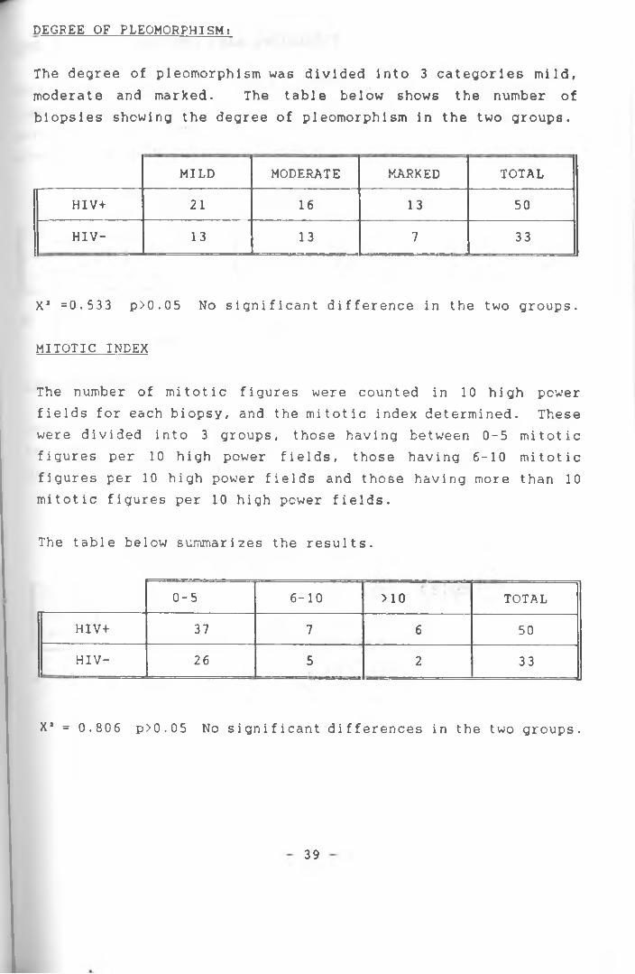
DEGREE OF PLEOMORPHISMt
The degree of pleomorphism was divided into 3 categories mild, moderate and marked. The table below shows the number of biopsies showing the degree of pleomorphism in the two groups.
MILD MODERATE MARKED TOTAL
HIV+ 21 16 13 50
HIV- 13 13 7 33
X* =0.533 p>0.05 No significant difference in the two groups.
MITOTIC INDEX
The number of mitotic figures were counted in 10 high power fields for each biopsy, and the mitotic index determined. These were divided into 3 groups, those having between 0-5 mitotic figures per 10 high power fields, those having 6-10 mitotic figures per 10 high power fields and those having more than 10 mitotic figures per 10 high power fields.
The table below summarizes the results.
0-5 6-10 >10 TOTAL
HIV+ 37 7 6 50
HIV- 26 5 2 33
X* = 0.806 p>0.05 No significant differences in the two groups.
39

ngfiREE OF INFLAMMATORY CELL INFILTRATE
This parameter was studied only in the skin lesions and excluded lymph node lesions. Thus 65 skin biopsies were studied for this parameter. The results are shown below. This parameter was graded as Mild, Moderate or Intense.
MILD MODERATE INTENSE TOTAL
HI Vi- 19 14 5 38
HIV- 7 10 10 27
X1 = 6.187 0.02<p< 0.05significant difference exist in the two groups
TYPE OF INFILTRATE
This parameter was studied only in skin lesions and was described as being either lymphocyte predominant or plasma cell predominant. Results for two groups are shown belcw:-
LYMPHOCYTE PLASMA CELL TOTAL
HIV+ 21 17 38
HIV- 17 10 27
X1 = 0.385 p>0.05 No significant differences in the two groups.
40

ULCERATION OR NECROSIS IN LESIONS
This was described as being either present or absent. The results are summarized below:-
PRESENT ABSENT TOTAL
HIV+ 5 45 50
HIV- 9 24 ' 33
X* = 4.230 p<0.05 significant difference at 95% confidence1 eve 1 .
DEGREE OF HAEMORRHAGE AND VASCULARITY
This was categorized into 3 groups: Mild, Moderate and Extensive Results are shown below*.-
MILD MODERATE EXTENSIVE TOTAL
HIV+ 20 13 17 50
HIV- 17 8 8 33
X* 1.244 p>0.1 No significant difference in the two groups

FIBROSIS
This parameter was recorded as being either present or absent for the two groups. Results are shown below:-
PRESENT ABSENT TOTAL
HIV+ 15 35 50
HIV- 25 8 , 33
X 2 = 16.67 p<0.01 significant differences present between thetwo groups

DISCUSSION
An average of 36 cases per year of Kaposi’s sarcoma were seen between January 1987 and December 1991, with an increase of over 40% between 1988 and 1989.
The largest number of patients were in their 3rd decade of life and were specifically in the 26-30 yrs age group. this is a younger age group when compared to the study by Kungu and Gatei in which the majority of the patients were in the 5th decade. (35)
There was a male preponderance with a male to female ratio of 4.5:1. This is a much lower ratio than that of the study by Kungu and Gatei in which the male to female ratio was 8.5:1.(35) This ratio is also much lower the earliest study by Rogoff in which the M-.F ratio was 7.2:1. Thus although there is a male predominance, this change in M:F ratio indicates an increasing incidence of the disease amongst females which may be attributed to the emergence of AIDS in this environment.
With regard to tribal distribution, the largest number of cases were seen amongst members of the Luo community. Slightly over 50% of the total number of patients originated from the Western sector of the country. In the earlier studies by Kungu and Gatei as well as the earliest study by Rogoff, the majority of the patients were from the Kikuyu, Embu and Meru group(35,43 ) . This "tribal’’ shift may be related to the emergence of AIDS related Kaposi's sarcoma and the tribal distribution of HIV disease. Statistics from the Kenya National AIDS control programme indicate that approximately 43% of AIDS cases classified by district of birth are from Nyan2a and Western province together. This high ethic prevalence may suggest a genetic predisposition.
This ethnic predisposition to the disease has been supported by the relatively high prevalence of the disease in certain ethnic groups in the United States and in whom there is a distinct
43

association with particular HLA-DR phenotypes and principally HLA-DR5(12,44). This association also supports the findings that within certain geographical areas, members of a particular ethnic group showed a considerably higher incidence of KS than others living in the same 1ocation.(45) Early in the AIDS epidemic, there was an frequency of HLA-DR5 in KS patients of European, Jewish or Italian background, while KS patients of Northern European background had a higher than expected frequency of HLA DR2.(46) /
110 patients had serology results for HIV antibodies performed by the ELISA method. Of these 68 were positive and 42 were negative for HIV. This gives an overall ratio of HIV positive to HIV negative as 1.6:1. However in the childhood group the ratio of HIV positive Kaposi's sarcoma to HIV negative Kaposi’s sarcoma is 1:7. This ratio for the adult group is 2.4:1. These ratios suggest that the major impact of AIDS on the incidence of Kaposi's sarcoma has been on the adult age groups. The ratio for the adult group is a bare minimum. This ratio could be much higher because many AIDS patients with Kaposi's sarcoma are not biopsied as they may be extremely moribund and may die before investigations are performed. Furthermore some patients may have the diagnosis made on clinical grounds only. In some patients the lesions may be missed altogether. Furthermore AIDS patients may have visceral KS without cutaneous lesions.( 22,23,24,25 ) Of the HIV negative cases of Kaposi’s sarcoma, there was one case of a renal transplant patient who was on immunosuppressive drugs. This patient was a 28 year old Kamba male who developed the lesions 3 years after transplant surgery. To date only 3 renal transplants have been performed at Kenyatta National Hospital, out of which one patient developed Kaposi's sarcoma. This is a relatively high incidence, even though the numbers are few. In Western countries the incidence of Kaposi's sarcoma in recipients of renal transplants is approximately 0.4%.(47) Despite identical management protocols, this high incidence suggests the presence of some etiologic agent in this environment which is yet to be ident i f i ed.
44

Although skin lesions were the commonest presentation (71.4%), lymph node presentation was not infrequent. Indeed 19.2% of the lesions were in lymph nodes. This incidence of lymph node involvement is more than twice that reported in the study by Kungu and Gatei. Furthermore 60% of Kaposi's sarcoma in lymph nodes were seen in adults, while only 40% were seen in children. In the earlier study the majority of lymph node presentation were seen in children below 10 years of age. KS involving the lymph nodes is seen in a large number of AIDS pat ient s. (48 ) The lymphadenopathic form of KS is rather intriguing. Bhana et al suggest two forms of lymph node involvement.(49) One occurs in younger patients and involves many groups of nodes and probably develops in situ. The other lymphademopathic form is as a result of metastases from an aggressive tumour to the regional lymph node. It is not clear from this study into which group the lymphadenorpathic form seen in adult patients (and especially those with AIDS), fall into since there were no follow up studies or post-mortem studies done. However the lymph node involvement in childhood KS probably fall into the former group.
The clinical features were studied from data retrieved from case record files of 100 patients. There were 63 HIV positive Kaposi’s sarcoma patients and 37 HIV negative patients. Most of the patients in the two groups presented with the development of nodules preceded by swelling. 25% of these patients in the HIV positive group described the lesions as being pruritic. In Western literature, Safai et al and other authors mention the lesions of Kaposi's sarcoma as being generally non pruritic.( 12,16,20,21, 36 ) This feature may be due to secondary fungal infections of the skin which are commonly present in HIV positive patients in this environment.
Other symptoms present in HIV positive Kaposi's sarcoma patients include cough, persistent diarrhoea and dysphagia. Their symptoms may be due to involvement of these sites by Kaposi's sarcoma lesions. 3 Patients with HIV positive Kaposi's sarcoma were asymptomatic. This emphasizes the need for clinicians to
45

look actively for lesions of Kaposi's sarcoma in AIDS patients.
71% of patients in the HIV positive group had duration of symptoms lasting less than 6 months while only 51% of HIV negative Kaposi's sarcoma patients had symptoms lasting less than 6 months. 25% of HIV negative Kaposi's sarcoma had symptoms lasting more than 2 years. There were no patients with AIDS KS having symptoms lasting more than 2 years. This observation suggests that the lesions develop more rapidly in HIV positive patients and could be related to the rate of development of immune dysfunction and the degree of immunodeficency.
In the HIV positive group 33.3% of the patients presented with multiple lesions, 27% had lesions involving the lower limbs only and 17.4% of the patients presented with lymphnode involvement 25% of the patients were noted to have oral lesions. There were no patients with visceral involvement seen, neither were there any FM follow up done to confirm visceral involvement-, FM studies may have shown visceral involvement. C. Hwangi showed visceral involvement is cutaneous form in 8C% of HIV positive patients. In the HIV negative group the lower limbs were involved in 62% of the cases while lymphnode enlargement was seen in 18.9% of the patients. The clinical features of HIV positive Kaposi's sarcoma differ from those described in Western literature by Safai et al and Amber son et al in which involvement of the lower limb is less common.(36,54) In this study involvement of the lower limbs is seen frequently in HIV positive Kaposi's sarcoma patients and may not be clinically distinguished' from the classical type of Kaposi's sarcoma. This observation has also been expressed by A.C. Bayley et al in which endemic form of KS was distinguished from atypical KS by HTLV III serology.(50) KS has been associated with other malignancies, especially lymphomas are also seen in AIDS. In this study No associated malignancies were observed occurring with AIDS-KS.
Histological studies were performed on 50 HIV positive KS lesions and 33 HIV negative KS lesions.
46

Slit forming type was seen in 80% of HIV positive patients while mixed and solid types were seen in 14% and 6% respectively. Slit forming type was seen in 51% of HIV negative KS lesions. This difference was statically significant at p value of less than0.05. This observation has also been expressed by C. Mwangi at KNH, D. Servadda et al, in Uganda and in Western literature by Heyer, Kahn and Volberding.(25,29,37) This slit forming type is synonymous with the mixed cell variant expressed by otherauthors. This variant is associated with nodular non aggressive
✓
lesions and thus may have better prognosis.(33) Meyer et al emphasize that HIV patients with KS live longer than these dagnosed with other HIV related i1lnesses.(37) Furthermore, Kaposi sarcoma lesions in latrogenical ly immunosuppressed patients, such as renal transplant patients, have been known to regress upon withdrawing therapy.(11) It can therefore be hypothetically inferred that KS lesions in HIV positive patients may regress if the immunsuppressed state is reversed. Currently no such treatment modality exist.
50% of HIV positive KS lesions shewed a mild degree of mononuclear cell infiltration. In the HIV negative group 75% shewed moderate to intense degree of infiltration while 25% showed mild degree of infiltration, around the lesions. These differences were statistically significant and may be explained by the relative differences in the degree of immunesuppression. Some histological features that suggest most immunuity include the infiltration of the tumour by lymphocytes and plasma cells.(7,53) A number of immunological studies have been done on KS. Masters et al found that antibody responses and immunoglobulin levels were normal but there was a striking impairment in delayed hypersensitivity in patients with aggressive lesions only.(51) Kestens et al found no evidence of immune suppression among patients with endemic African KS as compared with normal controls.(52)
Ulceration or necrosis was observed to be present in 10% of HIV positive lesions and 28% of HIV negative lesions. These
47

differences were statically significant. This observed difference may be explained as an immune response to the lesions of Kaposi's sarcoma and which are dimished in HIV positive pat ients.
Fibrosis was present in 30% of HIV positive biopsies and in 75% of HIV negative biopsies. This difference is statistically significant and may again be explained on the basis of theimpaired immunity in AIDS patients.
/
There were no statistical difference observed with respect to the degree of pleomorphism, mitotic index, degree of vascularity and haemorrhage, the degree of haemosiderin deposition and the type of infiltrate. Thus in this respect the two groups are essentially similar. There observation differs from those of Heyer et al in which AIDS-KS lesions are described as being more angiosarcomatous,(37) and other studies in which plasma cells, extravasated erythrocyte and haemosiderin deposition and cells with bizzare nuclei were seen.(55)
CONCLUSIONS
1. The full spectrum of Kaposi's sarcoma is present in this environment.
2. Major changes noted in the epidemiology of Kaposi's sarcoma. These are:-
(a) Younger age group with the largest number of patients in 26-30 years age group.
(b) Lower M:F ratio of 4.5:1
(c) Shift in tribal distribution with majority of patients from Luo community.
48
These changes in pattern of Kaposi’s sarcoma may be attributed to the emergence of AIDS in this setup.
3. Although skin presentation is the commonest form ofmanifestation there has been an increase in thelymphadenopathic form of Kaposi's sarcoma and specifically in the adult age group.
4. The disease is widespread in the patients with HIV, with a/
• shorter duration of symptoms. Leg involvement is notuncommon and may be indistinguishable clinically from the endemic and classical form of the disease.
5. Slit forming type predominates in HIV positive Kaposi's sarcoma.
6. Minor differences exist histologically and include the absences or presence of Necrosis/ulceration. sclerosis and the degree of lymphocyte or plasma cell infiltration.
MAJOR CONSTRAINTS
1. The retrieval of paraffin blocks was extremely cumbersome and time consuming as there was no organized filing system and because of this a number of blocks could not be traced.
2. A nuiTiber of patient record files could not be traced because there was no monitoring system as to the movement of files from the records department. In others although the IP numbers corresponded, the files were for different pat ient s.
3. The data presented on the request forms by the clinicians was in most cases grossly inadequate with regard to the clinical picture presented by the patient.
49

RECOMMENDATIONS
1. There is an urgent need to reorganize the filing system for the paraffin blocks so that retrospective histopathological studies such as this one can be made more convenient and easier.
2. There is an urgent need for computerization of the entirerecords department at Kenyatta National Hospital.
/
3. There is need to carry out studies to identify the aetiologic agents, if any, that are implicated in the pathogenesis of Kaposi's sarcoma.
4. There is a need to carry out immunogenet i c studies to identify high risk populations in the environment.
5. Transplant recipient patients may be prone to develop Kaposi's sarcoma and early recognition and therapeutic intervention may lead to a more favourable outcome.
50

CHILDHOOD LYMPHADENOPATHIC KS (HIV NEGATIVE)
- 51 -

ENDEMIC KS INVOLVING LOWER LIMBS (HIV NEGATIVE)
A

FLORID ORAL KS LESIONS IN HIV POSITIVE PATIENT
53

DISSEMINATED KS LESIONS IN HIV POSITIVE PATIENT
- 54 -
1

FLORID KS LESION ON LOWER LIMBS OF HIV POSITIVE PATIENT
KS: SLIT FORMING TYPE WITH MILD DEGREE OF PLEOMORPHISM (HE X400 )
- 55 -

KS: MIXED TYPE (HE X 100)
KS: SOLID TYPE SHOWING MARKED DEGREE OF PLEOMORPHISM(HE X 400)
56

KS: INTENSE HAEMOSIDERIN DEPOSITION(PERL'S PRUSSIAN BLUE STAIN X 100)
- 57 -
- *

KS: MILD DEGREE OF HAEMOSIDERIN DEPOSITION (PERL'S PRUSSIAN BLUE STAIN x 100)
- 58 -

PREDOMINANT PLASMA CELL INFILTRATION (HE X 400)
PREDOMINANT LYMPHOCYTE INFILTRATION (HE X 400)
- 59 -
L *

REFERENCES
1. Templeton A.C.Kaposi's sarcoma; chapter 52, Cancer of The Skin; Biology, Diagnosis Managements; Philadelphia Saunders 1976
2. Safai B, Mike V, Giraldo G, et al;Association of KS with Second Primary Malignancies; Cancer 1980 45: 1472-1479
3. Kaposi M;Idiopatiches multiple pigment sarkom der haut; Arch, for dermatologies and syphilis 1872 4:265-275 (Translated CA1982 32:342-7)
4. Cotrans, Kumar, Robbins;Robbins pathologic basis of disease, chapter 12, 4th Edition; Saunders W.B., Philadelphia 1989
5. Hutt M.S.R;Kaposi’s sarcoma; British Medical Bulletin 1984, Vol 40: 355-358
6. Bay ley A.C;An overview of Kaposi's sarcoma; Proceedings of The Association of Surgeons of East Africa 1984 ..
7. Slav in G, Cameron H, Singh H;KS in mainland Tanzania; Report of 117 cases; British Journal of Cancer 1970 24:349-357
8. Taylor J, Templeton A.C, Vogel C.L, et al;KS in Uganda; A clinico pathological study International Journal of Cancer 1971 8:122-138
9. O'ccnnell K.M;KS histopathological study of 159 cases from Malawi;
60

Journal of clinical pathology 1977 30: 687-695
10.
1 1 .
1 2 .
13.
14 .
15.
16.
17 .
18 .
Hutt M.S.R;Epidemiology of Kaposi's sarcoma, Antibiotics and chemotherapy Vol 29 S. Karger
Hardwood A.R, Osoba D, Hofstder S.L et al;KS in recipients of renal transplant; American Journal of Medicine 67:759-765(1979)
Fideldman-kein A .L , Laubenstein L.J, Rubienstein P et al; Disseminated KS in homosexual men; Annals of internal medicine 96:693-700 (1982)
Marmor M, Friedman-Kein A .E , Zolla-Pa2ner S et al;KS in homosexual men; Annals of Internal Medicine 1984 100 : 809-815
AIDS update:NIH Conference; Annals of Interal Medicine 1985 102:800-813
Adler M.W;AIDS an introduction; Medicine Internationa1 1988 2326-2330
AIDS-Epidemiologic, clinical, immunologic and therapeutic considerations-NIH conference; Annals of Internal Medicine 1984 100:92-106
Serwadda D, Mugerwa R .D , Sewankombo M.R et al;Slim disease; a new disease in Uganda and its association with HTLV III infection; Lancet 1985 11:849-852
Amayo E ;Clinical manifestations of AIDS in Adults as seen at KNH; M.Med thesis 1988
- 61 -
19. Owinn T.C, Plot P, Tadman H et al;AIDS in a heterosexual population in Zaire; Lancet 11:65-69 1981.
20. Mitsamyasu R.T, Taylor J.M, Gtaspy J, Fehey J.L;Heterogenicity of Epidemic KS; Cancer 1986 57:1657-61
21. Centre for Disease Control;KS and pneumocystis pneumonia among homosexual men in New York city and California MMWR 30:305-8
22. Niedt G.W, Schinella R . A ;AIDS, Clinicopathologic study of 56 autopsies; Arch Path lab med 1985 109:727-734
23. Reichert C.M, O'leary T.J, Levens D.L et al ;Autopsy Pathology in AIDS; American Jounal of Pathology 1983 112:357-382
24. Guarda L .A , Luma M .A , Leslie Smith J et al;AIDS Postmortem findings; American Journal of Clinical Pathology 1984 81:549-557
25. Hwangi C;Histopathology of Various Organs M.Med thesis 1990
26. Giraldo G, Beth E, Haguer.au F;KS A new model in the search for human malignancies; Journal of Institute 1972 49:1495-1507
27. Giraldo G, Beth E, Haguenau F;Herpes type virus particles in tissue culture of KS; Journal of The National Cancer Institute 1972 49:1509-1526
28. Gange R.W, Jones E.W;
in Patients with AIDS;
viruses associated with The National Cancer
62

29.
30.
31.
32.
33.
34 .
35.
36.
37 .
KS and immunosuppresive Therapy an appraisal; Clinical and Experimental Dermatology 1978 3:135-46
Giraldo G, Beth E, Kaurilsky F.M;Serological association of European KS with CMV; Int Journal of Cancer 1975 15:839-845
Marmor M, Laubenstein L, William D.C, et al?Risk factors for KS in homosexual men; Lancet 1982 1:1084-6
Vogel J, Hinrichs S.H, Reynolds R.K, et al ;The HIV tat gene induces dermal lesions resembling KS in transgenic mice; Nature 1988 355:606-611
Salahuddin S.Z, Nakamura S, Biberfeld P, et al;Angiogenic properties of KS derived cells after long term culture in vitro; Science 1988 242:430-3
Templeton A.C;soft tissue tumours in a tropical country; Recent results in cancer chemotherapy; Springer-Ver1ag Berlin. Heidelbery New York 1973
Hutt M.S.R;Pathology of KS; Antibiotics and chemotherapy Vol 29 S. Karger 1981 ' ' ~'s*ri'
Kungu A, Gatei D.G;KS in Kenya antibiotics and chemotherapy Vol 29 S. Karger 1981
Safai B, Johnson K.G, Myskowski P.L, et al;The natural history of KS in AIDS; annals of Internal Medicine 1985 103:744-750
Heyer D.M, Kahn J.O, Volberding P.A;
63

HIV related KS; AIDS knowledge base text book copy right 1989 Masscichusetts Medical Society.
38. Serwadda D, Carswell W, Ayuko W.O, et al;Further experience with KS in Uganda; British Journal of Cancer 1986 53:497-500
39. McNutt N.S, Fletcher V, Conant M.A;Early lesions of KS in homosexual men; American Journal of Pathology 1983 111:62-77
40. Paul Nunn;Kaposi's sarcoma; Belliere's clinical Tropical Medicine and communications Vol 3 No.l April 1988 73-87
41. Susan E, Krown;Editional review AIDS-association KS; Pathogensis, clinical course and treatment; AIDS 1988 2:71-80
42. Me lean N;Multiple haemorrhagic pigment sarcoma of Kaposi's in African natives; East African Medical Journal 1939 16:308-310
43. Rogoff M.A;KS age, sex and tribal incidence in Kenya; Cancer in Africa, copyright East Africa Medical Journal 1968 446-448
44. Pollack M.S, Safai B, Myskowski P.L, et al;Frequencies of HLA and CRM immunogent ic markers in KS; Tissue Antigens 1982 21:1-8
45. Keen P;The clinical features of KS in The South African Eantu; Acta Unio Int Cancer 1962 18:380-387
46. Pollack M.S, Safai B, Dupont B;
64
HLA-DR5 and DR2 are susceptibility factors for AIDS with KS in different ethnic populations; Disease Markers 1983 1:135-139
47. Enzinger F.M, Weiss S.W;Soft tissue tumours, 2nd Editions C.V Mosby Company Toronto 1988
48. Moskowitz L.B, Hensley G.T, Gould E.W, Weiss S.D;Frequency and anatomic distribution of lymphadenopathic KS in AIDS; An autopsy series; Human Pathology 1985 16:447-56
49. Bhana D, Templeton A.C, Master S.P, Kyalwazi S.K;British Journal of Cancer 1970 Vol 24:464-470
50. Bayley A.C, Downing R.G, Cheingsong-Popov R, et al;HTLV III serology distinguishes atypical and endemic KS in Africa; Lancet 1985 359-361
51. Master S.P, Taylor J.F, Kyalwazi S.K, Ziegler Z;Immunological studies in KS in Uganda British Medical Journal 1970 1:600-602
52. Kestens L, Melbye M, Biggar J, et al;Endemic African KS is not associated with immunodefiency; International Journal of Cancer 1985 36:49-54
53. Lothe F;KS in Uganda Africans 1963 Oslo University Sitetsfor 1aget
54. Amberson J.B, Dicarlo E.F, Mctroka C.E, et al;Diagnostic Pathology in AIDS; Arch Path lab Med 1985 109:345-351
55. Fir.kbeiner W. E, Egbert B.M, Coroundwater J.R, Sagebiel R.W;KS in young homosexual men; Arch of Path Med 1982 106:261-264
65

APPENDIX A PROFORMA SHEET
BIOPSY NUMBER:_____________
IN-PATIENT NUMBER:________
AGE:________
SEX:________ TRIBE:
HIV STATUS:________________
HISTORY:___________________
CLINICAL EXAMINATION
GENERAL EXAMINATION:____
DISTRIBUTION OF LESIONS:
OTHER CLINICAL FEATURES:
66

/ v r t C .K U J L /W D s
TABULATION OF HISTOLOGICAL FEATURES
BIOPSYNUMBER
HIVSTATUS
CLASSIFICATIONSLIT MIXED SOLID
DEGREEOFPLEO.
MITOTICINDEX _______lOhpf * DEGREE
j
INFILTERATIONLYMP. PLASMA
CELLULCER.NECR. HAEM. H/S
DEPOST
KEY+ MILD HAEM HAEMONHAGE♦+ MODERATE H/S DEPOSIT HAEMOSIDEKIN DEPOSITION++♦ SEVERE/INTENSE PLEO. PLEOMORHI3MX PRESENT ULCER. ULCERATIONo ABSENT NECR. NECROSIS
I

APPENDIX C
Staining by Haematoxylin and Eosin Procedure:-
1) Dewax and hydrate paraffin sections
2) Stain in Mayers haemalium for 5 minutes
3) Rinse and Blue tap water
4) Immerse slides in Eosin for 30s - lmin with agitation/
5) Wash in tap water
6) Dehydrate in 70%, 95% and two changes of absolute alcohol
7) Clear in Xylene and mount with DPX
- 68 -
1d
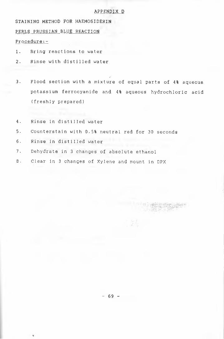
APPENDIX DSTAINING METHOD FOR HAEMOSIDERIN
PERLS PRUSSIAN BLUE REACTION
Procedure:-
1. Bring reactions to water
2. Rinse with distilled water
/3. Flood section with a mixture of equal parts of 4% aqueous
potassium ferrocyanide and 4% aqueous hydrochloric acid
(freshly prepared)
4. Rinse in distilled water
5. Counterstain with 0.5% neutral red for 30 seconds
6. Rinse in distilled water
7. Dehydrate in 3 changes of absolute ethanol
8. Clear in 3 changes of Xylene and mount in DPX
- 69 -

UMiVgaSITV 0? WASR0B11COLLEGE OF HEALTH SCIENCES
DEPARTMENT Of P**u Im.ACYRGSTAl ADDACSS RO So« l ;S 7 a SA.R03IFEN Y A .
TW»vho.i» 726770/1
T»**9' - ’ A V n J ty
Ov» A«fc
Youi M#:
Dr. P. Dave,Dept. of Pathology, University of Nairobi.
19th May, 1992.
Re: Research Proposal entitled "Some clinical histopatho— logical aspects of karposis sarcoma in patients with and without the acquired innu.no deficiency syndror e"---Jggl/i/S _
X am pleased to inform you that the KNH-ERC has reviewed 5Pproved the revis d version of your a’ ove cited
research proposal*On behalf of the Committee, I wish you luck in carrying out̂ your study and in addition request you to submit a summary of your research findings and recommendations 'if any) for future reference by the Conmittee.Thank you. —
Dr. Anastasia N. Guantai SECRETAP Y - KNi’.-ERCcc, Frof. F.B. Ony .ngo Chairman - KNK-ERC
Prof. Kungu - Dept, of Pathology Dir -ctor - KNH '
rI'e
1