sonographic detection of intrathyroidal branchial cleft ... · sonographic detection of...
TRANSCRIPT
Korean J Radiol 7(2), June 2006 149
Sonographic Detection of IntrathyroidalBranchial Cleft Cyst: A Case Report
We report here on an extremely rare case of an intrathyroidal branchial cleftcyst. Intrathyroidal branchial cleft cyst is rare disease entity and it has nonspecificfindings on sonography, so the diagnosis of the lesion is very difficult. However,during aspiration, if pus-like materials are aspirated from a thyroid cyst, we shouldconsider the possibility of intrathyroidal branchial cleft cyst in the differential diag-nosis.
ranchial cleft cysts are a well-described entity and branchial anomaliesare derived from the branchial cleft apparatus that persists after fetaldevelopment (1). A branchial cleft cyst is usually located in the lateral
areas of the head and neck, and an intrathyroidal branchial cleft cyst is extremely rare(2).
We report here on a case of intrathyroidal branchical cleft cyst presenting as abenign thyroid cystic lesion and its sonographic finding.
CASE REPORT
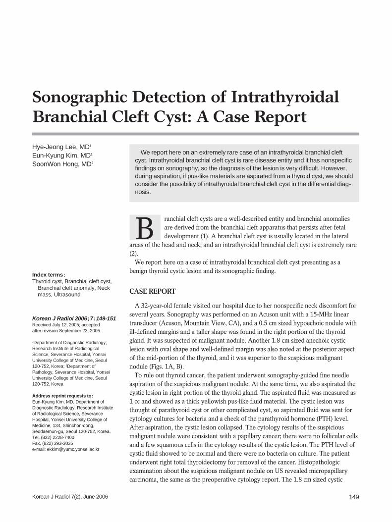
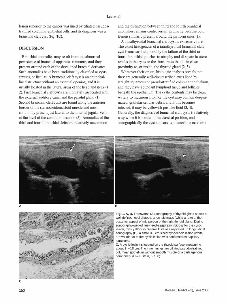
A 32-year-old female visited our hospital due to her nonspecific neck discomfort forseveral years. Sonography was performed on an Acuson unit with a 15-MHz lineartransducer (Acuson, Mountain View, CA), and a 0.5 cm sized hypoechoic nodule withill-defined margins and a taller shape was found in the right portion of the thyroidgland. It was suspected of malignant nodule. Another 1.8 cm sized anechoic cysticlesion with oval shape and well-defined margin was also noted at the posterior aspectof the mid-portion of the thyroid, and it was superior to the suspicious malignantnodule (Figs. 1A, B).
To rule out thyroid cancer, the patient underwent sonography-guided fine needleaspiration of the suspicious malignant nodule. At the same time, we also aspirated thecystic lesion in right portion of the thyroid gland. The aspirated fluid was measured as1 cc and showed as a thick yellowish pus-like fluid material. The cystic lesion wasthought of parathyroid cyst or other complicated cyst, so aspirated fluid was sent forcytology cultures for bacteria and a check of the parathyroid hormone (PTH) level.After aspiration, the cystic lesion collapsed. The cytology results of the suspiciousmalignant nodule were consistent with a papillary cancer; there were no follicular cellsand a few squamous cells in the cytology results of the cystic lesion. The PTH level ofcystic fluid showed to be normal and there were no bacteria on culture. The patientunderwent right total thyroidectomy for removal of the cancer. Histopathologicexamination about the suspicious malignant nodule on US revealed micropapillarycarcinoma, the same as the preoperative cytology report. The 1.8 cm sized cystic
Hye-Jeong Lee, MD1
Eun-Kyung Kim, MD1
SoonWon Hong, MD2
Index terms:Thyroid cyst, Branchial cleft cyst,
Branchial cleft anomaly, Neckmass, Ultrasound
Korean J Radiol 2006;7:149-151Received July 12, 2005; accepted after revision September 23, 2005.
1Department of Diagnostic Radiology,Research Institute of RadiologicalScience, Severance Hospital, YonseiUniversity College of Medicine, Seoul120-752, Korea; 2Department ofPathology, Severance Hospital, YonseiUniversity College of Medicine, Seoul120-752, Korea
Address reprint requests to:Eun-Kyung Kim, MD, Department ofDiagnostic Radiology, Research Instituteof Radiological Science, SeveranceHospital, Yonsei University College ofMedicine, 134, Shinchon-dong,Seodaemun-gu, Seoul 120-752, Korea.Tel. (822) 2228-7400Fax. (822) 393-3035e-mail: [email protected]
B
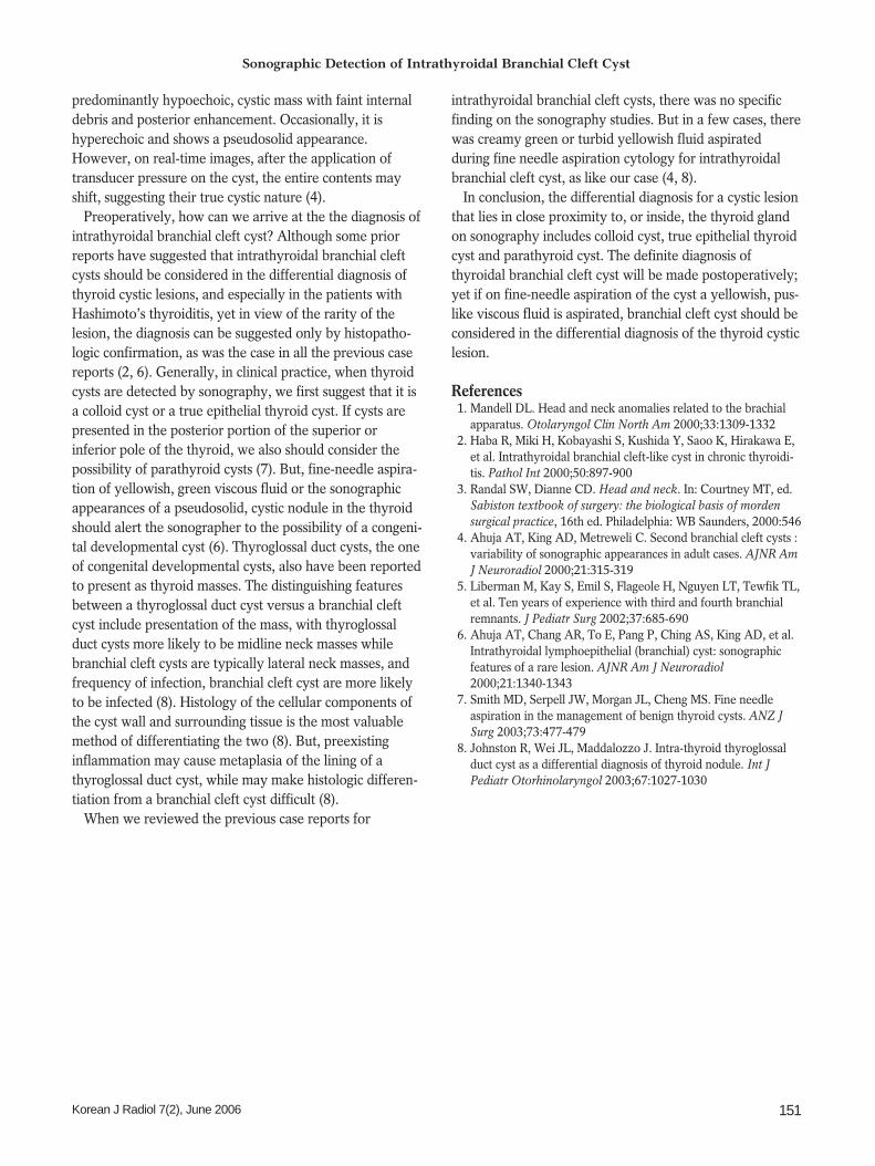
lesion superior to the cancer was lined by ciliated pseudos-tratified columnar epithelial cells, and its diagnosis was abranchial cleft cyst (Fig. 1C).
DISCUSSION
Branchial anomalies may result from the abnormalpersistence of branchial apparatus remnants, and theypresent around each of the developed brachial derivates.Such anomalies have been traditionally classified as cysts,sinuses, or fistulas. A branchial cleft cyst is an epithelial-lined structure without an external opening, and it isusually located in the lateral areas of the head and neck (1,2). First branchial cleft cysts are intimately associated withthe external auditory canal and the parotid gland (1).Second branchial cleft cysts are found along the anteriorborder of the sternocleidomastoid muscle and mostcommonly present just lateral to the internal jugular veinat the level of the carotid bifurcation (3). Anomalies of thethird and fourth branchial clefts are relatively uncommon
and the distinction between third and fourth branhcialanomalies remains controversial, primarily because bothlesions similarly present around the piriform sinus (1).
A intrathyroidal branchial cleft cyst is extremely rare.The exact histogenesis of a intrathyroidal branchial cleftcyst is unclear, but probably the failure of the third orfourth branchial pouches to atrophy and dissipate in uteroresults in the cysts or the sinus tracts that lie in closeproximity to, or inside, the thyroid gland (2, 5).
Whatever their origin, histologic analysis reveals thatthey are generally well-circumscribed cysts lined bystraight squamous or pseudostratified columnar epithelium,and they have abundant lymphoid tissue and folliclesbeneath the epithelium. The cystic contents may be clear,watery to mucinous fluid, or the cyst may contain desqua-mated, granular cellular debris and if this becomesinfected, it may be yellowish pus-like fluid (3, 4).Generally, the diagnosis of branchial cleft cysts is relativelyeasy when it is located in its classical position, andsonographically the cyst appears as an anechoic mass or a
Lee et al.
150 Korean J Radiol 7(2), June 2006
A B
Fig. 1. A, B. Transverse (A) sonography of thyroid glnad shows awell-defined, oval shaped, anechoic mass (white arrow) at theposterior aspect of mid portion of the right thyroid gland. Duringsonography-guided fine-needle aspiration biopsy for the cysticlesion, thick yellowish pus like fluid was aspirated. In longitudinalsonography (B), a small 0.5 cm sized hypoechoic lesion (whitearrow) inferior to the cystic lesion was confirmed as papillarycarcinoma. C. A cystic lesion is located on the thyroid surface, measuringabout 1 0.8 cm. The inner linings are ciliated pseudostratifiedcolumnar epithelium without smooth muscle or a cartilagenouscomponent (H & E stain, 100).
C
Sonographic Detection of Intrathyroidal Branchial Cleft Cyst
Korean J Radiol 7(2), June 2006 151
predominantly hypoechoic, cystic mass with faint internaldebris and posterior enhancement. Occasionally, it ishyperechoic and shows a pseudosolid appearance.However, on real-time images, after the application oftransducer pressure on the cyst, the entire contents mayshift, suggesting their true cystic nature (4).
Preoperatively, how can we arrive at the the diagnosis ofintrathyroidal branchial cleft cyst? Although some priorreports have suggested that intrathyroidal branchial cleftcysts should be considered in the differential diagnosis ofthyroid cystic lesions, and especially in the patients withHashimoto’s thyroiditis, yet in view of the rarity of thelesion, the diagnosis can be suggested only by histopatho-logic confirmation, as was the case in all the previous casereports (2, 6). Generally, in clinical practice, when thyroidcysts are detected by sonography, we first suggest that it isa colloid cyst or a true epithelial thyroid cyst. If cysts arepresented in the posterior portion of the superior orinferior pole of the thyroid, we also should consider thepossibility of parathyroid cysts (7). But, fine-needle aspira-tion of yellowish, green viscous fluid or the sonographicappearances of a pseudosolid, cystic nodule in the thyroidshould alert the sonographer to the possibility of a congeni-tal developmental cyst (6). Thyroglossal duct cysts, the oneof congenital developmental cysts, also have been reportedto present as thyroid masses. The distinguishing featuresbetween a thyroglossal duct cyst versus a branchial cleftcyst include presentation of the mass, with thyroglossalduct cysts more likely to be midline neck masses whilebranchial cleft cysts are typically lateral neck masses, andfrequency of infection, branchial cleft cyst are more likelyto be infected (8). Histology of the cellular components ofthe cyst wall and surrounding tissue is the most valuablemethod of differentiating the two (8). But, preexistinginflammation may cause metaplasia of the lining of athyroglossal duct cyst, while may make histologic differen-tiation from a branchial cleft cyst difficult (8).
When we reviewed the previous case reports for
intrathyroidal branchial cleft cysts, there was no specificfinding on the sonography studies. But in a few cases, therewas creamy green or turbid yellowish fluid aspiratedduring fine needle aspiration cytology for intrathyroidalbranchial cleft cyst, as like our case (4, 8).
In conclusion, the differential diagnosis for a cystic lesionthat lies in close proximity to, or inside, the thyroid glandon sonography includes colloid cyst, true epithelial thyroidcyst and parathyroid cyst. The definite diagnosis ofthyroidal branchial cleft cyst will be made postoperatively;yet if on fine-needle aspiration of the cyst a yellowish, pus-like viscous fluid is aspirated, branchial cleft cyst should beconsidered in the differential diagnosis of the thyroid cysticlesion.
References1. Mandell DL. Head and neck anomalies related to the brachial
apparatus. Otolaryngol Clin North Am 2000;33:1309-13322. Haba R, Miki H, Kobayashi S, Kushida Y, Saoo K, Hirakawa E,
et al. Intrathyroidal branchial cleft-like cyst in chronic thyroidi-tis. Pathol Int 2000;50:897-900
3. Randal SW, Dianne CD. Head and neck. In: Courtney MT, ed.Sabiston textbook of surgery: the biological basis of mordensurgical practice, 16th ed. Philadelphia: WB Saunders, 2000:546
4. Ahuja AT, King AD, Metreweli C. Second branchial cleft cysts :variability of sonographic appearances in adult cases. AJNR AmJ Neuroradiol 2000;21:315-319
5. Liberman M, Kay S, Emil S, Flageole H, Nguyen LT, Tewfik TL,et al. Ten years of experience with third and fourth branchialremnants. J Pediatr Surg 2002;37:685-690
6. Ahuja AT, Chang AR, To E, Pang P, Ching AS, King AD, et al.Intrathyroidal lymphoepithelial (branchial) cyst: sonographicfeatures of a rare lesion. AJNR Am J Neuroradiol2000;21:1340-1343
7. Smith MD, Serpell JW, Morgan JL, Cheng MS. Fine needleaspiration in the management of benign thyroid cysts. ANZ JSurg 2003;73:477-479
8. Johnston R, Wei JL, Maddalozzo J. Intra-thyroid thyroglossalduct cyst as a differential diagnosis of thyroid nodule. Int JPediatr Otorhinolaryngol 2003;67:1027-1030