sop: brain mri - rigshospitalet · sop: brain mri author(s) date changes approved by cornelia...
TRANSCRIPT

SOP: Brain MRI
Author(s) Date Changes Approved by
Cornelia Hagmann Manon Benders Anne Mette Plomgaard
23.09.2012
1.0 Inital version
Gorm Greisen
CONTENT
1.0 Timing of Brain MRI .......................................................................................1
2.0 Informed consent.............................................................................................1
3.0 Which sequences to take ...............................................................................1
4.0 The procedure .................................................................................................2
5.0 Saving and transferring data to CTU ............................................................3
6.0 Appendix A: scanning detales.........................................................................5
1.0 Timing of Brain MRI Brain MRI should take place between 40 -44 weeks of corrected gestational age.
2.0 Informed consent According to the SafeBoosC protocol separate informed consent must be obtained before conducting the brain MRI. Unless it is localy standard care to have an MRI at term equivalent age.
3.0 Which sequences to take The following sequences should be conducted (if software is available at the study site)
1 sagittal T1 SE
2 coronal T2W 1.2mm
3 coronal T1W 3D
4 DWI with ADC-MAP, either coronal or axial
5 DTI 45 directions axial (angulation left-right zero)
See scanning details in the attachment/appendix.

Some general remarks: 1. be sure the whole brain in scanned including the CSF until outside the skull 2. preferable on 3T, second choice 1.5T 3. for T2/T1 we prefer coronal slices, second choice is axonal/transverse slices 4. slice thickness <2mm, for T1 And T2, scan without a gap (gap zero) 5. Scans we prefer: T1 sagittal, t2W, T1W, DWI, DTI
Country AT
...1.5T, [email protected]
BE ....
CH GE, 3T
DE Siemens 1.5
DK Siemens 3T Magnetom Trio, [email protected]
ES GE1.5 and 3T, Fernando
FR Philips 1.5T, [email protected]
IE Siemens 1.5T
IT Philips, 3T, [email protected]
NL Philips 3T
SE ....
UK GE 1.5T
4.0 The Procedure These are suggested guidelines of how to perform a Brain MRI; however, local instructions may be used. There are two ways on performing the brain MRI: either the infant is scanned during natural sleep or under sedation. For sedation oral Cholralhydrate (50-60 mg/kg) may be used. Before starting:
Make sure that the baby’s body suits and clothes are metal free.
If any surgical metal (PDA clip, VP shunt etc) has been used, one needs to check with the surgeons / radiologists whether it is MR compatible metal.
4.1 Natural sleep: When scanning is done during natural sleep it is essential to plan well in advance:
30-60 minutes before the scanning the mother should feed the child
Change the nappy if necessary
Pulsoximetry is applied on the foot - check the signal.
Place MRI compatible electrodes for monitoring heart rate
Ear plugs and ear muffs are applied (a hat may be applied to keep the ear protection in place)
Wrap a blanket tightly around the baby so that the shoulders and arms are within the blanket.
Check the signal of the heart rate.
If you hav a belly-belt for respiratory rate, we recommend to use this as well

If using a vacuum mattress
Wait until the baby falls asleep.
Place the baby on the vacuum mattress on the scanner – please make sure that the baby’s head is in the midline before applying the vacuum.
If using an MRI compatible incubator (with / without a vacuum mattress)
Put the child in the MR-incubator
Wait until the child is a sleep
If using a vacuum mattress: please make sure that the baby’s head is in the midline before applying the vacuum
The Scanning can start. If the baby wakes up consider Glucose drops or feeding again. After the scanning is done, the parent(s) can take the baby home. 4.2 Sedation (with oral Cloralhydrate):
Sedation should be given 45 minutes before scanning starts – oral Cloralhydrate (50- 60 mg/kg).
Pulsoximetry is applied on the foot - check the signal.
Place electrodes for monitoring heart rate- check the signal.
(The baby can be fed as long as it still awake.)
The ear protection and the wrapping of the baby is as in the paragraph “natural sleep”.
After the scanning it is advised to keep the baby for 12-24 hours on the monitor when sedation has been used.
5.0 Saving and transferring data to CTU After the first MRI Manon Benders and Cornelia Hagmann must be alerted in order to check the quality of the images.
The MRI data must be saved in the local archive with patient id
Within one month after the MRI is condusted, a copy of the MRI file either anonymised or called by study-id must be send with FTP (File Transfer Protocol) to the CTU (Copenhagen Trial Unit)
For information regarding the data transferring please go to the SafeBoosC homepage and find the SOP: Data transfer to Copenhagen Trial Unit. Link to the SafeBoosC homepage: www.safeboosc.eu

6.0 Appendix A: scanning details
3T Philips Sagittal T1W SE (Philips 3T)
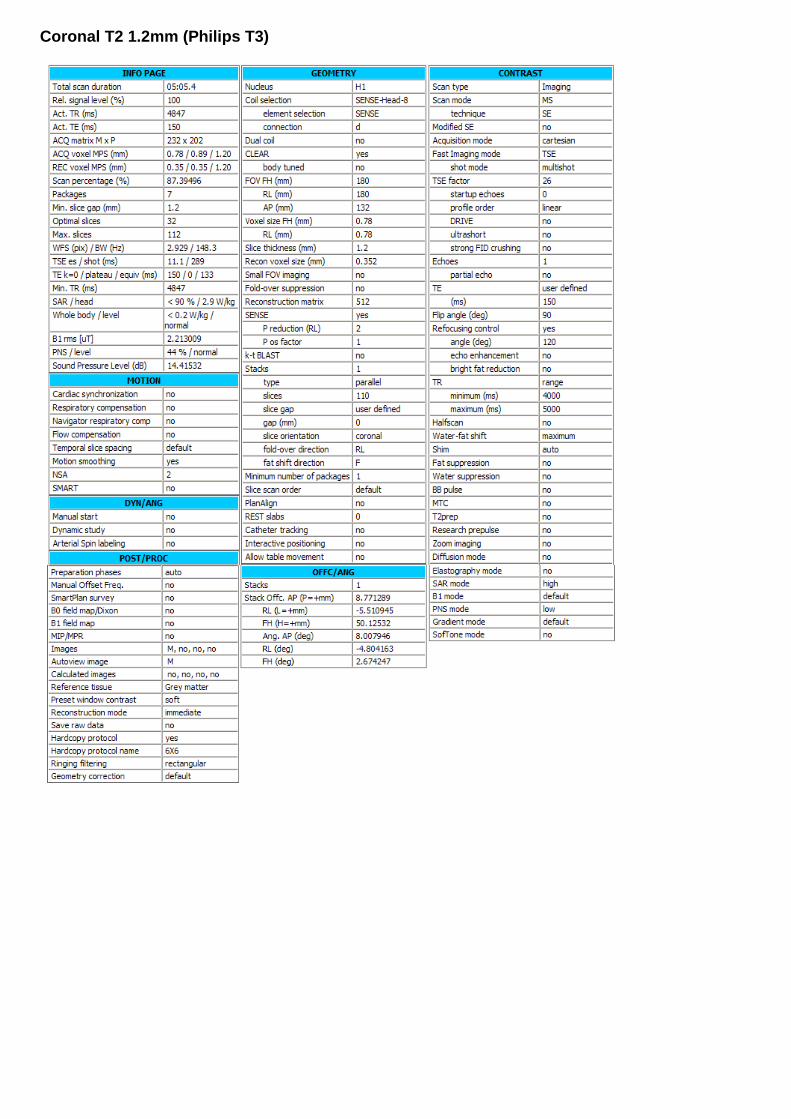
Coronal T2 1.2mm (Philips T3)
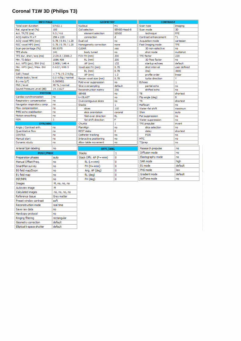
Coronal T1W 3D (Philips T3)

DWI: Diffusion weighted imaging with ADC-MAP (Philips T3)

DTI: Diffusion Tensor imaging with 45 directions (Philips T3) Multi-transmit = "no"; Nucleus = "H1"; Coil selection = "SENSE-Flex-S"; element selection = "12"; connection = "d"; Dual coil = "no"; CLEAR "yes"; body tuned = "no"; FOV RL (mm) = 160; AP (mm) = 160; FH (mm) = 90; Voxel size RL (mm) = 2; AP (mm) = 2; Slice thickness (mm) = 2; Recon voxel size (mm) =2; Small FOV imaging = "no"; Fold-over suppression = "no"; Reconstruction matrix = 80; SENSE = "yes"; P reduction (AP) = 1.39999998; P os factor = 1; k-t BLAST = "no"; Stacks = 1; type = "parallel"; slices = 45; slice gap = "user defined"; gap (mm) = 0; slice orientation = "transverse"; fold-over direction = "AP"; fat shift direction = "P"; Stack Offc. AP (P=+mm) = -35.4536819; RL (L=+mm) = 3.62022781; FH (H=+mm) = 8.57342815; Ang. AP (deg) = 2.41241336; RL (deg) = 18.1080379; FH (deg) = -5.14448023; Minimum number of packages = 1; Slice scan order = "default"; Large table movement ="no"; PlanAlign = "no"; REST slabs = 0; Shim Size AP (mm) = 87.5996399; RL (mm) = 58.2509651; FH (mm) = 54.9007835; Offc. AP (P=+mm) = -35.4573975; RL (L=+mm) = 4.14247704; FH (H=+mm) = 13.7650223; Ang. AP (deg) = 4.31364822; RL (deg) = 0; FH (deg) = -4.89545584; Catheter tracking = "no"; Interactive positioning = "no"; Allow table movement = "no"; Patient position = "head first"; orientation = "R decub"; Scan type = "Imaging"; Scan mode = "MS"; technique = "SE"; Modified SE = "no"; Acquisition mode = "cartesian"; Fast Imaging mode = "EPI"; shot mode = "single-shot"; Echoes = 1; partial echo = "no"; TE = "user defined"; (ms) = 80; Flip angle (deg) = 90;

TR = "user defined"; (ms) = 6500; Halfscan = "no"; Water-fat shift = "user defined"; (pixels) = 20; Shim = "PB-volume"; ShimAlign = "no"; mDIXON = "no"; Fat suppression = "SPIR"; strength = "strong"; frequency offset = "user defined"; offset (Hz) = 200; Water suppression = "no"; Grad. rev. offres. supp. ="no"; BB pulse = "no"; MTC = "no"; Research prepulse = "no"; Diffusion mode = "DTI"; sequence = "SE"; gradient duration = "maximum"; gradient overplus = "no"; directional resolution ="user defined"; nr of directions = 45; user defined dirs = 1, (2) 0, 0.456999987, 0.889999986, 0, -0.264999986, 0.485000014, 0.833999991, 0.936999977, -0.284000009, 0.202000007, 0.063000001, -0.805999994, 0.588, -0.67900002, -0.0939999968, 0.727999985, 0.686999977, 0.279000014, 0.671000004, -0.232999995, 0.949999988, 0.209000006, 0.922999978, 0.179000005, 0.342000008, -0.111000001, -0.980000019, -0.164000005, -0.131999999, -0.853999972, -0.503000021, 0.367000014, -0.768999994, -0.523000002, -0.291000009, -0.704999983, 0.647000015, -0.675999999, 0.713999987, 0.185000002, -0.598999977, 0.0780000016, -0.796999991, -0.73299998, -0.67900002, 0.0390000008, 0.800000012, -0.216000006, -0.559000015, 0.558000028, -0.340999991, -0.755999982, -0.435000002, -0.598999977, -0.671999991, -0.785000026, 0.596000016, -0.170000002, 0.495999992, 0.791000009, 0.356999993, -0.660000026, 0.433999985, -0.614000022, 0.842000008, -0.144999996, 0.519999981, 0.143999994, 0.430000007, 0.890999973, -0.301999986, 0.282000005, -0.911000013, 0.229000002, -0.930999994, 0.284000009, 0.943000019, 0.0120000001, -0.333999991, 0.0109999999, -0.0250000004, 0.999000013, 0.402999997, 0.84799999, -0.344999999, 0.921999991, 0.388000011, -0.00999999978, 0.0399999991, 0.486999989, -0.871999979, -0.296999991, 0.145999998, 0.944000006, -0.374000013, -0.165999994, -0.912, -0.284999996, -0.219999999, 0.933000028,

-0.856000006, -0.317999989, 0.407999992, -0.902999997, 0.382999986, 0.194999993, 0.538999975, -0.728999972, 0.421999991, -0.477999985, 0.878000021, -0.0399999991, 0.764999986, 0.531000018, 0.363999993, -0.518999994, -0.426999986, 0.74000001, 0.66900003, 0.606999993, -0.42899999, -0.104000002, -0.97299999, 0.204999998, -0.660000026, 0.569999993, 0.49000001, -0.0350000001, 0.690999985, 0.722000003, 0.333000004, -0.606000006, 0.722000003, (249) 0; nr of b-factors =2; b-factor order ="ascending"; max b-factor =800; average high b ="user defined"; b-factor averages =4, (15) 1; SAR mode = "high"; B1 mode = "default"; SAR Patient data = "auto"; PNS mode = "moderate"; Gradient mode = "enhanced"; SofTone mode = "no"; Cardiac synchronization ="no"; Heart rate > 250 bpm = "no"; Respiratory compensation ="no"; Navigator respiratory comp = "no"; Flow compensation = "no"; Temporal slice spacing = "default"; NSA = 1; Manual start = "no"; Dynamic study = "no"; dyn stabilization = "no"; Arterial Spin labeling = "no"; Preparation phases = "auto"; Interactive F0 = "no"; SmartPlan survey = "no"; B0 field map = "no"; B1 field map = "no"; MIP/MPR = "no"; Images = " M", (3) " no"; Autoview image = " M"; Calculated images = (4) " no"; Reference tissue = "User defined"; Tissue T1 = 2000; Tissue T2 = 150; Tissue rho = 100; EPI 2D phase correction = "no"; Preset window contrast = "soft"; Reconstruction mode = "immediate"; Save raw data = "no"; Hardcopy protocol = "no"; Ringing filtering = "default"; Geometry correction = "default"; IF_info_seperator = 1634755923; Total scan duration = "05:25.0"; Rel. signal level (%) = 100; Act. TR (ms) = "6500"; Act. TE (ms) = "80"; ACQ matrix M x P = "80 x 78"; ACQ voxel MPS (mm) = "2.00 / 2.03 / 2.00"; REC voxel MPS (mm) = "2.00 / 2.00 / 2.00"; Scan percentage (%) = 98.3871002; Packages = 1; Min. slice gap (mm) = 0;

Diffusion gradient timing DELTA / delta (ms) = "38.8 / 9.5"; EPI factor = 61; WFS (pix) / BW (Hz) = "20.000 / 21.7"; BW in EPI freq. dir. (Hz) = "2638.7"; Min. TR (ms) = "5346"; SPIR offset act./default (Hz) = "200 [135]" SAR / local torso = "< 54 %"; Whole body / level = "< 0.5 W/kg / normal"; B1 rms = "1.18 uT"; PNS / level = "73 % / normal"; Sound Pressure Level (dB) = 20.1907005;

1.5T Philips Sagittal T1W SE (Philips T1.5)

Coronal T2W (Philips T1.5): please change slice orientation in geometry from transverse into coronal:
Inverse recovery: IR (Philips T1.5) please change slice orientation in geometry from transverse into coronal:

\

DWI: Diffusion weighted imaging (Philips T1.5)

DTI: diffusion tensor imaging please scan axial. (Philips T1.5) We prefer the settings that are explained above for the 3T, however if you will not manage to get nice images, please try the settings below for 1.5T

Simens Magnetom trio – 3T

SIEMENS MAGNETOM TrioTim syngo MR B17
\\USER\Cerebrum\Projekter\SafeBoosC phase ll MRI.\localizer
TA: 0:25 PAT: Off Voxel size: 1.1×1.0×7.0 mm Rel. SNR: 1.00 SIEMENS: gre
Properties
Prio Recon Off
Before measurement
After measurement
Load to viewer On
Inline movie Off
Auto store images On
Load to stamp segments On
Load images to graphic On
segments
Auto open inline display Off
Phase partial Fourier Off
Interpolation On - - - - - - - - - - - - - - - - - - - - - - - - - - - - - - - - - - - - - - - - - - - - - - - - - - - - - - - - - - - - - - - - - - - - - - - - - - - - - - - - - - - -
PAT mode None
Matrix Coil Mode Auto (CP) - - - - - - - - - - - - - - - - - - - - - - - - - - - - - - - - - - - - - - - - - - - - - - - - - - - - - - - - - - - - - - - - - - - - - - - - - - - - - - - - - - - -
Image Filter Off
Distortion Corr. On
Mode 2D
Unfiltered images Off
Prescan Normalize Off
Normalize Off
Start measurement without
further preparation
Off B1 filter On
Intensity Medium
Wait for user to start Off
Start measurements single
Routine
Slice group 1
Slices 4
Dist. factor 20 %
Position R11.6 A103.7 H2.1
Orientation Transversal
Phase enc. dir. A >> P
Rotation 0.00 deg
Slice group 2
Slices 3
Dist. factor 20 %
Position R11.6 A104.7 F0.1
Orientation Coronal
Phase enc. dir. R >> L
Rotation 0.00 deg
Slice group 3
Slices 5
Dist. factor 30 %
Position R3.2 A104.7 F0.1
Orientation Sagittal
Phase enc. dir. A >> P
Rotation 0.00 deg
Phase oversampling 0 %
FoV read 250 mm
FoV phase 100.0 %
Slice thickness 7.0 mm
TR 8.6 ms
TE 4.00 ms
Averages 1
Concatenations 12
Filter Distortion Corr.(2D), Elliptical
filter, B1 filter
Coil elements NHA
Contrast
TD 0 ms
MTC Off
Magn. preparation None
Flip angle 20 deg
Fat suppr. None
Water suppr. None - - - - - - - - - - - - - - - - - - - - - - - - - - - - - - - - - - - - - - - - - - - - - - - - - - - - - - - - - - - - - - - - - - - - - - - - - - - - - - - - - - - -
Averaging mode Short term
Reconstruction Magnitude
Measurements 1
Multiple series Each measurement
Resolution
Base resolution 256
Phase resolution 90 %
Unfiltered images Off Raw filter Off
Elliptical filter On
Mode Inplane
Geometry
Multi-slice mode Sequential
Series Interleaved - - - - - - - - - - - - - - - - - - - - - - - - - - - - - - - - - - - - - - - - - - - - - - - - - - - - - - - - - - - - - - - - - - - - - - - - - - - - - - - - - - - -
Saturation mode Standard
Special sat. None - - - - - - - - - - - - - - - - - - - - - - - - - - - - - - - - - - - - - - - - - - - - - - - - - - - - - - - - - - - - - - - - - - - - - - - - - - - - - - - - - - - -
- - - - - - - - - - - - - - - - - - - - - - - - - - - - - - - - - - - - - - - - - - - - - - - - - - - - - - - - - - - - - - - - - - - - - - - - - - - - - - - - - - - -
Tim CT mode Off
System
Body Off
NBA Off
NHA On - - - - - - - - - - - - - - - - - - - - - - - - - - - - - - - - - - - - - - - - - - - - - - - - - - - - - - - - - - - - - - - - - - - - - - - - - - - - - - - - - - - -
Positioning mode REF
Table position H
Table position 0 mm
MSMA S - C - T
Sagittal R >> L
Coronal P >> A
Transversal H >> F
Save uncombined Off
Coil Combine Mode Adaptive Combine
AutoAlign ---
Auto Coil Select On - - - - - - - - - - - - - - - - - - - - - - - - - - - - - - - - - - - - - - - - - - - - - - - - - - - - - - - - - - - - - - - - - - - - - - - - - - - - - - - - - - - -
Shim mode Tune up
Adjust with body coil Off
Confirm freq. adjustment Off
Assume Silicone Off
? Ref. amplitude 1H 0.000 V
Adjustment Tolerance Auto
Adjust volume
Position Isocenter
Orientation Transversal
Rotation 0.00 deg
R >> L 350 mm
A >> P 263 mm
F >> H 350 mm
Physio
1st Signal/Mode None
Segments 1 - - - - - - - - - - - - - - - - - - - - - - - - - - - - - - - - - - - - - - - - - - - - - - - - - - - - - - - - - - - - - - - - - - - - - - - - - - - - - - - - - - - -
Tagging None
Dark blood Off - - - - - - - - - - - - - - - - - - - - - - - - - - - - - - - - - - - - - - - - - - - - - - - - - - - - - - - - - - - - - - - - - - - - - - - - - - - - - - - - - - - -
Resp. control Off
Inline
1/+

SIEMENS MAGNETOM TrioTim syngo MR B17
Subtract Off
Liver registration Off
Std-Dev-Sag Off
Std-Dev-Cor Off
Std-Dev-Tra Off
Std-Dev-Time Off
MIP-Sag Off
MIP-Cor Off
MIP-Tra Off
MIP-Time Off
Save original images On - - - - - - - - - - - - - - - - - - - - - - - - - - - - - - - - - - - - - - - - - - - - - - - - - - - - - - - - - - - - - - - - - - - - - - - - - - - - - - - - - - - -
Wash - In Off
Wash - Out Off
TTP Off
PEI Off
MIP - time Off
Sequence
Introduction On
Dimension 2D
Phase stabilisation Off
Asymmetric echo Allowed
Contrasts 1
Bandwidth 320 Hz/Px
Flow comp. No
Allowed delay 0 s - - - - - - - - - - - - - - - - - - - - - - - - - - - - - - - - - - - - - - - - - - - - - - - - - - - - - - - - - - - - - - - - - - - - - - - - - - - - - - - - - - - -
RF pulse type Normal
Gradient mode Normal
Excitation Slice-sel.
RF spoiling On
2/+

SIEMENS MAGNETOM TrioTim syngo MR B17
\\USER\Cerebrum\Projekter\SafeBoosC phase ll MRI.\t1_se_sag_3mm_200mm
TA: 3:48 PAT: Off Voxel size: 1.0×0.8×3.0 mm Rel. SNR: 1.00 SIEMENS: tse
Properties
Prio Recon Off
Before measurement
After measurement
Load to viewer On
Inline movie Off
Auto store images On
Load to stamp segments On
Load images to graphic On
segments
Auto open inline display Off
Start measurement without On
further preparation
Wait for user to start Off
Start measurements single
Routine
Slice group 1
Slices 31
Dist. factor 10 %
Position R4.2 A96.9 F9.9
Orientation S > C-5.2 > T-3.4
Phase enc. dir. A >> P
Rotation 0.00 deg
Phase oversampling 0 %
FoV read 200 mm
FoV phase 100.0 %
Slice thickness 3.0 mm
TR 550 ms
TE 12 ms
Averages 1
Concatenations 2
Filter Prescan Normalize, Elliptical
filter
Coil elements NHA
Contrast
TD 0.0 ms
MTC Off
Magn. preparation None
Flip angle 90 deg
Fat suppr. None
Water suppr. None
Restore magn. Off - - - - - - - - - - - - - - - - - - - - - - - - - - - - - - - - - - - - - - - - - - - - - - - - - - - - - - - - - - - - - - - - - - - - - - - - - - - - - - - - - - - -
Averaging mode Long term
Reconstruction Magnitude
Measurements 1
Multiple series Each measurement
Resolution
Base resolution 256
Phase resolution 80 %
Phase partial Fourier Off
Trajectory Cartesian
Interpolation On - - - - - - - - - - - - - - - - - - - - - - - - - - - - - - - - - - - - - - - - - - - - - - - - - - - - - - - - - - - - - - - - - - - - - - - - - - - - - - - - - - - -
PAT mode None
Matrix Coil Mode Auto (CP) - - - - - - - - - - - - - - - - - - - - - - - - - - - - - - - - - - - - - - - - - - - - - - - - - - - - - - - - - - - - - - - - - - - - - - - - - - - - - - - - - - - -
Image Filter Off
Distortion Corr. Off
Unfiltered images Off
Prescan Normalize On
Normalize Off
B1 filter Off
Raw filter Off
Elliptical filter On
Mode Inplane
Geometry
Multi-slice mode Interleaved
Series Interleaved - - - - - - - - - - - - - - - - - - - - - - - - - - - - - - - - - - - - - - - - - - - - - - - - - - - - - - - - - - - - - - - - - - - - - - - - - - - - - - - - - - - -
Special sat. None - - - - - - - - - - - - - - - - - - - - - - - - - - - - - - - - - - - - - - - - - - - - - - - - - - - - - - - - - - - - - - - - - - - - - - - - - - - - - - - - - - - -
- - - - - - - - - - - - - - - - - - - - - - - - - - - - - - - - - - - - - - - - - - - - - - - - - - - - - - - - - - - - - - - - - - - - - - - - - - - - - - - - - - - -
Tim CT mode Off
System
Body Off
NBA Off
NHA On - - - - - - - - - - - - - - - - - - - - - - - - - - - - - - - - - - - - - - - - - - - - - - - - - - - - - - - - - - - - - - - - - - - - - - - - - - - - - - - - - - - -
Positioning mode REF
Table position H
Table position 0 mm
MSMA S - C - T
Sagittal R >> L
Coronal P >> A
Transversal H >> F
Save uncombined Off
Coil Combine Mode Adaptive Combine
AutoAlign ---
Auto Coil Select Default - - - - - - - - - - - - - - - - - - - - - - - - - - - - - - - - - - - - - - - - - - - - - - - - - - - - - - - - - - - - - - - - - - - - - - - - - - - - - - - - - - - -
Shim mode Tune up
Adjust with body coil Off
Confirm freq. adjustment Off
Assume Silicone Off
? Ref. amplitude 1H 0.000 V
Adjustment Tolerance Auto
Adjust volume
Position Isocenter
Orientation Transversal
Rotation 0.00 deg
R >> L 350 mm
A >> P 263 mm
F >> H 350 mm
Physio
1st Signal/Mode None - - - - - - - - - - - - - - - - - - - - - - - - - - - - - - - - - - - - - - - - - - - - - - - - - - - - - - - - - - - - - - - - - - - - - - - - - - - - - - - - - - - -
Dark blood Off - - - - - - - - - - - - - - - - - - - - - - - - - - - - - - - - - - - - - - - - - - - - - - - - - - - - - - - - - - - - - - - - - - - - - - - - - - - - - - - - - - - -
Resp. control Off
Inline
Subtract Off
Std-Dev-Sag Off
Std-Dev-Cor Off
Std-Dev-Tra Off
Std-Dev-Time Off
MIP-Sag Off
MIP-Cor Off
MIP-Tra Off
MIP-Time Off
Save original images On
Sequence
Introduction On
Dimension 2D
Compensate T2 decay Off
Reduce Motion Sens. On
Contrasts 1 3/+

SIEMENS MAGNETOM TrioTim syngo MR B17
Bandwidth 215 Hz/Px
Flow comp. Slice
Allowed delay 60 s - - - - - - - - - - - - - - - - - - - - - - - - - - - - - - - - - - - - - - - - - - - - - - - - - - - - - - - - - - - - - - - - - - - - - - - - - - - - - - - - - - - -
Define Turbo factor
Turbo factor 1
Echo trains per slice 205
RF pulse type Low SAR
Gradient mode Normal
4/+

SIEMENS MAGNETOM TrioTim syngo MR B17
\\USER\Cerebrum\Projekter\SafeBoosC phase ll MRI.\t2_tse_cor_1,2mm_448_p2
TA: 5:10 PAT: 2 Voxel size: 0.9×0.7×1.2 mm Rel. SNR: 1.00 SIEMENS: tse
Properties
Prio Recon Off
Before measurement
After measurement
Load to viewer On
Inline movie Off
Auto store images On
Load to stamp segments On
Load images to graphic On
segments
Auto open inline display Off
Start measurement without On
further preparation
Wait for user to start Off
Start measurements single
Routine
Slice group 1
Slices 110
Dist. factor 20 %
Position R4.5 A101.2 F4.7
Orientation C > S3.5 > T-1.5
Phase enc. dir. R >> L
Rotation 0.00 deg
Phase oversampling 0 %
FoV read 180 mm
FoV phase 100.0 %
Slice thickness 1.2 mm
TR 4900 ms
TE 148 ms
Averages 2
Concatenations 7
Filter Prescan Normalize, Elliptical
filter
Coil elements NHA
Contrast
TD 0.0 ms
MTC Off
Magn. preparation None
Flip angle 120 deg
Fat suppr. None
Water suppr. None
Restore magn. Off - - - - - - - - - - - - - - - - - - - - - - - - - - - - - - - - - - - - - - - - - - - - - - - - - - - - - - - - - - - - - - - - - - - - - - - - - - - - - - - - - - - -
Averaging mode Long term
Reconstruction Magnitude
Measurements 1
Multiple series Each measurement
Resolution
Base resolution 256
Phase resolution 79 %
Phase partial Fourier Off
Trajectory Cartesian
Interpolation On - - - - - - - - - - - - - - - - - - - - - - - - - - - - - - - - - - - - - - - - - - - - - - - - - - - - - - - - - - - - - - - - - - - - - - - - - - - - - - - - - - - -
PAT mode GRAPPA
Accel. factor PE 2
Ref. lines PE 32
Matrix Coil Mode Auto (Triple)
Reference scan mode Self-calibration - - - - - - - - - - - - - - - - - - - - - - - - - - - - - - - - - - - - - - - - - - - - - - - - - - - - - - - - - - - - - - - - - - - - - - - - - - - - - - - - - - - -
Image Filter Off
Distortion Corr. Off
Unfiltered images Off
Prescan Normalize On
Normalize Off
B1 filter Off
Raw filter Off
Elliptical filter On
Mode Inplane
Geometry
Multi-slice mode Interleaved
Series Interleaved - - - - - - - - - - - - - - - - - - - - - - - - - - - - - - - - - - - - - - - - - - - - - - - - - - - - - - - - - - - - - - - - - - - - - - - - - - - - - - - - - - - -
Special sat. None - - - - - - - - - - - - - - - - - - - - - - - - - - - - - - - - - - - - - - - - - - - - - - - - - - - - - - - - - - - - - - - - - - - - - - - - - - - - - - - - - - - -
- - - - - - - - - - - - - - - - - - - - - - - - - - - - - - - - - - - - - - - - - - - - - - - - - - - - - - - - - - - - - - - - - - - - - - - - - - - - - - - - - - - -
Tim CT mode Off
System
Body Off
NBA Off
NHA On - - - - - - - - - - - - - - - - - - - - - - - - - - - - - - - - - - - - - - - - - - - - - - - - - - - - - - - - - - - - - - - - - - - - - - - - - - - - - - - - - - - -
Positioning mode REF
Table position H
Table position 0 mm
MSMA S - C - T
Sagittal R >> L
Coronal P >> A
Transversal H >> F
Save uncombined Off
Coil Combine Mode Adaptive Combine
AutoAlign ---
Auto Coil Select Default - - - - - - - - - - - - - - - - - - - - - - - - - - - - - - - - - - - - - - - - - - - - - - - - - - - - - - - - - - - - - - - - - - - - - - - - - - - - - - - - - - - -
Shim mode Standard
Adjust with body coil Off
Confirm freq. adjustment Off
Assume Silicone Off
? Ref. amplitude 1H 0.000 V
Adjustment Tolerance Auto
Adjust volume
Position R4.5 A101.2 F4.7
Orientation C > S3.5 > T-1.5
Rotation 0.00 deg
F >> H 180 mm
R >> L 180 mm
A >> P 159 mm
Physio
1st Signal/Mode None - - - - - - - - - - - - - - - - - - - - - - - - - - - - - - - - - - - - - - - - - - - - - - - - - - - - - - - - - - - - - - - - - - - - - - - - - - - - - - - - - - - -
Dark blood Off - - - - - - - - - - - - - - - - - - - - - - - - - - - - - - - - - - - - - - - - - - - - - - - - - - - - - - - - - - - - - - - - - - - - - - - - - - - - - - - - - - - -
Resp. control Off
Inline
Subtract Off
Std-Dev-Sag Off
Std-Dev-Cor Off
Std-Dev-Tra Off
Std-Dev-Time Off
MIP-Sag Off
MIP-Cor Off
MIP-Tra Off
MIP-Time Off
Save original images On
Sequence
Introduction On
Dimension 2D
5/+

SIEMENS MAGNETOM TrioTim syngo MR B17
Compensate T2 decay Off
Reduce Motion Sens. On
Contrasts 1
Bandwidth 254 Hz/Px
Flow comp. No
Allowed delay 60 s
Echo spacing 11.4 ms - - - - - - - - - - - - - - - - - - - - - - - - - - - - - - - - - - - - - - - - - - - - - - - - - - - - - - - - - - - - - - - - - - - - - - - - - - - - - - - - - - - -
Define Turbo factor
Turbo factor 26
Echo trains per slice 4
RF pulse type Normal
Gradient mode Normal
6/+
SIEMENS MAGNETOM TrioTim syngo MR B17
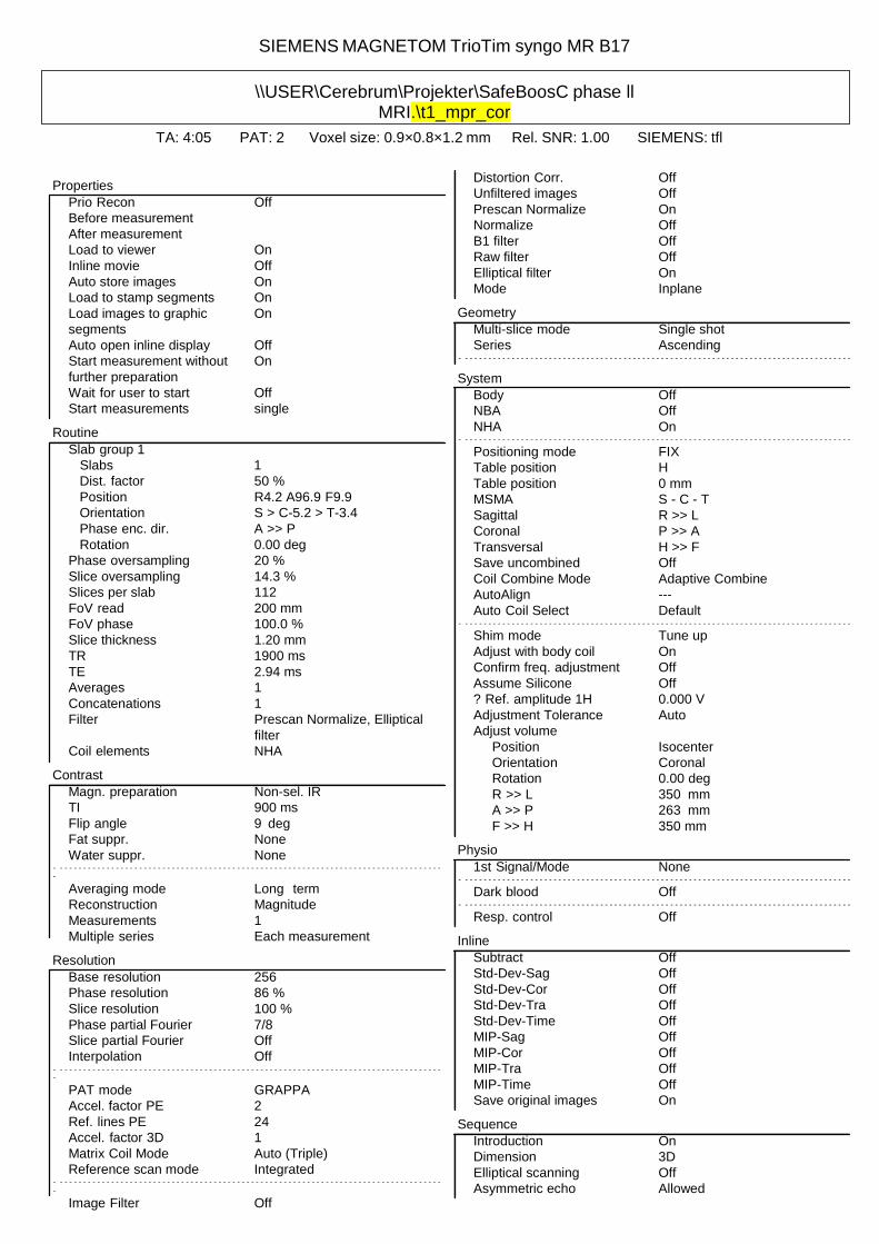
\\USER\Cerebrum\Projekter\SafeBoosC phase ll MRI.\t1_mpr_cor
TA: 4:05 PAT: 2 Voxel size: 0.9×0.8×1.2 mm Rel. SNR: 1.00 SIEMENS: tfl
Properties
Prio Recon Off
Before measurement
After measurement
Load to viewer On
Inline movie Off
Auto store images On
Load to stamp segments On
Load images to graphic On
segments
Auto open inline display Off
Start measurement without On
further preparation
Distortion Corr. Off
Unfiltered images Off
Prescan Normalize On
Normalize Off
B1 filter Off
Raw filter Off
Elliptical filter On
Mode Inplane
Geometry
Multi-slice mode Single shot
Series Ascending - - - - - - - - - - - - - - - - - - - - - - - - - - - - - - - - - - - - - - - - - - - - - - - - - - - - - - - - - - - - - - - - - - - - - - - - - - - - - - - - - - - -
System Wait for user to start Off
Start measurements single
Routine
Slab group 1
Slabs 1
Dist. factor 50 %
Position R4.2 A96.9 F9.9
Orientation S > C-5.2 > T-3.4
Phase enc. dir. A >> P
Rotation 0.00 deg
Phase oversampling 20 %
Slice oversampling 14.3 %
Slices per slab 112
FoV read 200 mm
FoV phase 100.0 %
Slice thickness 1.20 mm
TR 1900 ms
TE 2.94 ms
Averages 1
Concatenations 1
Filter Prescan Normalize, Elliptical
filter
Coil elements NHA
Contrast
Magn. preparation Non-sel. IR
TI 900 ms
Flip angle 9 deg
Fat suppr. None
Water suppr. None - - - - - - - - - - - - - - - - - - - - - - - - - - - - - - - - - - - - - - - - - - - - - - - - - - - - - - - - - - - - - - - - - - - - - - - - - - - - - - - - - - - -
Averaging mode Long term
Reconstruction Magnitude
Measurements 1
Multiple series Each measurement
Resolution
Base resolution 256
Phase resolution 86 %
Slice resolution 100 %
Phase partial Fourier 7/8
Slice partial Fourier Off
Interpolation Off - - - - - - - - - - - - - - - - - - - - - - - - - - - - - - - - - - - - - - - - - - - - - - - - - - - - - - - - - - - - - - - - - - - - - - - - - - - - - - - - - - - -
PAT mode GRAPPA
Accel. factor PE 2
Ref. lines PE 24
Accel. factor 3D 1
Matrix Coil Mode Auto (Triple)
Reference scan mode Integrated - - - - - - - - - - - - - - - - - - - - - - - - - - - - - - - - - - - - - - - - - - - - - - - - - - - - - - - - - - - - - - - - - - - - - - - - - - - - - - - - - - - -
Image Filter Off
Body Off
NBA Off
NHA On - - - - - - - - - - - - - - - - - - - - - - - - - - - - - - - - - - - - - - - - - - - - - - - - - - - - - - - - - - - - - - - - - - - - - - - - - - - - - - - - - - - -
Positioning mode FIX
Table position H
Table position 0 mm
MSMA S - C - T
Sagittal R >> L
Coronal P >> A
Transversal H >> F
Save uncombined Off
Coil Combine Mode Adaptive Combine
AutoAlign ---
Auto Coil Select Default - - - - - - - - - - - - - - - - - - - - - - - - - - - - - - - - - - - - - - - - - - - - - - - - - - - - - - - - - - - - - - - - - - - - - - - - - - - - - - - - - - - -
Shim mode Tune up
Adjust with body coil On
Confirm freq. adjustment Off
Assume Silicone Off
? Ref. amplitude 1H 0.000 V
Adjustment Tolerance Auto
Adjust volume
Position Isocenter
Orientation Coronal
Rotation 0.00 deg
R >> L 350 mm
A >> P 263 mm
F >> H 350 mm
Physio
1st Signal/Mode None - - - - - - - - - - - - - - - - - - - - - - - - - - - - - - - - - - - - - - - - - - - - - - - - - - - - - - - - - - - - - - - - - - - - - - - - - - - - - - - - - - - -
Dark blood Off - - - - - - - - - - - - - - - - - - - - - - - - - - - - - - - - - - - - - - - - - - - - - - - - - - - - - - - - - - - - - - - - - - - - - - - - - - - - - - - - - - - -
Resp. control Off
Inline
Subtract Off
Std-Dev-Sag Off
Std-Dev-Cor Off
Std-Dev-Tra Off
Std-Dev-Time Off
MIP-Sag Off
MIP-Cor Off
MIP-Tra Off
MIP-Time Off
Save original images On
Sequence
Introduction On
Dimension 3D
Elliptical scanning Off
Asymmetric echo Allowed

SIEMENS MAGNETOM TrioTim syngo MR B17
Bandwidth 140 Hz/Px
Flow comp. No
Echo spacing 9 ms - - - - - - - - - - - - - - - - - - - - - - - - - - - - - - - - - - - - - - - - - - - - - - - - - - - - - - - - - - - - - - - - - - - - - - - - - - - - - - - - - - - -
RF pulse type Normal
Gradient mode Normal
Excitation Non-sel.
RF spoiling On
8/+

SIEMENS MAGNETOM TrioTim syngo MR B17
\\USER\Cerebrum\Projekter\SafeBoosC phase ll MRI.\ep2d_diff_orth_p2_tra
TA: 1:31 PAT: 2 Voxel size: 1.2×1.2×4.0 mm Rel. SNR: 1.00 SIEMENS: ep2d_diff
Properties
Prio Recon Off
Before measurement
After measurement
Load to viewer On
Inline movie Off
Auto store images On
Load to stamp segments On
Load images to graphic On
segments
Auto open inline display Off
Start measurement without On
further preparation
Wait for user to start Off
Start measurements single
Routine
Slice group 1
Slices 28
Dist. factor 30 %
Position R3.2 A106.3 H9.7
Orientation T > S4.5 > C2.9
Phase enc. dir. A >> P
Rotation 0.00 deg
Phase oversampling 0 %
FoV read 230 mm
FoV phase 100.0 %
Slice thickness 4.0 mm
TR 4700 ms
TE 102 ms
Averages 4
Concatenations 1
Filter Raw filter, Distortion
Corr.(2D), Prescan Normalize
Coil elements NHA
Contrast
MTC Off
Magn. preparation None
Fat suppr. Fat sat. - - - - - - - - - - - - - - - - - - - - - - - - - - - - - - - - - - - - - - - - - - - - - - - - - - - - - - - - - - - - - - - - - - - - - - - - - - - - - - - - - - - -
Averaging mode Long term
Reconstruction Magnitude
Delay in TR 0 ms
Resolution
Base resolution 192
Phase resolution 100 %
Phase partial Fourier 6/8
Interpolation On - - - - - - - - - - - - - - - - - - - - - - - - - - - - - - - - - - - - - - - - - - - - - - - - - - - - - - - - - - - - - - - - - - - - - - - - - - - - - - - - - - - -
PAT mode GRAPPA
Accel. factor PE 2
Ref. lines PE 24
Matrix Coil Mode Auto (Triple)
Reference scan mode Separate - - - - - - - - - - - - - - - - - - - - - - - - - - - - - - - - - - - - - - - - - - - - - - - - - - - - - - - - - - - - - - - - - - - - - - - - - - - - - - - - - - - -
Distortion Corr. On
Mode 2D
Unfiltered images Off
Prescan Normalize On
Raw filter On
Intensity Weak
Slope 25
Elliptical filter Off
Hamming Off
Geometry
Multi-slice mode Interleaved
Series Interleaved - - - - - - - - - - - - - - - - - - - - - - - - - - - - - - - - - - - - - - - - - - - - - - - - - - - - - - - - - - - - - - - - - - - - - - - - - - - - - - - - - - - -
Special sat. None - - - - - - - - - - - - - - - - - - - - - - - - - - - - - - - - - - - - - - - - - - - - - - - - - - - - - - - - - - - - - - - - - - - - - - - - - - - - - - - - - - - -
System
Body Off
NBA Off
NHA On - - - - - - - - - - - - - - - - - - - - - - - - - - - - - - - - - - - - - - - - - - - - - - - - - - - - - - - - - - - - - - - - - - - - - - - - - - - - - - - - - - - -
Positioning mode REF
Table position H
Table position 0 mm
MSMA S - C - T
Sagittal R >> L
Coronal P >> A
Transversal H >> F
Coil Combine Mode Adaptive Combine
AutoAlign ---
Auto Coil Select Default - - - - - - - - - - - - - - - - - - - - - - - - - - - - - - - - - - - - - - - - - - - - - - - - - - - - - - - - - - - - - - - - - - - - - - - - - - - - - - - - - - - -
Shim mode Standard
Adjust with body coil Off
Confirm freq. adjustment Off
Assume Silicone Off
? Ref. amplitude 1H 0.000 V
Adjustment Tolerance Auto
Adjust volume
Position R3.2 A106.3 H9.7
Orientation T > S4.5 > C2.9
Rotation 0.00 deg
R >> L 230 mm
A >> P 230 mm
F >> H 145 mm
Physio
1st Signal/Mode None - - - - - - - - - - - - - - - - - - - - - - - - - - - - - - - - - - - - - - - - - - - - - - - - - - - - - - - - - - - - - - - - - - - - - - - - - - - - - - - - - - - -
Resp. control Off
Diff
Diffusion mode Orthogonal
Diff. weightings 2
b-value 1 0 s/mm²
b-value 2 800 s/mm²
Diff. weighted images Off
Trace weighted images On
Average ADC maps On
Individual ADC maps Off
FA maps Off
Mosaic Off
Tensor Off
Noise level 40
Diff. directions 3
Sequence
Introduction On
Bandwidth 1184 Hz/Px
Free echo spacing Off
Echo spacing 0.93 ms - - - - - - - - - - - - - - - - - - - - - - - - - - - - - - - - - - - - - - - - - - - - - - - - - - - - - - - - - - - - - - - - - - - - - - - - - - - - - - - - - - - -
EPI factor 192
RF pulse type Normal
Gradient mode Fast
9/+

SIEMENS MAGNETOM TrioTim syngo MR B17
\\USER\Cerebrum\Projekter\SafeBoosC phase ll MRI.\ep2d_diff_mddw_30_p2
TA: 9:28 PAT: 2 Voxel size: 2.0×2.0×2.0 mm Rel. SNR: 1.00 SIEMENS: ep2d_diff
Properties
Prio Recon Off
Before measurement
After measurement
Load to viewer On
Inline movie Off
Auto store images On
Load to stamp segments On
Load images to graphic On
segments
Auto open inline display Off
Start measurement without On
further preparation
Wait for user to start Off
Start measurements single
Routine
Slice group 1
Slices 49
Dist. factor 30 %
Position R3.2 A106.3 H9.7
Orientation T > S4.5 > C2.9
Phase enc. dir. A >> P
Rotation 0.00 deg
Phase oversampling 0 %
FoV read 250 mm
FoV phase 100.0 %
Slice thickness 2.0 mm
TR 5900 ms
TE 81 ms
Averages 3
Concatenations 1
Filter Prescan Normalize
Coil elements NHA
Contrast
MTC Off
Magn. preparation None
Fat suppr. Fat sat. - - - - - - - - - - - - - - - - - - - - - - - - - - - - - - - - - - - - - - - - - - - - - - - - - - - - - - - - - - - - - - - - - - - - - - - - - - - - - - - - - - - -
Averaging mode Long term
Reconstruction Magnitude
Delay in TR 0 ms
Multiple series Off
Resolution
Base resolution 122
Phase resolution 100 %
Phase partial Fourier 6/8
Interpolation Off - - - - - - - - - - - - - - - - - - - - - - - - - - - - - - - - - - - - - - - - - - - - - - - - - - - - - - - - - - - - - - - - - - - - - - - - - - - - - - - - - - - -
PAT mode GRAPPA
Accel. factor PE 2
Ref. lines PE 24
Matrix Coil Mode Auto (Triple)
Reference scan mode Separate - - - - - - - - - - - - - - - - - - - - - - - - - - - - - - - - - - - - - - - - - - - - - - - - - - - - - - - - - - - - - - - - - - - - - - - - - - - - - - - - - - - -
Distortion Corr. Off
Prescan Normalize On
Raw filter On
Elliptical filter Off
Hamming Off
Geometry
Multi-slice mode Interleaved
Series Interleaved - - - - - - - - - - - - - - - - - - - - - - - - - - - - - - - - - - - - - - - - - - - - - - - - - - - - - - - - - - - - - - - - - - - - - - - - - - - - - - - - - - - -
Special sat. None - - - - - - - - - - - - - - - - - - - - - - - - - - - - - - - - - - - - - - - - - - - - - - - - - - - - - - - - - - - - - - - - - - - - - - - - - - - - - - - - - - - -
System
Body Off
NBA Off
NHA On - - - - - - - - - - - - - - - - - - - - - - - - - - - - - - - - - - - - - - - - - - - - - - - - - - - - - - - - - - - - - - - - - - - - - - - - - - - - - - - - - - - -
Positioning mode FIX
Table position H
Table position 0 mm
MSMA S - C - T
Sagittal R >> L
Coronal P >> A
Transversal H >> F
Coil Combine Mode Adaptive Combine
AutoAlign ---
Auto Coil Select Default - - - - - - - - - - - - - - - - - - - - - - - - - - - - - - - - - - - - - - - - - - - - - - - - - - - - - - - - - - - - - - - - - - - - - - - - - - - - - - - - - - - -
Shim mode Standard
Adjust with body coil Off
Confirm freq. adjustment Off
Assume Silicone Off
? Ref. amplitude 1H 0.000 V
Adjustment Tolerance Auto
Adjust volume
Position R3.2 A106.3 H9.7
Orientation T > S4.5 > C2.9
Rotation 0.00 deg
R >> L 250 mm
A >> P 250 mm
F >> H 127 mm
Physio
1st Signal/Mode None - - - - - - - - - - - - - - - - - - - - - - - - - - - - - - - - - - - - - - - - - - - - - - - - - - - - - - - - - - - - - - - - - - - - - - - - - - - - - - - - - - - -
Resp. control Off
Diff
Diffusion mode MDDW
Diff. weightings 2
b-value 1 0 s/mm²
b-value 2 800 s/mm²
Diff. weighted images On
Trace weighted images On
Average ADC maps On
Individual ADC maps Off
FA maps On
Mosaic On
Tensor On
Noise level 30
Diff. directions 30
Sequence
Introduction On
Bandwidth 1518 Hz/Px
Free echo spacing Off
Echo spacing 0.74 ms - - - - - - - - - - - - - - - - - - - - - - - - - - - - - - - - - - - - - - - - - - - - - - - - - - - - - - - - - - - - - - - - - - - - - - - - - - - - - - - - - - - -
EPI factor 122
RF pulse type Normal
Gradient mode Fast 10/+

SIEMENS MAGNETOM TrioTim syngo MR B17
Table of contents
\\USER Cerebrum
Projekter
SafeBoosC phase ll MRI. localizer
t1_se_sag_3mm_200mm
t2_tse_cor_1,2mm_448_p2
t1_mpr_sag
ep2d_diff_orth_p2_tra
ep2d_diff_mddw_30_p2

SafeBoosC phase II - SOP - Brain MRI
Version 1.0 29