spa15 of shigella flexneri is secreted through the type iii...
TRANSCRIPT
INFECTION AND IMMUNITY, Dec. 2009, p. 5281–5290 Vol. 77, No. 120019-9567/09/$12.00 doi:10.1128/IAI.00800-09Copyright © 2009, American Society for Microbiology. All Rights Reserved.
Spa15 of Shigella flexneri Is Secreted through the Type III SecretionSystem and Prevents Staurosporine-Induced Apoptosis�
Christina S. Faherty and Anthony T. Maurelli*Department of Microbiology and Immunology, F. Edward Hebert School of Medicine, Uniformed Services University of the
Health Sciences, Bethesda, Maryland 20814-4799
Received 16 July 2009/Returned for modification 13 August 2009/Accepted 22 September 2009
Shigella flexneri is a gram-negative, facultative intracellular pathogen that invades the colonic epithelium andcauses bacillary dysentery. We previously demonstrated that S. flexneri inhibits staurosporine-induced apop-tosis in infected epithelial cells and that a �mxiE mutant is unable to inhibit apoptosis. Therefore, wehypothesized that an MxiE-regulated gene was responsible for protection of epithelial cells from apoptosis.Analysis of all MxiE-regulated genes yielded no mutants that lacked the ability to prevent apoptosis. Spa15,which is defined as a type III secretion system chaperone, was analyzed since it associates with MxiE. A �spa15mutant was unable to prevent staurosporine-induced apoptosis. C-terminal hemagglutinin-tagged spa15 wassecreted by S. flexneri within 2 h in the Congo red secretion assay, and secretion was dependent on the type IIIsecretion system. Spa15 was also secreted by Shigella in infected epithelial cells, as verified by immunofluo-rescence analysis. Spa15 secretion was decreased in the �mxiE mutant, which demonstrates why this mutantis unable to prevent staurosporine-induced apoptosis. Our data are the first to show that Spa15 is secreted ina type III secretion system-dependent fashion, and the absence of Spa15 in the �spa15 mutant results in theloss of protection from staurosporine-induced apoptosis in epithelial cells. Thus, Spa15 contributes to theintracellular survival of Shigella by blocking apoptosis in the infected host cell.
Shigella flexneri is a gram-negative, facultative intracellularpathogen that causes bacillary dysentery. Clinical symptoms ofdisease include watery diarrhea, severe abdominal pain, andbloody stools (39). Disease is a result of the ability of the patho-gen to invade the colonic epithelium. Once S. flexneri enters thecolon, the bacteria transit through M cells and encounter residentmacrophages. The bacteria escape the macrophages by inducingcell death and subsequently invade epithelial cells at the basolat-eral face (19). Proinflammatory signaling and a subsequent effluxof polymorphonuclear cells (PMNs) into the infected tissue allowthe bacteria to invade more epithelial cells at the basolateral pole,while the PMNs contribute to the disease severity by causing localtissue destruction (19). Once inside the cytoplasm of the host cell,S. flexneri induces actin polymerization, which allows the bacteriato move to adjacent cells without the need to enter the extracel-lular environment (5). The epithelial cells provide the bacteriawith an intracellular niche to multiply and spread to adjacentcells.
S. flexneri virulence requires a 220-kb virulence plasmid thatencodes a type III secretion system (T3SS), the Ipa proteinsessential for entry into the host cells, and other effector pro-teins. The T3SS is comprised of a needle complex that has aseven-ringed basal body and a protruding needle. The needlecomplex delivers proteins directly to the host cell from thebacterial cytoplasm (14). Temperature regulation of the geneson the virulence plasmid allows the needle complex to besynthesized and assembled at 37°C. The secretion of proteins isinduced upon contact of the bacteria with the host cell. The Ipa
proteins mediate entry of the bacteria into the epithelial cellthrough localized actin depolymerization and membrane en-gulfment. After engulfment, the bacteria are inside a vacuolethat is subsequently lysed, allowing the bacteria to enter thecytoplasm of the host cell (39). Once inside the cytoplasm, thebacteria spread and secrete additional effector proteins. Theseproteins are encoded by genes that are scattered throughoutthe 220-kb virulence plasmid and are subsequently secretedthrough the T3SS for postinvasion virulence (6).
We previously showed that S. flexneri inhibits staurosporine(STS)-induced apoptosis in epithelial cells by preventing the ac-tivation of caspase-3, a key protein in apoptotic cell death, andthat a �mxiE mutant is unable to prevent STS-induced apoptosis(7). MxiE is a transcriptional activator that induces the expressionof �20 bacterial genes when the bacteria are inside the cytosol ofthe host cell. The subsequent protein products are secretedthrough the T3SS (22, 24). We therefore hypothesized that anMxiE-regulated gene is responsible for protection. In this study,we analyzed all of the MxiE-regulated genes and found that nonewas required to inhibit STS-induced apoptosis. We also analyzeda �spa15 mutant since Spa15 associates with MxiE (35). Thisreport describes the inability of a �spa15 mutant to prevent STS-induced apoptosis and demonstrates, for the first time, that Spa15is secreted through the T3SS. Spa15 was originally described as aT3SS chaperone and a coantiactivator to MxiE (34, 35). We areproposing a new, third function in which Spa15 is involved inapoptosis inhibition in epithelial cells since Spa15 is secreted bythe T3SS and the �spa15 mutant is unable to prevent apoptosis inepithelial cells.
MATERIALS AND METHODS
Bacterial strains and growth conditions. The strains of S. flexneri used arelisted in Table 1. Bacteria were routinely cultured at 37°C either in Luria-Bertanibroth with aeration or on tryptic soy broth plates with 1.5% agar and 0.025%
* Corresponding author. Mailing address: Department of Microbi-ology and Immunology, Uniformed Services University of the HealthSciences, 4301 Jones Bridge Road, Bethesda, MD 20814-4799. Phone:(301) 295-3415. E-mail: [email protected].
� Published ahead of print on 5 October 2009.
5281
on July 13, 2018 by guesthttp://iai.asm
.org/D
ownloaded from
Congo red (CR; Sigma). Antibiotics were used at the following concentrations:kanamycin, 50 �g/ml; streptomycin, 50 �g/ml; chloramphenicol, 5 �g/ml; andampicillin, 100 �g/ml.
Strain and plasmid construction. BS902 was constructed using the � red linearrecombination method as previously described (7). PCR was used to amplify akanamycin resistance cassette gene (kan) from pKD4 (Table 1) with 5� and 3�overhangs identical to the 5� and 3� regions of spa15 internal to the start and stopcodons (Table 2). Kanamycin-resistant recombinants were purified and screenedvia PCR using primers (Table 2) that annealed �70 bp upstream and down-stream of the spa15 coding region to detect the size difference due to theinsertion of the kanamycin cassette. Next, this mutant was used as the donor
strain for transduction of 2457T using P1L4 and selection for kanamycin resis-tance.
Additional primers listed in Table 2 were used to construct the pBSKS-spa15,pSpa15-2HA, and pMxiE-2HA plasmids. spa15 was amplified by colony PCRusing Platinum Taq high-fidelity DNA polymerase (Invitrogen) and cloned intopGEM-T (Promega) according to the manufacturers’ protocols. Subsequently,spa15 was subcloned into pBSKS (Stratagene) via the Acc65I and BamHI re-striction enzyme sites using T4 DNA ligase (New England Biolabs). The ligationreaction was transformed into Escherichia coli DH5� for production of the newplasmid, pBSKS-spa15. The spa15 insert sequence was verified with primers thatanneal outside the multiple cloning site of the vector. pBSKS-spa15 was subse-
TABLE 1. Strains and plasmids used in this study
Strain or plasmid Description Source or reference
StrainsS. flexneri
2457T Wild-type serotype 2a 13BS611 2457T/�mxiE2::aphA-3 22BS613 BS611/pRRS13 (PlacmxiE�) 22BS652 2457T/�spa47::aadA Lab stockBS766 2457T transformed with pKM208 7BS902 2457T/�spa15::aphA-3 This studyBS904 BS902 transformed with pBSKS-spa15 This studyBS905 2457T transformed with pSpa15-2HA This studyBS906 BS611 transformed with pSpa15-2HA This studyBS907 BS613 transformed with pSpa15-2HA This studyBS908 BS652 transformed with pSpa15-2HA This studyBS909 2457T transformed with pMxiE-2HA This studyBS914 BS902 transformed with pSpa15-2HA This study
E. coliDH5� endA1 hsdR17 supE44 thi-1 recA1 gyrA96 relA1 (�lacIZYA-argF) U169 deoR
(�80dlac�lacZM15)17
ATM951 E. coli expressing a plasmid that contains the Shigella entry region Lab stock (in preparation)
PlasmidspKD4 oriR6K; bla aphA-3 10pKM208 Temperature-sensitive red-, gam-, and lacI-expressing plasmid driven by PTac promoter; bla 10pBSKS(�) pBluescript cloning vector; bla StratagenepBSKS-spa15 spa15 cloned into pBSKS This studypDZ1 Cloning intermediate for 2HA fusions; cat 46pSpa15-2HA spa15 cloned into pDZ1; cat This studypMxiE-2HA mxiE cloned into pDZ1; cat This study
TABLE 2. Primers used in this study
PurposeForward primer Reverse primer
Name Sequencea Name Sequencea
Amplify aphA-3 cassettefor spa15 deletion
S15kanF 5�-AGTAACATTAATTTAGTTCAATTAGTTAGAGATAGTCTTTTCACGATTGGTGTGTAGGCTGGAGCTGCTTC-3�
S15kanR 5�-TAAGACCCCATTTAAGATTTCCATCCTCTGATAAAACTCATGCAGAATCTCATATGAATACCTCCTTAG-3�
Confirm �spa15deletion
Spa15F2 5�-GTTATATCTATGCTGAGATTG-3� Spa15R2 5�-CCAATCGAAACATCGCTAAG-3�
Clone spa15 intopBSKS
spaF 5�-GATCGGTACCATGAGTAACATTAATTTAGTTC-3�
spaR2 5�-GATCGGATCCATTATAAGACCCCATTTAAGATTTC-3�
Sequence spa15 inpBSKS
pBSKF 5�-AGCGGATAACAATTTCACACAGGAAAC-3�
pBSKR 5�-GTTTTCCCAGTCACGACGTTG-3�
Clone spa15 into pDZ1 spaF 5�-GATCGGTACCATGAGTAACATTAATTTAGTTC-3�
spaR 5�-GATCAGATCTTAAGACCCCATTTAAGATTTC-3�
Clone mxiE into pDZ1 mxiE2HAF 5�-CTAGGGTACCATGGAAGGGTTTTTTTTTGTCCG-3�
mxiE2HAR 5�-AGATCGGATCCAATTTTTTCATTTATTTTTTTCAC-3�
Sequence spa15 ormxiE in pDZ1
pDZ1F 5�-CTGGGTTGAAGGCTCTCAAG-3� pDZ1R 5�-TCAGCCCCATACGATATAAG-3�
a Restriction enzyme sites are underlined.
5282 FAHERTY AND MAURELLI INFECT. IMMUN.
on July 13, 2018 by guesthttp://iai.asm
.org/D
ownloaded from

quently transformed into BS902 to generate BS904. To create C-terminal hem-agglutinin (HA) tags of spa15 and mxiE, vector pDZ1 was used. pDZ1 containstwo HA epitopes and is a low-copy vector with a pACYC184 origin of replication(46). spa15 and mxiE were amplified by colony PCR as mentioned above, clonedinto pGEM-T, and subcloned into pDZ1 using Acc65I and BglII for spa15 andAcc65I and BamHI for mxiE. The ligation reactions were transformed into E.coli DH5� for production of the new plasmids pSpa15-2HA and pMxiE-2HA.The inserts were sequenced using primers that anneal outside the multiplecloning site of pDZ1. The plasmids were transformed into the various strainslisted in Table 1 and selected on medium with the appropriate antibiotic.
Virulence assays. The invasion, plaque, and apoptosis assays were performedas previously described (7, 18, 32). Briefly for the apoptosis assay, bacterialcultures were grown to mid-log phase, standardized to an optical density at 600nm of 0.72, washed in 1� phosphate buffered saline (PBS), resuspended in 1�Dulbecco’s modified Eagle’s medium (DMEM), and applied to a semiconfluentmonolayer of HeLa cells. The plates were centrifuged at 3,000 rpm for 10 min at37°C to facilitate the invasion process by allowing the bacteria to make contactwith the HeLa cells. The plates were incubated at 37°C with 5% CO2 for 30 min.Both infected and uninfected controls were then washed with 1� PBS, DMEMplus 50 �g/ml gentamicin was added, and the plates were incubated for 3 h at37°C with 5% CO2. Infected and uninfected cells were then washed again, andDMEM plus 50 �g/ml gentamicin and 4 �M STS (Calbiochem) was added for anadditional 3 h. After the assay, the cells were processed for immunofluorescenceor Western blot analysis.
CR secretion assay. The CR secretion assay was used to identify the proteinssecreted by the bacteria through the T3SS and was performed as previouslydescribed (4). Briefly, bacteria were grown to mid-log phase, resuspended in 1�PBS, and CR was added to a final concentration of 30 �g/ml. The bacteria wereincubated at 37°C for 30 min for secretion of early T3SS proteins or at 1-hintervals for secretion of late T3SS effector proteins (41). After incubation, thebacteria were pelleted by centrifugation, and the supernatant was collected andfiltered through a 0.22-�m-pore filter and then stored at �20°C. The proteins inthe supernatant represent the secreted proteins and were concentrated by tri-chloroacetic acid precipitation. Trichloroacetic acid pellets were resuspended in50 �l sodium dodecyl sulfate (SDS) loading buffer for protein analysis and storedat �20°C. The bacterial pellets, representing the nonsecreted proteins, wereresuspended in 500 �l of SDS loading buffer and stored at �20°C.
Immunofluorescence and Western blot analysis. The same procedures werefollowed for immunofluorescence or Western blot analysis as previously de-scribed (7). For the immunofluorescence analysis, infected monolayers werefixed with 1� PBS with 3% formaldehyde (36% stock; Sigma) and 0.2% glutar-aldehyde (25% stock; Sigma). To visualize nuclei, 5 mg/ml of 4�,6-diamido-2-phenylindole (DAPI; Molecular Probes) was diluted 1:1,000 in 1� PBS. Nucleiwere monitored for a characteristic apoptotic phenotype, namely DNA fragmen-tation and chromatin condensation, as demonstrated previously (7). To detectactivated caspase-3, a rabbit anti-human cleaved caspase-3 antibody (Cell Sig-naling Technologies) was used with a secondary goat anti-rabbit immunoglobulinG (IgG) antibody conjugated to Alexa 488 (Invitrogen). To measure secretion ofthe HA-tagged Spa15, infected cells were stained with a mouse monoclonalanti-HA antibody (Covance) followed by a goat anti-mouse IgG antibody con-jugated to Alexa 488 (Invitrogen). After the staining procedure, antifade reagent(Molecular Probes) was applied to all samples, which were then overlaid withcoverslips and stored at 4°C in the dark.
For Western blot analyses, each sample was resolved by SDS-polyacrylamidegel electrophoresis, and Coomassie blue staining verified equal loading of totalprotein for all samples. Caspase-3 was detected with rabbit anti-human caspase-3or cleaved caspase-3 antibodies (Cell Signaling Technology) followed by a don-key anti-rabbit IgG antibody conjugated to horseradish peroxidase (AmershamBiosciences). The HA-tagged Spa15 was detected using a mouse monoclonalanti-HA antibody in a 1:1,000 dilution with a sheep anti-mouse IgG antibodyconjugated to horseradish peroxidase (Amersham Biosciences). IpaB and IpaCwere detected with mouse monoclonal anti-IpaB (1:20,000 dilution) and anti-IpaC (1:5,000 dilution) antibodies as previously described (31). The same sec-ondary antibodies were used as with the anti-HA antibody. Western blots weredeveloped using the Visualizer developing system (Upstate Cell Signaling Solu-tions) according to the protocol provided, and the blots were imaged using a FujiIntelligent Dark Box with a Fujinon lens and Image Reader LAS-3000 software(Fuji) on the chemiluminescence setting in increment mode at 10-s intervals.Densitometry comparisons were made using the Image Gauge V4.22 softwareprovided.
Statistical analysis. The densitometry results for the caspase-3 Western blotwere analyzed using Tukey’s analysis of variance (ANOVA) post hoc test on theSPSS program, version 12.0.1, for Windows. For the strain comparison in the CR
secretion assay, the ratios of Spa15 secretion in wild-type bacteria (strain BS905)to the �mxiE mutant (strain BS906) and to the �mxiE/mxiE�-complementedstrain (strain BS907) were compared. A repeated-measures ANOVA was per-formed using Tukey’s post hoc analysis for all time points on the SPSS programversion 12.0.1 for Windows. Ratios were analyzed on a log scale to comply withthe assumptions of ANOVA, and the summaries are reported as geometric meanratios relative to BS905, averaging across three repetitions.
RESULTS
Identifying spa15 as the antiapoptosis gene. We constructeddeletion mutants in most of the MxiE-regulated genes (Table3), and we did not find any genes that were responsible forapoptosis protection since all of the mutants prevented STS-induced apoptosis (data not shown). We next utilized a strainof E. coli expressing the Shigella entry region (SER), whichcontains all of the genes necessary for epithelial cell invasion(27), in order to rule out the MxiE-regulated genes carried onthe chromosome (3). This strain of E. coli also preventedSTS-induced apoptosis (data not shown). Thus, Shigella-spe-cific chromosomal genes are not required for the antiapoptoticphenotype. Since this strain of bacteria expresses only Shigellagenes carried within the SER, we hypothesized that the pro-tective gene/protein is either mxiE itself or a protein that as-sociates with MxiE, namely IpgC, OspD1, or Spa15. IpgC is acoactivator needed for the induction of MxiE-regulated genes,while OspD1 and Spa15 have been described as antiactivatorsof MxiE (35, 36). We found that a �ipgC mutant was notinvasive, and it was not further pursued. OspD1 was eliminatedas a candidate since the E. coli strain carrying the SER blocksapoptosis and ospD1 is not located within the SER (6). Finally,the spa15 gene was a likely candidate since it is located within
TABLE 3. Shigella genes analyzed that had no effect onapoptosis protectiona
MxiE-regulateddeletion mutant
gene(s)
MxiE-regulatedgenes via E.coli strainb
Deletion mutantgene whose
product associateswith Spa15
Gene whoseproduct
associates withSpa15 (via E.coli strain)b
ospBc ipaH_1 ipaA ospC2ospC1 ipaH_2 ipgB1 ospC3ospD3 ipaH_5 ospB ospD1ospE1 ipaH_7 ipgB2ospE2ospFospGvirAospE1 ospE2d
ospC1 ospB ospFe
ipaH1.4ipaH2.5ipaH4.5ipaH7.8ipaH9.8ipaH_4
a Strains were analyzed for the ability to inhibit STS-induced apoptosis byDAPI staining of the nuclei of infected cells to determine if the nuclei had thecharacteristic apoptotic phenotype—namely DNA fragmentation and/or chro-matin condensation (7).
b The E. coli strain expresses the Shigella entry region and invades epithelialcells (see text).
c Gene product also associates with Spa15.d Double mutant.e Triple mutant.
VOL. 77, 2009 Spa15 INHIBITS APOPTOSIS IN SHIGELLA-INFECTED CELLS 5283
on July 13, 2018 by guesthttp://iai.asm
.org/D
ownloaded from
the SER and encodes a T3SS chaperone that has the ability tobind to several T3SS effector proteins (6, 34). Therefore, weconstructed BS902, a �spa15 mutant of wild-type S. flexneristrain 2457T. The mutant showed a slightly (10%) reducedentry efficiency, similar to published data on a �spa15 mutantconstructed in S. flexneri strain M90T (34). In addition, themutant produced plaque sizes about half the size as those ofwild-type bacteria in the plaque assay (data not shown).
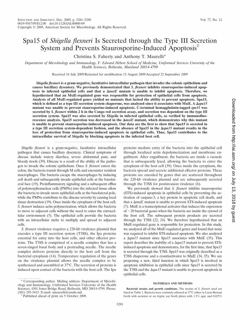
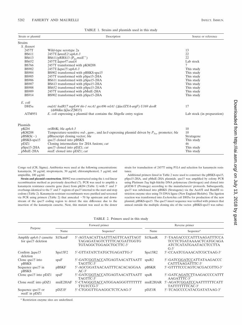
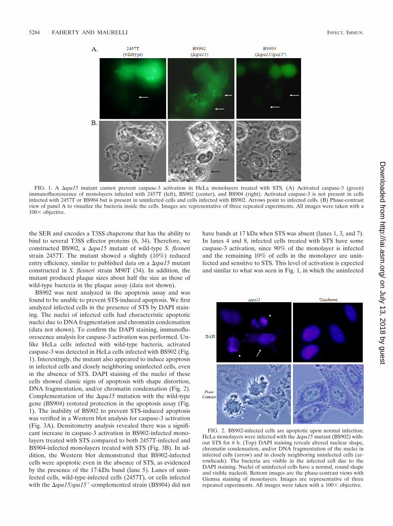
BS902 was next analyzed in the apoptosis assay and wasfound to be unable to prevent STS-induced apoptosis. We firstanalyzed infected cells in the presence of STS by DAPI stain-ing. The nuclei of infected cells had characteristic apoptoticnuclei due to DNA fragmentation and chromatin condensation(data not shown). To confirm the DAPI staining, immunoflu-orescence analysis for caspase-3 activation was performed. Un-like HeLa cells infected with wild-type bacteria, activatedcaspase-3 was detected in HeLa cells infected with BS902 (Fig.1). Interestingly, the mutant also appeared to induce apoptosisin infected cells and closely neighboring uninfected cells, evenin the absence of STS. DAPI staining of the nuclei of thesecells showed classic signs of apoptosis with shape distortion,DNA fragmentation, and/or chromatin condensation (Fig. 2).Complementation of the �spa15 mutation with the wild-typegene (BS904) restored protection in the apoptosis assay (Fig.1). The inability of BS902 to prevent STS-induced apoptosiswas verified in a Western blot analysis for caspase-3 activation(Fig. 3A). Densitometry analysis revealed there was a signifi-cant increase in caspase-3 activation in BS902-infected mono-layers treated with STS compared to both 2457T-infected andBS904-infected monolayers treated with STS (Fig. 3B). In ad-dition, the Western blot demonstrated that BS902-infectedcells were apoptotic even in the absence of STS, as evidencedby the presence of the 17-kDa band (lane 5). Lanes of unin-fected cells, wild-type-infected cells (2457T), or cells infectedwith the �spa15/spa15�-complemented strain (BS904) did not
have bands at 17 kDa when STS was absent (lanes 1, 3, and 7).In lanes 4 and 8, infected cells treated with STS have somecaspase-3 activation, since 90% of the monolayer is infectedand the remaining 10% of cells in the monolayer are unin-fected and sensitive to STS. This level of activation is expectedand similar to what was seen in Fig. 1, in which the uninfected
FIG. 1. A �spa15 mutant cannot prevent caspase-3 activation in HeLa monolayers treated with STS. (A) Activated caspase-3 (green)immunofluorescence of monolayers infected with 2457T (left), BS902 (center), and BS904 (right). Activated caspase-3 is not present in cellsinfected with 2457T or BS904 but is present in uninfected cells and cells infected with BS902. Arrows point to infected cells. (B) Phase-contrastview of panel A to visualize the bacteria inside the cells. Images are representative of three repeated experiments. All images were taken with a100� objective.
FIG. 2. BS902-infected cells are apoptotic upon normal infection.HeLa monolayers were infected with the �spa15 mutant (BS902) with-out STS for 6 h. (Top) DAPI staining reveals altered nuclear shape,chromatin condensation, and/or DNA fragmentation of the nuclei ininfected cells (arrow) and in closely neighboring uninfected cells (ar-rowheads). The bacteria are visible in the infected cell due to theDAPI staining. Nuclei of uninfected cells have a normal, round shapeand visible nucleoli. Bottom images are the phase-contrast views withGiemsa staining of monolayers. Images are representative of threerepeated experiments. All images were taken with a 100� objective.
5284 FAHERTY AND MAURELLI INFECT. IMMUN.
on July 13, 2018 by guesthttp://iai.asm
.org/D
ownloaded from
cells in the population have a positive signal for activatedcaspase-3.
Finally, we constructed mutations in ipaA and ipgB1, sincetheir products are known to associate with Spa15 and aresecreted into epithelial cells (33, 34, 42). The mutants hadreduced invasion efficiencies, as previously described (33, 42).We found that neither ipaA nor ipgB1 was required for apop-tosis inhibition since both of the mutants protected cells in theapoptosis assay (data not shown). The four other proteins thatassociate with Spa15 were ruled out as candidate protectiveproteins since the genes encoding these proteins are notpresent in the E. coli strain expressing the SER (Table 3).Therefore, the �spa15 mutant was the only strain besides the�mxiE mutant that was unable to protect HeLa cells in theapoptosis assay.
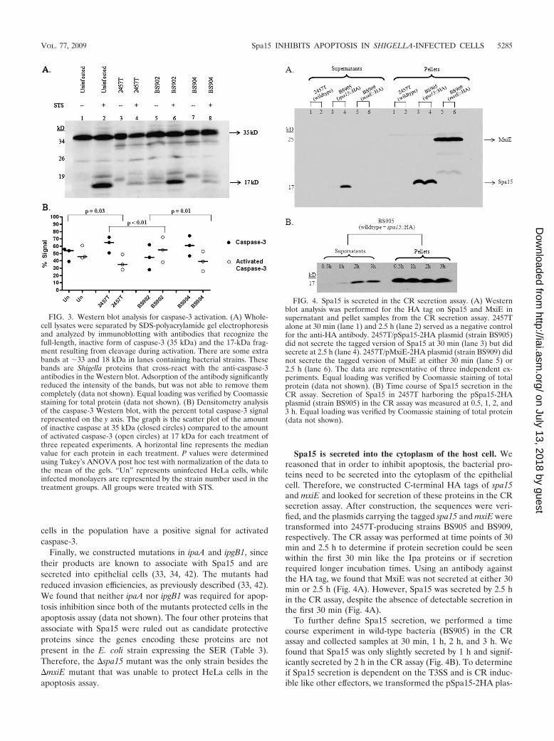
Spa15 is secreted into the cytoplasm of the host cell. Wereasoned that in order to inhibit apoptosis, the bacterial pro-teins need to be secreted into the cytoplasm of the epithelialcell. Therefore, we constructed C-terminal HA tags of spa15and mxiE and looked for secretion of these proteins in the CRsecretion assay. After construction, the sequences were veri-fied, and the plasmids carrying the tagged spa15 and mxiE weretransformed into 2457T-producing strains BS905 and BS909,respectively. The CR assay was performed at time points of 30min and 2.5 h to determine if protein secretion could be seenwithin the first 30 min like the Ipa proteins or if secretionrequired longer incubation times. Using an antibody againstthe HA tag, we found that MxiE was not secreted at either 30min or 2.5 h (Fig. 4A). However, Spa15 was secreted by 2.5 hin the CR assay, despite the absence of detectable secretion inthe first 30 min (Fig. 4A).
To further define Spa15 secretion, we performed a timecourse experiment in wild-type bacteria (BS905) in the CRassay and collected samples at 30 min, 1 h, 2 h, and 3 h. Wefound that Spa15 was only slightly secreted by 1 h and signif-icantly secreted by 2 h in the CR assay (Fig. 4B). To determineif Spa15 secretion is dependent on the T3SS and is CR induc-ible like other effectors, we transformed the pSpa15-2HA plas-
FIG. 3. Western blot analysis for caspase-3 activation. (A) Whole-cell lysates were separated by SDS-polyacrylamide gel electrophoresisand analyzed by immunoblotting with antibodies that recognize thefull-length, inactive form of caspase-3 (35 kDa) and the 17-kDa frag-ment resulting from cleavage during activation. There are some extrabands at �33 and 18 kDa in lanes containing bacterial strains. Thesebands are Shigella proteins that cross-react with the anti-caspase-3antibodies in the Western blot. Adsorption of the antibody significantlyreduced the intensity of the bands, but was not able to remove themcompletely (data not shown). Equal loading was verified by Coomassiestaining for total protein (data not shown). (B) Densitometry analysisof the caspase-3 Western blot, with the percent total caspase-3 signalrepresented on the y axis. The graph is the scatter plot of the amountof inactive caspase at 35 kDa (closed circles) compared to the amountof activated caspase-3 (open circles) at 17 kDa for each treatment ofthree repeated experiments. A horizontal line represents the medianvalue for each protein in each treatment. P values were determinedusing Tukey’s ANOVA post hoc test with normalization of the data tothe mean of the gels. “Un” represents uninfected HeLa cells, whileinfected monolayers are represented by the strain number used in thetreatment groups. All groups were treated with STS.
FIG. 4. Spa15 is secreted in the CR secretion assay. (A) Westernblot analysis was performed for the HA tag on Spa15 and MxiE insupernatant and pellet samples from the CR secretion assay. 2457Talone at 30 min (lane 1) and 2.5 h (lane 2) served as a negative controlfor the anti-HA antibody. 2457T/pSpa15-2HA plasmid (strain BS905)did not secrete the tagged version of Spa15 at 30 min (lane 3) but didsecrete at 2.5 h (lane 4). 2457T/pMxiE-2HA plasmid (strain BS909) didnot secrete the tagged version of MxiE at either 30 min (lane 5) or2.5 h (lane 6). The data are representative of three independent ex-periments. Equal loading was verified by Coomassie staining of totalprotein (data not shown). (B) Time course of Spa15 secretion in theCR assay. Secretion of Spa15 in 2457T harboring the pSpa15-2HAplasmid (strain BS905) in the CR assay was measured at 0.5, 1, 2, and3 h. Equal loading was verified by Coomassie staining of total protein(data not shown).
VOL. 77, 2009 Spa15 INHIBITS APOPTOSIS IN SHIGELLA-INFECTED CELLS 5285
on July 13, 2018 by guesthttp://iai.asm
.org/D
ownloaded from
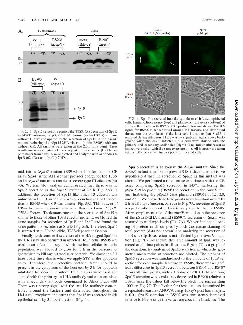
mid into a �spa47 mutant (BS908) and performed the CRassay. Spa47 is the ATPase that provides energy for the T3SS,and a �spa47 mutant is unable to secrete type III effectors (40,43). Western blot analysis demonstrated that there was noSpa15 secretion in the �spa47 mutant at 2.5 h (Fig. 5A). Inaddition, the secretion of Spa15 like other T3 effectors wasinducible with CR since there was a reduction in Spa15 secre-tion in BS905 when CR was absent (Fig. 5A). This pattern ofCR-inducible secretion is the same as those for known ShigellaT3SS effectors. To demonstrate that the secretion of Spa15 issimilar to those of other T3SS effector proteins, we blotted thesame samples for secretion of IpaB and IpaC and found thesame pattern of secretion as Spa15 (Fig. 5B). Therefore, Spa15is secreted in a CR-inducible, T3SS-dependent fashion.
Finally, to determine if secretion of the HA-tagged Spa15 inthe CR assay also occurred in infected HeLa cells, BS905 wasused in an infection assay in which the intracellular bacterialpopulation was allowed to grow for 3 h in the presence ofgentamicin to kill any extracellular bacteria. We chose the 3-htime point since this is when we apply STS in the apoptosisassay. Therefore, the protective bacterial factor should bepresent in the cytoplasm of the host cell by 3 h for apoptosisinhibition to occur. The infected monolayers were fixed andstained with the primary anti-HA antibody and counterstainedwith a secondary antibody conjugated to Alexa Fluor 488.There was a strong signal with the anti-HA antibody concen-trated around the bacteria and distributed throughout theHeLa cell cytoplasm, indicating that Spa15 was secreted insideepithelial cells by 3 h postinfection (Fig. 6).
Spa15 secretion is delayed in the �mxiE mutant. Since the�mxiE mutant is unable to prevent STS-induced apoptosis, wehypothesized that the secretion of Spa15 in this mutant wasaltered. We performed a time course experiment with the CRassay comparing Spa15 secretion in 2457T harboring thepSpa15-2HA plasmid (BS905) to secretion in the �mxiE mu-tant harboring the pSpa15-2HA plasmid (BS906) at 1.5, 2.0,and 2.5 h. We chose these time points since secretion occurs by2 h in wild-type bacteria. As seen in Fig. 7A, secretion of Spa15is significantly reduced in BS906 compared to that in BS905.After complementation of the �mxiE mutation in the presenceof the pSpa15-2HA plasmid (BS907), secretion of Spa15 wasrestored to wild-type levels (Fig. 7A). We verified equal load-ing of protein in all samples by both Coomassie staining oftotal protein (data not shown) and analyzing the secretion ofIpaB since IpaB secretion is not affected by the �mxiE muta-tion (Fig. 7B). As shown, the same amount of IpaB was se-creted at all time points in all strains. Figure 7C is a graph ofthe densitometry analysis of Spa15 secretion in which the geo-metric mean ratios of secretion are plotted. The amount ofSpa15 secretion was standardized to the amount of IpaB se-cretion for each sample. Relative to BS905, there was a signif-icant difference in Spa15 secretion between BS906 and BS907across all time points, with a P value of 0.001. In addition,Spa15 secretion was consistently decreased in BS906 relative toBS905 since the values fall below the black line representing100% in Fig. 7C. The P value for these data, as determined bya repeated-measures ANOVA using Tukey’s post hoc analysis,is 0.01. Spa15 secretion in BS907 was consistently increasedrelative to BS905 since the values are above the black line. The
FIG. 5. Spa15 secretion requires the T3SS. (A) Secretion of Spa15in 2457T harboring the pSpa15-2HA plasmid (strain BS905) with andwithout CR was compared to the secretion of Spa15 in the �spa47mutant harboring the pSpa15-2HA plasmid (strain BS908) with andwithout CR. All samples were taken at the 2.5-h time point. Theseresults are representative of three repeated experiments. (B) The su-pernatants from panel A were blotted and analyzed with antibodies toIpaB (62 kDa) and IpaC (42 kDa).
FIG. 6. Spa15 is secreted into the cytoplasm of infected epithelialcells. Immunofluorescence (top) and phase-contrast views (bottom) ofHeLa cells infected with BS905 at 3 h postinfection are shown. The HAsignal for BS905 is concentrated around the bacteria and distributedthroughout the cytoplasm of the host cell, indicating that Spa15 issecreted during infection. There was no significant signal above back-ground when the 2457T-infected HeLa cells were stained with theprimary and secondary antibodies (right). The immunofluorescenceimages were taken with the same exposure time. All images were takenwith a 100� objective. Arrows point to infected cells.
5286 FAHERTY AND MAURELLI INFECT. IMMUN.
on July 13, 2018 by guesthttp://iai.asm
.org/D
ownloaded from
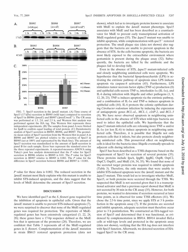
P value for these data is 0.002. The reduced secretion in the�mxiE mutant most likely explains why this mutant is unable toinhibit STS-induced apoptosis, and the data suggest that thelevels of MxiE determine the amount of Spa15 secretion.
DISCUSSION
We have identified spa15 as the S. flexneri gene required forthe inhibition of apoptosis in epithelial cells. Given that the�mxiE mutant is unable to prevent STS-induced apoptosis (7),we were surprised to discover that none of the MxiE-regulatedgenes was required for protection. The repertoire of MxiE-regulated genes has been extensively categorized (3, 22, 24,28); these genes have a 17-bp sequence defined as the MxiEbox that is upstream of the promoter (28). Therefore, we areconfident that Table 3 represents all of the MxiE-regulatedgenes in S. flexneri. Complementation of the �mxiE mutationin strain BS613 restored apoptosis protection (data not
shown), which led us to investigate proteins known to associatewith MxiE to explain the �mxiE mutant phenotype. Spa15associates with MxiE and has been described as a coantiacti-vator for MxiE to prevent early transcriptional activation ofMxiE-regulated genes (35). The �spa15 mutant was unable toinhibit apoptosis, while complementation with spa15� restoredprotection. The small plaque size (data not shown) also sug-gests that the bacteria are unable to prevent apoptosis in theabsence of STS. As the cells become apoptotic, the bacteria aremost likely exposed to the extracellular environment wheregentamicin is present during the plaque assay (32). Subse-quently, the bacteria are killed by the antibiotic and theplaques fail to develop fully.
Even in the absence of STS, �spa15 mutant-infected cellsand closely neighboring uninfected cells were apoptotic. Wehypothesize that the bacterial lipopolysaccharide (LPS) is ac-tivating the extrinsic pathway of apoptosis since LPS inducesapoptosis via caspase-8 activation (45). Furthermore, LPSstimulates tumor necrosis factor alpha (TNF-�) production (8)and epithelial cells secrete TNF-�, interleukin-1� (IL-1�), andIL-6 during infection with Shigella and other pathogens (1, 9,12, 20, 23). TNF-� induces apoptosis via the extrinsic pathway,and a combination of IL-1� and TNF-� induces apoptosis inepithelial cells (44). IL-6 protects the colonic epithelium dur-ing Citrobacter rodentium infection by inducing the expressionof antiapoptotic genes BCL-xL, MCL-1, cIAP-2, and BCL-3(9). We have never observed apoptosis in neighboring unin-fected cells in the absence of STS when wild-type bacteria areused to infect the epithelial cells. We hypothesize that the�spa15 mutant-infected cells are releasing more TNF-� and/orIL-1� (or less IL-6) to induce apoptosis in neighboring unin-fected cells. Therefore, it is possible that Shigella not onlyprotects cells that it infects, but also protects closely neighbor-ing uninfected cells. This prosurvival effect on neighboringcells is ideal for the bacteria since Shigella eventually spreads toadjacent cells during infection.
Spa15 has been described as a T3SS chaperone based on therequirement of Spa15 for secretion of several proteins (35).These proteins include IpaA, IpgB1, IpgB2, OspB, OspC2,OspC3, OspD1, and MxiE (16, 34, 35). We found that none ofthe secreted target proteins was required to inhibit apoptosis(Table 3). Therefore, the only mutants that were unable toinhibit STS-induced apoptosis were the �mxiE mutant and the�spa15 mutant. This result led us to investigate whether MxiE,Spa15, or both proteins were secreted through the T3SS. Wesuspected that MxiE is not secreted given that it is a transcrip-tional activator and that a previous report showed that MxiE isnot secreted by 30 min in the CR assay (35). However, for bothproteins, we wanted to determine if secretion occurred at latertime points, as is observed for some T3SS effectors (41). Wechose the 2.5-h time point, since we apply STS at 3 h postin-fection in the apoptosis assay (7). If the proteins are secretedand inhibit apoptosis, adequate secretion must therefore occurprior to 3 h postinfection. We constructed an HA-tagged ver-sion of Spa15 and determined that it was functional, as evi-denced by complementation in BS914. BS914 invaded HeLacells at wild-type levels and protected in the apoptosis assay(data not shown), verifying that the HA tag does not interferewith Spa15 function. Afterwards, we detected secretion of HA-tagged Spa15 in the CR assay.
FIG. 7. Spa15 secretion in the �mxiE mutant. (A) Time course ofSpa15 (17 kDa) secretion in BS905 (wild type) compared to secretionof Spa15 in BS906 (�mxiE) and BS907 (�mxiE/mxiE�). The CR assaywas performed at 1.5, 2.0, and 2.5 h, and Western blot analysis wasperformed against the HA tag. This Western blot represents threeindependent experiments. (B) The samples from panel A were blottedfor IpaB to confirm equal loading of total protein. (C) Densitometryanalysis of Spa15 secretion in BS905, BS906, and BS907. The geomet-ric mean ratios of Spa15 secretion from the Western blot in panel A inBS906 and BS907 are plotted relative to the secretion of Spa15 inBS905 (set at 100% and represented by the black line). The amount ofSpa15 secretion was standardized to the amount of IpaB secretion inpanel B for each sample. Error bars represent the standard error forthe three repeated experiments. A repeated-measures ANOVA usingTukey’s post hoc analysis demonstrated that the P value for Spa15secretion in BS906 relative to BS905 is 0.01. The P value for Spa15secretion in BS907 relative to BS905 is 0.002. The P value for thedifference in Spa15 secretion between BS906 and BS907 is 0.001.
VOL. 77, 2009 Spa15 INHIBITS APOPTOSIS IN SHIGELLA-INFECTED CELLS 5287
on July 13, 2018 by guesthttp://iai.asm
.org/D
ownloaded from
This report is the first to demonstrate that Spa15 is secreted bythe T3SS. We were able to detect Spa15 secretion because weassayed for the tagged protein, and we analyzed later time pointsin the CR secretion assay. A previous report only analyzed Coo-massie-stained protein gels for secreted products of S. flexneriM90T and a �spa15 mutant at the 30-min time point (35). It istherefore not surprising that secretion of Spa15 was not detectedprior to our work. One could argue that sample processing afterthe CR assay resulted in the appearance of secretion, perhaps dueto lysis of the bacteria. The absence of Spa15 secretion in the�spa47 mutant argues against this possibility since Spa15, likeIpaB and IpaC, remained in the pellet fraction and was notsecreted due to the absence of Spa47. In addition, the HA tagdoes not cause secretion of an otherwise nonsecreted proteinsince the HA-tagged MxiE was not secreted. We are thereforeconfident that Spa15 is secreted into epithelial cells based on theCR assay and the immunofluorescence analysis of Spa15 secre-tion in infected epithelial cells (Fig. 6).
Since Spa15 is the antiapoptosis factor and is secreted, wehypothesized that the �mxiE mutant did not prevent STS-induced apoptosis because Spa15 secretion was altered. Wedetected a significant decrease in Spa15 secretion in the �mxiEmutant (Fig. 7). Secretion was restored above wild-type levelswhen the �mxiE mutation was complemented (BS907), whichis verified by the fact that BS613 inhibits STS-induced apop-tosis (data not shown). The comparison of Spa15 secretionbetween strains BS905, BS906, and BS907 is valid, since theHA tag does not interfere with function. Therefore, notenough Spa15 is secreted from the �mxiE mutant in order forthis strain to inhibit apoptosis in the presence of STS. Inter-estingly, epithelial cells infected with the �mxiE mutant neverappeared apoptotic in the absence of STS (data not shown).The low levels of Spa15 secreted by the �mxiE mutant aremost likely sufficient to inhibit apoptosis normally during in-fection but insufficient to inhibit apoptosis in infected cells inthe presence of a strong apoptosis inducer like STS.
Based on our data, it appears that the levels of MxiE affectthe amount of Spa15 that is secreted. With MxiE absent instrain BS906, Spa15 is not needed to prevent early activation ofMxiE-regulated genes. Spa15 secretion above wild-type levelsin BS907 suggests that when more MxiE is present, moreSpa15 is needed to prevent early transcriptional activation ofMxiE-regulated genes. Once this larger amount of Spa15 isreleased from MxiE, Spa15 is available for secretion by theT3SS. It is also possible that MxiE contributes to the stabilityof Spa15. The absence of MxiE may cause Spa15 to remainattached to another protein, possibly OspD1, which wouldreduce the level of Spa15 secretion in the �mxiE mutant.Future studies analyzing how MxiE modulates the secretion ofSpa15 are clearly needed.
Most T3SS chaperones bind to one or two targets and areencoded next to the genes for these targets. For example, ipgCis immediately upstream of ipaB and ipaC on the Shigellavirulence plasmid, and IpgC binds to both IpaB and IpaC toprevent early association of these proteins (30). Spa15 is anatypical chaperone, since it is encoded in the middle of themxi-spa operon and the genes that encode the Spa15 targetsare scattered throughout the 220-kb virulence plasmid (6, 34).Homology searches with BLAST revealed several homologuesto Spa15 in other pathogens, including InvB from Salmonella
enterica subsp. enterica serovar Typhimurium, SpaK from S.enterica subsp. enterica serovar Paratyphi A, BsaR from severalBurkholderia species, and YsaK from Yersinia enterocolitica. Atleast two of the homologues, InvB and YsaK, are also atypicalT3SS chaperones in that they bind to many targets and are notencoded near the genes for those targets (26, 34). These ho-mologues are similar in structure, and there are six conservedresidues in the chaperones that define a binding motif (26).Given the sequence identities and similarities, it is possible thatsome of the homologues are also secreted into eukaryotic cells.The percent identity between Spa15 and the homologues is�30%. It remains to be seen if the homologues have additionalfunctions. These T3SS chaperones may truly represent a new,unique class of chaperones.
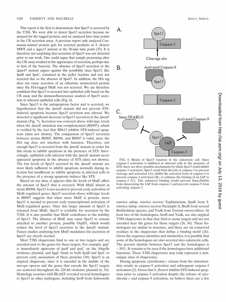
During apoptosis, cytochrome c release from the mitochon-dria results in caspase-9 activation and subsequent caspase-3activation (2). Given that S. flexneri inhibits STS-induced apop-tosis prior to caspase-3 activation despite the release of cyto-chrome c and caspase-9 activation, we believe there are a few
FIG. 8. Model of Spa15 function in the eukaryotic cell. Sincecaspase-3 activation is inhibited in infected cells in the presence ofSTS, there are three possible mechanisms by which Spa15 could inhibitcaspase-3 activation. Spa15 could bind directly to caspase-3 to preventcleavage and activation (A), inhibit the activated form of caspase-9 toprevent caspase-3 activation (B), or enhance the binding of an IAP tocaspase-3 (C). This enhanced binding would prevent Smac/Diablofrom dissociating the IAP from caspase-3 and prevent caspase-9 fromactivating caspase-3.
5288 FAHERTY AND MAURELLI INFECT. IMMUN.
on July 13, 2018 by guesthttp://iai.asm
.org/D
ownloaded from
likely eukaryotic targets for Spa15 to directly inhibit caspase-3(Fig. 8). First, Spa15 could bind directly to caspase-3 and blockthe cleavage site. Alternatively, Spa15 could associate withcaspase-9 to prevent it from activating caspase-3. This associ-ation would not prevent caspase-9 from being activated by theapoptosome (2) but would prevent the activated form ofcaspase-9 from cleaving caspase-3. Finally, Spa15 could en-hance the binding of an inhibitor of apoptosis protein (IAP) tocaspase-3. This enhanced binding would prevent the dissocia-tion of the IAP from caspase-3 by proteins like Smac/Diablothat are released from the mitochondria (11). The binding ofSpa15 to an IAP would also prevent activated caspase-9 fromcleaving caspase-3 since the caspase-3 cleavage site would beblocked by the IAP. We hypothesize that the simple binding ofSpa15 to a eukaryotic target would be sufficient for inhibitingcaspase-3 activation. We are in the process of identifying po-tential eukaryotic targets through protein interaction screens.
In conclusion, we have identified Spa15 as the antiapoptosisfactor that inhibits apoptosis in epithelial cells during S. flexneriinfection. Of equal importance, we were able to demonstratefor the first time that Spa15 is secreted through the T3SS andhas effector function in addition to being a T3SS chaperone.Prevention of epithelial cell apoptosis is important for thebacteria to survive inside the host. During infection, manyapoptosis stimuli are present. These stimuli include the cyto-kines TNF-� (21) and Fas ligand (37), leukocyte elastases (15),and the transmigration of PMNs across the colonic epithelium(25) that occurs during Shigella infection (29). If S. flexneri isunable to prevent epithelial cell apoptosis and the infectedcells die, the bacteria will be exposed to the extracellular en-vironment that is infiltrated with immune cells. In fact, in vivostudies have verified that only immune cells are apoptoticduring Shigella infection (37, 38, 47). Therefore, the inhibitionof epithelial cell apoptosis is vital for S. flexneri to establish areplicative niche inside the host in order to survive.
ACKNOWLEDGMENTS
This work was supported by grant AI24656 from the National Insti-tute of Allergy and Infectious Diseases. We thank the Henry M. Jack-son Foundation and the Uniformed Services University for the ValHemming Fellowship awarded to C.S.F., as well as the Graduate Ed-ucation Office at USU for stipend support.
We also thank members of the Maurelli lab for technical assistanceand Cara Olsen for help with statistical analysis.
The opinions or assertions contained herein are the private ones ofthe authors and are not to be construed as official or reflecting theviews of the Department of Defense or the Uniformed Services Uni-versity of the Health Sciences.
REFERENCES
1. Arondel, J., M. Singer, A. Matsukawa, A. Zychlinsky, and P. J. Sansonetti.1999. Increased interleukin-1 (IL-1) and imbalance between IL-1 and IL-1receptor antagonist during acute inflammation in experimental shigellosis.Infect. Immun. 67:6056–6066.
2. Ashe, P. C., and M. D. Berry. 2003. Apoptotic signaling cascades. Prog.Neuropsychopharmacol. Biol. Psychiatry 27:199–214.
3. Ashida, H., T. Toyotome, T. Nagai, and C. Sasakawa. 2007. Shigella chro-mosomal IpaH proteins are secreted via the type III secretion system and actas effectors. Mol. Microbiol. 63:680–693.
4. Bahrani, F. K., P. J. Sansonetti, and C. Parsot. 1997. Secretion of Ipaproteins by Shigella flexneri: inducer molecules and kinetics of activation.Infect. Immun. 65:4005–4010.
5. Bernardini, M. L., J. Mounier, H. d’Hauteville, M. Coquis-Rondon, and P. J.Sansonetti. 1989. Identification of icsA, a plasmid locus of Shigella flexnerithat governs bacterial intra- and intercellular spread through interaction withF-actin. Proc. Natl. Acad. Sci. USA 86:3867–3871.
6. Buchrieser, C., P. Glaser, C. Rusniok, H. Nedjari, H. d’Hauteville, F. Kunst,P. Sansonetti, and C. Parsot. 2000. The virulence plasmid pWR100 and therepertoire of proteins secreted by the type III secretion apparatus of Shigellaflexneri. Mol. Microbiol. 38:760–771.
7. Clark, C. S., and A. T. Maurelli. 2007. Shigella flexneri inhibits staurosporine-induced apoptosis in epithelial cells. Infect. Immun. 75:2531–2539.
8. Comstock, K. L., K. A. Krown, M. T. Page, D. Martin, P. Ho, M. Pedraza,E. N. Castro, N. Nakajima, C. C. Glembotski, P. J. Quintana, and R. A.Sabbadini. 1998. LPS-induced TNF-alpha release from and apoptosis in ratcardiomyocytes: obligatory role for CD14 in mediating the LPS response. J.Mol. Cell. Cardiol. 30:2761–2775.
9. Dann, S. M., M. E. Spehlmann, D. C. Hammond, M. Iimura, K. Hase, L. J.Choi, E. Hanson, and L. Eckmann. 2008. IL-6-dependent mucosal protec-tion prevents establishment of a microbial niche for attaching/effacing lesion-forming enteric bacterial pathogens. J. Immunol. 180:6816–6826.
10. Datsenko, K. A., and B. L. Wanner. 2000. One-step inactivation of chromo-somal genes in Escherichia coli K-12 using PCR products. Proc. Natl. Acad.Sci. USA 97:6640–6645.
11. Ekert, P. G., and D. L. Vaux. 2005. The mitochondrial death squad: hard-ened killers or innocent bystanders? Curr. Opin. Cell Biol. 17:626–630.
12. Fahey, J. V., T. M. Schaefer, J. Y. Channon, and C. R. Wira. 2005. Secretionof cytokines and chemokines by polarized human epithelial cells from thefemale reproductive tract. Hum. Reprod. 20:1439–1446.
13. Formal, S. B., G. J. Dammin, E. H. LaBrec, and H. Schneider. 1958. Exper-imental Shigella infections: characteristics of a fatal infection produced inguinea pigs. J. Bacteriol. 75:604–610.
14. Ghosh, P. 2004. Process of protein transport by the type III secretion system.Microbiol. Mol. Biol. Rev. 68:771–795.
15. Ginzberg, H. H., P. T. Shannon, T. Suzuki, O. Hong, E. Vachon, T. Moraes,M. T. Abreu, V. Cherepanov, X. Wang, C. W. Chow, and G. P. Downey. 2004.Leukocyte elastase induces epithelial apoptosis: role of mitochondrial per-meability changes and Akt. Am. J. Physiol. Gastrointest. Liver Physiol. 287:G286–G298.
16. Hachani, A., L. Biskri, G. Rossi, A. Marty, R. Menard, P. Sansonetti, C.Parsot, G. T. Van Nhieu, M. L. Bernardini, and A. Allaoui. 2008. IpgB1 andIpgB2, two homologous effectors secreted via the Mxi-Spa type III secretionapparatus, cooperate to mediate polarized cell invasion and inflammatorypotential of Shigella flexenri. Microbes Infect. 10:260–268.
17. Hanahan, D. 1983. Studies on transformation of Escherichia coli with plas-mids. J. Mol. Biol. 166:557–580.
18. Hromockyj, A. E., and A. T. Maurelli. 1989. Identification of Shigella inva-sion genes by isolation of temperature-regulated inv::lacZ operon fusions.Infect. Immun. 57:2963–2970.
19. Jennison, A. V., and N. K. Verma. 2004. Shigella flexneri infection: patho-genesis and vaccine development. FEMS Microbiol. Rev. 28:43–58.
20. Johnson, R. M. 2004. Murine oviduct epithelial cell cytokine responses toChlamydia muridarum infection include interleukin-12–p70 secretion. Infect.Immun. 72:3951–3960.
21. Jung, H. C., L. Eckmann, S. K. Yang, A. Panja, J. Fierer, E. Morzycka-Wroblewska, and M. F. Kagnoff. 1995. A distinct array of proinflammatorycytokines is expressed in human colon epithelial cells in response to bacterialinvasion. J. Clin. Investig. 95:55–65.
22. Kane, C. D., R. Schuch, W. A. Day, Jr., and A. T. Maurelli. 2002. MxiEregulates intracellular expression of factors secreted by the Shigella flexneri2a type III secretion system. J. Bacteriol. 184:4409–4419.
23. Le-Barillec, K., J. G. Magalhaes, E. Corcuff, A. Thuizat, P. J. Sansonetti, A.Phalipon, and J. P. Di Santo. 2005. Roles for T and NK cells in the innateimmune response to Shigella flexneri. J. Immunol. 175:1735–1740.
24. Le Gall, T., M. Mavris, M. C. Martino, M. L. Bernardini, E. Denamur, andC. Parsot. 2005. Analysis of virulence plasmid gene expression defines threeclasses of effectors in the type III secretion system of Shigella flexneri. Mi-crobiology 151:951–962.
25. Le’Negrate, G., E. Selva, P. Auberger, B. Rossi, and P. Hofman. 2000.Sustained polymorphonuclear leukocyte transmigration induces apoptosis inT84 intestinal epithelial cells. J. Cell Biol. 150:1479–1488.
26. Lilic, M., M. Vujanac, and C. E. Stebbins. 2006. A common structural motifin the binding of virulence factors to bacterial secretion chaperones. Mol.Cell 21:653–664.
27. Maurelli, A. T., B. Baudry, H. d’Hauteville, T. L. Hale, and P. J. Sansonetti.1985. Cloning of plasmid DNA sequences involved in invasion of HeLa cellsby Shigella flexneri. Infect. Immun. 49:164–171.
28. Mavris, M., P. J. Sansonetti, and C. Parsot. 2002. Identification of thecis-acting site involved in activation of promoters regulated by activity of thetype III secretion apparatus in Shigella flexneri. J. Bacteriol. 184:6751–6759.
29. McCormick, B. A., A. M. Siber, and A. T. Maurelli. 1998. Requirement of theShigella flexneri virulence plasmid in the ability to induce trafficking of neu-trophils across polarized monolayers of the intestinal epithelium. Infect.Immun. 66:4237–4243.
30. Menard, R., P. Sansonetti, C. Parsot, and T. Vasselon. 1994. Extracellularassociation and cytoplasmic partitioning of the IpaB and IpaC invasins of S.flexneri. Cell 79:515–525.
31. Mills, J. A., J. M. Buysse, and E. V. Oaks. 1988. Shigella flexneri invasion
VOL. 77, 2009 Spa15 INHIBITS APOPTOSIS IN SHIGELLA-INFECTED CELLS 5289
on July 13, 2018 by guesthttp://iai.asm
.org/D
ownloaded from
plasmid antigens B and C: epitope location and characterization with mono-clonal antibodies. Infect. Immun. 56:2933–2941.
32. Oaks, E. V., M. E. Wingfield, and S. B. Formal. 1985. Plaque formation byvirulent Shigella flexneri. Infect. Immun. 48:124–129.
33. Ohya, K., Y. Handa, M. Ogawa, M. Suzuki, and C. Sasakawa. 2005. IpgB1is a novel Shigella effector protein involved in bacterial invasion of host cells.Its activity to promote membrane ruffling via Rac1 and Cdc42 activation.J. Biol. Chem. 280:24022–24034.
34. Page, A. L., P. Sansonetti, and C. Parsot. 2002. Spa15 of Shigella flexneri, athird type of chaperone in the type III secretion pathway. Mol. Microbiol.43:1533–1542.
35. Parsot, C., E. Ageron, C. Penno, M. Mavris, K. Jamoussi, H. d’Hauteville, P.Sansonetti, and B. Demers. 2005. A secreted anti-activator, OspD1, and itschaperone, Spa15, are involved in the control of transcription by the type IIIsecretion apparatus activity in Shigella flexneri. Mol. Microbiol. 56:1627–1635.
36. Pilonieta, M. C., and G. P. Munson. 2008. The chaperone IpgC copurifieswith the virulence regulator MxiE. J. Bacteriol. 190:2249–2251.
37. Raqib, R., C. Ekberg, P. Sharkar, P. K. Bardhan, A. Zychlinsky, P. J.Sansonetti, and J. Andersson. 2002. Apoptosis in acute shigellosis is associ-ated with increased production of Fas/Fas ligand, perforin, caspase-1, andcaspase-3 but reduced production of Bcl-2 and interleukin-2. Infect. Immun.70:3199–3207.
38. Sansonetti, P. J., J. Arondel, J. R. Cantey, M.-C. Prevost, and M. Huerre.1996. Infection of rabbit Peyer’s patches by Shigella flexneri: effect of adhe-sive or invasive bacterial phenotypes on follicle-associated epithelium. Infect.Immun. 64:2752–2764.
39. Schroeder, G. N., and H. Hilbi. 2008. Molecular pathogenesis of Shigellaspp.: controlling host cell signaling, invasion, and death by type III secretion.Clin. Microbiol. Rev. 21:134–156.
40. Tamano, K., S. Aizawa, E. Katayama, T. Nonaka, S. Imajoh-Ohmi, A. Ku-wae, S. Nagai, and C. Sasakawa. 2000. Supramolecular structure of theShigella type III secretion machinery: the needle part is changeable in lengthand essential for delivery of effectors. EMBO J. 19:3876–3887.
41. Toyotome, T., T. Suzuki, A. Kuwae, T. Nonaka, H. Fukuda, S. Imajoh-Ohmi,T. Toyofuku, M. Hori, and C. Sasakawa. 2001. Shigella protein IpaH9.8 issecreted from bacteria within mammalian cells and transported to the nu-cleus. J. Biol. Chem. 276:32071–32079.
42. Tran Van Nhieu, G., A. Ben-Ze’ev, and P. J. Sansonetti. 1997. Modulation ofbacterial entry into epithelial cells by association between vinculin and theShigella IpaA invasin. EMBO J. 16:2717–2729.
43. Venkatesan, M. M., J. M. Buysse, and E. V. Oaks. 1992. Surface presentationof Shigella flexneri invasion plasmid antigens requires the products of the spalocus. J. Bacteriol. 174:1990–2001.
44. Wright, K., G. Kolios, J. Westwick, and S. G. Ward. 1999. Cytokine-inducedapoptosis in epithelial HT-29 cells is independent of nitric oxide formation.Evidence for an interleukin-13-driven phosphatidylinositol 3-kinase-depen-dent survival mechanism. J. Biol. Chem. 274:17193–17201.
45. Yu, L. C., A. N. Flynn, J. R. Turner, and A. G. Buret. 2005. SGLT-1-mediatedglucose uptake protects intestinal epithelial cells against LPS-induced apop-tosis and barrier defects: a novel cellular rescue mechanism? FASEB J.19:1822–1835.
46. Zurawski, D. V., C. Mitsuhata, K. L. Mumy, B. A. McCormick, and A. T.Maurelli. 2006. OspF and OspC1 are Shigella flexneri type III secretionsystem effectors that are required for postinvasion aspects of virulence.Infect. Immun. 74:5964–5976.
47. Zychlinsky, A., K. Thirumalai, J. Arondel, J. R. Cantey, A. O. Aliprantis, andP. J. Sansonetti. 1996. In vivo apoptosis in Shigella flexneri infections. Infect.Immun. 64:5357–5365.
Editor: S. M. Payne
5290 FAHERTY AND MAURELLI INFECT. IMMUN.
on July 13, 2018 by guesthttp://iai.asm
.org/D
ownloaded from