sparc independent drug delivery and antitumour - gut - bmj
TRANSCRIPT

ORIGINAL ARTICLE
SPARC independent drug delivery and antitumoureffects of nab-paclitaxel in genetically engineeredmiceAlbrecht Neesse,1,2 Kristopher K Frese,1 Derek S Chan,1 Tashinga E Bapiro,1
William J Howat,1 Frances M Richards,1 Volker Ellenrieder,2 Duncan I Jodrell,1
David A Tuveson1,3
▸ Additional material ispublished online only. To viewplease visit the journal online(http://dx.doi.org/10.1136/gutjnl-2013-305559).1Cancer Research UKCambridge Institute, TheUniversity of Cambridge,Cambridge, UK2Department ofGastroenterology,Endocrinology, Infectiology andMetabolism, Philipps UniversityMarburg, Marburg, Germany3Cold Spring HarborLaboratory, Cold SpringHarbor, New York, USA
Correspondence toDr David A Tuveson, ColdSpring Harbor Laboratory,1 Bungtown Road,Cold Spring Harbor,NY 11724, USA;[email protected]
Received 1 July 2013Revised 24 July 2013Accepted 28 July 2013
To cite: Neesse A,Frese KK, Chan DS, et al.Gut Published Online First:[please include Day MonthYear] doi:10.1136/gutjnl-2013-305559
ABSTRACTDesign Pharmacokinetic and pharmacodynamicparameters of cremophor-paclitaxel, nab-paclitaxel(human-albumin-bound paclitaxel, Abraxane) and anovel mouse-albumin-bound paclitaxel (m-nab-paclitaxel)were evaluated in genetically engineered mouse models(GEMMs) by liquid chromatography-tandem massspectrometry (LC-MS/MS), histological and biochemicalanalysis. Preclinical evaluation of m-nab-paclitaxelincluded assessment by three-dimensional high-resolution ultrasound and molecular analysis in a novelsecreted protein acidic and rich in cysteine (SPARC)-deficient GEMM of pancreatic ductal adenocarcinoma(PDA).Results nab-Paclitaxel exerted its antitumoural effectsin a dose-dependent manner and was associated withless toxicity compared with cremophor-paclitaxel. SPARCnullizygosity in a GEMM of PDA, KrasG12D;p53flox/−;p48Cre (KPfC), resulted in desmoplastic ductal pancreastumours with impaired collagen maturation. Paclitaxelconcentrations were significantly decreased in SPARCnull plasma samples and tissues when administered aslow-dose m-nab-paclitaxel. At the maximally tolerateddose, SPARC deficiency did not affect the intratumouralpaclitaxel concentration, stromal deposition and theimmediate therapeutic response.Conclusions nab-Paclitaxel accumulates and acts in adose-dependent manner. The interaction of plasmaSPARC and albumin-bound drugs is observed at lowdoses of nab-paclitaxel but is saturated at therapeuticdoses in murine tumours. Thus, this study providesimportant information for future preclinical and clinicaltrials in PDA using nab-paclitaxel in combination withnovel experimental and targeted agents.
INTRODUCTIONPancreatic ductal adenocarcinoma (PDA) is consid-ered one of the most aggressive and lethal humanmalignancies with increasing incidence and a 5-yearsurvival rate of less than 5%.1 Over the pastdecades, intensive efforts to develop novel targetedtherapies in combination with various chemothera-pies have not yielded a significant breakthrough,and the standard of care chemotherapy hasremained gemcitabine despite only marginal effectson patient survival.2 Oxaliplatin, irinotecan, leu-covorin and 5-fluorouracil (FOLFIRINOX) hasrecently been reported to achieve a significant
Significance of this study
What is already known about this subject▸ Pancreatic ductal adenocarcinoma (PDA) is a
stroma-rich tumour that is highly refractory tosystemic therapies.
▸ nab-paclitaxel (Abraxane) alone and incombination with gemcitabine has recentlyshown significant clinical activity in human andmurine PDA.
▸ Secreted protein acidic and rich in cysteine(SPARC) has been proposed as a biomarker fornab-paclitaxel-based chemotherapies bysequestering nab-paclitaxel to concentrate thedrug intratumourally and to deplete tumourstroma.
What are the new findings▸ nab-paclitaxel acts in a dose-dependent but
SPARC saturable manner in geneticallyengineered mouse models (GEMM).
▸ SPARC ablation in KPfC mice results indesmoplastic ductal pancreas tumours withimpaired collagen maturation.
▸ A preclinical trial using a novel formulation ofmouse-nab-paclitaxel demonstrates robusttherapeutic effects independent of SPARCexpression.
▸ Prolonged treatment with mouse-nab-paclitaxeldoes not lead to stromal depletion in the KPCmouse model.
How might it impact on clinical practice inthe foreseeable future▸ nab-paclitaxel could be investigated with
additional stromal depletion agents tomaximise treatment effects.
▸ Preclinical platforms usingmouse-nab-paclitaxel can be employed toidentify more powerful combination therapiesfor early clinical testing.
▸ SPARC serum levels should be measured inPDA patients prior to startingnab-paclitaxel-based chemotherapies todetermine whether high plasma SPARC levelsaffect plasma pharmacokinetics of albuminformulated drugs.
Neesse A, et al. Gut 2013;0:1–10. doi:10.1136/gutjnl-2013-305559 1
Pancreatic cancer Gut Online First, published on September 25, 2013 as 10.1136/gutjnl-2013-305559
Copyright Article author (or their employer) 2013. Produced by BMJ Publishing Group Ltd (& BSG) under licence.
on 15 Novem
ber 2018 by guest. Protected by copyright.
http://gut.bmj.com
/G
ut: first published as 10.1136/gutjnl-2013-305559 on 25 Septem
ber 2013. Dow
nloaded from

survival benefit for patients with metastatic PDA compared withgemcitabine (11.1 vs 6.8 months); however, increased rates oftoxicity will likely limit the frequent clinical use of this regimenin PDA patients.3 Histologically, PDA is characterised by abun-dant tumour stroma with activated cancer-associated fibroblasts(CAFs) and immune cells that contribute to a dense and highlydynamic tumour microenvironment (TME) around neoplasticductal cells.4 The distinct stromal architecture has increasinglybeen appreciated to create physical barriers for drug deliveryand also to provide additional biochemical signals that collect-ively promote the resistance of PDA cells to systemic and tar-geted therapies.5–13 Secreted protein acidic and rich in cysteine(SPARC/osteonectin/BM40) is an albumin-binding 42-kDamatricellular glycoprotein that has recently gained significantclinical interest as a potential biomarker in PDA. Indeed,SPARC is overexpressed by fibroblasts in the TME of humanPDA and has been shown to inversely correlate with survival.1415 A novel drug formulation of paclitaxel bound to albumin(nab-paclitaxel, Abraxane) has been hypothesised to accumulatein and potentially deplete PDA tumour stroma via binding ofalbumin to SPARC.16 A phase I/II trial of gemcitabine in com-bination with nab-paclitaxel showed promising overall survivalrates and suggested the potential usefulness of SPARC as a pre-dictive biomarker.17 More recently, results from the first rando-mised phase III study of nab-paclitaxel and gemcitabineMetastatic Pancreatic Adenocarcinoma Clinical Trial, MPACT)have confirmed initial observations of clinical benefit andshowed a significant median survival improvement over gemcita-bine monotherapy (8.5 vs 6.7 months) in patients with meta-static PDA.18 In parallel, our laboratory has confirmed theantitumour efficacy of nab-paclitaxel and gemcitabine and dis-covered a synergistic drug–drug interaction where nab-paclitaxelimpaired gemcitabine metabolism due to reactive oxygen species(ROS)-mediated degradation of cytidine deaminase.19 Followingthis encouraging data, nab-paclitaxel plus gemcitabine is cur-rently being evaluated for registration as novel first-line treat-ment for PDA patients. Furthermore, a recent phase II studyfound preliminary activity of nab-paclitaxel monotherapy inpatients who progressed on gemcitabine-based therapy.20
The use of biomarkers to select effective targeted and cyto-toxic therapies and predict response to treatment for individuals(‘personalised medicine’) is becoming a reality in certain typesof cancer but has remained elusive in PDA. Recent clinical datasuggested that expression of SPARC in the stroma of archivedtumour tissue could predict for response to nab-paclitaxel andgemcitabine,17 and a detailed analysis of tumour specimen fromthe MPACT study is still pending. Preclinical and clinical dataon the potential of stromal SPARC to determine treatmentresponse to nab-paclitaxel, sequester nab-paclitaxel to concen-trate the drug intratumourally, and to deplete tumour stromahave been conflicting in different tumour entities and hamperedby methodological differences.20–23 A preclinical study inpatient-derived xenografts reported stromal depletion upon nab-paclitaxel treatment,17 whereas our own results in a geneticallyengineered mouse model (GEMM) of PDA did not indicatesuch effects following short-term treatment with nab-paclitaxel.19 A limitation of our prior study was anaphylaxis dueto the use of human-albumin-bound paclitaxel, and thus, didnot allow us to investigate longer term treatment effects such asstromal depletion.19 Furthermore, the interaction of murineSPARC and human albumin does not recapitulate the genuinebiological situation and may possibly bias the results.
Here, we determine the pharmacokinetic and pharmacody-namic profile of nab-paclitaxel in murine PDA in vivo and
assess the preclinical effectiveness in GEMMs of PDA in aSPARC-dependent manner by using a novel experimentalmouse-albumin paclitaxel (m-nab-paclitaxel) that was specific-ally formulated for this purpose. Thus, our study providesimportant information for future preclinical and clinical trials inPDA using nab-paclitaxel in combination with novel experimen-tal and targeted agents.
MATERIALS AND METHODSAdditional details are provided in online supplementary materi-als and methods.
Genetically engineered miceThe following genetically engineered mice were used for this study:SPARC−/− mice (B6;129S-Sparctm1Hwe/J) were purchased fromCharles River (Margate, UK).24 LSL-KrasG12D/+;LSL-Trp53R172H/+;Pdx-1-Cre (KPC) and KrasG12D/+;Trp53flox/−;p48-Cre (KPfC) micewere used for the experiments, and both models develop advancedand metastatic PDAwith 100% penetrance at an early age recapitu-lating the full spectrum of histopathological and clinical features ofhuman PDA.25 Mice were housed at a 12-h light, 12-h dark cycle.All procedures were conducted in accordance with the institutionaland national guidelines.
Drugsnab-paclitaxel (Abraxane) and mouse (m)-nab-paclitaxel were for-mulated and provided by Celgene Corporation (USA) and resus-pended in sterile normal saline at 24 mg/mL, 12 mg/mL or 6 mg/mL, and intravenously administered at 120 mg/kg, 60 mg/kg or30 mg/kg. Paclitaxel was provided by Addenbrooke’s HospitalPharmacy (Cambridge, UK), resuspended in Cremophor at 6 mg/mL or 2 mg/mL and administered at 10 mg/kg or 30 mg/kg. Theamount of paclitaxel is equal in 30 mg/kg nab-paclitaxel and 30 mg/kg cremophor-paclitaxel; 60 mg/kg nab-paclitaxel in mice corre-sponds to approximately 180 mg/m2 nab-paclitaxel in humans.26
LC-MS/MS of paclitaxelFresh frozen tumour samples from different time points were pro-cessed and analysed for paclitaxel concentrations using liquidchromatography tandem mass spectrometry (LC-MS/MS). Briefly,samples were extracted with 100% acetonitrile containing2H5-paclitaxel as internal standard, and chromatography was doneon an acquity ultra performance liquid chromatography (UPLC),high strength silica (HSS) T3 50 mm×2.1 mm id., 1.8 mm column(Waters, Hertfordshire, UK). The mobile phase was (A) 0.1%acetic acid: (acetonitrile:methanol (1:1)) 70:30 and (B) 0.1%acetic acid: (acetonitrile:methanol (1:1)) 10:90. The gradient, at aflow rate of 0.3 mL/min, was 100% A for 0.6 min changed to over0.2 min to 100% B and maintained for 2 min, changed back to100% A over 0.2 min and held for 1.5 min to give a total run timeof 4.5 min. LC-MS/MS was performed on a triple stage quadru-pole (TSQ) Vantage mass spectrometer (Thermo Scientific,Waltham, MA) fitted with a heated electrospray ionisation(HESI-II) probe operated in positive mode at a spray voltage of5 KV and capillary temperature of 350°C. Quantitation was doneby multiple reaction monitoring of the transitions 876.4–308.1and 881.4–308.1 for paclitaxel and the internal standard, respect-ively, and data acquisition was done using LC Quan2.5.6 (ThermoFisher Scientific, Waltham, MA).
Therapeutic intervention studies with m-nab-paclitaxelFollowing weekly manual palpation from 2 months of age, KPCand KPfC mice were subjected to high-contrast ultrasound screen-ing using the Vevo 2100 System with a MS250, 13–24MHz
2 Neesse A, et al. Gut 2013;0:1–10. doi:10.1136/gutjnl-2013-305559
Pancreatic cancer
on 15 Novem
ber 2018 by guest. Protected by copyright.
http://gut.bmj.com
/G
ut: first published as 10.1136/gutjnl-2013-305559 on 25 Septem
ber 2013. Dow
nloaded from

scanhead (Visual Sonics, Inc, Amsterdam, NL). Mice with tumourdiameters of 5–9 mm were randomised and enrolled into thetherapeutic intervention studies: m-nab-paclitaxel was intraven-ously administered at 60 mg/kg in tumour-bearing mice accordingto the outlined treatment schedules. During the study, tumourgrowth was quantified on day 3 and day 7 by measuring tumourvolumes using reconstructed three-dimensional (3D) ultrasonog-raphy with the integrated Vevo 2100 software package. The lastadministration of m-nab-paclitaxel was given 2 h prior toendpoint.
Western blot analysisWestern blots were performed as previously described.27 Thefollowing primary antibodies were used: Hsp90 (Cell Signaling)and murine SPARC (R&D Systems). Membranes were incubatedwith secondary HRP-antibodies ( Jackson ImmunoResearch) anddeveloped using the enhanced chemiluminescence (ECL) detec-tion system (GE Healthcare).
Histological examinationTissues were fixed in 10% neutral buffered formalin for 24 h andtransferred to 70% ethanol. Tissues were embedded in paraffin,and 3–5 mm sections were processed for H&E staining, Herovici(Sigma) and immunohistochemistry using standard protocols aspreviously described.10 The following antibodies were used:murine SPARC (R&D Systems, AF942), Cleaved Caspase-3 (CellSignaling Technology, 9661), CD31 (BD Pharmingen, 553370),Ki-67 (Dako, M7249) and phospho-histone-3 (Upstate, 06-570).Images were acquired on an Olympus BX51 microscope(Olympus) or Aperio XT automated scanning system andImagescope 10 software (Leica). More information can be foundin online supplementary material and methods.
Statistical analysisStatistical analysis was carried out using GraphPad Prism V.5.01(GraphPad Software). The Mann–Whitney non-parametric t testwas used and results are presented as mean±SE. p<0.05 wasconsidered to be significant.
RESULTSPharmacokinetics and pharmacodynamics ofcremophor-paclitaxel and nab-paclitaxel in KPC tumoursSPARC is overexpressed in the pancreatic tumour microenviron-ment in KPC mice,19 predominantly by cancer-associated fibro-blasts, and to a lesser extent in tumour cell lines (see onlinesupplementary figure 1A). Since this expression pattern issimilar to that described with PDA patients,14 15 this mousemodel represents a tractable experimental platform to interro-gate the interaction of SPARC and nab-paclitaxel.
To systematically compare pharmacokinetics and pharmaco-dynamics of nab-paclitaxel and cremophor-paclitaxel, tumour-bearing KPC mice were intravenously administered a single doseof equimolar (30 mg/kg) cremophor-paclitaxel (n=12), nab-paclitaxel (n=5) and equitoxic 120 mg/kg nab-paclitaxel (n=10).Notably, 5/12 mice (43%) experienced acute toxicity-relatedhealth issues with physical inactivity, loss of body temperature andrespiratory distress following the administration of 30 mg/kgcremophor-paclitaxel, and those mice had to be culled prior to thedefined endpoint. In contrast, 120 mg/kg nab-paclitaxel was welltolerated in all 10 mice treated. Tissues were collected 4 h post-dose and assessed for intratumoural paclitaxel concentrations byLC-MS/MS and pharmacodynamic parameters by immunohisto-chemistry. Strikingly, equimolar doses of cremophor-paclitaxel andnab-paclitaxel (30 mg/kg) resulted in similar intratumoural
paclitaxel concentrations (mean: 6.9 ng/mg, SD:±4.5 vs 4.3 ng/mg±1.75, p=0.28, figure 1A), suggesting that intratumoural pacli-taxel delivery was independent of the albumin nanoformulation.The maximum tolerated dose (MTD) of 120 mg/kg nab-paclitaxelresulted in approximately fourfold higher paclitaxel levels(30.8 ng/mg±10.4, figure 1A), indicating a nearly linear intratu-moural accumulation of nab-paclitaxel with significantly less acutesystemic toxicity compared with 30 mg/kg cremophor–paclitaxel.
Paclitaxel has been shown to elicit its antitumoural effectsthrough induction of apoptosis and cell cycle arrest in G2-M. Wetherefore assessed the effects of the different dosing regimens inKPC tumours by immunohistochemistry for proliferation markerki-67, apoptosis marker cleaved caspase 3 (CC3) and phosphory-lated histone H3 (pH3) as a marker for aberrant mitotic figures. Atthe MTD of 120 mg/kg, nab-paclitaxel induced significant levels ofapoptosis compared with untreated (p<0.001), cremophor-paclitaxel (p<0.001) and low-dose nab-paclitaxel (p<0.01) treatedtumours (figure 1B). Notably, cremophor-paclitaxel and low-dosenab-paclitaxel had no significant effects on tumour cell death com-pared with untreated controls. The appearance of aberrant mitoticfigures that contained an abundance of phosphorylated histone H3was increased following treatment with cremophor-paclitaxel,low-dose nab-paclitaxel and most significantly, in the 120 mg/kgnab-paclitaxel cohort (figure 1C, p<0.001). Again, equimolardoses of nab-paclitaxel and cremophor-paclitaxel did not show sig-nificant differences in the percentage of pH3-positive cells(p=0.15, figure 1C). nab-Paclitaxel and cremophor-paclitaxeldecreased overall proliferation rate of KPC tumours marginally, andalthough we found a significant difference in tumour proliferationbetween low-dose and high-dose nab-paclitaxel (p<0.02), compari-son against untreated KPC tumours failed to reach significance(figure 1D; p=0.09).
Overall, these results suggest that nab-paclitaxel andcremophor-paclitaxel have comparable pharmacokinetic andpharmacodynamic properties in murine pancreas tumours withendogenous SPARC expression. However, nab-paclitaxel can bedosed at least four times higher than cremophor-paclitaxel dueto improved tolerability and therefore exerts significantly betterantitumoural effects in vivo.
m-nab-Paclitaxel extended intervention study in the KPCmouse modelnab-Paclitaxel is formulated with human albumin and thus causesfatal anaphylactic reactions in mice following repeated intravenousinjections.19 Therefore, mechanisms such as stromal depletion thatrequire longer term follow-up and repeated injections could notbe properly investigated in the past. In order to circumvent theseissues, we preclinically assessed a novel, mouse albumin-boundpaclitaxel that was specifically formulated for experimental pur-poses and termed murine (m)-nab-paclitaxel. First, we comparedthe bioavailability of 30 mg/kg nab-paclitaxel (n=5) and 30 mg/kgm-nab-paclitaxel (n=5) in healthy C57B6 wild-type mice anddemonstrated that plasma and tissue concentrations of paclitaxelwere comparable between the two compounds following i.v.administration (see online supplementary figure 2A,B).
We then determined that 60 mg/kg q3d m-nab-paclitaxel wasthe MTD in wild-type mice when weight loss became apparenton day 15 (data not shown). To assess whether this dose ofm-nab-paclitaxel depletes tumour stroma in KPC mice, tumour-bearing mice (tumour diameter 5–9 mm by 3D ultrasound)were identified and treated over the course of 15 days (seeonline supplementary figure 2C). Immunohistochemical analysesdemonstrated that treatment with m-nab-paclitaxel significantlyincreased the number of aberrant mitotic figures that contained
Neesse A, et al. Gut 2013;0:1–10. doi:10.1136/gutjnl-2013-305559 3
Pancreatic cancer
on 15 Novem
ber 2018 by guest. Protected by copyright.
http://gut.bmj.com
/G
ut: first published as 10.1136/gutjnl-2013-305559 on 25 Septem
ber 2013. Dow
nloaded from

an abundance of phosphorylated histone H3 compared withuntreated KPC tumours (figure 2A, p<0.001). Furthermore,m-nab-paclitaxel significantly increased the number of apoptoticneoplastic cells while sparing stromal fibroblasts in the tumour(figure 2B, p<0.001). Histological examination of the tumourstroma showed that there was no difference in stromal depos-ition or SPARC expression following m-nab-paclitaxel adminis-tration (figure 2C). Although we cannot exclude the possibilitythat further prolongation of treatment may affect the stromalcompartment, it is worth noting that stromal depletion inpatient-derived xenografts required only five doses of nab-paclitaxel.17 Taken together, our results suggest that extendedtreatment with 60 mg/kg m-nab-paclitaxel is well tolerated for15 days and elicits substantial neoplastic cell death while sparingthe stromal compartment in KPC tumours.
SPARC-dependent pharmacokinetics of m-nab-paclitaxelTo investigate the potential interaction between the albumin-binding protein SPARC and m-nab-paclitaxel in SPARC-deficient
mice, a pharmacokinetic tissue uptake study was undertaken. InC57B6 wild-type mice, SPARC can be predominantly detected inplasma (n=7) and testis tissue (n=2), whereas kidney (n=2),liver (n=2) and normal pancreas tissues (n=2) show very low orundetectable levels of SPARC protein (see online supplementaryfigure 3A,B). Paclitaxel concentrations were measured in plasma,kidney and testis samples at seven different time points (0.5 h,1 h, 2 h, 4 h, 8 h, 20 h and 48 h) in SPARC+/+ (n≥3 mice pertime point, n=30 in total) and SPARC−/− mice (n≥3 mice pertime point, n=31) following a single intravenous injection of30 mg/kg m-nab-paclitaxel. Strikingly, the initial phase of pacli-taxel distribution from the blood circulation to peripheral com-partments (alpha phase) was delayed in SPARC+/+ micecompared with SPARC−/− mice (figure 3A). Plasma paclitaxellevels were significantly increased after 1 h (p<0.01) and 2 h(p<0.02) postinjection of m-nab-paclitaxel, and plasma paclitaxelarea under the curve (AUC) was increased by 61.7% in SPARC+/+
versus SPARC−/− (24 100 ng h/mL vs 14 900 ng h/mL)(figure 3A). Accordingly, paclitaxel concentrations were also
Figure 1 Single-dose pharmacokinetic and pharmacodynamic analysis of cremophor-paclitaxel and nab-paclitaxel in KPC tumours(A) Tumour-bearing KPC mice were treated with a single dose of cremophor-paclitaxel (30 mg/kg), nab-paclitaxel at 30 mg/kg or nab-paclitaxel at120 mg/kg. Tumour tissues were harvested and assessed for intratumoural paclitaxel levels 4 h later by liquid chromatography tandem massspectrometry (LC-MS/MS) (n≥5; *p=0.001; ** p<0.001). (B) Automated quantification of 30 randomly chosen fields for cleaved caspase-3(CC3, *p<0.01), (C) phospho-histone H3 (*p<0.02) and (D) ki67 proliferation (*p<0.02) immunohistochemistry. Cremophor-paclitaxel LC-MS/MSresults from figure 1A have been previously published by Frese et al.19
4 Neesse A, et al. Gut 2013;0:1–10. doi:10.1136/gutjnl-2013-305559
Pancreatic cancer
on 15 Novem
ber 2018 by guest. Protected by copyright.
http://gut.bmj.com
/G
ut: first published as 10.1136/gutjnl-2013-305559 on 25 Septem
ber 2013. Dow
nloaded from

found to be increased in SPARC+/+ kidney and testis tissue at 1 h(p<0.01) and 2 h (p<0.005) compared with SPARC−/−. TheAUC was increased by 18.3% (218.4 vs 184.5 ng h/mL) forkidney, and only marginally increased by 2.7% (70.8 vs68.9 ng h/mL) in testis tissue (figure 3B,C). Notably, the pacli-taxel AUC increase in kidney and testis did not correlate withSPARC expression as SPARC is only expressed in testis but not inkidneys of SPARC+/+ mice (see online supplementary figure 3A).Importantly, SPARC expression in testis tissue did not affect themean vessel density (see online supplementary figure 3C), andtherefore cannot explain the increased AUC. Since kidney and
testis samples were not terminally perfused with saline prior toanalysis, we hypothesise that increased paclitaxel concentrationsin SPARC+/+ organs are caused by higher quantities of blood-borne paclitaxel.
To investigate whether SPARC deficiency accounts for differ-ences in paclitaxel clearance, we treated SPARC+/+ (n=20) andSPARC−/− mice (n=20) with low-dose cremophor-paclitaxel(10 mg/kg) at four different time points (0.5 h, 1 h, 2 h and 4 h,n=5 per time point) and detected identical plasma paclitaxelconcentrations (figure 3D), suggesting that the increased levelsof nab-paclitaxel do not stem from altered paclitaxel metabolism
Figure 2 m-nab-Paclitaxel extended (five doses) intervention study in the KPC model. (A) Automated quantification of 30 randomly chosen fieldsfor phospho-histone H3 and (B) cleaved caspase-3 immunohistochemistry (*p<0.001) following treatment with 60 mg/kg every 3 days for five doses(q3d×5) m-nab-paclitaxel and compared with untreated (UNtx) KPC tumours. (C) Representative H&E stainings from untreated versus long-termm-nab-paclitaxel-treated KPC tumours show no relevant stromal collapse or differences in secreted protein acidic and rich in cysteine expression.
Neesse A, et al. Gut 2013;0:1–10. doi:10.1136/gutjnl-2013-305559 5
Pancreatic cancer
on 15 Novem
ber 2018 by guest. Protected by copyright.
http://gut.bmj.com
/G
ut: first published as 10.1136/gutjnl-2013-305559 on 25 Septem
ber 2013. Dow
nloaded from
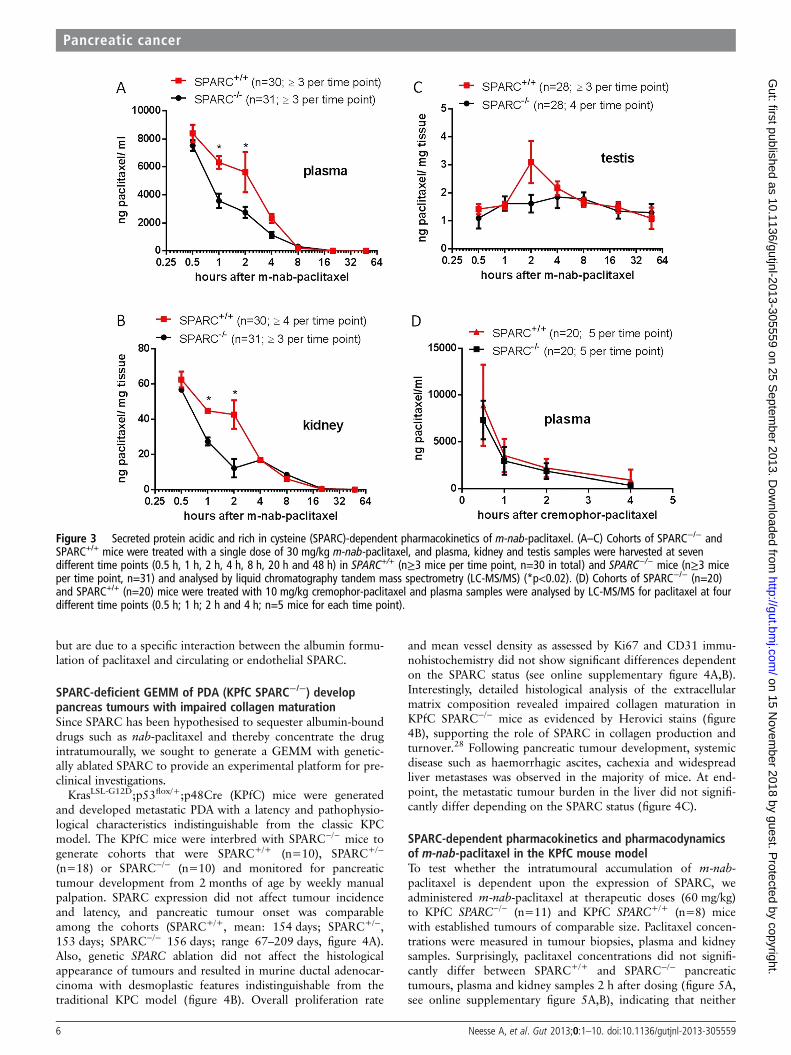
but are due to a specific interaction between the albumin formu-lation of paclitaxel and circulating or endothelial SPARC.
SPARC-deficient GEMM of PDA (KPfC SPARC−/−) developpancreas tumours with impaired collagen maturationSince SPARC has been hypothesised to sequester albumin-bounddrugs such as nab-paclitaxel and thereby concentrate the drugintratumourally, we sought to generate a GEMM with genetic-ally ablated SPARC to provide an experimental platform for pre-clinical investigations.
KrasLSL-G12D;p53flox/+;p48Cre (KPfC) mice were generatedand developed metastatic PDA with a latency and pathophysio-logical characteristics indistinguishable from the classic KPCmodel. The KPfC mice were interbred with SPARC−/− mice togenerate cohorts that were SPARC+/+ (n=10), SPARC+/−
(n=18) or SPARC−/− (n=10) and monitored for pancreatictumour development from 2 months of age by weekly manualpalpation. SPARC expression did not affect tumour incidenceand latency, and pancreatic tumour onset was comparableamong the cohorts (SPARC+/+, mean: 154 days; SPARC+/−,153 days; SPARC−/− 156 days; range 67–209 days, figure 4A).Also, genetic SPARC ablation did not affect the histologicalappearance of tumours and resulted in murine ductal adenocar-cinoma with desmoplastic features indistinguishable from thetraditional KPC model (figure 4B). Overall proliferation rate
and mean vessel density as assessed by Ki67 and CD31 immu-nohistochemistry did not show significant differences dependenton the SPARC status (see online supplementary figure 4A,B).Interestingly, detailed histological analysis of the extracellularmatrix composition revealed impaired collagen maturation inKPfC SPARC−/− mice as evidenced by Herovici stains (figure4B), supporting the role of SPARC in collagen production andturnover.28 Following pancreatic tumour development, systemicdisease such as haemorrhagic ascites, cachexia and widespreadliver metastases was observed in the majority of mice. At end-point, the metastatic tumour burden in the liver did not signifi-cantly differ depending on the SPARC status (figure 4C).
SPARC-dependent pharmacokinetics and pharmacodynamicsof m-nab-paclitaxel in the KPfC mouse modelTo test whether the intratumoural accumulation of m-nab-paclitaxel is dependent upon the expression of SPARC, weadministered m-nab-paclitaxel at therapeutic doses (60 mg/kg)to KPfC SPARC−/− (n=11) and KPfC SPARC+/+ (n=8) micewith established tumours of comparable size. Paclitaxel concen-trations were measured in tumour biopsies, plasma and kidneysamples. Surprisingly, paclitaxel concentrations did not signifi-cantly differ between SPARC+/+ and SPARC−/− pancreatictumours, plasma and kidney samples 2 h after dosing (figure 5A,see online supplementary figure 5A,B), indicating that neither
Figure 3 Secreted protein acidic and rich in cysteine (SPARC)-dependent pharmacokinetics of m-nab-paclitaxel. (A–C) Cohorts of SPARC−/− andSPARC+/+ mice were treated with a single dose of 30 mg/kg m-nab-paclitaxel, and plasma, kidney and testis samples were harvested at sevendifferent time points (0.5 h, 1 h, 2 h, 4 h, 8 h, 20 h and 48 h) in SPARC+/+ (n≥3 mice per time point, n=30 in total) and SPARC−/− mice (n≥3 miceper time point, n=31) and analysed by liquid chromatography tandem mass spectrometry (LC-MS/MS) (*p<0.02). (D) Cohorts of SPARC−/− (n=20)and SPARC+/+ (n=20) mice were treated with 10 mg/kg cremophor-paclitaxel and plasma samples were analysed by LC-MS/MS for paclitaxel at fourdifferent time points (0.5 h; 1 h; 2 h and 4 h; n=5 mice for each time point).
6 Neesse A, et al. Gut 2013;0:1–10. doi:10.1136/gutjnl-2013-305559
Pancreatic cancer
on 15 Novem
ber 2018 by guest. Protected by copyright.
http://gut.bmj.com
/G
ut: first published as 10.1136/gutjnl-2013-305559 on 25 Septem
ber 2013. Dow
nloaded from

Figure 4 Secreted protein acidic andrich in cysteine (SPARC) deficientgenetically engineered mouse model(GEMM) of pancreatic ductaladenocarcinoma develop pancreastumours with impaired collagenmaturation. (A) Tumour incidencefollowing weekly manual palpation ofKPfC mice from 2 months of age.(B) Representative H&E from KPfCSPARC+/+ and KPfC SPARC−/−miceshows desmoplastic ducaladenocarcinoma (upper panel).Immunohistochemistry for SPARCconfirms genetic ablation of SPARC(middle panel), and Herovici stainsshow decrease in mature collagenfibres (pink) in KPfC SPARC−/− mice(lower panel). (C) Quantification of fiveserial liver sections (100 mm apart)shows no significant difference inmetastatic burden upon SPARCheterozygous or homozygous deletionin KPfC mice.
Neesse A, et al. Gut 2013;0:1–10. doi:10.1136/gutjnl-2013-305559 7
Pancreatic cancer
on 15 Novem
ber 2018 by guest. Protected by copyright.
http://gut.bmj.com
/G
ut: first published as 10.1136/gutjnl-2013-305559 on 25 Septem
ber 2013. Dow
nloaded from

circulating nor tumoural SPARC sequesters nab-paclitaxel toaccumulate the drug intratumourally during this timeframe.Furthermore, we conducted a short-term intervention study thatcompared the tumour growth in KPfC SPARC+/+ (n=6) andKPfC SPARC−/− mice (n=5) with 60 mg/kg m-nab-paclitaxeldosed 3× over 7 days (figure 5B, see online supplementary figure5C,D). Final tumour volumes in KPfC SPARC+/+ versus KPfCSPARC−/− mice did not significantly differ from each other fol-lowing 7 days of 60 mg/kg m-nab-paclitaxel treatment (mean:152 mm3±43 vs 193 mm3±27; figure 5C). In line with treat-ment responses observed by 3D ultrasound, levels of intratu-moural apoptosis were not significantly different in KPfCSPARC+/+ and KPfC SPARC−/− pancreatic tumours (figure 5D).Furthermore, weight loss (not shown) or hematological toxicitywas not affected by the SPARC status upon m-nab-paclitaxeltreatment (see online supplementary figure 5E). In summary, ourdata suggest that although m-nab-paclitaxel exerts significantantitumour effects at 60 mg/kg by induction of apoptotic celldeath, drug delivery and response to treatment are independentof SPARC expression.
DISCUSSIONNanoparticle albumin-bound (nab)-paclitaxel, an albumin-bound-stabilised paclitaxel formulation, has recently demon-strated significant improvements in median survival of patientswith metastatic PDA when combined with gemcitabine.18 Sincethis drug combination represents the first therapeutic regimenthat significantly extends survival of advanced stage PDApatients and is accompanied by an acceptable toxicity profile,nab-paclitaxel and gemcitabine will likely become the newstandard of care chemotherapy and will shortly be implementedin national and international PDA treatment guidelines. Apartfrom the striking antitumour effect of nab-paclitaxel alone inKPC tumours, our group previously discovered a potential syn-ergistic drug–drug interaction of gemcitabine and nab-paclitaxeldue to ROS-mediated degradation of cytidine deaminase thatresulted in higher levels of activated intratumoural gemcitabinemetabolites.19 However, the limited availability of patient tissueand the lack of murine albumin-bound paclitaxel have hamperedour ability to address the mechanism of action of nab-paclitaxelin human and murine PDA. Importantly, preliminary data
Figure 5 Pharmacokinetics and pharmacodynamics of m-nab-paclitaxel in the KPfC secreted protein acidic and rich in cysteine (SPARC) mousemodel. (A) Cohorts of KPfC SPARC+/+ (n=8) and KPC SPARC−/− mice (n=11) were treated with 60 mg/kg m-nab-paclitaxel, and tumour sampleswere taken 2 h after dosing and analysed by liquid chromatography tandem mass spectrometry (LC-MS/MS). (B) Representative high-resolutionultrasound picture of typical KPfC murine pancreatic tumour. (C) Quantification of tumour volume growth using biweekly three-dimensionalhigh-resolution ultrasound shows no significant decrease in tumour burden on day 7 in KPfC SPARC+/+ versus KPfC SPARC−/− pancreatic tumoursfollowing three injections of 60 mg/kg m-nab-paclitaxel over 7 days. (D) Computer-based quantification of apoptosis (cleaved caspase-3) inpancreatic tumours from KPfC SPARC+/+ (n=6) and KPfC SPARC−/− mice (n=5) treated with 60 mg/kg m-nab-paclitaxel. All animals were sacrificed2 h after the last dose of m-nab-paclitaxel.
8 Neesse A, et al. Gut 2013;0:1–10. doi:10.1136/gutjnl-2013-305559
Pancreatic cancer
on 15 Novem
ber 2018 by guest. Protected by copyright.
http://gut.bmj.com
/G
ut: first published as 10.1136/gutjnl-2013-305559 on 25 Septem
ber 2013. Dow
nloaded from

suggest that SPARC may play a pivotal role as a biomarker fornab-paclitaxel-based chemotherapeutic regimens and maypredict responses to treatment.17
Our results suggest that intratumoural concentrations of pacli-taxel following either nab-paclitaxel or cremophor-paclitaxel arecomparable in vivo; however, the dramatically reduced toxicity ofwater-soluble nab-paclitaxel increases the maximum tolerated pacli-taxel dose more than fourfold, thus resulting in significantly higherneoplastic cell death rates compared with cremophor-paclitaxel.
To determine whether SPARC would sequester nab-paclitaxelto concentrate the drug intratumourally, we employed twoapproaches. First, we used equimolar doses of nab-paclitaxeland cremophor-paclitaxel in the KPC model with endogenousSPARC expression and assessed intratumoural paclitaxel concen-trations by LC-MS/MS. Second, we genetically ablated SPARCin the closely related KPfC mouse model and used a novelmouse albumin-bound paclitaxel formulation to account forinterspecies differences of human albumin and murine SPARC.Both approaches showed that SPARC plays no role in sequester-ing nab-paclitaxel intratumourally. Despite reports that SPARCis associated with poor survival in PDA patients14 15 and thatpancreatic tumours grown orthotopically in SPARC−/− mice aremore metastatic and less vascular,29 SPARC deficiency did notchange the kinetics of tumour onset, growth rate, angiogenesisand metastasis in the KPfC model in our study. However, exten-sive pharmacokinetic analysis in SPARC+/+ and SPARC−/− micerevealed a SPARC-specific distribution pattern of low-dosem-nab-paclitaxel that was independent from paclitaxel metabol-ism and saturable at higher concentrations of nab-paclitaxel intumour-bearing mice. Notably, increased paclitaxel concentra-tions were observed in plasma and point towards a potentialinteraction between circulating SPARC and albumin-bounddrugs. Alternatively, SPARC has also been shown to beexpressed in vascular endothelial cells and platelets,30–32 andhas frequently been implicated in the modulation of vascularbiology such as endothelial cell proliferation as well as pericyterecruitment and regulation.33–35 It has been previously reportedin vitro that endothelial binding and transcytosis of albumin andnab-paclitaxel may be mediated by endothelial cells that expressSPARC and other albumin-binding proteins such as gp60.16 36
However, using therapeutic concentration of m-nab-paclitaxel(60 mg/kg) in tumour-bearing KPfC mice with geneticallyablated SPARC, this effect was saturable and both plasma andintratumoural paclitaxel concentration did not show significantdifferences in the context of SPARC expression. Also, PDA neo-plastic cell apoptosis and tumour volume increases followingtreatment with m-nab-paclitaxel for 1 week were not substan-tially affected by SPARC deletion. Therefore, we conclude thatalthough circulating SPARC may directly or indirectly increasethe intravascular concentration of low-dose nab-paclitaxel,stromal-derived SPARC does not strongly influence the accumu-lation of nab-paclitaxel intratumourally in a PDA mouse model.However, we cannot exclude the possibility that extremely hightissue or plasma SPARC concentrations in PDA patients mayincrease drug concentrations and improve antitumour efficacy,and therefore, the measurement of SPARC plasma levels priorto the initiation of nab-paclitaxel–based chemotherapies shouldbe investigated in pancreatic cancer trials.
Lastly, we examined whether prolonged treatment withm-nab-paclitaxel was able to deplete tumour stroma in KPCmice. Previous work from our group has shown that the exten-sive desmoplastic reaction of endogenous murine pancreatictumours can be successfully depleted following 10–14 days ofcontinuous pharmacological inhibition of the sonic hedgehog
pathway by IPI-926.10 Therefore, and due to increasing toxicitybeyond 2 weeks, we chose to treat tumour-bearing KPC miceover 2 weeks with m-nab-paclitaxel. In contrast to previous datapublished in patient-derived xenografts,17 we found thatm-nab-paclitaxel predominantly induced apoptotic cell death intumour rather than stromal cells, and the stromal content ofdesmoplastic murine pancreatic tumours was unchanged follow-ing 2 weeks of treatment. This finding should prompt the designof clinical trials that combine nab-paclitaxel with other stromalaltering agents such as PEGPH20 (hyaluronidase) to increaseintratumoural delivery of nab-paclitaxel.5–12
In conclusion, we present a pharmacokinetic and pharmaco-dynamic analysis of cremophor-paclitaxel, nab-paclitaxel and anovel m-nab-paclitaxel in various GEMMs of PDA that haspotential value for future preclinical and clinical drug develop-ment. We found that nab-paclitaxel accumulation and antitu-mour effects were dose-dependent, but SPARC independent inGEMMs of PDA. Although stromal-derived SPARC may notincrease drug accumulation or induce stromal depletion uponnab-paclitaxel treatment in our model system, plasma SPARClevels may play a role in drug retention and therefore tissuedelivery in patients with baseline elevated SPARC levels. Futureclinical studies will be required to evaluate the potential valueof plasma SPARC as a non-invasive and predictive biomarkerfor nab-paclitaxel-based chemotherapeutic regimens.
Acknowledgements We thank Manuel Hidalgo for sharing his data beforepublication. We thank Dr. Aarthi Gopinathan for reviewing ultrasound scans. Wethank Frances Connor, Paul Mackin, Lisa Young and Steven Kupczak formaintenance and management of mouse colonies, as well as staff from theCambridge Research Institute Biological Resource Unit, histology core andpharmacokinetics core.
Contributors AN, KKF and DAT conceived and designed the experiments. AN, KKFand DSC performed animal experiments. AN and KKF performed cell cultureexperiments. AN, DSC, WJH and VE performed histology stainings. TEB, FMR andDIJ designed and carried out paclitaxel pharmacokinetic experiments. AN and DATwrote the manuscript. All authors reviewed the manuscript.
Funding This research was supported by the University of Cambridge and CancerResearch UK, The Li Ka Shing Foundation and Hutchison Whampoa Limited and theNational Institute for Health Research Cambridge Biomedical Research Centre. KKFand DAT were supported by the European Community Grant EPC-TM-Net 256974.AN was supported by the Deutsche Krebshilfe Mildred Scheel PostdoctoralFellowship. DIJ is a Group Leader in the Cancer Research UK Cambridge Institute.TEB and FMR are supported by Cancer Research UK. DAT is also supported by theLustgarten Foundation for Pancreatic Cancer Research and by the Cold SpringHarbor Laboratory Association.
Competing interests nab-Paclitaxel and m-nab-paclitaxel were formulated andprovided by Celgene Corporation.
Provenance and peer review Not commissioned; externally peer reviewed.
Open Access This is an Open Access article distributed in accordance with theCreative Commons Attribution Non Commercial (CC BY-NC 3.0) license, whichpermits others to distribute, remix, adapt, build upon this work non-commercially,and license their derivative works on different terms, provided the original work isproperly cited and the use is non-commercial. See: http://creativecommons.org/licenses/by-nc/3.0/
REFERENCES1 Siegel R, Naishadham D, Jemal A. Cancer statistics, 2013. CA Cancer J Clin
2013;63:11–30.2 Burris HA, Moore MJ, Andersen J, et al. Improvements in survival and clinical
benefit with gemcitabine as first-line therapy for patients with advanced pancreascancer: a randomized trial. J Clin Oncol 1997;15:2403–13.
3 Conroy T, Desseigne F, Ychou M, et al. FOLFIRINOX versus gemcitabine formetastatic pancreatic cancer. N Engl J Med 2011;364:1817–25.
4 Neesse A, Michl P, Frese KK, et al. Stromal biology and therapy in pancreaticcancer. Gut 2011;60:861–8.
5 Beatty GLChiorean EG, Fishman MP, et al. CD40 agonists alter tumor stroma and showefficacy against pancreatic carcinoma in mice and humans. Science 2011;331:1612–16.
6 Clark CE, Hingorani SR, Mick R, et al. Dynamics of the immune reaction topancreatic cancer from inception to invasion. Cancer Res 2007;67:9518–27.
Neesse A, et al. Gut 2013;0:1–10. doi:10.1136/gutjnl-2013-305559 9
Pancreatic cancer
on 15 Novem
ber 2018 by guest. Protected by copyright.
http://gut.bmj.com
/G
ut: first published as 10.1136/gutjnl-2013-305559 on 25 Septem
ber 2013. Dow
nloaded from

7 Bayne LJ, Beatty GL, Jhala N, et al. Tumor-derived granulocyte-macrophagecolony-stimulating factor regulates myloid inflammation and T cell immunity inpancreatic cancer. Cancer Cell 2012;21:822–35.
8 Rhim AD, Mirek ET, Aiello NM, et al. EMT and dissemination precede pancreatictumor formation. Cell 2012;148:349–61.
9 Straussman R, Morikawa T, Shee K, et al. Tumour micro-environment elicits innateresistance to RAF inhibitors through HGF secretion. Nature 2012;487:500–4.
10 Olive KP, Jacobetz MA, Davidson CJ, et al. Inhibition of Hedgehog signalingenhances delivery of chemotherapy in a mouse model of pancreatic cancer. Science2009;324:1457–61.
11 Provenzano PP, Cuevas C, Chang AE, et al. Enzymatic targeting of the stromaablates physical barriers to treatment of pancreatic ductal adenocarcinoma. CancerCell 2012;21:418–29.
12 Jacobetz MA, Chan DS, Neesse A, et al. Hyaluronan impairs vascular function anddrug delivery in a mouse model of pancreatic cancer. Gut 2012;62:112–20.
13 Neesse A, Frese KK, Bapiro TE, et al. CTGF antagonism with mAb FG-3019enhances chemotherapy response without increasing drug delivery in murine ductalpancreas cancer. Proc Natl Acad Sci USA Published Online First: 8 July 2013.doi:10.1073/pnas.1300415110
14 Mantoni TS, Schendel RR, Rodel F, et al. Stromal SPARC expression and patientsurvival after chemoradiation for non-resectable pancreatic adenocarcinoma. CancerBiol Ther 2008;7:1806–15.
15 Infante JR, Matsubayashi H, Sato N, et al. Peritumoral fibroblast SPARC expressionand patient outcome with resectable pancreatic adenocarcinoma. J Clin Oncol2007;25:319–25.
16 Desai N, Trieu V, Yao Z, et al. Increased antitumor activity, intratumor paclitaxelconcentrations, and endothelial cell transport of cremophor-free, albumin-boundpaclitaxel, ABI-007, compared with cremophor-based paclitaxel. Clin Cancer Res2006;12:1317–24.
17 Von Hoff DD, Ramanathan RK, Borad MJ, et al. Gemcitabine plus nab-paclitaxel isan active regimen in patients with advanced pancreatic cancer: a phase I/II trial.J Clin Oncol 2011;29:4548–54.
18 Von Hoff DD, Ervin TJ, Arena FP, et al. Randomized phase III study of weeklynab-paclitaxel plus gemcitabine versus gemcitabine alone in patients withmetastatic adenocarcinoma of the pancreas (MPACT). J Clin Oncol 2013;30(Suppl34): abstr LBA148.
19 Frese KK, Neesse A, Cook N, et al. nab-Paclitaxel potentiates gemcitabine activityby reducing cytidine deaminase levels in a mouse model of pancreatic cancer.Cancer Discovery 2012;2:260–9.
20 Hosein PJ, de Lima Lopes G, Pastorini VH, et al. A phase II trial of nab-Paclitaxel assecond-line therapy in patients with advanced pancreatic cancer. Am J Clin Oncol2013;36:151–6.
21 Desai NP, Trieu V, Hwang LY, et al. Improved effectiveness of nanoparticlealbumin-bound (nab) paclitaxel versus polysorbate-based docetaxel in multiple
xenografts as a function of HER2 and SPARC status. Anticancer Drugs2008;19:899–909.
22 Shao H, Tang H, Salavaggione OE, et al. Improved response to nab-paclitaxelcompared with cremophor-solubilized paclitaxel is independent of secreted proteinacidic and rich in cysteine expression in non-small cell lung cancer. J Thorac Oncol2011;6:998–1005.
23 Desai N, Trieu V, Damascelli B, et al. SPARC expression correlates with tumorresponse to albumin-bound paclitaxel in head and neck cancer patients. TranslOncol 2009;2:59–64.
24 Gilmour DT, Lyon GJ, Carlton MB, et al. Mice deficient for the secreted glycoproteinSPARC/osteonectin/BM40 develop normally but show severe age-onset cataractformation and disruption of the lens. EMBO J 1998;17:1860–70.
25 Hingorani SR, Wang L, Multani AS, et al. Trp53R172H and KrasG12D cooperate topromote chromosomal instability and widely metastatic pancreatic ductaladenocarcinoma in mice. Cancer Cell 2005;7:469–83.
26 Freireich EJ, Gehan EA, Rall DP, et al. Quantitative comparison of toxicity ofanticancer agents in mouse, rat, hamster, dog, monkey, and man. CancerChemother Rep 1966;50:219–44.
27 Karreth FA, DeNicola GM, Winter SP, et al. C-Raf inhibits MAPK activation andtransformation by B-Raf(V600E). Mol Cell 2009;36:477–86.
28 Schellings MW, Vanhoutte D, Swinnen M, et al. Absence of SPARC results inincreased cardiac rupture and dysfunction after acute myocardial infarction. J ExpMed 2009;206:113–23.
29 Arnold SA, Rivera LB, Miller AF, et al. Lack of host SPARC enhances vascularfunction and tumor spread in an orthotopic murine model of pancreatic carcinoma.Dis Model Mech 2010;3:57–72.
30 Stenner DD, Tracy RP, Riggs BL, et al. Human platelets contain and secreteosteonectin, a major protein of mineralized bone. Proc Natl Acad Sci USA1986;83:6892–6.
31 Sage H, Johnson C, Bornstein P. Characterization of a novel serum albumin-bindingglycoprotein secreted by endothelial cells in culture. J Biol Chem 1984;259:3993–4007.
32 Tiruppathi C, Finnegan A, Malik AB. Isolation and characterization of a cell surfacealbumin-binding protein from vascular endothelial cells. Proc Natl Acad Sci USA1996;93:250–4.
33 Nozaki M, Sakurai E, Raisler BJ, et al. Loss of SPARC-mediated VEGFR-1suppression after injury reveals a novel antiangiogenic activity of VEGF-A. J ClinInvest 2006;116:422–9.
34 Rivera LB, Bradshaw AD, Brekken RA. The regulatory function of SPARC in vascularbiology. Cell Mol Life Sci 2011;68:3165–73.
35 Rivera LB, Brekken RA. SPARC promotes pericyte recruitment via inhibition ofendoglin-dependent TGF-beta1 activity. J Cell Biol 2011;193:1305–19.
36 Schnitzer JE, Oh P. Antibodies to SPARC inhibit albumin binding to SPARC, gp60,and microvascular endothelium. Am J Physiol 1992;263:H1872–9.
10 Neesse A, et al. Gut 2013;0:1–10. doi:10.1136/gutjnl-2013-305559
Pancreatic cancer
on 15 Novem
ber 2018 by guest. Protected by copyright.
http://gut.bmj.com
/G
ut: first published as 10.1136/gutjnl-2013-305559 on 25 Septem
ber 2013. Dow
nloaded from